User login
Unneeded CT Scans Magnify Radiation Risks
CHICAGO — Patients undergoing abdominal/pelvic computed tomography frequently receive unindicated additional scans, resulting in excess radiation exposure, a review of 978 CT scans suggests.
The review, which included exams done in 500 patients, revealed that 263 exams (52.6% of the total) were not indicated based on American College of Radiology Appropriateness Criteria. The exams, performed at institutions in Illinois, Michigan, Minnesota, and Wisconsin, involved patients aged 9 months to 91 years.
One in five patients received at least 50 millisieverts (mSv) of radiation in a single examination. Seven (1.4%) of the 500 patients received greater than 100 mSv—a dose that is associated with an increased cancer risk. The mean radiation dose per patient was 32.9 mSv, which is equivalent to the dose from about 1,000 chest X-rays. The 308 exams that consisted of multiple phases exposed patients to a mean dose of 43 mSv, Dr. Kristie Guite reported at the annual meeting of the Radiological Society of North America.
Radiologists are encouraged to follow the standard of “As Low as Reasonably Achievable” or ALARA, noted Dr. Guite, a resident at the University of Wisconsin, Madison. “The most important finding in this study is that adherence to ALARA is not widespread at this time,” she said at a press briefing during the meeting.
“At the doses seen in our study, 1 in 1,000 patients could get a radiation-induced cancer,” she said. If the data are extrapolated to all CT scans of the abdomen/pelvis performed in the United States, “this would lead to 23,000 radiation-induced cancer cases per year.”
As for why radiologists may order extra scans, coauthor Dr. J. Louis Hinshaw, also with the UW-Madison, said it may be that radiologists simply have the ability and tools available to perform the tests. Also, protocols are set up on CT scanners beforehand and are often designed to answer “what if” scenarios, in which additional views may be needed. “There is some risk-aversion that comes into play,” he said at the briefing.
Of the 500 patients, 7% had findings that would not have been identified without the extra scans, but most of these were not clinically relevant, Dr. Hinshaw said.
The Radiological Society of North America has partnered with the American College of Radiology to create a task force on adult radiation protection to increase awareness of cumulative dose and radiation risks.
Both researchers stressed that CT is a valuable diagnostic tool and that the study findings should not impede use of the technology.
Press briefing moderator Dr. Robert Zimmerman, professor of radiology at Weill Cornell Medical College in New York, concurred. He suggested that patients investigate the radiation protocol at their institution and ask their physicians what steps are being taken to minimize radiation dose.
“We don't want to damage patients, but we know we have the technology that is very useful in saving people's lives,” Dr. Zimmerman said. “We are trying to balance the two.”
In the current study, patients with a malignancy were 22% more likely to receive excess radiation, Dr. Guite said.
Delayed-phase imaging, performed after a contrast agent has accumulated in the kidneys/bladder, accounted for 77% of the unnecessary scans.
There was no study sponsorship, but one coauthor disclosed being a stock holder with NeuWave Medical Inc. and a patent holder with Covidien AG.
'At the doses seen in our study, 1 in 1,000 patients could get a radiation-induced cancer.'
Source DR. GUITE
The Value of Echocardiography in the RUSH Protocol
Sudden Onset of Pleuritic Chest Pain
Severe Left Shoulder Pain While Weightlifting
The Subxiphoid and Apical Views
Four Patients With a Focal Neurological Deficit
Differences Between Neurosurgeons and Orthopedic Surgeons in Classifying Cervical Dislocation Injuries and Making Assessment and Treatment Decisions: A Multicenter Reliability Study
Imaging Modalities for Osteoarthritis
Radiographs are no longer de rigeur in making the diagnosis of knee osteoarthritis, according to guidelines released by the European League Against Rheumatism last month. Clinical signs, symptoms, and risk factors are sufficient to make the diagnosis.
Yet x-rays and other imaging modalities continue to have a role in osteoarthritis, according to Dr. Ali Guermazi and Dr. David J. Hunter. At present, imaging should primarily be used in research rather than in clinical practice (where its use should be limited to ruling out other likely diagnoses), they agreed.
Imaging conveys information about the pathophysiology of OA and has provided insight into symptoms and progression, said Dr. Hunter, chief of the research division of New England Baptist Hospital in Boston. Ultimately, “it will help to define the best treatments for osteoarthritis,” he added. Imaging has the potential to “have wider clinical application, when we have an opportunity to intervene in earlier arthritis through modifying joint structural changes.”
Yet, there continues to be “widespread use of different imaging modalities in the clinical setting, where it's clear there is osteoarthritis.” Dr. Hunter estimated that 60%–70% of patients who present to him in the clinic have an MRI on CD with them. “It doesn't change the diagnosis or what I'm going to do for them, so I'm not sure that there's much rationale for having that at present.”
Each imaging modality has a role to play when assessing the pathophysiology of the whole joint. “Some of that role is complementary, but much of it is unique to that particular modality. … Each has its strengths and weaknesses.”
Dr. Guermazi and Dr. Hunter shared their thoughts on how various imaging modalities can further understanding of the pathophysiology of OA.
Radiograph
An x-ray is typically used only to rule out other diagnoses, said Dr. Hunter. These could include rheumatoid arthritis, gout, intra-articular loose bodies, and trauma. “These would be reasons to think about doing additional imaging.”
X-ray is still the most widely used imaging modality because it's relatively inexpensive, it's available, and it's relatively easy to interpret. X-rays are useful primarily for outlining the two-dimensional geometry of the bones. “You infer from the x-ray what the joint space is, and from that, the health and integrity of the cartilage, but it's an indirect inference and you really can't make any direct representations of other tissues,” Dr. Hunter said.
In addition, x-ray evaluates features that don't contribute to pain, which is the primary symptom of OA, said Dr. Guermazi, director of the quantitative imaging center and section chief of musculoskeletal imaging at Boston University. “If we look at joint space narrowing, thinking that this is the cartilage measure, [x-ray is] useless for many reasons. Arthritis is defined clinically as pain. Cartilage can't be painful because there are no nerves in it.”
Synovitis, effusions, and bone marrow lesions, however, can all be painful. “The only features that are not painful are the ones we are looking at on a radiograph, which are cartilage [inferred from the joint space narrowing] and osteophytes.”
Dr. Hunter noted that the imaging community is divided about whether the best way to monitor the structural progression of underlying disease is to measure joint space on x-ray or to measure other features—such as the cartilage itself—on an MRI.
MRI
The main strength of MRI as a research tool is its ability to provide information on many tissues in the joint. “On MRI, you're able to … see changes in the curvature of the bone, lesions within the bone [which demonstrate where focal loading is occurring, and] alterations within the cartilage itself. … You can see inflammation within the joint [such as synovitis or an effusion], the ligaments, and the muscle,” said Dr. Hunter.
“The more we're learning about osteoarthritis, the more we're realizing that much of the reason why a person has pain [and] functional limitation—and much of the reason why the joint progresses—has a lot more to do with the tissues in the joint other than cartilage,” Dr. Hunter said. “Bone marrow lesions, synovitis, and effusions appear to account for the majority of the reason why a person has pain. MRIs provide a lot of insight there,” he said.
Beyond the local-tissue reasons for a person to have symptoms, there is also some local alteration in the neurophysiology. “The communication between different nerve pathways that leads to sensitization of nerves occurs both peripherally and centrally, “Dr. Hunter said.
“If we are focused on symptomatic response, it's helpful to think about the contribution of all of those factors.” Nerve endings are present in specific tissues, particularly the bone and synovium.
In MRI, gadolinium contrast can be given intravenously or intra-articularly, said Dr. Guermazi. Intravenous contrast is useful for evaluating synovitis and differentiating it from effusions. Intra-articular contrast is useful for evaluating cartilage and meniscal lesions.
Among the several downsides of MRI are its cost and the time needed to acquire the images and to interpret them. MRIs of the knee or hip are acquired with the patient in a lying down or supine position; however, physiologically, the knee is probably best understood when those joints are bearing weight.
CT
CT is widely available and generally less expensive than MRI. Unlike dual-energy x-ray absorptiometry, CT imaging can provide information about volumetric density of the bone via changes in the periarticular density, Dr. Hunter said.
The chosen modality may depend on the joint to be imaged, said Dr. Guermazi. In the case of OA of the facet joints, osteophytosis and bone are better viewed with CT than MRI. However, much other information is lost. Although meniscal and anterior/posterior cruciate ligament lesions can be seen on contrast CT, bone marrow lesions cannot.
Ultrasound
“Intra-articular steroids appear to reduce the extent of inflammation in joints. That can be appreciated on ultrasound,” said Dr. Hunter. Ultrasound can also be used to guide the injection of corticosteroids. “Some of the therapies that are being developed are likely to be intra-articular, and ultrasound may be helpful in guiding the needle to the right spot.”
Ultrasound also is being used in some clinical trials to visualize synovitis, according to Dr. Guermazi. “I think it's promising, especially if you use Doppler and can see vascularity.” Doppler ultrasound can be used to identify and monitor active synovitis, after treatment.
Ultrasound is also able to assess the effect of biologic drugs—currently in testing for OA—on synovitis.
Ultrasound is inexpensive, but “it's very operator dependent,” said Dr. Guermazi. “Ultrasound tends to be used more widely outside the United States, where MRI may not be so readily available.”
Ultrasound allows visualization of ligaments, muscles, and tendons, but not bone, and it can visualize only tissues close to the skin and near the probe.
Therapy
Research into potential OA therapies now focuses on tissues that are likely to play a role symptomatically and structurally, rather than just concentrating on cartilage. Just as bBiologic drugs have made huge inroads in RA, “we're right at the cusp of that” with OA, said Dr. Guermazi. When “therapies do become available, the ability to identify OA in the early stages will be very important.”
X-rays can supply information about the structural progression of underlying disease, as evidenced by the osteophytic changes, subchondral bone sclerosis, and absence of joint space seen in an anteroposterior radiograph of the left knee in a 65-year-old woman with secondary arthritis at 9 years after treated fractures (at left). A lateral view (at right) confirms severe tibiofemoral OA and shows severe patellofemoral OA with large posterior femoral condyles (arrowhead) and tibial plateaus (arrow) not seen on the anteroposterior view.
Sagittal fat-suppressed proton density-weighted MRI shows diffuse cartilage loss of the lateral central weight-bearing tibia and femur. Also seen are a central lateral femur bone marrow lesion (arrow) and a small tibiofibular ganglion cyst (arrowhead).
CT can visualize volumetric density. Medial narrowing plus subchondral medial tibiofemoral bone sclerosis (arrows) and cystic changes are seen on coronal reformatted CT.
Source Images courtesy Dr. Ali Guermazi
Role of MRI in breast cancer management
A 52-year-old woman presents to your outpatient clinic with a newly palpable marble-sized lump in the upper outer quadrant of her right breast. The mass is firm and non-tender. She performs breast self-examinations regularly and first noticed the mass 3 weeks ago. Your examination confirms a palpable lesion. Annual screening mammograms at another facility have been negative, including the last one 6 months ago. The woman is otherwise in good health.
When she was younger, she had two pregnancies and gave birth to two children, whom she did not breastfeed. She reached menopause at age 49 and has never been on hormone replacement therapy. Neither she nor anyone in her family has had breast cancer, and she has never undergone breast biopsy.
She says that a woman she spoke with in the waiting room, a 2-year breast cancer survivor, told her that her primary cancer had been “finally diagnosed” with magnetic resonance imaging (MRI). This patient is now urging you to order an MRI. What should you do?
DIFFERENT IMAGING TESTS FOR DIFFERENT INDICATIONS
Different imaging tests are indicated in different situations. Screening mammography is the standard of care for women who have no signs or symptoms of breast cancer. When a screening examination shows abnormal or equivocal findings or when a patient presents with symptoms (eg, a palpable lesion, breast pain, nipple discharge), further characterization with a tailored imaging examination is warranted. Such an examination might include diagnostic mammography, ultrasonography, or MRI.
This article reviews MRI’s role in breast cancer management with respect to the other imaging tests currently in use.
MAMMOGRAPHY IS THE SCREENING TEST OF CHOICE
Screening mammography, as distinguished from diagnostic mammography, is an x-ray examination of breast tissue that obtains high-quality images while using a very low dose of ionizing radiation. It is performed with the breast tissue compressed to optimize image quality. The examination time is short, usually 15 to 20 minutes. No contrast agents are used. The malignant lesions it detects differ from normal fibroglandular tissue in their x-ray attenuation, appearing as asymmetric soft-tissue densities, architectural distortion, masses, or abnormal calcifications.
The American Cancer Society1 recommends mammography as the method of choice to screen for nonpalpable, clinically occult breast cancers in women over age 40 and in younger women with certain risk factors.
The rationale for screening mammography is supported by evidence of significant reductions in death rates from breast cancer in patients who undergo routine mammographic screening. Tabar et al,2 in the Swedish Two-County Trial, found a 30% lower rate of death from breast cancer in women ages 40 to 74 who were invited to undergo screening: the reduction was 34% for women age 50 to 74 and 12% for women age 40 to 49. The authors attributed the smaller benefit in the younger group to a tendency toward more rapid tumor progression in that age group.
Mammography is somewhat less sensitive and specific in women with dense breasts,3 in younger women, and in women on hormone replacement therapy.4 For example, a recent population-based study of seven mammography registries in the United States5 reported that the sensitivity of mammography for detecting breast cancer, adjusted for breast density and age, ranged from 63% for extremely dense breast tissue to 87% for entirely fatty breast tissue, and from 69% for women age 40 to 44 to 83% for women age 80 to 89. The adjusted specificities ranged from 89% for extremely dense breast tissue to 97% for entirely fatty breast tissue. The adjusted specificities in women not on hormone replacement therapy ranged from 91% for those age 40 to 44 to 94% for those age 80 to 89. In women on hormone replacement therapy, the adjusted specificity was about 92% for all ages taken together.
Digital is often better than film
Screening mammography has improved with the development of digital technology,6 in which images are acquired directly from an x-ray-sensitive solid-state receptor, without film. This process differs from digital computer-aided detection techniques, in which conventional analog films are optically scanned, generating a secondary digital image, which is then used for subsequent computerized analysis.
In 2005, the first large national trial comparing digital mammography with conventional mammography—the Digital Mammographic Imaging Screening Trial6—showed that digital mammography held a statistically significant advantage over conventional film-screen mammography in three subgroups7:
- Women under age 50
- Women with radiographically dense breast tissue
- Premenopausal or perimenopausal women. It is hoped that these encouraging results will be confirmed and extended by other trials now in progress.
DIAGNOSTIC MAMOGRAPHY FOR FURTHER WORKUP
Screening mammography yields findings of uncertain significance or frank concern in roughly 1 out of 10 examinations. In these cases, the examination is considered to be incomplete. If the finding is of unclear significance, previous mammograms, if available, may reveal whether the finding has remained the same or changed over time. If a worrisome change has occurred or if no prior films can be obtained, a diagnostic study with additional imaging must be carried out so that the radiologist can decide if a lesion is actually present.
Diagnostic mammography is a tailored examination that may include special projections to better visualize a specific region of concern, spot-compression views to disperse dense breast tissue, or magnification views to characterize microcalcifications. In cases of known breast cancer, diagnostic mammography helps detect additional foci of cancer in the same or in the contralateral breast.8
DIRECTED ULTRASONOGRAPHY TO EVALUATE REGIONS OF CONCERN
While ultrasonography is not part of the standard breast cancer screening protocol,9 directed or “targeted” breast ultrasonography is routinely used in the diagnostic workup to evaluate particular regions of concern. Ultrasonography is used in combination with diagnostic mammography to evaluate mammographic masses, palpable lumps, asymmetric tissue, and architectural distortions.
Breast ultrasonography can usually distinguish cystic lesions from solid lesions, and it is used to guide core biopsy or fine needle aspiration of suspicious breast lesions. It is relatively inexpensive, widely available, and reliable when performed by a skilled and knowledgeable operator.
HOW MRI WORKS
Magnetic resonance imaging takes advantage of the magnetic properties of hydrogen nuclei (protons) in breast tissue. A small fraction of the protons in the patient are brought into alignment with a strong magnetic field within the MRI scanner. Then, the protons are exposed to a brief pulse of radiofrequency energy, which displaces their magnetic vectors. As the protons “relax” and realign along the applied magnetic field, energy is released. This energy, the electromagnetic magnetic resonance signal, is detected and electronically processed to construct an image, exploiting the different “relaxation times” of the different tissues in the breast to generate image contrast.
A standard breast MRI examination requires an intravenous paramagnetic contrast agent, usually a gadolinium chelate, to increase the sensitivity of the study. Gadolinium-based contrast material causes shortening of the T1 relaxation time of tissues in which the contrast agent accumulates, thereby increasing signal intensity (or “enhancement”) in those tissues.
Contrast enhancement may occur in malignant tissues with defective or “leaky” capillaries, but it also can occur in benign tissues, such as normal lymph nodes or benign proliferative processes. Thus, the finding of contrast enhancement does not by itself establish the diagnosis of breast cancer.
The patient must remain still
The patient is positioned prone for about 30 to 40 minutes inside the MRI scanner with the breasts encompassed by specially designed imaging coils, which maximize the signal strength and achieve high spatial resolution. The prone position also minimizes motion of breast tissue and transmitted physiologic motions, further ensuring good image quality.
Contrast enhancement over time
To display how contrast enhancement resolves over time, a series of scans must be obtained. First, a baseline scan is recorded. Then, the contrast material is given, and multiple postcontrast scans are obtained at equally spaced time intervals, typically 1 to 1.5 minutes apart. Usually five to seven postcontrast scans are recorded. During this time, the patient must continue to lie still without moving.
For each individual volume element (voxel) of breast tissue, which may measure 1 mm3 or less, a curve representing contrast enhancement vs time can be constructed. Such curves tend to show one of three typical trajectories or patterns, known as “washout,” “plateau,” and “progressive.” With additional postprocessing, these three contrast enhancement patterns are reduced to color coding and are mapped onto the gray-scale MRI image in the form of a color overlay, so that overall enhancement patterns in both breasts can be discerned at a glance by the radiologist.
These enhancement patterns initially were believed to be reliable indicators of malignant and benign conditions, but further experience has shown considerable overlap of the enhancement patterns between benign and malignant tissues. Thus, the diagnostic value of enhancement patterns is limited. As a rule of thumb, the washout pattern of enhancement (rapid uptake of contrast material followed by rapid washout) is thought to indicate malignancy in 60% to 70% of lesions that are suspicious in other respects.
Abnormal contrast enhancement of the suspicious region must be considered along with morphologic features, the degree of enhancement in adjacent normal-appearing tissue, and the correlation with mammographic or ultrasonographic findings.
Better for invasive ductal carcinoma than invasive lobular carcinoma or ductal carcinoma in situ
At present, we have no foolproof method of diagnosing cancer by MRI alone, though in many cases invasive ductal carcinoma can be predicted with a high degree of confidence. The accuracy of breast MRI is lower for “non-mass-like” enhancement, as is often seen in invasive lobular carcinoma and ductal carcinoma in situ.
Contraindications, problems
MRI for breast cancer evaluation is contraindicated in women with cardiac pacemakers, implanted neurostimulators, and certain older models of aneurysm clips and cardiac prosthetic valves. However, this is becoming less of a problem as MRI-compatible devices of recent design become more prevalent. To ensure safety, patients should complete a screening questionnaire for ferromagnetic devices before they are allowed to undergo breast MRI.
Claustrophobia may preclude an MRI study, but this is less of an issue now, as the newer “short-bore” magnet designs reduce the sensation of confinement.
A problem of increasing importance today is patient obesity: obese patients may not fit into the MRI scanner. So-called “open” MRI scanners are not a good alternative, as they cannot provide the high-resolution images of uniform quality required for breast MRI.
Another factor affecting the use of MRI for breast cancer diagnosis is the limited number of facilities that offer it. Head, spine, and orthopedic MRI services are more widely available.
Cost may be an issue. A breast MRI evaluation costs about 10 times more than screening mammography and may not be covered by health insurance, although coverage for this indication appears to be improving gradually.
MRI IS SENSITIVE, BUT NOT SO SPECIFIC
The role of MRI in evaluating breast disease has been studied and debated since contrast-enhanced breast MRI was introduced in 1985.10 Interest has grown steadily as evidence of its usefulness has accumulated. Improvements in MRI scanners have included better image resolution, dedicated breast coils, and rapid dynamic contrast-enhanced imaging.
The overall sensitivity of MRI for breast cancer is relatively high, with estimates ranging from 85% to 100%.11 In invasive ductal carcinoma, its sensitivity approaches 100%.12 Sensitivities for invasive lobular carcinoma and ductal carcinoma in situ are lower and not yet well defined.
In contrast, MRI’s specificity for breast cancer is much more variable, ranging from 37% to 100%. The discrepancies among estimates of specificity are attributed to multiple confounding methodologic factors in the studies to date, such as differences in imaging protocols, patient selection criteria, patient ages, interpretation criteria, and the level of experience of the interpreting radiologist.12
False-positive results may be caused by benign conditions such as fibroadenomas, intramammary lymph nodes, proliferative and nonproliferative fibrocystic changes, and mastitis, as well as by radial scars, atypical ductal hyperplasia, and lobular carcinoma in situ.13–15 In premenopausal women, the menstrual cycle may bring about regional physiologic variation in enhancement of the normal breast parenchyma, which may either simulate the appearance of a lesion or obscure a true lesion.16
Thus, breast MRI may detect cancer that is occult to mammography, but it also carries the risk of worrisome incidental findings that may only be resolved by biopsy. Such uncertain findings are troubling for both the radiologist and the patient when mammography, ultrasonography, and the physical examination are all normal. Clearly, breast MRI cannot be counted on to reassure the “worried well” patient.
MRI is not for screening in the general population
While its high sensitivity for invasive ductal carcinoma17 would seem to make breast MRI attractive for breast cancer screening, it has the disadvantages of lower sensitivity for invasive lobular carcinoma and ductal carcinoma in situ,17,18 as well as the potential to raise suspicions of breast cancer that may be difficult to resolve. For these reasons, MRI is not suitable for routine breast cancer screening in asymptomatic women, although it is recommended for patents in some high-risk groups, as we discuss later.
Data from the Memorial Sloan-Kettering Cancer Center suggest that MRI can detect mammographically occult breast cancer in high-risk populations.19 This study evaluated 367 women at high risk (ie, with a personal history of breast cancer, lobular carcinoma in situ, or atypia, or with a family history of breast cancer). Biopsy was recommended in 64 (17%) of the women on the basis of MRI findings. Biopsy revealed cancer in 14 (24%) of 59 women who underwent biopsy. Subgroup analysis further suggested a 50% positive predictive value of biopsy based on MRI findings in women with both a positive family history and a personal history of breast cancer.
Further studies of breast MRI for screening high-risk populations are under way in North America and Europe.
CLINICAL APPLICATIONS OF MRI OF THE BREAST
MRI has been shown to be useful in:
- Staging biopsy-proven primary breast carcinoma
- Detecting an occult primary breast cancer in a patient with proven axillary node involvement but negative results on mammography and ultrasonography
- Ascertaining the extent of disease after lumpectomy with positive margins or close margins
- Investigating suspected pectoralis muscle invasion
- Assessing response to chemotherapy, including preoperative chemotherapy
- Looking for suspected recurrent disease, such as in a postsurgical scar
- A compelling clinical presentation with negative or equivocal imaging results
- Problem solving, ie, workup of uncertain imaging findings that could not be resolved even after special mammographic and ultrasonographic techniques were used
- Needle localization and guided biopsy
- Known or suspected rupture of breast implants
- Screening patients with certain well-defined risk factors for breast cancer.
The current standard of practice does not support the use of MRI to replace problem-solving mammography and ultrasonography. A negative MRI study does not preclude biopsy of a suspicious lesion found with mammography or directed ultrasonography.
Lesion characterization and staging
Surgical options for treating breast cancer are breast-conserving surgery and mastectomy, taking into account the tumor size, multifocality or multicentricity, local extent vs distant spread, nodal status, and patient preference. Studies have shown that MRI is more accurate than mammography and ultrasonography in defining the extent of tumor burden as characterized by tumor size and multifocality or multicentricity.20,21
Preoperative MRI also has been shown to change therapeutic decisions when additional disease was detected and then proven by image-guided biopsy.19 In a study by Fischer et al22 in 336 women with breast cancer, MRI led to a change in therapy in 19.6% of patients by demonstrating unsuspected multifocal or multicentric ipsilateral lesions or contralateral carcinomas. In all cases, a confirming tissue diagnosis, either before or after MRI, was obtained before surgery. Given the potential for false-positive findings on breast MRI, biopsy of newly detected suspicious lesions is generally necessary before mastectomy is contemplated.
MRI in the follow-up assessment
After excisional biopsy, MRI may help determine the presence or absence of residual tumor if there are positive or close margins shown by surgical pathology, or if residual microcalcifications persist on the postbiopsy mammogram.23 The time between surgical biopsy and follow-up MRI affects the sensitivity of MRI for residual tumor. Frei et al24 reported a sensitivity of 89% to 94% when imaging was done at least 28 days after excision.21
MRI also is useful in identifying and differentiating tumor recurrence from postsurgical or postradiation scar when conventional imaging is indeterminate.25 In a study of 45 women with suspected tumor recurrence after lumpectomy, with or without radiotherapy and chemotherapy, Lewis-Jones et al26 reported a sensitivity of 100% and a specificity of 94% for MRI in detecting new tumor vs posttreatment fibrosis.26
Inflammatory changes in the breast tissue after surgery and radiation therapy limit the accuracy of MRI. Tissue enhancement can be seen in the operative bed for up to 6 months after surgery and for up to 24 months after radiation therapy. In general, local tumor recurrence appears after this interval. Therefore, the postsurgical timing of the MRI examination is important.
Problem solving
At times, mammographic findings are unclear as to whether a suspected lesion is truly present, often because of the radiographic density of fibroglandular breast tissue. In some cases, a lesion’s morphology is indeterminate for malignancy. These equivocal findings can usually be resolved with the combined use of tailored mammographic views, as noted above (eg, magnification or compression views), and directed ultrasonography.
If the findings are still inconclusive after this additional workup, MRI may be useful. Newer, improved MRI scanners can show structures as small as 0.5 mm, which helps the radiologist discern lesion morphology. Moreover, contrast-enhanced and temporally resolved imaging provides estimates of spatially localized enhancement patterns and kinetics, which in turn may offer clues as to whether a lesion is benign or malignant.
Screening patients at high risk
The National Comprehensive Cancer Network27 currently recommends screening with both mammography and MRI starting at age 20 to 25 for women at high risk for hereditary breast cancer and ovarian cancer. Risk factors include the following:
- A known BRCA1 or BRCA2 mutation in the patient or a family member
- A personal history of breast cancer, with two or more close blood relatives with breast or epithelial ovarian cancer at any age
- A close male blood relative with breast cancer
- A personal history of epithelial ovarian cancer
- Being in an ethnic group with a higher frequency of deleterious mutations (eg, Ashkenazi Jews)
- Mutations in p53 (Li-Fraumeni syndrome) or PTEN (Cowden syndrome).
MRI IN THE PREOPERATIVE EVALUATION: THE DEBATE
Numerous reports have shown that MRI can detect additional foci of breast cancer in a substantial number of women with a new diagnosis of breast cancer. While some argue that detecting these additional lesions should improve outcomes after the first operation and, hopefully, lead to lower rates of recurrence, the long-term consequences of MRI-directed changes in treatment have not been fully studied. Below is a summary of the arguments both against and for the use of breast MRI in staging.
The argument against preoperative MRI
Mastectomy was the routine treatment for breast cancer into the 1980s. The arrival of breast conservation surgery combined with radiation therapy offered major advantages with similarly low recurrence rates. Based on the results of controlled clinical trials with mortality as the end point, breast conservation therapy and mastectomy confer equivalent risk to the patient. Any increase in the rate of mastectomy prompted by MRI findings would represent a setback in the standard of care. And since radiation therapy is presumed to eradicate or delay progression of residual disease in most women who undergo conservation therapy, preoperative MRI would have little or no impact on rates of recurrence or death. Thus, MRI should not be used routinely in the workup of new breast cancers.28
The argument for preoperative MRI
The upper threshold amount of residual disease that can be eradicated by radiation therapy is not yet well established. There are as yet no MRI criteria for assessing the likelihood of standard treatment failure in individual patients with multifocal or multicentric disease, or with occult cancer in the contralateral breast. Although the rate of recurrence after breast conservation is low, it is not zero, and each patient should be offered the best possible chance for successful treatment. Detecting widespread disease can obviate inappropriate attempts at conservation, in which both lumpectomy with positive margins and re-excision with positive margins are carried out before the full extent of the disease burden is understood. Knowledge of the extent of disease at presentation will help the patient to make a more informed decision when presented with treatment options. A staging MRI examination showing only a single cancer lesion may permit the patient to choose conservation therapy with a high degree of confidence that no macroscopic disease will be missed at surgery.29
Challenges for future clinical trials
These issues will not be easy to resolve. Definitive answers can only come from controlled clinical trials with mortality as the end point, but for the data from these trials to be useful, the trials must use standardized MRI technique and interpretation criteria. Such standardization has yet to be accomplished.
In the absence of such guidance, it seems reasonable to use MRI for staging within the known limitations of the technique and with secure histologic confirmation whenever widespread disease is suspected from the MRI findings. In this way, the patient and her surgeon can select a treatment plan based on the most realistic assessment of disease burden.
CASE RESOLVED
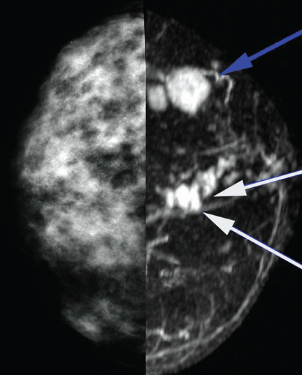
MRI to assess extent of disease (Figure 1) showed two enhancing lesions with irregular borders in the region of the proven cancer. The MRI enhancement kinetics of the lesions were consistent with malignancy. MRI also showed several additional, unsuspected, small, irregular lesions in the 12-o’clock region.
On the basis of these findings, a second ultrasonographic examination of the right breast was carried out, targeting the 12-o’clock region. One of the MRI-detected lesions was located, and biopsy showed invasive breast cancer of the same cell type as the palpable mass. With this evidence of multiple malignant lesions in the same breast, it was concluded that breast-conserving surgery would not be feasible. The patient underwent mastectomy with pathologic confirmation of the MRI findings.
Comment. This case demonstrates how breast MRI, when used appropriately, can lead to objective pathologic results that support the clinical decision to perform a mastectomy rather than breast conservation therapy.
- American Cancer Society. Breast cancer facts and figures 2008–2009. Atlanta: American Cancer Society. http:/www.cancer.org/docroot/STT/content/STT_1x_Breast_Cancer_Facts_Figures_2007-2008_08.asp. Accessed 10/9/2009.
- Tabar L, Fagerberg G, Chen HH, et al. Efficacy of breast cancer screening by age: new results from the Swedish Two-County Trial. Cancer 1995; 75:2507–2517.
- Vachon CM, van Gils CH, Sellers TA, et al. Mammographic density, breast cancer risk and risk prediction. Breast Cancer Res 2007; 9:217.
- Warren R. Hormones and mammographic breast density. Maturitas 2004; 49:67–78.
- Carney PA, Miglioretti DL, Yankaskas BC, et al. Individual and combined effects of age, breast density, and hormone replacement therapy use on the accuracy of screening mammography. Ann Intern Med 2003; 138:168–175.
- Pisano ED, Gatsonis C, Hendrick RE, et al. Diagnostic performance of digital versus film mammography for breast-cancer screening. N Engl J Med 2005; 53:1773–1783.
- Pisano ED, Hendrick RE, Yaffe MJ, et al. Diagnostic accuracy of digital versus film mammography: exploratory analysis of selected population subgroups in DMIST. Radiology 2008; 246:376–383.
- Barlow WE, Lehman CD, Zheng Y, et al. Performance of diagnostic mammography for women with signs or symptoms of breast cancer. J Natl Cancer Inst 2002; 94:1151–1159.
- Berg WA, Blume JD, Cormack JB, et al. Combined screening with ultrasound and mammography vs mammography alone in women at elevated risk of breast cancer. JAMA 2008; 299:2151–2163.
- Heywang SH, Hahn D, Schmid H, et al. MR imaging of the breast using gadolinium-DTPA. J Comput Assist Tomogr 1986; 10:199–204.
- Heywang-Kobrunner SH, Viehweg P, Heinig A, Kuchler CH. Contrast-enhanced MRI of the breast: accuracy, value, controversies, solutions. Eur J Radiol 1997; 24:94–108.
- Lee CH. Problem solving MR imaging of the breast. Radiol Clin North Am 2004; 42:919–934.
- Heywang SH, Wolf A, Pruss E, Hilbertz T, Eiermann W, Permanetter W. MR imaging of the breast with Gd-DTPA: use and limitations. Radiology 1989; 171:95–103.
- Gilles R, Guinebretière JM, Lucidarme O, et al. Nonpalpable breast tumors: diagnosis with contrast-enhanced subtraction dynamic MR imaging. Radiology 1994; 191:625–631.
- Fobben ES, Rubin CZ, Kalisher L, Dembner AG, Seltzer MH, Santoro EJ. Breast MR imaging with commercially available techniques: radiologic-pathologic correlation. Radiology 1995; 196:143–152.
- Müller-Schimpfle M, Ohmenhaüser K, Stoll P, Dietz K, Claussen CD. Menstrual cycle and age: influence on parenchymal contrast medium enhancement in MR imaging of the breast. Radiology 1997; 203:145–149.
- Orel SG, Schnall MD, Powell CM, et al. Staging of suspected breast cancer: effect of MR imaging and MR-guided biopsy. Radiology 1995; 196:115–122.
- Weinstein SP, Orel SG, Heller R, et al. MR imaging of the breast in patients with invasive lobular carcinoma. AJR Am J Roentgenol 2001; 176:399–406.
- Morris EA, Liberman L, Ballon DJ, et al. MRI of occult breast carcinoma in a high-risk population. AJR Am J Roentgenol 2003; 181:619–626.
- Esserman L, Hylton N, Yassa L, Barclay J, Frankel S, Sickles E. Utility of magnetic resonance imaging in the management of breast cancer: evidence for improved preoperative staging. J Clin Oncol 1999; 17:110–119.
- Boetes C, Mus RD, Holland R, et al. Breast tumors: comparative accuracy of MR imaging relative to mammography and US for demonstrating extent. Radiology 1995; 197:743–747.
- Fischer U, Kopa L, Grabbe E. Breast carcinoma: effect of preoperative contrast-enhanced MR imaging on the therapeutic approach. Radiology 1999; 213:881–888.
- Orel SG, Reynolds C, Schnall MD, Solin LJ, Fraker DL, Sullivan DC. Breast carcinoma: MR imaging before re-excisional biopsy. Radiology 1997; 205:429–436.
- Frei KA, Kinkel K, Bonel HM, Lu Y, Esserman LJ, Hylton NM. MR imaging of the breast in patients with positive margins after lumpectomy: influence of the time interval between lumpectomy and MR imaging. AJR Am J Roentgenol 2000; 175:1577–1584.
- Gilles R, Guinebretière JM, Shapeero LG, et al. Assessment of breast cancer recurrence with contrast-enhanced subtraction MR imaging: preliminary results in 26 patients. Radiology 1993; 188:473–478.
- Lewis-Jones HG, Whitehouse GH, Leinster SJ. The role of magnetic resonance imaging in the assessment of local recurrent breast carcinoma. Clinical Radiol 1991; 43:197–204.
- National Comprehensive Cancer Network. Clinical practice guidelines in oncology. Genetic/familial high-risk assessment: breast and ovarian cancer. www.nccn.org/professionals/physician_gls/f_guidelines.asp. Accessed May 4, 2009.
- Morrow M, Freedman G. A clinical oncology perspective on the use of breast MR. Magn Reson Imaging Clin N Am 2006; 14:363–378.
- Schnall M. MR imaging of cancer extent: is there clinical relevance? Magn Reson Imaging Clin N Am 2006; 14:379–381.
A 52-year-old woman presents to your outpatient clinic with a newly palpable marble-sized lump in the upper outer quadrant of her right breast. The mass is firm and non-tender. She performs breast self-examinations regularly and first noticed the mass 3 weeks ago. Your examination confirms a palpable lesion. Annual screening mammograms at another facility have been negative, including the last one 6 months ago. The woman is otherwise in good health.
When she was younger, she had two pregnancies and gave birth to two children, whom she did not breastfeed. She reached menopause at age 49 and has never been on hormone replacement therapy. Neither she nor anyone in her family has had breast cancer, and she has never undergone breast biopsy.
She says that a woman she spoke with in the waiting room, a 2-year breast cancer survivor, told her that her primary cancer had been “finally diagnosed” with magnetic resonance imaging (MRI). This patient is now urging you to order an MRI. What should you do?
DIFFERENT IMAGING TESTS FOR DIFFERENT INDICATIONS
Different imaging tests are indicated in different situations. Screening mammography is the standard of care for women who have no signs or symptoms of breast cancer. When a screening examination shows abnormal or equivocal findings or when a patient presents with symptoms (eg, a palpable lesion, breast pain, nipple discharge), further characterization with a tailored imaging examination is warranted. Such an examination might include diagnostic mammography, ultrasonography, or MRI.
This article reviews MRI’s role in breast cancer management with respect to the other imaging tests currently in use.
MAMMOGRAPHY IS THE SCREENING TEST OF CHOICE
Screening mammography, as distinguished from diagnostic mammography, is an x-ray examination of breast tissue that obtains high-quality images while using a very low dose of ionizing radiation. It is performed with the breast tissue compressed to optimize image quality. The examination time is short, usually 15 to 20 minutes. No contrast agents are used. The malignant lesions it detects differ from normal fibroglandular tissue in their x-ray attenuation, appearing as asymmetric soft-tissue densities, architectural distortion, masses, or abnormal calcifications.
The American Cancer Society1 recommends mammography as the method of choice to screen for nonpalpable, clinically occult breast cancers in women over age 40 and in younger women with certain risk factors.
The rationale for screening mammography is supported by evidence of significant reductions in death rates from breast cancer in patients who undergo routine mammographic screening. Tabar et al,2 in the Swedish Two-County Trial, found a 30% lower rate of death from breast cancer in women ages 40 to 74 who were invited to undergo screening: the reduction was 34% for women age 50 to 74 and 12% for women age 40 to 49. The authors attributed the smaller benefit in the younger group to a tendency toward more rapid tumor progression in that age group.
Mammography is somewhat less sensitive and specific in women with dense breasts,3 in younger women, and in women on hormone replacement therapy.4 For example, a recent population-based study of seven mammography registries in the United States5 reported that the sensitivity of mammography for detecting breast cancer, adjusted for breast density and age, ranged from 63% for extremely dense breast tissue to 87% for entirely fatty breast tissue, and from 69% for women age 40 to 44 to 83% for women age 80 to 89. The adjusted specificities ranged from 89% for extremely dense breast tissue to 97% for entirely fatty breast tissue. The adjusted specificities in women not on hormone replacement therapy ranged from 91% for those age 40 to 44 to 94% for those age 80 to 89. In women on hormone replacement therapy, the adjusted specificity was about 92% for all ages taken together.
Digital is often better than film
Screening mammography has improved with the development of digital technology,6 in which images are acquired directly from an x-ray-sensitive solid-state receptor, without film. This process differs from digital computer-aided detection techniques, in which conventional analog films are optically scanned, generating a secondary digital image, which is then used for subsequent computerized analysis.
In 2005, the first large national trial comparing digital mammography with conventional mammography—the Digital Mammographic Imaging Screening Trial6—showed that digital mammography held a statistically significant advantage over conventional film-screen mammography in three subgroups7:
- Women under age 50
- Women with radiographically dense breast tissue
- Premenopausal or perimenopausal women. It is hoped that these encouraging results will be confirmed and extended by other trials now in progress.
DIAGNOSTIC MAMOGRAPHY FOR FURTHER WORKUP
Screening mammography yields findings of uncertain significance or frank concern in roughly 1 out of 10 examinations. In these cases, the examination is considered to be incomplete. If the finding is of unclear significance, previous mammograms, if available, may reveal whether the finding has remained the same or changed over time. If a worrisome change has occurred or if no prior films can be obtained, a diagnostic study with additional imaging must be carried out so that the radiologist can decide if a lesion is actually present.
Diagnostic mammography is a tailored examination that may include special projections to better visualize a specific region of concern, spot-compression views to disperse dense breast tissue, or magnification views to characterize microcalcifications. In cases of known breast cancer, diagnostic mammography helps detect additional foci of cancer in the same or in the contralateral breast.8
DIRECTED ULTRASONOGRAPHY TO EVALUATE REGIONS OF CONCERN
While ultrasonography is not part of the standard breast cancer screening protocol,9 directed or “targeted” breast ultrasonography is routinely used in the diagnostic workup to evaluate particular regions of concern. Ultrasonography is used in combination with diagnostic mammography to evaluate mammographic masses, palpable lumps, asymmetric tissue, and architectural distortions.
Breast ultrasonography can usually distinguish cystic lesions from solid lesions, and it is used to guide core biopsy or fine needle aspiration of suspicious breast lesions. It is relatively inexpensive, widely available, and reliable when performed by a skilled and knowledgeable operator.
HOW MRI WORKS
Magnetic resonance imaging takes advantage of the magnetic properties of hydrogen nuclei (protons) in breast tissue. A small fraction of the protons in the patient are brought into alignment with a strong magnetic field within the MRI scanner. Then, the protons are exposed to a brief pulse of radiofrequency energy, which displaces their magnetic vectors. As the protons “relax” and realign along the applied magnetic field, energy is released. This energy, the electromagnetic magnetic resonance signal, is detected and electronically processed to construct an image, exploiting the different “relaxation times” of the different tissues in the breast to generate image contrast.
A standard breast MRI examination requires an intravenous paramagnetic contrast agent, usually a gadolinium chelate, to increase the sensitivity of the study. Gadolinium-based contrast material causes shortening of the T1 relaxation time of tissues in which the contrast agent accumulates, thereby increasing signal intensity (or “enhancement”) in those tissues.
Contrast enhancement may occur in malignant tissues with defective or “leaky” capillaries, but it also can occur in benign tissues, such as normal lymph nodes or benign proliferative processes. Thus, the finding of contrast enhancement does not by itself establish the diagnosis of breast cancer.
The patient must remain still
The patient is positioned prone for about 30 to 40 minutes inside the MRI scanner with the breasts encompassed by specially designed imaging coils, which maximize the signal strength and achieve high spatial resolution. The prone position also minimizes motion of breast tissue and transmitted physiologic motions, further ensuring good image quality.
Contrast enhancement over time
To display how contrast enhancement resolves over time, a series of scans must be obtained. First, a baseline scan is recorded. Then, the contrast material is given, and multiple postcontrast scans are obtained at equally spaced time intervals, typically 1 to 1.5 minutes apart. Usually five to seven postcontrast scans are recorded. During this time, the patient must continue to lie still without moving.
For each individual volume element (voxel) of breast tissue, which may measure 1 mm3 or less, a curve representing contrast enhancement vs time can be constructed. Such curves tend to show one of three typical trajectories or patterns, known as “washout,” “plateau,” and “progressive.” With additional postprocessing, these three contrast enhancement patterns are reduced to color coding and are mapped onto the gray-scale MRI image in the form of a color overlay, so that overall enhancement patterns in both breasts can be discerned at a glance by the radiologist.
These enhancement patterns initially were believed to be reliable indicators of malignant and benign conditions, but further experience has shown considerable overlap of the enhancement patterns between benign and malignant tissues. Thus, the diagnostic value of enhancement patterns is limited. As a rule of thumb, the washout pattern of enhancement (rapid uptake of contrast material followed by rapid washout) is thought to indicate malignancy in 60% to 70% of lesions that are suspicious in other respects.
Abnormal contrast enhancement of the suspicious region must be considered along with morphologic features, the degree of enhancement in adjacent normal-appearing tissue, and the correlation with mammographic or ultrasonographic findings.
Better for invasive ductal carcinoma than invasive lobular carcinoma or ductal carcinoma in situ
At present, we have no foolproof method of diagnosing cancer by MRI alone, though in many cases invasive ductal carcinoma can be predicted with a high degree of confidence. The accuracy of breast MRI is lower for “non-mass-like” enhancement, as is often seen in invasive lobular carcinoma and ductal carcinoma in situ.
Contraindications, problems
MRI for breast cancer evaluation is contraindicated in women with cardiac pacemakers, implanted neurostimulators, and certain older models of aneurysm clips and cardiac prosthetic valves. However, this is becoming less of a problem as MRI-compatible devices of recent design become more prevalent. To ensure safety, patients should complete a screening questionnaire for ferromagnetic devices before they are allowed to undergo breast MRI.
Claustrophobia may preclude an MRI study, but this is less of an issue now, as the newer “short-bore” magnet designs reduce the sensation of confinement.
A problem of increasing importance today is patient obesity: obese patients may not fit into the MRI scanner. So-called “open” MRI scanners are not a good alternative, as they cannot provide the high-resolution images of uniform quality required for breast MRI.
Another factor affecting the use of MRI for breast cancer diagnosis is the limited number of facilities that offer it. Head, spine, and orthopedic MRI services are more widely available.
Cost may be an issue. A breast MRI evaluation costs about 10 times more than screening mammography and may not be covered by health insurance, although coverage for this indication appears to be improving gradually.
MRI IS SENSITIVE, BUT NOT SO SPECIFIC
The role of MRI in evaluating breast disease has been studied and debated since contrast-enhanced breast MRI was introduced in 1985.10 Interest has grown steadily as evidence of its usefulness has accumulated. Improvements in MRI scanners have included better image resolution, dedicated breast coils, and rapid dynamic contrast-enhanced imaging.
The overall sensitivity of MRI for breast cancer is relatively high, with estimates ranging from 85% to 100%.11 In invasive ductal carcinoma, its sensitivity approaches 100%.12 Sensitivities for invasive lobular carcinoma and ductal carcinoma in situ are lower and not yet well defined.
In contrast, MRI’s specificity for breast cancer is much more variable, ranging from 37% to 100%. The discrepancies among estimates of specificity are attributed to multiple confounding methodologic factors in the studies to date, such as differences in imaging protocols, patient selection criteria, patient ages, interpretation criteria, and the level of experience of the interpreting radiologist.12
False-positive results may be caused by benign conditions such as fibroadenomas, intramammary lymph nodes, proliferative and nonproliferative fibrocystic changes, and mastitis, as well as by radial scars, atypical ductal hyperplasia, and lobular carcinoma in situ.13–15 In premenopausal women, the menstrual cycle may bring about regional physiologic variation in enhancement of the normal breast parenchyma, which may either simulate the appearance of a lesion or obscure a true lesion.16
Thus, breast MRI may detect cancer that is occult to mammography, but it also carries the risk of worrisome incidental findings that may only be resolved by biopsy. Such uncertain findings are troubling for both the radiologist and the patient when mammography, ultrasonography, and the physical examination are all normal. Clearly, breast MRI cannot be counted on to reassure the “worried well” patient.
MRI is not for screening in the general population
While its high sensitivity for invasive ductal carcinoma17 would seem to make breast MRI attractive for breast cancer screening, it has the disadvantages of lower sensitivity for invasive lobular carcinoma and ductal carcinoma in situ,17,18 as well as the potential to raise suspicions of breast cancer that may be difficult to resolve. For these reasons, MRI is not suitable for routine breast cancer screening in asymptomatic women, although it is recommended for patents in some high-risk groups, as we discuss later.
Data from the Memorial Sloan-Kettering Cancer Center suggest that MRI can detect mammographically occult breast cancer in high-risk populations.19 This study evaluated 367 women at high risk (ie, with a personal history of breast cancer, lobular carcinoma in situ, or atypia, or with a family history of breast cancer). Biopsy was recommended in 64 (17%) of the women on the basis of MRI findings. Biopsy revealed cancer in 14 (24%) of 59 women who underwent biopsy. Subgroup analysis further suggested a 50% positive predictive value of biopsy based on MRI findings in women with both a positive family history and a personal history of breast cancer.
Further studies of breast MRI for screening high-risk populations are under way in North America and Europe.
CLINICAL APPLICATIONS OF MRI OF THE BREAST
MRI has been shown to be useful in:
- Staging biopsy-proven primary breast carcinoma
- Detecting an occult primary breast cancer in a patient with proven axillary node involvement but negative results on mammography and ultrasonography
- Ascertaining the extent of disease after lumpectomy with positive margins or close margins
- Investigating suspected pectoralis muscle invasion
- Assessing response to chemotherapy, including preoperative chemotherapy
- Looking for suspected recurrent disease, such as in a postsurgical scar
- A compelling clinical presentation with negative or equivocal imaging results
- Problem solving, ie, workup of uncertain imaging findings that could not be resolved even after special mammographic and ultrasonographic techniques were used
- Needle localization and guided biopsy
- Known or suspected rupture of breast implants
- Screening patients with certain well-defined risk factors for breast cancer.
The current standard of practice does not support the use of MRI to replace problem-solving mammography and ultrasonography. A negative MRI study does not preclude biopsy of a suspicious lesion found with mammography or directed ultrasonography.
Lesion characterization and staging
Surgical options for treating breast cancer are breast-conserving surgery and mastectomy, taking into account the tumor size, multifocality or multicentricity, local extent vs distant spread, nodal status, and patient preference. Studies have shown that MRI is more accurate than mammography and ultrasonography in defining the extent of tumor burden as characterized by tumor size and multifocality or multicentricity.20,21
Preoperative MRI also has been shown to change therapeutic decisions when additional disease was detected and then proven by image-guided biopsy.19 In a study by Fischer et al22 in 336 women with breast cancer, MRI led to a change in therapy in 19.6% of patients by demonstrating unsuspected multifocal or multicentric ipsilateral lesions or contralateral carcinomas. In all cases, a confirming tissue diagnosis, either before or after MRI, was obtained before surgery. Given the potential for false-positive findings on breast MRI, biopsy of newly detected suspicious lesions is generally necessary before mastectomy is contemplated.
MRI in the follow-up assessment
After excisional biopsy, MRI may help determine the presence or absence of residual tumor if there are positive or close margins shown by surgical pathology, or if residual microcalcifications persist on the postbiopsy mammogram.23 The time between surgical biopsy and follow-up MRI affects the sensitivity of MRI for residual tumor. Frei et al24 reported a sensitivity of 89% to 94% when imaging was done at least 28 days after excision.21
MRI also is useful in identifying and differentiating tumor recurrence from postsurgical or postradiation scar when conventional imaging is indeterminate.25 In a study of 45 women with suspected tumor recurrence after lumpectomy, with or without radiotherapy and chemotherapy, Lewis-Jones et al26 reported a sensitivity of 100% and a specificity of 94% for MRI in detecting new tumor vs posttreatment fibrosis.26
Inflammatory changes in the breast tissue after surgery and radiation therapy limit the accuracy of MRI. Tissue enhancement can be seen in the operative bed for up to 6 months after surgery and for up to 24 months after radiation therapy. In general, local tumor recurrence appears after this interval. Therefore, the postsurgical timing of the MRI examination is important.
Problem solving
At times, mammographic findings are unclear as to whether a suspected lesion is truly present, often because of the radiographic density of fibroglandular breast tissue. In some cases, a lesion’s morphology is indeterminate for malignancy. These equivocal findings can usually be resolved with the combined use of tailored mammographic views, as noted above (eg, magnification or compression views), and directed ultrasonography.
If the findings are still inconclusive after this additional workup, MRI may be useful. Newer, improved MRI scanners can show structures as small as 0.5 mm, which helps the radiologist discern lesion morphology. Moreover, contrast-enhanced and temporally resolved imaging provides estimates of spatially localized enhancement patterns and kinetics, which in turn may offer clues as to whether a lesion is benign or malignant.
Screening patients at high risk
The National Comprehensive Cancer Network27 currently recommends screening with both mammography and MRI starting at age 20 to 25 for women at high risk for hereditary breast cancer and ovarian cancer. Risk factors include the following:
- A known BRCA1 or BRCA2 mutation in the patient or a family member
- A personal history of breast cancer, with two or more close blood relatives with breast or epithelial ovarian cancer at any age
- A close male blood relative with breast cancer
- A personal history of epithelial ovarian cancer
- Being in an ethnic group with a higher frequency of deleterious mutations (eg, Ashkenazi Jews)
- Mutations in p53 (Li-Fraumeni syndrome) or PTEN (Cowden syndrome).
MRI IN THE PREOPERATIVE EVALUATION: THE DEBATE
Numerous reports have shown that MRI can detect additional foci of breast cancer in a substantial number of women with a new diagnosis of breast cancer. While some argue that detecting these additional lesions should improve outcomes after the first operation and, hopefully, lead to lower rates of recurrence, the long-term consequences of MRI-directed changes in treatment have not been fully studied. Below is a summary of the arguments both against and for the use of breast MRI in staging.
The argument against preoperative MRI
Mastectomy was the routine treatment for breast cancer into the 1980s. The arrival of breast conservation surgery combined with radiation therapy offered major advantages with similarly low recurrence rates. Based on the results of controlled clinical trials with mortality as the end point, breast conservation therapy and mastectomy confer equivalent risk to the patient. Any increase in the rate of mastectomy prompted by MRI findings would represent a setback in the standard of care. And since radiation therapy is presumed to eradicate or delay progression of residual disease in most women who undergo conservation therapy, preoperative MRI would have little or no impact on rates of recurrence or death. Thus, MRI should not be used routinely in the workup of new breast cancers.28
The argument for preoperative MRI
The upper threshold amount of residual disease that can be eradicated by radiation therapy is not yet well established. There are as yet no MRI criteria for assessing the likelihood of standard treatment failure in individual patients with multifocal or multicentric disease, or with occult cancer in the contralateral breast. Although the rate of recurrence after breast conservation is low, it is not zero, and each patient should be offered the best possible chance for successful treatment. Detecting widespread disease can obviate inappropriate attempts at conservation, in which both lumpectomy with positive margins and re-excision with positive margins are carried out before the full extent of the disease burden is understood. Knowledge of the extent of disease at presentation will help the patient to make a more informed decision when presented with treatment options. A staging MRI examination showing only a single cancer lesion may permit the patient to choose conservation therapy with a high degree of confidence that no macroscopic disease will be missed at surgery.29
Challenges for future clinical trials
These issues will not be easy to resolve. Definitive answers can only come from controlled clinical trials with mortality as the end point, but for the data from these trials to be useful, the trials must use standardized MRI technique and interpretation criteria. Such standardization has yet to be accomplished.
In the absence of such guidance, it seems reasonable to use MRI for staging within the known limitations of the technique and with secure histologic confirmation whenever widespread disease is suspected from the MRI findings. In this way, the patient and her surgeon can select a treatment plan based on the most realistic assessment of disease burden.
CASE RESOLVED
MRI to assess extent of disease (Figure 1) showed two enhancing lesions with irregular borders in the region of the proven cancer. The MRI enhancement kinetics of the lesions were consistent with malignancy. MRI also showed several additional, unsuspected, small, irregular lesions in the 12-o’clock region.
On the basis of these findings, a second ultrasonographic examination of the right breast was carried out, targeting the 12-o’clock region. One of the MRI-detected lesions was located, and biopsy showed invasive breast cancer of the same cell type as the palpable mass. With this evidence of multiple malignant lesions in the same breast, it was concluded that breast-conserving surgery would not be feasible. The patient underwent mastectomy with pathologic confirmation of the MRI findings.
Comment. This case demonstrates how breast MRI, when used appropriately, can lead to objective pathologic results that support the clinical decision to perform a mastectomy rather than breast conservation therapy.
- American Cancer Society. Breast cancer facts and figures 2008–2009. Atlanta: American Cancer Society. http:/www.cancer.org/docroot/STT/content/STT_1x_Breast_Cancer_Facts_Figures_2007-2008_08.asp. Accessed 10/9/2009.
- Tabar L, Fagerberg G, Chen HH, et al. Efficacy of breast cancer screening by age: new results from the Swedish Two-County Trial. Cancer 1995; 75:2507–2517.
- Vachon CM, van Gils CH, Sellers TA, et al. Mammographic density, breast cancer risk and risk prediction. Breast Cancer Res 2007; 9:217.
- Warren R. Hormones and mammographic breast density. Maturitas 2004; 49:67–78.
- Carney PA, Miglioretti DL, Yankaskas BC, et al. Individual and combined effects of age, breast density, and hormone replacement therapy use on the accuracy of screening mammography. Ann Intern Med 2003; 138:168–175.
- Pisano ED, Gatsonis C, Hendrick RE, et al. Diagnostic performance of digital versus film mammography for breast-cancer screening. N Engl J Med 2005; 53:1773–1783.
- Pisano ED, Hendrick RE, Yaffe MJ, et al. Diagnostic accuracy of digital versus film mammography: exploratory analysis of selected population subgroups in DMIST. Radiology 2008; 246:376–383.
- Barlow WE, Lehman CD, Zheng Y, et al. Performance of diagnostic mammography for women with signs or symptoms of breast cancer. J Natl Cancer Inst 2002; 94:1151–1159.
- Berg WA, Blume JD, Cormack JB, et al. Combined screening with ultrasound and mammography vs mammography alone in women at elevated risk of breast cancer. JAMA 2008; 299:2151–2163.
- Heywang SH, Hahn D, Schmid H, et al. MR imaging of the breast using gadolinium-DTPA. J Comput Assist Tomogr 1986; 10:199–204.
- Heywang-Kobrunner SH, Viehweg P, Heinig A, Kuchler CH. Contrast-enhanced MRI of the breast: accuracy, value, controversies, solutions. Eur J Radiol 1997; 24:94–108.
- Lee CH. Problem solving MR imaging of the breast. Radiol Clin North Am 2004; 42:919–934.
- Heywang SH, Wolf A, Pruss E, Hilbertz T, Eiermann W, Permanetter W. MR imaging of the breast with Gd-DTPA: use and limitations. Radiology 1989; 171:95–103.
- Gilles R, Guinebretière JM, Lucidarme O, et al. Nonpalpable breast tumors: diagnosis with contrast-enhanced subtraction dynamic MR imaging. Radiology 1994; 191:625–631.
- Fobben ES, Rubin CZ, Kalisher L, Dembner AG, Seltzer MH, Santoro EJ. Breast MR imaging with commercially available techniques: radiologic-pathologic correlation. Radiology 1995; 196:143–152.
- Müller-Schimpfle M, Ohmenhaüser K, Stoll P, Dietz K, Claussen CD. Menstrual cycle and age: influence on parenchymal contrast medium enhancement in MR imaging of the breast. Radiology 1997; 203:145–149.
- Orel SG, Schnall MD, Powell CM, et al. Staging of suspected breast cancer: effect of MR imaging and MR-guided biopsy. Radiology 1995; 196:115–122.
- Weinstein SP, Orel SG, Heller R, et al. MR imaging of the breast in patients with invasive lobular carcinoma. AJR Am J Roentgenol 2001; 176:399–406.
- Morris EA, Liberman L, Ballon DJ, et al. MRI of occult breast carcinoma in a high-risk population. AJR Am J Roentgenol 2003; 181:619–626.
- Esserman L, Hylton N, Yassa L, Barclay J, Frankel S, Sickles E. Utility of magnetic resonance imaging in the management of breast cancer: evidence for improved preoperative staging. J Clin Oncol 1999; 17:110–119.
- Boetes C, Mus RD, Holland R, et al. Breast tumors: comparative accuracy of MR imaging relative to mammography and US for demonstrating extent. Radiology 1995; 197:743–747.
- Fischer U, Kopa L, Grabbe E. Breast carcinoma: effect of preoperative contrast-enhanced MR imaging on the therapeutic approach. Radiology 1999; 213:881–888.
- Orel SG, Reynolds C, Schnall MD, Solin LJ, Fraker DL, Sullivan DC. Breast carcinoma: MR imaging before re-excisional biopsy. Radiology 1997; 205:429–436.
- Frei KA, Kinkel K, Bonel HM, Lu Y, Esserman LJ, Hylton NM. MR imaging of the breast in patients with positive margins after lumpectomy: influence of the time interval between lumpectomy and MR imaging. AJR Am J Roentgenol 2000; 175:1577–1584.
- Gilles R, Guinebretière JM, Shapeero LG, et al. Assessment of breast cancer recurrence with contrast-enhanced subtraction MR imaging: preliminary results in 26 patients. Radiology 1993; 188:473–478.
- Lewis-Jones HG, Whitehouse GH, Leinster SJ. The role of magnetic resonance imaging in the assessment of local recurrent breast carcinoma. Clinical Radiol 1991; 43:197–204.
- National Comprehensive Cancer Network. Clinical practice guidelines in oncology. Genetic/familial high-risk assessment: breast and ovarian cancer. www.nccn.org/professionals/physician_gls/f_guidelines.asp. Accessed May 4, 2009.
- Morrow M, Freedman G. A clinical oncology perspective on the use of breast MR. Magn Reson Imaging Clin N Am 2006; 14:363–378.
- Schnall M. MR imaging of cancer extent: is there clinical relevance? Magn Reson Imaging Clin N Am 2006; 14:379–381.
KEY POINTS
- Whether rates of death and local recurrence are reduced when additional breast tumors found by MRI are treated remains to be seen.
- MRI contrast enhancement occurs in many cancers, but it may occur for benign reasons; thus, the finding of contrast enhancement does not establish the diagnosis of breast cancer.
- The National Comprehensive Cancer Network currently recommends screening with both mammography and MRI starting at age 20 to 25 for women at high risk of hereditary breast cancer and ovarian cancer.
- A breast MRI evaluation costs about 10 times more than screening mammography and may not be covered by health insurance, but coverage for this indication appears to be improving gradually.
