User login
Cloud-Based Patient Data Holds Allure, Risks
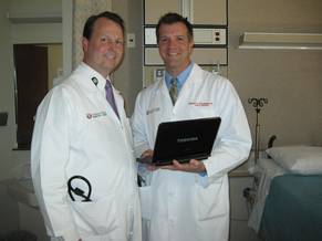
For years, hospitalists at University of Utah Healthcare have yearned for a fast, elegant way to share information with referring physicians.
When patients are referred from nonaffiliated practices, "they arrive with medical records burned onto a CD," said Dr. Mike Strong, a hospitalist and the chief medical information officer for the university’s hospitals and clinics, which are based in Salt Lake City.
"One of the big frustrations is the referring physicians say ‘we love the care that you give us, but it feels like our patients are going into a black hole,’ " Dr. Strong said. "We take care of them, and the patients come back, but the physicians feel that they don’t see the whole story."
For Utah Healthcare and others grappling with similar problems, the solution may lie in the "cloud," medical IT experts say. With cloud technology, electronic medical records (EMRs) and images, and the programs used to access them, can be stored and processed on the Web.
The cloud can be likened to a public utility. Under the cloud model, computing power, like electricity, draws from the equivalent of a grid. Giant server "farms" – such as those now supported by Amazon.com and other cloud vendors – can provide not only storage but also processing power, making applications and information accessible at high speed to anyone with a computer or even a smartphone.
"A CD takes 2 or 3 days to arrive," Dr. Strong said. If a referring hospital were able to upload images into a cloud, and we would have the records up and running before a patient even gets here, it would be great, he said.
Cloud-based services are already widespread in business and personal use – think Gmail, Facebook, Google Docs, or online banking. Proponents of cloud in health care suggest that its flexibility (hospitals can use as little or as much storage and computing capacity as they need), its processing power and speed, which can help smaller hospitals perform sophisticated modeling programs the way large ones do and allowing them to run the kind of algorithms that would tie up or crash their own servers.
Those less keen on the cloud cite its vulnerability to crashes and privacy violations.
Freedom and Flexibility
Cloud applications promise greater physical freedom to hospitalists, who need to gather information on patients from a wide variety of sources and to retrieve or deliver information from wherever they happen to be in the hospital. And hospitalists, members of one of the younger-demographic medical specialties, are hardly technology-averse.
"As a practicing hospitalist working in a system that has computer-order entry and electronic records, I live in the IT world," said Dr. Robert Pendleton, Dr. Strong’s colleague at University of Utah and codirector of its hospitalist program. EMRs and portable devices "have changed my work flow. When we were paper chart–based, I had to go to the patient’s unit." Now, Dr. Pendleton said, "we pull up data as we’re rounding, and I come back to my office and put in my notes electronically."
A shift to the cloud and mobile technologies could eliminate the trip back to the office or, better still, offer hospitalists more intuitive approaches to information sharing.
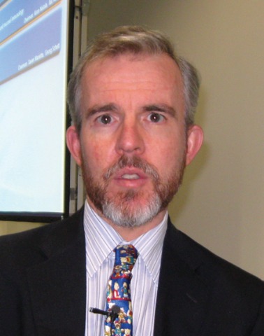
"Health care is about teamwork," says Dr. Jonathan Bloor, the cofounder, along with fellow surgeon Jonathon Shaw of DocCom, a U.K.-based company that sells cloud applications modeled after such social networks as Facebook, as an alternative to traditional e-mail in hospitals. Dr. Bloor and Dr. Shaw developed the platform while working together at University Hospitals Bristol, part of the U.K.’s National Health Service, where they noticed gaps in team communication that they felt threatened patient care.
Yet while hospitalists’ work flow could greatly benefit from Web-based applications designed for physicians, cloud dependence has a flip side: The April crash of Internet behemoth Amazon’s cloud server was a stark and devastating reminder that the cloud is hardly invulnerable. Some high-profile websites that rely on Amazon’s server were left with minimal or no processing power for a week.
Safety or Speed?
With the cloud, security and encryption are also handled offsite, and information is only as secure as an individual user’s password.
This in part is why hospitals like University of Utah have been hesitant to embrace it. Migrating to a cloud model – in which many businesses and institutions, not just hospitals, store information – could collaterally expose hospitals to hackers aiming at banks or other businesses. "If you’re a hacker, are you going to target a hospital system?" Utah Healthcare’s Dr. Strong said. "If we combine storage onto a cloud [used by other types of business], we could get hacked for other reasons."
Last year, when Utah Healthcare needed to create a way to move information between its two electronic medical records systems (a legacy of the hospital’s decade-old acquisition of another health care company), its IT department chose to design a Web-based bridge – a cloud-style technology. But the server hosting the bridge is the hospital’s own. In May, Utah introduced a portal allowing referring physicians to have read-only access to records on its dedicated servers, in effect approximating some of what a cloud can do, except keeping it all in house. Cloud "is still an in-vogue term," Dr. Pendleton said. "But though the majority of health care systems have Web-based elements, they still don’t capitalize on the concept in terms of the sharing and dissemination of patient information."
A few are taking the plunge anyway. This spring, the University of California San Diego Medical Center’s trauma department began using a cloud system to move radiology files, making it one of the first major hospitals in the country to switch from CDs.
The reason, said trauma surgeon Jeanne Lee, is that UC-San Diego receives referrals from trauma centers at two smaller hospitals that can take from 45 minutes to 2 hours to complete. "It’s a lot of wasted time," Dr. Lee said, which could be better used if the hospital had the radiology information before the patient arrives.
Dr. Lee said that while there had been a debate at San Diego about whether to move to a cloud model, "any system you use is liable to some sort of breach," and so far, "security hasn’t been an issue."
One Password Away
DocCom’s Dr. Bloor said that an increasing number of U.K. hospitals are using cloud-based applications, though not yet for patient records. DocCom’s own products aren’t designed to exchange patient-identifiable information but rather to coordinate teamwork within networks of National Health Service hospitals.
Still, the company aims to produce a platform capable of allowing teams to consolidate and communicate about patients, including the sharing of records – but this is probably years or even a decade before it is likely to be adopted wide scale, Dr. Bloor and Dr. Shaw concede. In the United Kingdom as in the United States, the cloud is a long way from being accepted in a hospital setting as quickly as it has been in business, they said.
One NHS hospital in London has recently begun experimenting with a cloud model for patient records that would allow both clinicians and patients to access them from Internet-connected devices. But the "records" being used are simulations, and the project has attracted some controversy – mostly over privacy and security – even before its official rollout in August.
David Sansom and Brent Hicks, codirectors of clinical IT solutions at the Cleveland Clinic, say that despite concerns about patient information on the cloud, there’s already more of it there – at least in the United States – than people realize. They point to Surescripts, one of the country’s largest e-prescription networks, which uses a Web-based system for its 220 million member records on the cloud.
The Cleveland Clinic’s innovations department is currently working on a number of inventive cloud-based technologies, Mr. Hicks and Mr. Sansom say, including some that both supply data to clinicians to aid patient care and feed clinical data back into models. Recently, Cleveland Clinic’s innovations department spun off a company called Explorys, whose cloud-based product aggregates data on 10 million patients for use in population-based studies – information that, Explorys insists, is HIPAA secure.
While Mr. Hicks and Mr. Sansom are strong advocates for using the cloud in hospital settings, neither dismisses the privacy and security concerns the technology raises.
Hospitals must have the capacity to cache months worth of data in house as a safeguard against Internet connection failures, they say, and the cloud itself can become another point of failure, as the Amazon crash showed. Meanwhile, "HIPAA requirements were never designed for cloud-based computing," Mr. Sansom said. HIPAA requires two methods of identification, which is still not secure enough. When an EMR system is run in house, Mr. Sansom said, someone has to physically come into the hospital, find an unguarded PC, and enter a password to access sensitive patient information. With the cloud, of course, it’s only a password, he points out: "And that’s the problem."
I think Apple’s introduction of iCloud is great news.
Apple’s
use of the word "cloud" should help overcome fears that people may have
of the word, and the most significant impact will be to make "cloud
computing" mainstream. It will become a core service in the same way
that everyone chooses a particular GSM [Global System for Mobile
Communications] provider for their cell phone. Personal computing needs
now demand syncronised content across multiple devices that are backed
up elsewhere. Many people are already doing this but probably do not
realize that they are already using cloud services.
The knock-on effect in health care will be to raise expectations from both health care professionals and patients.
If
they can manage their personal life by synchronising documents on the
cloud to be accessed anywhere, or they can share a calendar with their
friends, then they will soon expect this same level of functionality in
health care.
For clinicians, being able to access and share
content on the move is hugely advantageous. For patients, I can see them
asking that, "If I can share photos with my family, why can’t the
hospital easily share x-rays with my family physician?!"
Interestingly,
there are many health care companies that have already adopted cloud
strategies but are not necessarily marketing them as such, for fear of
creating concerns that people currently have about offsite, cloud-based
data storage.
As far as a health care specific–strategy with
Apple’s iCloud is concerned, then it certainly will be an interesting
space to watch. One of the biggest changes we will all see is the
"consumerisation" of health care, in which more people will start to
manage their health themselves independently through health care apps
that will make it easier and easier to self-diagnose and monitor disease
and manage lifestyle.
This will be an interesting space as it starts to bypass many of the regulatory systems in place for traditional health care.
Dr.
Jonathon Shaw, MD, is a cofounder of DocCom, an early provider of
cloud-based applications and communication services and for hospitals
within the U.K. National Health System.
For years, hospitalists at University of Utah Healthcare have yearned for a fast, elegant way to share information with referring physicians.
When patients are referred from nonaffiliated practices, "they arrive with medical records burned onto a CD," said Dr. Mike Strong, a hospitalist and the chief medical information officer for the university’s hospitals and clinics, which are based in Salt Lake City.
"One of the big frustrations is the referring physicians say ‘we love the care that you give us, but it feels like our patients are going into a black hole,’ " Dr. Strong said. "We take care of them, and the patients come back, but the physicians feel that they don’t see the whole story."
For Utah Healthcare and others grappling with similar problems, the solution may lie in the "cloud," medical IT experts say. With cloud technology, electronic medical records (EMRs) and images, and the programs used to access them, can be stored and processed on the Web.
The cloud can be likened to a public utility. Under the cloud model, computing power, like electricity, draws from the equivalent of a grid. Giant server "farms" – such as those now supported by Amazon.com and other cloud vendors – can provide not only storage but also processing power, making applications and information accessible at high speed to anyone with a computer or even a smartphone.
"A CD takes 2 or 3 days to arrive," Dr. Strong said. If a referring hospital were able to upload images into a cloud, and we would have the records up and running before a patient even gets here, it would be great, he said.
Cloud-based services are already widespread in business and personal use – think Gmail, Facebook, Google Docs, or online banking. Proponents of cloud in health care suggest that its flexibility (hospitals can use as little or as much storage and computing capacity as they need), its processing power and speed, which can help smaller hospitals perform sophisticated modeling programs the way large ones do and allowing them to run the kind of algorithms that would tie up or crash their own servers.
Those less keen on the cloud cite its vulnerability to crashes and privacy violations.
Freedom and Flexibility
Cloud applications promise greater physical freedom to hospitalists, who need to gather information on patients from a wide variety of sources and to retrieve or deliver information from wherever they happen to be in the hospital. And hospitalists, members of one of the younger-demographic medical specialties, are hardly technology-averse.
"As a practicing hospitalist working in a system that has computer-order entry and electronic records, I live in the IT world," said Dr. Robert Pendleton, Dr. Strong’s colleague at University of Utah and codirector of its hospitalist program. EMRs and portable devices "have changed my work flow. When we were paper chart–based, I had to go to the patient’s unit." Now, Dr. Pendleton said, "we pull up data as we’re rounding, and I come back to my office and put in my notes electronically."
A shift to the cloud and mobile technologies could eliminate the trip back to the office or, better still, offer hospitalists more intuitive approaches to information sharing.
"Health care is about teamwork," says Dr. Jonathan Bloor, the cofounder, along with fellow surgeon Jonathon Shaw of DocCom, a U.K.-based company that sells cloud applications modeled after such social networks as Facebook, as an alternative to traditional e-mail in hospitals. Dr. Bloor and Dr. Shaw developed the platform while working together at University Hospitals Bristol, part of the U.K.’s National Health Service, where they noticed gaps in team communication that they felt threatened patient care.
Yet while hospitalists’ work flow could greatly benefit from Web-based applications designed for physicians, cloud dependence has a flip side: The April crash of Internet behemoth Amazon’s cloud server was a stark and devastating reminder that the cloud is hardly invulnerable. Some high-profile websites that rely on Amazon’s server were left with minimal or no processing power for a week.
Safety or Speed?
With the cloud, security and encryption are also handled offsite, and information is only as secure as an individual user’s password.
This in part is why hospitals like University of Utah have been hesitant to embrace it. Migrating to a cloud model – in which many businesses and institutions, not just hospitals, store information – could collaterally expose hospitals to hackers aiming at banks or other businesses. "If you’re a hacker, are you going to target a hospital system?" Utah Healthcare’s Dr. Strong said. "If we combine storage onto a cloud [used by other types of business], we could get hacked for other reasons."
Last year, when Utah Healthcare needed to create a way to move information between its two electronic medical records systems (a legacy of the hospital’s decade-old acquisition of another health care company), its IT department chose to design a Web-based bridge – a cloud-style technology. But the server hosting the bridge is the hospital’s own. In May, Utah introduced a portal allowing referring physicians to have read-only access to records on its dedicated servers, in effect approximating some of what a cloud can do, except keeping it all in house. Cloud "is still an in-vogue term," Dr. Pendleton said. "But though the majority of health care systems have Web-based elements, they still don’t capitalize on the concept in terms of the sharing and dissemination of patient information."
A few are taking the plunge anyway. This spring, the University of California San Diego Medical Center’s trauma department began using a cloud system to move radiology files, making it one of the first major hospitals in the country to switch from CDs.
The reason, said trauma surgeon Jeanne Lee, is that UC-San Diego receives referrals from trauma centers at two smaller hospitals that can take from 45 minutes to 2 hours to complete. "It’s a lot of wasted time," Dr. Lee said, which could be better used if the hospital had the radiology information before the patient arrives.
Dr. Lee said that while there had been a debate at San Diego about whether to move to a cloud model, "any system you use is liable to some sort of breach," and so far, "security hasn’t been an issue."
One Password Away
DocCom’s Dr. Bloor said that an increasing number of U.K. hospitals are using cloud-based applications, though not yet for patient records. DocCom’s own products aren’t designed to exchange patient-identifiable information but rather to coordinate teamwork within networks of National Health Service hospitals.
Still, the company aims to produce a platform capable of allowing teams to consolidate and communicate about patients, including the sharing of records – but this is probably years or even a decade before it is likely to be adopted wide scale, Dr. Bloor and Dr. Shaw concede. In the United Kingdom as in the United States, the cloud is a long way from being accepted in a hospital setting as quickly as it has been in business, they said.
One NHS hospital in London has recently begun experimenting with a cloud model for patient records that would allow both clinicians and patients to access them from Internet-connected devices. But the "records" being used are simulations, and the project has attracted some controversy – mostly over privacy and security – even before its official rollout in August.
David Sansom and Brent Hicks, codirectors of clinical IT solutions at the Cleveland Clinic, say that despite concerns about patient information on the cloud, there’s already more of it there – at least in the United States – than people realize. They point to Surescripts, one of the country’s largest e-prescription networks, which uses a Web-based system for its 220 million member records on the cloud.
The Cleveland Clinic’s innovations department is currently working on a number of inventive cloud-based technologies, Mr. Hicks and Mr. Sansom say, including some that both supply data to clinicians to aid patient care and feed clinical data back into models. Recently, Cleveland Clinic’s innovations department spun off a company called Explorys, whose cloud-based product aggregates data on 10 million patients for use in population-based studies – information that, Explorys insists, is HIPAA secure.
While Mr. Hicks and Mr. Sansom are strong advocates for using the cloud in hospital settings, neither dismisses the privacy and security concerns the technology raises.
Hospitals must have the capacity to cache months worth of data in house as a safeguard against Internet connection failures, they say, and the cloud itself can become another point of failure, as the Amazon crash showed. Meanwhile, "HIPAA requirements were never designed for cloud-based computing," Mr. Sansom said. HIPAA requires two methods of identification, which is still not secure enough. When an EMR system is run in house, Mr. Sansom said, someone has to physically come into the hospital, find an unguarded PC, and enter a password to access sensitive patient information. With the cloud, of course, it’s only a password, he points out: "And that’s the problem."
Just What the Doctor Didn't Order: More Regs
Shortness of Breath and Fatigue
Appropriate Use of CT in the Emergency Department
Right Ventricular Structure Differs by Patient Age, Sex, Race
Right ventricular mass, volume, and ejection fraction differ significantly according to patient age, sex, and race, according to a prospective imaging study of more than 4,000 healthy people that was reported in Circulation on June 6.
Moreover, the effect of these patient characteristics on the right ventricle is independent of body size, blood pressure level, diabetes status, and other conventional cardiovascular risk factors that are known to be associated with right ventricular morphology, said Dr. Steven M. Kawut of the Penn Cardiovascular Institute at the University of Pennsylvania, Philadelphia, and his associates.
Right ventricular structure and function have been "challenging" to measure with standard echocardiography techniques, and to date studies examining possible determinants of RV morphology have been small, have focused on young adults of homogeneous ethnicity, and have produced mixed findings, the investigators said.
In what they described as "the largest study of RV morphology ever performed," Dr. Kawut and his colleagues used cardiac MRI to assess ventricular morphology in a subset of subjects participating in MESA (Multi-Ethnic Study of Atherosclerosis). Their ancillary study specifically included a large number of subjects (4,123) from six communities across the United States who were of four major ethnicities and who ranged in age from 45 to 84 years.
The investigators found a consistent, approximately 5% decrease in RV mass for every 10-year increase in patient age, with men showing significantly larger age-related decrements in RV mass (1.0 g per decade) than did women (0.8 g per decade). These findings "may be surprising," as previous studies have been all over the map, with reports that aging increases, decreases, or makes no changes in RV mass, the investigators said.
Older age also was associated with smaller RV volume, and again the decreases in volume were greater among men than among women. Increasing age also was associated with increasing right ventricular ejection fraction of approximately 1% per decade (in all ethnicities except white), and decreasing right ventricular stroke volume of approximately 3% per decade.
It is possible that both the loss of cardiac myocytes and diminished quality of cardiac myocytes occur with aging, and may account for these changes, they said.
Sex also was independently associated with RV morphology. Men had approximately 8% higher RV mass than did women, as well as approximately 10% larger RV end-diastolic volume, approximately 4% lower RV ejection fraction, and approximately 7% larger RV stroke volume.
These sex differences "appeared to persist despite adjustment for the respective left ventricular parameter, suggesting that these findings were not solely due to secondary effects from the left ventricle or residual confounding by body size or composition." It seems likely that hormonal influences play a role in these sex-based differences, the researchers added.
"To our knowledge, ours is the first study to examine differences in RV morphology between races and ethnicities," they noted.
Blacks and Chinese Americans had a lower RV mass than did whites, whereas Hispanics had higher RV mass. Blacks had smaller RV end-diastolic volume than did whites, whereas Hispanics had larger RV end-diastolic volume than whites. Similarly, RV stroke volume was smallest in blacks, intermediate in whites, and larger in Hispanics. These differences could be the result of genetic variation among ethnic groups and may explain the poorer outcomes that are noted in blacks who have cardiopulmonary disease.
Based on their findings, Dr. Kawut and his associates devised new normative equations for measures of right ventricular morphology based on patient age, sex, and race. When these new equations were applied to the entire study cohort, they "were effective at indexing right ventricular measures to body size, [which is] important for clinical care and research since the outcomes of patients with congestive heart failure and pulmonary arterial hypertension depend on RV morphology and function," they said.
The new equations must be validated in other populations before they can be used clinically, the investigators noted.
This study was limited in that many subjects in MESA were unable to tolerate cardiac MRI, and many of the results for subjects who did undergo the procedure were "uninterpretable," they added.
The authors had no disclosures.
FROM CIRCULATION
Major Finding: Right ventricular mass, volume, and stroke volume decrease with age, whereas RV ejection fraction increases; these and other parameters of RV structure and function also differ by sex and race.
Data Source: An ancillary study of the prospective, multicenter MESA, in which cardiac MRI was used to assess right ventricular morphology and function in 4,123 adults aged 45-84.
Disclosures: The authors had no disclosures.
Zoledronic Acid Relieves Knee OA Pain and Shrinks Bone Marrow Lesions
LONDON – A single 5-mg infusion of zoledronic acid, a bisphosphonate, in patients with knee osteoarthritis led to significant pain reduction and shrinkage of bone marrow lesions in a randomized, placebo-controlled study with 59 patients.
The zoledronic acid treatment led to an average 15-point drop in pain (on a visual analog scale of 0-100) beyond what occurred in the placebo group, and the active treatment was also linked with an average 170-mm2 reduction in maximal bone marrow lesion (BML) area beyond the placebo-treated patients, which was a cut in BML area of about 37%, compared with the starting BML area, Dr. Graeme Jones said at the annual European Congress of Rheumatology. Dr. Jones visualizes BMLs using MRI knee scans.
"This is the first intervention shown to work on BMLs" in patients with osteoarthritis (OA), said Dr. Jones, professor of rheumatology and epidemiology and head of the musculoskeletal unit at the Menzies Research Institute Tasmania, Hobart, Australia.
"This is exciting for treating existing OA. It is one of the first positive structure modification trials," commented Dr. Philip Conaghan, professor and chairman of musculoskeletal medicine at the University of Leeds (England).
"Results from several studies have linked BMLs with pain and cartilage damage in OA patients. The larger the BML, the faster the cartilage loss and the worse the pain," Dr. Jones said in an interview. Based on studies his group has done, about 20% of BMLs that are associated with knee OA spontaneously enlarge over the course of 3 years, another 20% shrink in size, and about 60% remain the same, he said. Their earlier research findings also showed that BMLs are independently linked with fast progression of OA and the need for knee replacement. "If you reduce BMLs, it should produce good outcomes in patients," he said.
"The next step is to show that treatment with zoledronic acid not only reduces BML size but also slows cartilage loss. Sixty patients followed for 12 months were not enough to assess cartilage. We will need about 400 patients followed for 2 years," Dr. Jones added.
Despite not yet having information on cartilage effects, he said that his results so far have convinced him that treatment with zoledronic acid is reasonable for patients with painful knee OA and BMLs.
"I use it off label. Patients need to know it’s off label, and they [therefore] must be willing to pay for it, but I use it. It’s been shown to work, and nothing else works. Zoledronic acid [Reclast] is available, we know about its safety, and it’s been used for a long time to treat osteoporosis and cancers. If you have OA patients with BMLs, this is something to actively consider for them. Patients with OA have very limited treatment options. This can make a large difference in their pain, and it has long-lasting benefit so it can be given once a year," Dr. Jones said.
He recommended an infusion of 5 mg of zoledronic acid for patients who are at least 50 years old with knee OA that fulfills the American College of Rheumatology clinical criteria, and knee BMLs that are visible on an MRI scan of the affected knee. In his experience, 88% of these knee OA patients have BMLs. Dr. Jones noted that he does not use a maximal BML area threshold for initiating treatment, although in his study the average maximal BML area was about 465 mm2. About one-third of people aged 50 years or older with no clinical evidence of OA also have BMLs, he noted.
The benefits of zoledronic acid that were seen in his study might be a class effect that may be replicated by treatment with another bisphosphonate, but zoledronic acid is more potent than oral bisphosphonates and hence the drug’s beneficial effect on pain and BML shrinkage may exceed the effect that other bisphosphonates might have, he said.
The pain benefit appeared to start wearing off about a year after the zoledronic acid injection. Dr. Jones said that he has a small number of patients whom he has infused a second time, which produced a second round of pain reduction. He has not yet given any OA patients a third dose of the drug.
The 59 patients who were enrolled in the study had an average age of about 60 years, with an average knee pain score of about 52 on the visual analog scale; all patients had BMLs as seen on MRI scans of their affected knees. In all, 31 patients received a 5-mg infusion of zoledronic acid and 28 patients received a placebo infusion. All patients also continued their conventional pain medication regimens. The zoledronic acid infusion was well tolerated: Although infusion reactions were reported in 90% of the patients who received the drug and in 43% of the placebo patients, serious adverse effects occurred in only 19% of the patients who received zoledronic acid and in 4% of the placebo patients.
The Food and Drug Administration has approved zoledronic acid under a number of brand names to prevent or treat osteoporosis in postmenopausal women or patients who are at risk for osteoporosis because they are taking or have taken corticosteroid therapy; to manage Paget disease; and to prevent chemotherapy-induced bone fractures or fractures in patients with multiple myeloma or cancer that has metastasized to the bones from other locations.
The study was funded by Novartis, which markets zoledronic acid (Reclast). Dr. Jones and Dr. Conaghan said that they had no disclosures.
*Correction, 6/6/2011: An earlier version of this story incorrectly stated that Dr. Jones had no disclosures. Dr. Jones has received speaker fees, travel sponsorship, and research support from Novartis and from several other drug companies.
FROM THE ANNUAL EUROPEAN CONGRESS OF RHEUMATOLOGY
Major Finding: A 5-mg infusion of zoledronic acid given to 31 patients with knee OA and associated bone marrow lesions reduced pain by 15 points more on a visual analogue scale than did placebo, and reduced maximal bone marrow lesion area by 170-mm2 after 6 months, compared with patients who received a placebo infusion.
Data Source: A single-center, randomized study with 31 patients who received a zoledronic acid infusion and 28 patients who received a placebo infusion.
Disclosures: The study was funded by Novartis, which markets zoledronic acid. Dr. Jones and Dr. Conaghan said that they had no disclosures.
The MRI Geyser Sign: Acromioclavicular Joint Cysts in the Setting of a Chronic Rotator Cuff Tear
Tenosynovial Giant Cell Tumor of the Thigh: Positron Emission Tomography Findings
BMD and FRAX Need Refinement for Patients With Type 2 Diabetes
Two measures of fracture risk – bone mineral density T score and FRAX score – are clinically useful in patients with type 2 diabetes, even though they tend to underestimate the increased fracture risk in this patient population, according to the results of a large study reported in the June 1 issue of JAMA.
But to be more accurate for older adults with diabetes, these treatment and diagnostic algorithms should undergo "refinements," said Ann V. Schwartz, Ph.D., of the department of epidemiology and biostatistics, University of California, San Francisco, and her associates (JAMA 2011;305:2184-92).
Type 2 diabetes is known to be associated with a higher bone mineral density (BMD) but, paradoxically, also with a higher risk of fracture. There has been concern that established methods for predicting fractures "may not perform adequately in patients with type 2 diabetes," the researchers said.
In what they described as the first study to prospectively examine the relationship between BMD and fractures in older adults with type 2 diabetes, Dr. Schwartz and her colleagues analyzed data from three large, prospective, observational studies that all used radiology reports and radiographs to confirm fracture diagnoses. The three studies also ascertained subjects’ diabetes status.
The Study of Osteoporotic Fractures (SOF) involved 7,926 white women at four U.S. clinical centers who underwent femoral neck-hip BMD measurement and were followed for the occurrence of fractures from 1988 through 2008. The Osteoporotic Fractures in Men Study (MrOS) involved 5,994 men at six U.S. clinical centers who underwent hip BMD measurement and were followed from 2000 through 2009. And the Health, Aging, and Body Composition (Health ABC) study involved 1,523 women and 1,442 men in their 70s, approximately half of whom were white and half black, who underwent hip BMD measurement and were followed from 1997 through 2007.
The FRAX (Fracture Risk Algorithm) scores were calculated for subjects in the SOF and MrOS studies by the World Health Organization (WHO) Collaborating Center for Metabolic Bone Disease, but not for subjects in the Health ABC study.
Dr. Schwartz and her associates found that both low BMD T score and high FRAX score were predictive of hip and nonspinal fracture in patients with diabetes. "However, for a given T score and age, those adults with diabetes had a higher risk of fracture than those without diabetes," they said.
For example, the risk of hip fracture in a woman with diabetes was equivalent to that of a nondiabetic woman with a T score of about 0.5 units lower.
The subjects with diabetes showed higher rates of fracture at any given FRAX score than did subjects who didn’t have diabetes, they noted.
"Interpretation of T score or FRAX score in an older patient with diabetes must take into account the higher fracture risk associated with diabetes. For example, ... a T score in a woman with diabetes is associated with hip fracture risk equivalent to a woman without diabetes with a T score of approximately 0.5 units lower," the investigators said.
"The FRAX score has been incorporated into U.S. guidelines for prevention and treatment of osteoporosis. The FRAX algorithm does not currently include type 2 diabetes as a risk factor for fracture, and our results indicate that use of the FRAX score in patients with diabetes will likely underestimate risk.
"An adjustment of this algorithm for type 2 diabetes seems justified, given the prevalence of diabetes among older adults," Dr. Schwartz and her associates said.
The reason diabetes raises fracture risk even as it appears to protect against loss of BMD is not understood. "Bone strength may be compromised through changes that are not captured with dual-energy x-ray absorptiometry, such as higher levels of advanced glycation end products in bone collagen. More frequent falls in older adults with diabetes could also increase fracture risk for a given BMD," they said.
This study was funded by an investigator-initiated grant from Amgen to Dr. Schwartz. She and her associates also reported receiving support from Novartis, Merck, Pfizer, Nycomed, and Roche.
FROM JAMA
Major Finding: In older adults with diabetes, any given BMD T score or FRAX score carried a higher fracture risk than did the same score in those without diabetes. The risk of hip fracture in a woman with diabetes was equivalent to that of a nondiabetic woman with a T score about 0.5 units lower.
Data Source: Analysis of data from three prospective observational studies involving more than 16,000 older subjects in whom fractures were tracked for about 10 years.
Disclosures: This study was funded by an investigator-initiated grant from Amgen to Dr. Schwartz. She and her associates also reported receiving support from Novartis, Merck, Pfizer, Nycomed, and Roche.
A Closer Look at Role of MRI in SpA
Magnetic resonance imaging is at least as sensitive as other modalities at predicting treatment response in patients with new onset spondyloarthritis, according to data presented on May 25 by Dr. Mikkel Østergaard at the annual European Congress of Rheumatology.
In discussing research that pointed to the usefulness of MRI in managing patients with ankylosing spondylitis (SpA), Dr. Østergaard reviewed the varied manifestations SpA can take on MRI, and how imaging can be a useful tool for predicting the formation of new bone.
“MRI has brought a major improvement in the evaluation and management of patients with spondyloarthritis,” he said. “It allows earlier detection of sacroiliitis than [does] any other modality, and has thereby enabled physicians to diagnose spondyloarthritis up to 5-10 years earlier than they could before.” He added that MRI provides physicians with an objective way to monitor disease activity in SpA.
Dr. Østergaard explained how to use MRI to identify disease in the sacroiliac joints, the spine, and the peripheral joints. “Given the major improvement in evaluation and management of spondyloarthritis patients that MRI has brought, it is very important that rheumatologists are aware of the findings that indicate actual spondyloarthritis and of the advantages of MRI so that they can order the correct examinations,” he said.
“It is important that not only rheumatologists but also radiologists are aware of the large impact of early diagnosis and treatment of patients with SpA concerning pain relief and better function,” he noted. Radiologists should prioritize to do early and adequate MRI examinations in patients with suspected spondyloarthritis. Ordering MRI “at an earlier and more appropriate phase in the patient’s disease” could help those patients get a quick diagnosis and allow early initiation of the relevant therapy, Dr. Østergaard added.
He discussed a study in which he participated that involved 50 patients with SpA who had an MRI done once at baseline and again on follow-up at an average of 19.2 months later. Patients also underwent spinal radiography at baseline and follow-up more than 2 years later. The goal of the study was to use imaging to evaluate the effectiveness of anti–tumor necrosis factor (TNF)–alpha agents for treatment of vertebral corner inflammatory lesions. The study aimed to evaluate whether MRI could be used to predict the growth of new syndesmophytes after anti-TNF-alpha therapy, said Dr. Østergaard of the department of rheumatology at the University of Copenhagen and Glostrup (Denmark) Hospital, who was a coauthor.
The study showed that in patients who receive anti-TNF-alpha therapy, new syndesmophytes developed more frequently from vertebral corners where follow-up MRI had shown a completely resolved corner inflammatory lesion, compared with vertebral corners with no inflammatory lesions (42.9 % vs. 2.4 %). For patients who received standard treatment, the study reported a similar pattern (20 % vs. 3.3 %), he added.
The study findings indicated that MRI can predict new bone formation on radiograph, and supported the theory that anti-TNF-alpha acts to inhibit new bone formation. However, because the sample size was so small, further study will be needed to validate these findings, Dr. Østergaard said.
Dr. Østergaard had no disclosures to report.
FROM THE ANNUAL EUROPEAN CONGRESS OF RHEUMATOLOGY
Major Findings: The study showed that in patients who receive anti-TNF-alpha therapy,
new syndesmophytes developed more frequently from vertebral corners
where follow-up MRI had shown a completely resolved corner inflammatory
lesion, compared with vertebral corners with no inflammatory lesions
(42.9 % vs. 2.4 %).
Data Source: A study of 50 patients with SpA who had an MRI done
once at baseline and again an average of 19.2 months later.
Patients also underwent spinal radiography at baseline and again more than
2 years later.
Disclosures: Dr. Østergaard had no disclosures to report.