User login
One-year outcomes support emergency department CCTA
SAN FRANCISCO – Coronary CT angiography works better than traditional rule-out approaches for identifying emergency department patients with chest pain who are free of significant coronary disease, according to 1-year results from a randomized comparison of more than 1,300 patients.
The results also show that relying on coronary CT angiography (CCTA) for emergency department assessment of coronary disease is safe, with a 1-year rate of death or myocardial infarction substantially below 1% in patients judged negative for coronary disease by CCTA, Dr. Judd E. Hollander said at the annual meeting of the American College of Cardiology.
The findings confirmed and extended the 30-day results reported a year ago from the same study, the American College of Radiology Imaging Network (ACRIN) PA 4005 study, which randomized 1,370 low- to intermediate-risk patients who presented with chest pain and possible acute coronary syndrome at any of five U.S. emergency departments.
Two-thirds of the patients were randomized to assessment that included CCTA when deemed necessary, while the remaining third underwent more traditional assessment that could include an exercise treadmill test, a stress test with imaging, and stress echocardiography.
While the results supported a role for routine use of CCTA for assessing the types of patients enrolled in the study, Dr. Hollander stressed that a CCTA is not needed for every ED patient with questionable chest pain.
"We try to use this as an exclusionary test for patients who need an exclusionary test," said Dr. Hollander, professor and clinical research director in the department of emergency medicine at the University of Pennsylvania in Philadelphia.
"We want to avoid making this like a d-dimer test [for thrombus] or CT perfusion [for stroke patients], where everyone who comes into the ED with appropriate signs and symptoms gets one. We don’t use CCTA on patients we normally discharge. There is the risk from radiation. You need to ask how much do you need to find out what is going on with the patient sooner rather than later," he said.
During year-long follow-up on 1,368 of the starting ACRIN patients (one patient was lost to follow-up from each randomization group), one cardiac death and one myocardial infarction occurred among the 825 patients randomized to the CCTA arm and declared free of coronary disease, a 0.2% rate that fell within the study’s prespecified safety criterion of less than 1%.
CCTA’s 1-year safety came without any boost in resource use compared with ED chest-pain patients assessed by conventional means. Overall use of cardiac testing, hospital readmissions, revascularization, and ED visits was similar during 1-year follow-up in the two treatment arms, Dr. Hollander said. Further breakdown of the resource-use data showed that the majority of medical activity following the index hospitalization occurred in the 9% of the initial patients identified with coronary disease by CCTA. Fewer resources were used in patients who had questionable coronary disease following CCTA, and the least amount of resource use occurred in patients without coronary disease by CCTA.
Because CCTA categorized 9% of patients in that study arm as having coronary disease compared with 3% of the control group, and since 77% of patients in the CCTA arm were judged free of coronary disease compared with 61% in the control arm, CCTA "shifts resources to where they are most useful, with more coronary artery disease identified and treated and more patients without disease not receiving treatment," Dr. Hollander said.
The initial, 30-day ACRIN report said that 50% of patients managed in the CCTA arm were discharged early from the ED, compared with 23% of the control group (N. Engl. J. Med. 2012;366:1393-403). Patients in the CCTA group also had a substantially shorter average length of stay during their initial hospital visit.
The ACRIN PA 4005 study was sponsored by the Pennsylvania Department of Health and the ACRIN Foundation. Dr. Hollander said that he has also been a consultant to Behring, Janssen, Luitpold, and Radiometer, and that he has received research funding from Abbott, Alere, Brahms, and Siemens.
On Twitter @mitchelzoler
AT ACC 13
Major finding: Patients negative for coronary disease by CT angiography had a 0.2% rate of cardiac death or myocardial infarction at 1-year follow-up.
Data source: The ACRIN PA 4005 study, which followed 1,368 chest-pain patients for 1 year after randomization to coronary CT angiography or conventional emergency department assessment.
Disclosures: The ACRIN PA 4005 study was sponsored by the Pennsylvania Department of Health and the ACRIN Foundation. Dr. Hollander said that he has also been a consultant to Behring, Janssen, Luitpold, and Radiometer, and that he has received research funding from Abbott, Alere, Brahms, and Siemens.

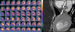
Enhanced cardiac MR indicates dilated myopathy risk, identifies salvageable LV walls
By identifying cardiac fibrosis, cardiovascular magnetic resonance imaging with late gadolinium enhancement proved to be a useful cardiac assessment tool in two studies published in the March 6 edition of JAMA.
In the first, British researchers from London’s Royal Brompton Hospital and elsewhere used cardiovascular magnetic resonance imaging with late gadolinium enhancement [LGE-CMR] to detect and quantify midwall fibrosis in patients with dilated cardiomyopathy. They found that doing so "provided independent prognostic information beyond LVEF" – left ventricular ejection fraction, the basis of current risk stratification schemes –in patients with nonischemic dilated cardiomyopathy.
Separately, American investigators used the same technology in coronary artery disease patients to assess the extent of scarring in their thinned left ventricular walls. They found that "myocardial regions with severe wall thinning do not necessarily consist entirely of scar tissue but instead may have minimal or no scarring," which is inconsistent with current assumptions that thinned regions are made of permanent scar tissue and have no residual viability.
The British group, led by Dr. Ankur Gulati of Royal Brompton Hospital in London, followed 472 patients with dilated cardiomyopathy, assessed at baseline for midwall fibrosis, for a median of 5.3 years.
Thirty-eight of the 142 patients (27%) found to have midwall fibrosis – but only 35 of 330 (11%) without it – died during the trial. Adjusted for LVEF and other conventional prognostic factors, both fibrosis presence (hazard ratio 2.43) and extent (HR, 1.11) were independently and incrementally associated with all-cause mortality. Midwall fibrosis increased by more than five times the likelihood of sudden or aborted cardiac death (JAMA 2013;309:896-908).
LGE-CMR also appeared to "facilitate identification of high-risk patients with milder degrees of left ventricular dysfunction who are currently overlooked by assessment of global left ventricular function alone, Dr. Gulati and colleagues wrote, noting that use of the technology could help guide patient selection for implantable cardioverter defibrillators. Addition of fibrosis to LVEF also significantly improved risk reclassification, they said.
The Duke team, led by Dr. Dipan J. Shah of the Duke Cardiovascular Magnetic Resonance Center in Durham, N.C., followed 201 ischemic heart disease patients with LV wall thinning spanning a mean of 34% of LV surface area; thinning was defined as a diastolic wall thickness at or below 5.5 mm. Thirty seven patients (18%) had had limited or no scar burden, defined as no more than 50% involvement.
Seventy-two of the 201 patients underwent revascularization, including revascularization of the coronary artery supplying the thinned region. Among the 14 limited-scar-burden patients who had repeat CMR-LGE afterward, diastolic wall thickness increased significantly, from 4.4 mm to 7.5 mm; their LV walls were no longer thin. A multivariate analysis showed that the extent of scarring was the strongest predictor of improvement. (JAMA 2013;309:909-18).
The function of the thin walls was also related scar burden. Limited scar burden was "strongly associated with contractile improvement and reversal of wall thinning after revascularization." The results, taken together, showed that as long there is limited scarring, the myocardial wall may thin and revert back to full thickness. Thus, myocardial thinning should not be considered permanent, they concluded.
Most of the 201 patients had significant LV dysfunction and multivessel coronary artery disease. Neither age, sex, cardiac risk factors, angina, heart failure symptoms, nor Q wave presence predicted the amount of scarring.
"The findings suggest that these clinical characteristics should not be used to assess viability in a region of thinning." They also indicate that "the end-stage of remodeling is better determined by tissue composition (i.e., scarring) rather than any set level of morphological changes to the LV cavity or LV wall," the Duke team concluded.
Members of the British team disclosed grants, consulting arrangements, and other commercial relationships with Biotronik, Boston Scientific, Roche, Servier, Celladon, AstraZeneca, GlaxoSmithKline, GE Healthcare, Bayer, ResMed, Roche Diagnostics, Pfizer, Boehringer, Novartis, Medtronic, Siemens, ApoPharma, AMAG, and Cardiovascular Imaging Solutions.
Two authors on the Duke and Northwestern team are named inventors on a Northwestern University patent for delayed-enhancement cardiovascular magnetic resonance imaging. Another reported speaker fees, consulting deals, or pending grants from Astellas, Siemens, AstraZeneca, Lantheus Medical Imaging, and Takeda.
Although these two imaging studies add to our knowledge of how supplemental noninvasive imaging studies can help cardiovascular specialists, "the clinical challenge remains in deciding which patients to evaluate with CMR and LGE and what to do with the findings," wrote Dr. Deepak Gupta, Dr. Raymond Kwong, and Dr. Marc Pfeffer.
The dilated cardiomyopathy (DCM) study, they note, did not take into consideration "proven and readily available" risk markers, such as recent hospitalization, renal function, and medication use. In addition, the study "was not powered nor designed to investigate the additional benefit of CMR beyond current class I indications for cardioverter-defibrillator therapy in DCM patients." Therefore, the data are "not sufficiently robust" to support the authors’ suggestion that LGE-CMR "may refine" the sudden cardiac death risk estimate ... [and] could guide [cardioverter defibrillator] implantation."
Regarding the thinned left ventricular wall study, the investigators failed to make clear whether all of the thinned segments assessed in this analysis were also akinetic, "which is generally considered as part of the criteria for nonviable myocardium. Furthermore, and perhaps most important, in the context of the results from [a recent] trial addressing the value of viability assessments in guiding revascularization decisions (N. Engl. J. Med. 2011;364:1617-25), the clinician is still left trying to decide what to do with a finding of viable myocardium.
Together, the two studies "provide a consistent message that detailed assessments of tissue composition, in particular fibrosis by LGE, may provide superior information than morphologic parameters, in both ischemic and nonischemic cardiomyopathies. CMR with LGE imaging adds to the practitioner’s armamentarium for assessment of cardiac structure and function and augments diagnostic and prognostic capabilities."
Dr. Gupta, Dr. Kwong, and Dr. Pfeffer, of the Division of Cardiovascular Medicine at Brigham and Women’s Hospital in Boston, made these comments in an editorial accompanying the two imaging studies (JAMA. 2013;309:929-30). They reported no disclosures.
By identifying cardiac fibrosis, cardiovascular magnetic resonance imaging with late gadolinium enhancement proved to be a useful cardiac assessment tool in two studies published in the March 6 edition of JAMA.
In the first, British researchers from London’s Royal Brompton Hospital and elsewhere used cardiovascular magnetic resonance imaging with late gadolinium enhancement [LGE-CMR] to detect and quantify midwall fibrosis in patients with dilated cardiomyopathy. They found that doing so "provided independent prognostic information beyond LVEF" – left ventricular ejection fraction, the basis of current risk stratification schemes –in patients with nonischemic dilated cardiomyopathy.
Separately, American investigators used the same technology in coronary artery disease patients to assess the extent of scarring in their thinned left ventricular walls. They found that "myocardial regions with severe wall thinning do not necessarily consist entirely of scar tissue but instead may have minimal or no scarring," which is inconsistent with current assumptions that thinned regions are made of permanent scar tissue and have no residual viability.
The British group, led by Dr. Ankur Gulati of Royal Brompton Hospital in London, followed 472 patients with dilated cardiomyopathy, assessed at baseline for midwall fibrosis, for a median of 5.3 years.
Thirty-eight of the 142 patients (27%) found to have midwall fibrosis – but only 35 of 330 (11%) without it – died during the trial. Adjusted for LVEF and other conventional prognostic factors, both fibrosis presence (hazard ratio 2.43) and extent (HR, 1.11) were independently and incrementally associated with all-cause mortality. Midwall fibrosis increased by more than five times the likelihood of sudden or aborted cardiac death (JAMA 2013;309:896-908).
LGE-CMR also appeared to "facilitate identification of high-risk patients with milder degrees of left ventricular dysfunction who are currently overlooked by assessment of global left ventricular function alone, Dr. Gulati and colleagues wrote, noting that use of the technology could help guide patient selection for implantable cardioverter defibrillators. Addition of fibrosis to LVEF also significantly improved risk reclassification, they said.
The Duke team, led by Dr. Dipan J. Shah of the Duke Cardiovascular Magnetic Resonance Center in Durham, N.C., followed 201 ischemic heart disease patients with LV wall thinning spanning a mean of 34% of LV surface area; thinning was defined as a diastolic wall thickness at or below 5.5 mm. Thirty seven patients (18%) had had limited or no scar burden, defined as no more than 50% involvement.
Seventy-two of the 201 patients underwent revascularization, including revascularization of the coronary artery supplying the thinned region. Among the 14 limited-scar-burden patients who had repeat CMR-LGE afterward, diastolic wall thickness increased significantly, from 4.4 mm to 7.5 mm; their LV walls were no longer thin. A multivariate analysis showed that the extent of scarring was the strongest predictor of improvement. (JAMA 2013;309:909-18).
The function of the thin walls was also related scar burden. Limited scar burden was "strongly associated with contractile improvement and reversal of wall thinning after revascularization." The results, taken together, showed that as long there is limited scarring, the myocardial wall may thin and revert back to full thickness. Thus, myocardial thinning should not be considered permanent, they concluded.
Most of the 201 patients had significant LV dysfunction and multivessel coronary artery disease. Neither age, sex, cardiac risk factors, angina, heart failure symptoms, nor Q wave presence predicted the amount of scarring.
"The findings suggest that these clinical characteristics should not be used to assess viability in a region of thinning." They also indicate that "the end-stage of remodeling is better determined by tissue composition (i.e., scarring) rather than any set level of morphological changes to the LV cavity or LV wall," the Duke team concluded.
Members of the British team disclosed grants, consulting arrangements, and other commercial relationships with Biotronik, Boston Scientific, Roche, Servier, Celladon, AstraZeneca, GlaxoSmithKline, GE Healthcare, Bayer, ResMed, Roche Diagnostics, Pfizer, Boehringer, Novartis, Medtronic, Siemens, ApoPharma, AMAG, and Cardiovascular Imaging Solutions.
Two authors on the Duke and Northwestern team are named inventors on a Northwestern University patent for delayed-enhancement cardiovascular magnetic resonance imaging. Another reported speaker fees, consulting deals, or pending grants from Astellas, Siemens, AstraZeneca, Lantheus Medical Imaging, and Takeda.
FROM JAMA
A Snowstorm, a Shovel, and a Thumb in the Eye
Proximal Biceps Tendon Tear in an Adolescent Tennis Player
Stress Injuries of the Ischiopubic Synchondrosis
Cardiac stress testing underutilized in stroke patients
HONOLULU – Ischemic stroke patients at high risk of coronary artery disease rarely receive guideline-supported cardiac stress testing, a study found.
Among 2,377 veterans with stroke, 28% were at high risk of coronary artery disease (CAD), and only 6.2% received CAD screening within 6 months of discharge.
Moreover, 1-year all-cause mortality was significantly lower among high-risk patients who received CAD screening than among their counterparts who did not (5% vs. 19%; P = .018), Dr. Jason Sico said at the International Stroke Conference.

American Heart Association/American Stroke Association guidelines (Circulation 2003;108:1278-90) recommend that acute ischemic stroke patients at high risk of CAD, defined by a Framingham Risk Score of at least 20%, should receive cardiac screening for occult disease.
Studies have shown that 20%-40% of stroke patients have silent cardiac ischemia, and up to 6% of stroke patients die from cardiac causes or are readmitted with a myocardial infarction in the first 3 months following a stroke, said Dr. Sico, of the department of neurology at Yale New Haven (Conn.) Hospital.
Cardiac stress testing may be underutilized because most medical professionals caring for stroke survivors are not aware of the recommendation, he said in an interview. When they are made aware, one study found that providers may not be convinced that screening for cardiac disease within the stroke population will help their patients or is cost-effective (Stroke 2009;40:3407-9).
"In their favor, there has not been a large prospective study that has demonstrated that cardiac screening for stroke patients improves such important outcomes as mortality and hospital readmission," he added.
The investigators reviewed medical records for a sample of 3,965 patients from 131 Veterans Health Administration facilities admitted for a confirmed diagnosis of ischemic stroke in 2007. Framingham Risk Scores were calculated for 2,377 patients after exclusion of 1,588 patients with a prior cardiac stress test or a known history of CAD, or if they died during hospitalization or had unaccountable data.
In all, 676 (28%) patients had a high Framingham Risk Score of 20% or more, and 1,701 (72%) had a low/intermediate Framingham Risk Score.
Cardiac stress testing within 6 months of discharge from the index stroke was not performed more frequently among high-risk than among low/intermediate-risk patients (6.2% vs. 7.5%; odds ratio, 0.81), Dr. Sico said.
Patients who underwent screening had significantly lower baseline National Institutes of Health Stroke Severity scores than those who did not (mean, 3.3 vs. 4.1; P = .003) and were younger by about 2 years (64.5 years vs. 66.4 years; P = .01). Rates of hypertension, hyperlipidemia, diabetes, and white race were similar between groups, he said at the meeting, which was sponsored by the American Heart Association.
Among all patients, 1-year mortality was significantly lower at 5% in cases where screening was performed, compared with 14% when it was not (P = .001).
Dr. Sico said the strength of the study was the relatively large cohort but that the study was limited by its makeup of primarily male veterans, data from fiscal year 2007, and the inability to explain the reasons for the underutilization of guideline-concordant cardiac screening.
Future work includes understanding the barriers to cardiac testing, the reasons behind the mortality differences among patients who did and did not receive CAD screening, and how implementation of CAD screening guidelines affects outcomes.
"To borrow a page from the diabetes literature, it was dogma that if you were diabetic because it is a coronary equivalent, you should get cardiac stress testing, but when it was prospectively looked at [in the DIAD trial] it really didn’t affect outcome; so we don’t have any prospective [study] in the stroke population to answer this question," he said.
Session moderator Dr. Jennifer Juhl Majersik, of the University of Utah, Salt Lake City, said, "This is a great example of stroke neurologists’ need to look beyond the brain."
The Veterans Health Administration provided funding for the study. Dr. Sico and his coauthors reported no disclosures.
HONOLULU – Ischemic stroke patients at high risk of coronary artery disease rarely receive guideline-supported cardiac stress testing, a study found.
Among 2,377 veterans with stroke, 28% were at high risk of coronary artery disease (CAD), and only 6.2% received CAD screening within 6 months of discharge.
Moreover, 1-year all-cause mortality was significantly lower among high-risk patients who received CAD screening than among their counterparts who did not (5% vs. 19%; P = .018), Dr. Jason Sico said at the International Stroke Conference.
American Heart Association/American Stroke Association guidelines (Circulation 2003;108:1278-90) recommend that acute ischemic stroke patients at high risk of CAD, defined by a Framingham Risk Score of at least 20%, should receive cardiac screening for occult disease.
Studies have shown that 20%-40% of stroke patients have silent cardiac ischemia, and up to 6% of stroke patients die from cardiac causes or are readmitted with a myocardial infarction in the first 3 months following a stroke, said Dr. Sico, of the department of neurology at Yale New Haven (Conn.) Hospital.
Cardiac stress testing may be underutilized because most medical professionals caring for stroke survivors are not aware of the recommendation, he said in an interview. When they are made aware, one study found that providers may not be convinced that screening for cardiac disease within the stroke population will help their patients or is cost-effective (Stroke 2009;40:3407-9).
"In their favor, there has not been a large prospective study that has demonstrated that cardiac screening for stroke patients improves such important outcomes as mortality and hospital readmission," he added.
The investigators reviewed medical records for a sample of 3,965 patients from 131 Veterans Health Administration facilities admitted for a confirmed diagnosis of ischemic stroke in 2007. Framingham Risk Scores were calculated for 2,377 patients after exclusion of 1,588 patients with a prior cardiac stress test or a known history of CAD, or if they died during hospitalization or had unaccountable data.
In all, 676 (28%) patients had a high Framingham Risk Score of 20% or more, and 1,701 (72%) had a low/intermediate Framingham Risk Score.
Cardiac stress testing within 6 months of discharge from the index stroke was not performed more frequently among high-risk than among low/intermediate-risk patients (6.2% vs. 7.5%; odds ratio, 0.81), Dr. Sico said.
Patients who underwent screening had significantly lower baseline National Institutes of Health Stroke Severity scores than those who did not (mean, 3.3 vs. 4.1; P = .003) and were younger by about 2 years (64.5 years vs. 66.4 years; P = .01). Rates of hypertension, hyperlipidemia, diabetes, and white race were similar between groups, he said at the meeting, which was sponsored by the American Heart Association.
Among all patients, 1-year mortality was significantly lower at 5% in cases where screening was performed, compared with 14% when it was not (P = .001).
Dr. Sico said the strength of the study was the relatively large cohort but that the study was limited by its makeup of primarily male veterans, data from fiscal year 2007, and the inability to explain the reasons for the underutilization of guideline-concordant cardiac screening.
Future work includes understanding the barriers to cardiac testing, the reasons behind the mortality differences among patients who did and did not receive CAD screening, and how implementation of CAD screening guidelines affects outcomes.
"To borrow a page from the diabetes literature, it was dogma that if you were diabetic because it is a coronary equivalent, you should get cardiac stress testing, but when it was prospectively looked at [in the DIAD trial] it really didn’t affect outcome; so we don’t have any prospective [study] in the stroke population to answer this question," he said.
Session moderator Dr. Jennifer Juhl Majersik, of the University of Utah, Salt Lake City, said, "This is a great example of stroke neurologists’ need to look beyond the brain."
The Veterans Health Administration provided funding for the study. Dr. Sico and his coauthors reported no disclosures.
AT THE INTERNATIONAL STROKE CONFERENCE
Major Finding: Only 6.2% of high-risk stroke patients received guideline-concordant cardiac stress testing.
Data Source: Retrospective cohort study of 2,337 ischemic stroke patients.
Disclosures: The Veterans Health Administration provided funding for the study. Dr. Sico and his coauthors reported no disclosures.

Fractional flow reserve the best tool for assessing intermediate stenoses
SNOWMASS, COLO. – Fractional flow reserve measurement has rapidly emerged as the tool of choice for deciding in the catheterization laboratory whether to revascularize an intermediate 50%-70% stenosis in a patient with stable ischemic heart disease, according to Dr. E. Murat *Tuzcu.
The evidence is now compelling that fractional flow reserve (FFR) calculated invasively from coronary pressure measurement is superior to angiographic assessment, intravascular ultrasound, or optical coherence tomography for this purpose.

"When the question is, ‘What should we do with the borderline lesion?’ the answer is fractional flow reserve, even in the left main coronary artery," Dr. Tuzcu, professor of medicine at the Cleveland Clinic, asserted at the Annual Cardiovascular Conference at Snowmass.
FFR is the method par excellence for determining if an intermediate stenosis is hemodynamically significant; that is, whether the lesion is responsible for reversible ischemia. If it is, then coronary stenting will improve the patient’s functional status and reduce the likelihood of acute MI and all-cause mortality down the road. If FFR indicates that the stenosis is not responsible for reversible ischemia, however, then PCI won’t improve the patient’s prognosis. FFR has the additional virtues of being fast and simple, and it enables immediate decision-making in the cath lab, he explained.
For Dr. Tuzcu, the game changer for FFR was the DEFER study, which he considers to be one of the most important clinical trials in the field of interventional cardiology in the past decade. It showed that, by using FFR, cardiologists could be more selective in their use of PCI in the setting of stable ischemic heart disease.
DEFER was a multicenter Dutch/Belgian study in which 325 patients underwent FFR measurement just prior to planned PCI for an intermediate stenosis. If the FFR value was less than 0.75 – indicative of reversible ischemia – then PCI was performed as planned. If it was 0.75 or greater, patients were randomized to PCI or to deferred PCI.
At 5 years of follow-up, the rate of cardiac death or acute MI was 3.3% in the 91 patients with an FFR of 0.75 or more in the deferred PCI group – less than 1% per year. That wasn’t significantly different from the 7.9% rate among the 90 patients with an FFR of at least 0.75 who underwent prompt PCI. In contrast, the combined endpoint occurred in 15.7% of the 144 PCI-treated patients with FFR evidence of reversible ischemia due to the target lesion (J. Am. Coll. Cardiol. 2007;49:2105-11.)
DEFER was conducted in the bare metal–stent era. The next major clinical trial advancing FFR was the FAME (Fractional Flow Reserve Versus Angiography for Multivessel Evaluation) study, in which 1,005 patients with multivessel CAD were randomized to PCI with drug-eluting stents guided by FFR or by angiography alone. The FFR definition of reversible ischemia used in FAME and subsequent trials was a value of 0.80 or less.
The 2-year rate of death or MI was 8.4% in the FFR-guided group, significantly less than the 12.9% rate in the angiography-guided patients. For patients with stenotic lesions deferred from PCI on the basis of an FFR greater than 0.80, the 2-year rate of MI was just 0.2%, with a revascularization rate of 3.2% (J. Am. Coll. Cardiol. 2010;56:177-84).
Nearly half of all stenoses were categorized angiographically as intermediate, 50%-70% lesions. FFR classified 35% of such lesions as functionally significant, while 65% were not associated with reversible ischemia (J. Am. Coll. Cardiol. 2010;55:2816-21).
In the FAME-2 trial, 888 patients with stable CAD and at least one functionally significant stenosis with an FFR of 0.80 or less were randomized to PCI plus optimal medical therapy or to optimal medical therapy alone. Recruitment was halted prematurely by the data safety monitoring board because the combined endpoint of death, MI, or urgent revascularization had occurred in 4.3% of the PCI group versus 12.7% of those assigned to optimal medical management alone (N. Engl. J. Med. 2012;367:991-1001).
Dr. Tuzcu noted that optical coherence tomography (OCT) has drawn much interest as a tool for identifying hemodynamically severe coronary stenoses. "It provides great pictures with tremendous resolution. For looking at stent strut coverage, OCT is a star tool. It’s almost like histology," he said.
It can’t, however, hold a candle to FFR for hemodynamic assessment of stenoses, he added, pointing to a recent Spanish study comparing FFR, OCT, and intravascular ultrasound (IVUS) for this purpose. OCT and IVUS displayed moderate diagnostic efficiency, with no clinically meaningful difference between them, but Dr. Tuzcu concurred with the investigators that the low specificity of OCT and IVUS precludes their use in lieu of FFR for functional assessment (J. Am. Coll. Cardiol. 2012;59:1080-9).
The most recent significant development on the FFR front was Dr. Gregg W. Stone’s presentation of the pooled results of the VERDICT and FIRST trials at the Transcatheter Cardiovascular Therapeutics conference in Miami last October. VERDICT and FIRST included 516 patients with 544 intermediate coronary stenoses evaluated by both FFR and IVUS at 24 centers in nine countries. The bottom line was that IVUS-determined minimum luminal cross-sectional area was only modestly correlated with FFR, according to Dr. Stone, professor of medicine and director of cardiovascular research and education at Columbia University Medical Center/New York Presbyterian Hospital.
"I think this clearly showed – and probably conclusively showed – that, while IVUS is useful in many, many settings, it’s probably not the best tool when FFR is available for deciding which lesion is significant and which is not," Dr. Tuzcu said.
"IVUS is a pretty good tool, sometimes, for morphologic assessment. I like it when there’s an issue with the left main coronary artery. I can size the artery, I can understand an ostial lesion, and I can certainly understand better a bifurcation or trifurcation of the left main coronary artery," he added.
The current American College of Cardiology/American Heart Association guidelines give IVUS a class IIa rating as "reasonable" to assess angiographically intermediate stenoses of the left main coronary artery. FFR gets the same relatively tepid IIa rating for assessment of intermediate stenoses in any coronary arteries. In contrast, the latest European Society of Cardiology guidelines on coronary revascularization have bumped up FFR to a class Ia rating, making it the standard for this assessment.
Dr. Tuzcu reported having no financial conflicts.
*CORRECTION, 3/1/2013: In an earlier version of this story, the name of Dr. E. Murat Tuzcu was spelled incorrectly. This version has been updated.
SNOWMASS, COLO. – Fractional flow reserve measurement has rapidly emerged as the tool of choice for deciding in the catheterization laboratory whether to revascularize an intermediate 50%-70% stenosis in a patient with stable ischemic heart disease, according to Dr. E. Murat *Tuzcu.
The evidence is now compelling that fractional flow reserve (FFR) calculated invasively from coronary pressure measurement is superior to angiographic assessment, intravascular ultrasound, or optical coherence tomography for this purpose.
"When the question is, ‘What should we do with the borderline lesion?’ the answer is fractional flow reserve, even in the left main coronary artery," Dr. Tuzcu, professor of medicine at the Cleveland Clinic, asserted at the Annual Cardiovascular Conference at Snowmass.
FFR is the method par excellence for determining if an intermediate stenosis is hemodynamically significant; that is, whether the lesion is responsible for reversible ischemia. If it is, then coronary stenting will improve the patient’s functional status and reduce the likelihood of acute MI and all-cause mortality down the road. If FFR indicates that the stenosis is not responsible for reversible ischemia, however, then PCI won’t improve the patient’s prognosis. FFR has the additional virtues of being fast and simple, and it enables immediate decision-making in the cath lab, he explained.
For Dr. Tuzcu, the game changer for FFR was the DEFER study, which he considers to be one of the most important clinical trials in the field of interventional cardiology in the past decade. It showed that, by using FFR, cardiologists could be more selective in their use of PCI in the setting of stable ischemic heart disease.
DEFER was a multicenter Dutch/Belgian study in which 325 patients underwent FFR measurement just prior to planned PCI for an intermediate stenosis. If the FFR value was less than 0.75 – indicative of reversible ischemia – then PCI was performed as planned. If it was 0.75 or greater, patients were randomized to PCI or to deferred PCI.
At 5 years of follow-up, the rate of cardiac death or acute MI was 3.3% in the 91 patients with an FFR of 0.75 or more in the deferred PCI group – less than 1% per year. That wasn’t significantly different from the 7.9% rate among the 90 patients with an FFR of at least 0.75 who underwent prompt PCI. In contrast, the combined endpoint occurred in 15.7% of the 144 PCI-treated patients with FFR evidence of reversible ischemia due to the target lesion (J. Am. Coll. Cardiol. 2007;49:2105-11.)
DEFER was conducted in the bare metal–stent era. The next major clinical trial advancing FFR was the FAME (Fractional Flow Reserve Versus Angiography for Multivessel Evaluation) study, in which 1,005 patients with multivessel CAD were randomized to PCI with drug-eluting stents guided by FFR or by angiography alone. The FFR definition of reversible ischemia used in FAME and subsequent trials was a value of 0.80 or less.
The 2-year rate of death or MI was 8.4% in the FFR-guided group, significantly less than the 12.9% rate in the angiography-guided patients. For patients with stenotic lesions deferred from PCI on the basis of an FFR greater than 0.80, the 2-year rate of MI was just 0.2%, with a revascularization rate of 3.2% (J. Am. Coll. Cardiol. 2010;56:177-84).
Nearly half of all stenoses were categorized angiographically as intermediate, 50%-70% lesions. FFR classified 35% of such lesions as functionally significant, while 65% were not associated with reversible ischemia (J. Am. Coll. Cardiol. 2010;55:2816-21).
In the FAME-2 trial, 888 patients with stable CAD and at least one functionally significant stenosis with an FFR of 0.80 or less were randomized to PCI plus optimal medical therapy or to optimal medical therapy alone. Recruitment was halted prematurely by the data safety monitoring board because the combined endpoint of death, MI, or urgent revascularization had occurred in 4.3% of the PCI group versus 12.7% of those assigned to optimal medical management alone (N. Engl. J. Med. 2012;367:991-1001).
Dr. Tuzcu noted that optical coherence tomography (OCT) has drawn much interest as a tool for identifying hemodynamically severe coronary stenoses. "It provides great pictures with tremendous resolution. For looking at stent strut coverage, OCT is a star tool. It’s almost like histology," he said.
It can’t, however, hold a candle to FFR for hemodynamic assessment of stenoses, he added, pointing to a recent Spanish study comparing FFR, OCT, and intravascular ultrasound (IVUS) for this purpose. OCT and IVUS displayed moderate diagnostic efficiency, with no clinically meaningful difference between them, but Dr. Tuzcu concurred with the investigators that the low specificity of OCT and IVUS precludes their use in lieu of FFR for functional assessment (J. Am. Coll. Cardiol. 2012;59:1080-9).
The most recent significant development on the FFR front was Dr. Gregg W. Stone’s presentation of the pooled results of the VERDICT and FIRST trials at the Transcatheter Cardiovascular Therapeutics conference in Miami last October. VERDICT and FIRST included 516 patients with 544 intermediate coronary stenoses evaluated by both FFR and IVUS at 24 centers in nine countries. The bottom line was that IVUS-determined minimum luminal cross-sectional area was only modestly correlated with FFR, according to Dr. Stone, professor of medicine and director of cardiovascular research and education at Columbia University Medical Center/New York Presbyterian Hospital.
"I think this clearly showed – and probably conclusively showed – that, while IVUS is useful in many, many settings, it’s probably not the best tool when FFR is available for deciding which lesion is significant and which is not," Dr. Tuzcu said.
"IVUS is a pretty good tool, sometimes, for morphologic assessment. I like it when there’s an issue with the left main coronary artery. I can size the artery, I can understand an ostial lesion, and I can certainly understand better a bifurcation or trifurcation of the left main coronary artery," he added.
The current American College of Cardiology/American Heart Association guidelines give IVUS a class IIa rating as "reasonable" to assess angiographically intermediate stenoses of the left main coronary artery. FFR gets the same relatively tepid IIa rating for assessment of intermediate stenoses in any coronary arteries. In contrast, the latest European Society of Cardiology guidelines on coronary revascularization have bumped up FFR to a class Ia rating, making it the standard for this assessment.
Dr. Tuzcu reported having no financial conflicts.
*CORRECTION, 3/1/2013: In an earlier version of this story, the name of Dr. E. Murat Tuzcu was spelled incorrectly. This version has been updated.
EXPERT ANALYSIS FROM THE CARDIOVASCULAR CONFERENCE AT SNOWMASS

Comparing imaging technologies for chest pain in ED
Coronary computed tomographic angiography for rule-out of acute coronary syndrome in the emergency department may have moved ahead of SPECT myocardial perfusion imaging – its main noninvasive imaging rival – on the strength of recent evidence of advantages in time-to-discharge and radiation exposure, according to Dr. Christopher M. Kramer.
Myocardial perfusion imaging (MPI) has for roughly a decade been seen as the standard of care for imaging assistance in ED triage of patients presenting with chest pain and a nondiagnostic ECG.
It attained this status based upon the favorable results of the very large randomized ERASE (ER Assessment of Sestamibi for Evaluation of Chest Pain) trial (JAMA 2002;288:2693-700) and other studies in which MPI was compared to usual ED care.
But in more recent randomized trials comparing computed tomographic angiography (CTA) to standard ED evaluation protocols, which now often include MPI, CTA has carried the day, Dr. Kramer said at the annual cardiovascular conference at Snowmass sponsored by the American College of Cardiology.
For example, the multicenter ACRIN-PA trial randomized 1,370 low- to intermediate-risk patients presenting with possible ACS to EDs to either early CTA or standard care, which in most cases included MPI. The CTA group had a higher rate of discharge from the ED: 50%, compared with 23%. They also had a median 18.0-hour length of stay, significantly shorter than the 24.8 hours in the control group (N. Engl. J. Med. 2012;366:1393-403).
Moreover, the CTA group had a higher rate of detection of CAD – 9.0% compared to 3.5% because CTA can detect nonobstructive as well as obstructive plaque, added Dr. Kramer, professor of cardiology and of radiology and director of the cardiovascular imaging center at the University of Virginia, Charlottesville.
The ROMICAT II (Rule Out Myocardial Ischemia/Infarction by Computer-Assisted Tomography) trial was a similar story. ROMICAT II was a multicenter trial in which 1,000 patients who presented to an ED with symptoms suggestive of ACS but a nondiagnostic ECG were randomized to CTA or a standard ED evaluation, most often including MPI. A total of 47% of patients in the CTA group were discharged directly from the ED, compared with just 12% in the control arm. Most notably, the median length of stay in the ED was 8.6 hours with CTA vs. 26.7 hours with a standard evaluation, and the mean length of stay in the hospital was reduced from 30.8 hours with standard evaluation to 23.2 hours, a 7.6-hour reduction (N. Engl. J. Med. 2012;367:299-308).
Follow-up demonstrated no cases of undetected ACS occurred in either group.
The average radiation dose to patients in the CTA arm was 14.3 mSv, compared with about 10 mSv for those patients in the standard evaluation group who got MPI. However, ROMICAT II didn’t use the latest CTA equipment.

"With the modern CTA units, the radiation doses have come down quite a lot. With our flash 256 detector in the ED, we can do a study in a patient with a controlled heart rate and a reasonable BMI [body mass index] with about 1 mSv of radiation now," Dr. Kramer recalled.
Costs in the ED were significantly lower in the CTA group than in controls in ROMICAT II because of the faster patient throughput. However, this savings was neutralized by higher in-hospital costs because the CTA group had more cardiac catheterizations and revascularization procedures because more cases of CAD were detected, as in ACRIN-PA.
"The real advantage to CTA is the rapid discharge from the ED. That’s what patients appreciate. They get their CT angiogram and they can go home within an hour or 2. It really improves their experience in the ED, rather than spending all night waiting for their SPECT MPI results. I don’t think you’re saving costs by doing CTA, though, because of this catch-up phenomenon," Dr. Kramer said.
Aside from the fact that MPI takes 3-5 hours, its other main limitations are that it has difficulty in differentiating acute ischemia from acute infarction or an old infarct, according to Dr. Kramer.
The ability of CTA to detect more cases of CAD by identifying nonobstructive plaque may prove to be of clinical import. That’s the underlying hypothesis of the ongoing National Heart, Lung, and Blood Institute–funded PROMISE (Prospective Multicenter Imaging Study for Evaluation of Chest Pain) study, in which 10,000 patients with symptoms suggestive of CAD are being randomized to an initial CTA or to usual care with a functional test, either MPI, stress echocardiography, or exercise ECG.
This study is being conducted in the offices of primary care physicians and cardiologists rather than in EDs. PROMISE has clinical endpoints as its primary outcomes. The study hypothesis is that intervening in patients identified as having nonobstructive CAD will yield improved outcomes. Results remain several years off, Dr. Kramer said.
ED physicians are eager for high-tech help in quickly and reliably ruling out ACS. Acute chest pain accounts for more than 8 million ED visits annually, but in only 1.19 million admissions for ACS.
Besides MPI and CTA, the other two noninvasive imaging technologies available for use in the ED are cardiac magnetic resonance (CMR) and contrast echocardiography. Neither utilizes radiation – a big plus. Yet neither is as widely used as MPI or CTA. That’s because magnetic resonance takes longer than CTA does, and the scanner may not be available at a moment’s notice, as it really needs to be, when a patient presents with chest pain to the ED. Also, expertise in CMR is not widely available. This is also an issue for contrast echo in the ED.
"The problem with contrast echo is that there are really very few centers around the world that can do it well. It really hasn’t caught on in terms of utilization in the ED," according to the cardiologist.
He reported that he serves as a consultant to Synarc and receives research support from Siemens Medical Solutions.
Coronary computed tomographic angiography for rule-out of acute coronary syndrome in the emergency department may have moved ahead of SPECT myocardial perfusion imaging – its main noninvasive imaging rival – on the strength of recent evidence of advantages in time-to-discharge and radiation exposure, according to Dr. Christopher M. Kramer.
Myocardial perfusion imaging (MPI) has for roughly a decade been seen as the standard of care for imaging assistance in ED triage of patients presenting with chest pain and a nondiagnostic ECG.
It attained this status based upon the favorable results of the very large randomized ERASE (ER Assessment of Sestamibi for Evaluation of Chest Pain) trial (JAMA 2002;288:2693-700) and other studies in which MPI was compared to usual ED care.
But in more recent randomized trials comparing computed tomographic angiography (CTA) to standard ED evaluation protocols, which now often include MPI, CTA has carried the day, Dr. Kramer said at the annual cardiovascular conference at Snowmass sponsored by the American College of Cardiology.
For example, the multicenter ACRIN-PA trial randomized 1,370 low- to intermediate-risk patients presenting with possible ACS to EDs to either early CTA or standard care, which in most cases included MPI. The CTA group had a higher rate of discharge from the ED: 50%, compared with 23%. They also had a median 18.0-hour length of stay, significantly shorter than the 24.8 hours in the control group (N. Engl. J. Med. 2012;366:1393-403).
Moreover, the CTA group had a higher rate of detection of CAD – 9.0% compared to 3.5% because CTA can detect nonobstructive as well as obstructive plaque, added Dr. Kramer, professor of cardiology and of radiology and director of the cardiovascular imaging center at the University of Virginia, Charlottesville.
The ROMICAT II (Rule Out Myocardial Ischemia/Infarction by Computer-Assisted Tomography) trial was a similar story. ROMICAT II was a multicenter trial in which 1,000 patients who presented to an ED with symptoms suggestive of ACS but a nondiagnostic ECG were randomized to CTA or a standard ED evaluation, most often including MPI. A total of 47% of patients in the CTA group were discharged directly from the ED, compared with just 12% in the control arm. Most notably, the median length of stay in the ED was 8.6 hours with CTA vs. 26.7 hours with a standard evaluation, and the mean length of stay in the hospital was reduced from 30.8 hours with standard evaluation to 23.2 hours, a 7.6-hour reduction (N. Engl. J. Med. 2012;367:299-308).
Follow-up demonstrated no cases of undetected ACS occurred in either group.
The average radiation dose to patients in the CTA arm was 14.3 mSv, compared with about 10 mSv for those patients in the standard evaluation group who got MPI. However, ROMICAT II didn’t use the latest CTA equipment.
"With the modern CTA units, the radiation doses have come down quite a lot. With our flash 256 detector in the ED, we can do a study in a patient with a controlled heart rate and a reasonable BMI [body mass index] with about 1 mSv of radiation now," Dr. Kramer recalled.
Costs in the ED were significantly lower in the CTA group than in controls in ROMICAT II because of the faster patient throughput. However, this savings was neutralized by higher in-hospital costs because the CTA group had more cardiac catheterizations and revascularization procedures because more cases of CAD were detected, as in ACRIN-PA.
"The real advantage to CTA is the rapid discharge from the ED. That’s what patients appreciate. They get their CT angiogram and they can go home within an hour or 2. It really improves their experience in the ED, rather than spending all night waiting for their SPECT MPI results. I don’t think you’re saving costs by doing CTA, though, because of this catch-up phenomenon," Dr. Kramer said.
Aside from the fact that MPI takes 3-5 hours, its other main limitations are that it has difficulty in differentiating acute ischemia from acute infarction or an old infarct, according to Dr. Kramer.
The ability of CTA to detect more cases of CAD by identifying nonobstructive plaque may prove to be of clinical import. That’s the underlying hypothesis of the ongoing National Heart, Lung, and Blood Institute–funded PROMISE (Prospective Multicenter Imaging Study for Evaluation of Chest Pain) study, in which 10,000 patients with symptoms suggestive of CAD are being randomized to an initial CTA or to usual care with a functional test, either MPI, stress echocardiography, or exercise ECG.
This study is being conducted in the offices of primary care physicians and cardiologists rather than in EDs. PROMISE has clinical endpoints as its primary outcomes. The study hypothesis is that intervening in patients identified as having nonobstructive CAD will yield improved outcomes. Results remain several years off, Dr. Kramer said.
ED physicians are eager for high-tech help in quickly and reliably ruling out ACS. Acute chest pain accounts for more than 8 million ED visits annually, but in only 1.19 million admissions for ACS.
Besides MPI and CTA, the other two noninvasive imaging technologies available for use in the ED are cardiac magnetic resonance (CMR) and contrast echocardiography. Neither utilizes radiation – a big plus. Yet neither is as widely used as MPI or CTA. That’s because magnetic resonance takes longer than CTA does, and the scanner may not be available at a moment’s notice, as it really needs to be, when a patient presents with chest pain to the ED. Also, expertise in CMR is not widely available. This is also an issue for contrast echo in the ED.
"The problem with contrast echo is that there are really very few centers around the world that can do it well. It really hasn’t caught on in terms of utilization in the ED," according to the cardiologist.
He reported that he serves as a consultant to Synarc and receives research support from Siemens Medical Solutions.
EXPERT ANALYSIS FROM THE ANNUAL CARDIOVASCULAR CONFERENCE AT SNOWMASS

It’s all in the P wave
A 49-year-old man with rheumatic mitral valve stenosis, which had been diagnosed 3 years previously, presented to the outpatient department with worsening exertional dyspnea, fatigue, and cough.
At rest, he appeared comfortable; his pulse rate was 94 bpm and his blood pressure was 117/82 mm Hg. Cardiac auscultation revealed a loud first heart sound, a mid-diastolic murmur with presystolic accentuation at the cardiac apex, and a pansystolic murmur at the left lower sternal border that increased in intensity with inspiration. A prominent left parasternal heave was present.
His 12-lead electrocardiogram is shown in Figure 1.
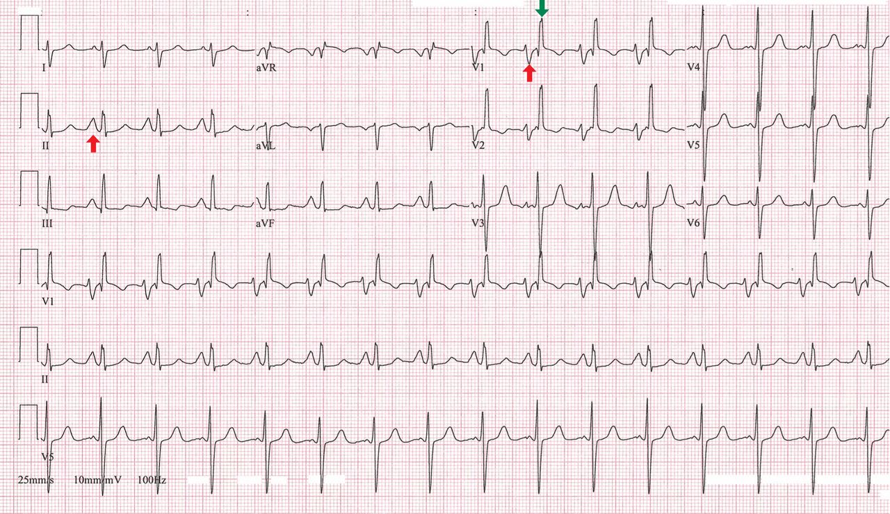
Transthoracic echocardiography confirmed severe mitral stenosis with an estimated mitral valve area of 0.7 cm2 without significant mitral regurgitation. In addition, right ventricular dilatation with moderately severe systolic dysfunction and 4+ (severe) tricuspid regurgitation were present. On the basis of the peak tricuspid regurgitant velocity, the right ventricular systolic pressure was calculated to be 80 mm Hg, consistent with severe pulmonary hypertension. The left ventricular end-diastolic volume was reduced and the ejection fraction was normal.
On right heart catheterization, the pulmonary artery pressure was 92/51 mm Hg.
Q: Electrocardiographic findings that support a diagnosis of pulmonary hypertension include which of the following?
- QRS complex axis of +110°
- R/S (QRS complex) ratio greater than 1 in lead V1
- Sum of the amplitudes of the R wave in lead V1 and the S wave in lead V6 greater than 1.0 mV
- All of the above
A: The correct answer is all of the above. Regardless of the cause, patients with long-standing pulmonary hypertension possess varying degrees of right ventricular hypertrophy that may be accompanied by right ventricular enlargement and systolic dysfunction. A QRS complex axis of 110° or more, an R/S (QRS complex) ratio greater than 1 in lead V1, and the sum of the amplitudes of the R wave in lead V1 and the S wave in lead V6 greater than 1.0 mV all support right ventricular hypertrophy.1
As noted in this electrocardiogram, T-wave inversion in leads V1 and V2 supports a right ventricular repolarization abnormality secondary to the hypertrophy.2
Q: Important electrocardiographic findings in this patient that support secondary pulmonary hypertension due to mitral stenosis include which of the following?
- Tall peaked P waves in lead II of at least 0.25 mV and positive P waves in V1 greater than 0.15 mV
- Prolonged P waves of at least 120 ms in lead II and terminal negative P waves in V1 greater than 40 ms
- Right ventricular hypertrophy
- All of the above
A: The correct answer is prolonged P waves of at least 120 ms in lead II and terminal negative P waves in V1 greater than 40 ms.
Abnormal surface electrocardiographic findings reflecting atrial enlargement or slowed atrial conduction are difficult to differentiate and are best characterized as “atrial abnormalities.” On surface electrocardiography, an atrial abnormality is represented by a P wave morphology that is best studied in leads II and V1. In lead II, a tall peaked P wave of at least 0.25 mV supports right atrial abnormality, and a prolonged P wave (≥ 120 ms) supports left atrial abnormality. In lead V1, right atrial abnormality is suggested by a positive P wave in V1 greater than 0.15 mV, and a terminally negative P wave greater than 40 ms in duration and greater than 0.1 mV deep supports left atrial abnormality.3
It is well recognized that the pathophysiology of pulmonary hypertension involves both the right ventricle and the right atrium.4,5 Therefore, irrespective of the cause of pulmonary hypertension, electrocardiography may additionally reveal right atrial abnormality.6
When the findings suggest pulmonary hypertension (ie, right ventricular hypertrophy with or without right atrial abnormality), it is also important to evaluate for concurrent left atrial abnormality. If present, concomitant left atrial abnormality is a valuable, more specific clue that may help characterize secondary pulmonary hypertension from left-sided heart disease, as illustrated in this example with long-standing severe mitral stenosis.2
- Hancock EW, Deal BJ, Mirvis DM, et al; American Heart Association Electrocardiography and Arrhythmias Committee, Council on Clinical Cardiology. AHA/ACCF/HRS recommendations for the standardization and interpretation of the electrocardiogram: part V: electrocardiogram changes associated with cardiac chamber hypertrophy: a scientific statement from the American Heart Association Electrocardiography and Arrhythmias Committee, Council on Clinical Cardiology; the American College of Cardiology Foundation; and the Heart Rhythm Society. Endorsed by the International Society for Computerized Electrocardiology. J Am Coll Cardiol 2009; 53:992–1002.
- Goldberger AL. Atrial and ventricular enlargement. In: Clinical Electrocardiography: A Simplified Approach. 7th ed. Philadelphia, PA: Mosby Elsevier; 2006:59–71.
- Bayés-de-Luna A, Goldwasser D, Fiol M, Bayés-Genis A. Surface electrocardiography. In: Hurst’s The Heart. 13th ed. New York, NY: McGraw-Hill Medical; 2011.
- Cioffi G, de Simone G, Mureddu G, Tarantini L, Stefenelli C. Right atrial size and function in patients with pulmonary hypertension associated with disorders of respiratory system or hypoxemia. Eur J Echocardiogr 2007; 8:322–331.
- Raymond RJ, Hinderliter AL, Willis PW, et al. Echocardiographic predictors of adverse outcomes in primary pulmonary hypertension. J Am Coll Cardiol 2002; 39:1214–1219.
- Al-Naamani K, Hijal T, Nguyen V, Andrew S, Nguyen T, Huynh T. Predictive values of the electrocardiogram in diagnosing pulmonary hypertension. Int J Cardiol 2008; 127:214–218.
A 49-year-old man with rheumatic mitral valve stenosis, which had been diagnosed 3 years previously, presented to the outpatient department with worsening exertional dyspnea, fatigue, and cough.
At rest, he appeared comfortable; his pulse rate was 94 bpm and his blood pressure was 117/82 mm Hg. Cardiac auscultation revealed a loud first heart sound, a mid-diastolic murmur with presystolic accentuation at the cardiac apex, and a pansystolic murmur at the left lower sternal border that increased in intensity with inspiration. A prominent left parasternal heave was present.
His 12-lead electrocardiogram is shown in Figure 1.
Transthoracic echocardiography confirmed severe mitral stenosis with an estimated mitral valve area of 0.7 cm2 without significant mitral regurgitation. In addition, right ventricular dilatation with moderately severe systolic dysfunction and 4+ (severe) tricuspid regurgitation were present. On the basis of the peak tricuspid regurgitant velocity, the right ventricular systolic pressure was calculated to be 80 mm Hg, consistent with severe pulmonary hypertension. The left ventricular end-diastolic volume was reduced and the ejection fraction was normal.
On right heart catheterization, the pulmonary artery pressure was 92/51 mm Hg.
Q: Electrocardiographic findings that support a diagnosis of pulmonary hypertension include which of the following?
- QRS complex axis of +110°
- R/S (QRS complex) ratio greater than 1 in lead V1
- Sum of the amplitudes of the R wave in lead V1 and the S wave in lead V6 greater than 1.0 mV
- All of the above
A: The correct answer is all of the above. Regardless of the cause, patients with long-standing pulmonary hypertension possess varying degrees of right ventricular hypertrophy that may be accompanied by right ventricular enlargement and systolic dysfunction. A QRS complex axis of 110° or more, an R/S (QRS complex) ratio greater than 1 in lead V1, and the sum of the amplitudes of the R wave in lead V1 and the S wave in lead V6 greater than 1.0 mV all support right ventricular hypertrophy.1
As noted in this electrocardiogram, T-wave inversion in leads V1 and V2 supports a right ventricular repolarization abnormality secondary to the hypertrophy.2
Q: Important electrocardiographic findings in this patient that support secondary pulmonary hypertension due to mitral stenosis include which of the following?
- Tall peaked P waves in lead II of at least 0.25 mV and positive P waves in V1 greater than 0.15 mV
- Prolonged P waves of at least 120 ms in lead II and terminal negative P waves in V1 greater than 40 ms
- Right ventricular hypertrophy
- All of the above
A: The correct answer is prolonged P waves of at least 120 ms in lead II and terminal negative P waves in V1 greater than 40 ms.
Abnormal surface electrocardiographic findings reflecting atrial enlargement or slowed atrial conduction are difficult to differentiate and are best characterized as “atrial abnormalities.” On surface electrocardiography, an atrial abnormality is represented by a P wave morphology that is best studied in leads II and V1. In lead II, a tall peaked P wave of at least 0.25 mV supports right atrial abnormality, and a prolonged P wave (≥ 120 ms) supports left atrial abnormality. In lead V1, right atrial abnormality is suggested by a positive P wave in V1 greater than 0.15 mV, and a terminally negative P wave greater than 40 ms in duration and greater than 0.1 mV deep supports left atrial abnormality.3
It is well recognized that the pathophysiology of pulmonary hypertension involves both the right ventricle and the right atrium.4,5 Therefore, irrespective of the cause of pulmonary hypertension, electrocardiography may additionally reveal right atrial abnormality.6
When the findings suggest pulmonary hypertension (ie, right ventricular hypertrophy with or without right atrial abnormality), it is also important to evaluate for concurrent left atrial abnormality. If present, concomitant left atrial abnormality is a valuable, more specific clue that may help characterize secondary pulmonary hypertension from left-sided heart disease, as illustrated in this example with long-standing severe mitral stenosis.2
- Hancock EW, Deal BJ, Mirvis DM, et al; American Heart Association Electrocardiography and Arrhythmias Committee, Council on Clinical Cardiology. AHA/ACCF/HRS recommendations for the standardization and interpretation of the electrocardiogram: part V: electrocardiogram changes associated with cardiac chamber hypertrophy: a scientific statement from the American Heart Association Electrocardiography and Arrhythmias Committee, Council on Clinical Cardiology; the American College of Cardiology Foundation; and the Heart Rhythm Society. Endorsed by the International Society for Computerized Electrocardiology. J Am Coll Cardiol 2009; 53:992–1002.
- Goldberger AL. Atrial and ventricular enlargement. In: Clinical Electrocardiography: A Simplified Approach. 7th ed. Philadelphia, PA: Mosby Elsevier; 2006:59–71.
- Bayés-de-Luna A, Goldwasser D, Fiol M, Bayés-Genis A. Surface electrocardiography. In: Hurst’s The Heart. 13th ed. New York, NY: McGraw-Hill Medical; 2011.
- Cioffi G, de Simone G, Mureddu G, Tarantini L, Stefenelli C. Right atrial size and function in patients with pulmonary hypertension associated with disorders of respiratory system or hypoxemia. Eur J Echocardiogr 2007; 8:322–331.
- Raymond RJ, Hinderliter AL, Willis PW, et al. Echocardiographic predictors of adverse outcomes in primary pulmonary hypertension. J Am Coll Cardiol 2002; 39:1214–1219.
- Al-Naamani K, Hijal T, Nguyen V, Andrew S, Nguyen T, Huynh T. Predictive values of the electrocardiogram in diagnosing pulmonary hypertension. Int J Cardiol 2008; 127:214–218.
Bullets no match for dual-energy CT scrutiny
CHICAGO – Dual-energy computed tomography may have a role in clearing patients with embedded bullets and other metallic objects for magnetic resonance imaging who would otherwise be excluded because of safety concerns.
A Swiss analysis of bullets and shotgun pellets found that dual-energy CT had a high discriminatory power to distinguish between projectiles with and without ferromagnetic properties. MR safety depends on whether a metallic object has ferromagnetic properties – something that most patients don’t know or may be unable to communicate during an emergency evaluation, study author Dr. Sebastian Winklhofer said at the annual meeting of the Radiological Society of North America.
"Dual-energy measurements may contribute to MR safety and allow for MR imaging of patients with retained projectiles," he said.
Retained metallic objects such as bullets, shrapnel, and medical devices are frequently contraindicated for MRI, because they might move, dislodge, or accelerate at dangerously high velocities toward the scanner’s magnet. This has led to patient injury, and in some cases death, if the object is located near or in a critical anatomic structure.
Session comoderator Dr. Seppo Koskinen of Helsinki University Central Hospital, Finland, said he recently had to forgo MR imaging in a patient with a bullet lodged in the spinal canal because the patient didn’t know if it was lead or steel.
"We didn’t do an MR, but maybe with this system we could have," he said in an interview. "So it has a real clinical impact."
Unsafe exams and misinformed refusals to refer or scan a patient are also occurring as a result of confusion over the ever increasing number of metallic orthopedic and cardiovascular implants. For example, a device that is known to be safe on a 1.5 tesla scanner may be contraindicated on a more powerful 3T scanner or on other scanners using different settings.
When the American College of Radiology penned its Safe MR Practices guidelines in 2007, no cardiac pacemakers or implantable defibrillators were labeled as safe or conditionally safe for MR imaging, prompting the college to recommend that routine MRI is inadvisable in patients with these devices.
In its scientific statement on the controversial topic, the American Heart Association says data are available to support MR imaging in patients with cardiovascular devices, but recommends a series of precautions, including careful patient screening and an accurate determination of the device and its properties (Circulation 2007:116:2878-91).
Dual-energy CT has been used in research and clinical settings to assess the composition of various objects, said Dr. Winklhofer of University Hospital Zurich, Switzerland. He highlighted one study showing that dual-energy CT had a 98% specificity in differentiating between urinary stones that did or did not contain uric acid (Invest. Radiology 2010;45:1-6).
In the current study, nine bullets and two shotgun pellets were scanned in an ex vivo chest phantom using second-generation dual-source CT (SOMATOM Definition Flash, Siemens Healthcare), with tube voltages set at 80, 100, 120, and 140 kVp. Two readers, blinded to the ferromagnetic properties of the projectiles, independently assessed CT numbers on 44 images reconstructed using the extended CT scale technique, which helps overcome artifacts arising from metallic objects. A CT number is the density assigned to a voxel in a CT scan.
Dual-energy indices (DEI) were calculated from 80/140 kVP and 100/140 kVP pairs, and receiver operating characteristics analyses were fitted to predict ferromagnetic properties by means of DEI and CT numbers.
The bullets/pellets ranged in diameter from 2 mm to 14 mm; five were ferromagnetic and six nonferromagnetic.

Intrareader agreement was significantly correlated with mean CT number measurements (P less than .001), with excellent intraclass correlation coefficient agreement (Reader 1: ICC = 0.998; Reader 2: ICC = 0.963), Dr. Winklhofer said.
Interreader agreement was also significantly correlated (P less than .001), and again, there was excellent ICC agreement (both readers ICC = 0.988).
In contrast, when the same projectiles were scanned with single-energy CT, no significant differences in CT numbers for ferromagnetic vs. nonferromagnetic projectiles were observed, said Dr. Winklhofer, who received the RSNA Trainee Research prize for his work.
"Single-energy does not allow for differentiation between those types of projectiles, whereas the dual-energy results show it is possible to differentiate between ferromagnetic and nonferromagnetic projectiles," he said.
Finally, the dual-energy indices of ferromagnetic projectiles were significantly higher than were those of nonferromagnetic projectiles for both the 80/140 kVP and 100/140 kVP energy pairs (at a P value less than .10).
Before dual-energy CT is ready for prime time, however, additional studies are needed to prove that the technique is valid in phantoms of other body regions and in humans, Dr. Winklhofer said in an interview. The prospective study is currently underway.
Dr. Winklhofer and Dr. Koskinen reported no conflicts of interest.
CHICAGO – Dual-energy computed tomography may have a role in clearing patients with embedded bullets and other metallic objects for magnetic resonance imaging who would otherwise be excluded because of safety concerns.
A Swiss analysis of bullets and shotgun pellets found that dual-energy CT had a high discriminatory power to distinguish between projectiles with and without ferromagnetic properties. MR safety depends on whether a metallic object has ferromagnetic properties – something that most patients don’t know or may be unable to communicate during an emergency evaluation, study author Dr. Sebastian Winklhofer said at the annual meeting of the Radiological Society of North America.
"Dual-energy measurements may contribute to MR safety and allow for MR imaging of patients with retained projectiles," he said.
Retained metallic objects such as bullets, shrapnel, and medical devices are frequently contraindicated for MRI, because they might move, dislodge, or accelerate at dangerously high velocities toward the scanner’s magnet. This has led to patient injury, and in some cases death, if the object is located near or in a critical anatomic structure.
Session comoderator Dr. Seppo Koskinen of Helsinki University Central Hospital, Finland, said he recently had to forgo MR imaging in a patient with a bullet lodged in the spinal canal because the patient didn’t know if it was lead or steel.
"We didn’t do an MR, but maybe with this system we could have," he said in an interview. "So it has a real clinical impact."
Unsafe exams and misinformed refusals to refer or scan a patient are also occurring as a result of confusion over the ever increasing number of metallic orthopedic and cardiovascular implants. For example, a device that is known to be safe on a 1.5 tesla scanner may be contraindicated on a more powerful 3T scanner or on other scanners using different settings.
When the American College of Radiology penned its Safe MR Practices guidelines in 2007, no cardiac pacemakers or implantable defibrillators were labeled as safe or conditionally safe for MR imaging, prompting the college to recommend that routine MRI is inadvisable in patients with these devices.
In its scientific statement on the controversial topic, the American Heart Association says data are available to support MR imaging in patients with cardiovascular devices, but recommends a series of precautions, including careful patient screening and an accurate determination of the device and its properties (Circulation 2007:116:2878-91).
Dual-energy CT has been used in research and clinical settings to assess the composition of various objects, said Dr. Winklhofer of University Hospital Zurich, Switzerland. He highlighted one study showing that dual-energy CT had a 98% specificity in differentiating between urinary stones that did or did not contain uric acid (Invest. Radiology 2010;45:1-6).
In the current study, nine bullets and two shotgun pellets were scanned in an ex vivo chest phantom using second-generation dual-source CT (SOMATOM Definition Flash, Siemens Healthcare), with tube voltages set at 80, 100, 120, and 140 kVp. Two readers, blinded to the ferromagnetic properties of the projectiles, independently assessed CT numbers on 44 images reconstructed using the extended CT scale technique, which helps overcome artifacts arising from metallic objects. A CT number is the density assigned to a voxel in a CT scan.
Dual-energy indices (DEI) were calculated from 80/140 kVP and 100/140 kVP pairs, and receiver operating characteristics analyses were fitted to predict ferromagnetic properties by means of DEI and CT numbers.
The bullets/pellets ranged in diameter from 2 mm to 14 mm; five were ferromagnetic and six nonferromagnetic.
Intrareader agreement was significantly correlated with mean CT number measurements (P less than .001), with excellent intraclass correlation coefficient agreement (Reader 1: ICC = 0.998; Reader 2: ICC = 0.963), Dr. Winklhofer said.
Interreader agreement was also significantly correlated (P less than .001), and again, there was excellent ICC agreement (both readers ICC = 0.988).
In contrast, when the same projectiles were scanned with single-energy CT, no significant differences in CT numbers for ferromagnetic vs. nonferromagnetic projectiles were observed, said Dr. Winklhofer, who received the RSNA Trainee Research prize for his work.
"Single-energy does not allow for differentiation between those types of projectiles, whereas the dual-energy results show it is possible to differentiate between ferromagnetic and nonferromagnetic projectiles," he said.
Finally, the dual-energy indices of ferromagnetic projectiles were significantly higher than were those of nonferromagnetic projectiles for both the 80/140 kVP and 100/140 kVP energy pairs (at a P value less than .10).
Before dual-energy CT is ready for prime time, however, additional studies are needed to prove that the technique is valid in phantoms of other body regions and in humans, Dr. Winklhofer said in an interview. The prospective study is currently underway.
Dr. Winklhofer and Dr. Koskinen reported no conflicts of interest.
AT THE ANNUAL MEETING OF THE RADIOLOGICAL SOCIETY OF NORTH AMERICA
Major Finding: Intrareader agreement was significantly correlated with mean CT number measurements (P less than .001), with excellent intraclass correlation coefficient agreement.
Data Source: Prospective analysis of ballistic projectiles.
Disclosures: Dr. Winklhofer and Dr. Koskinen reported no conflicts of interest.
