User login
VIDEO: Immune therapy effective, durable in treatment-naive melanoma brain metastases
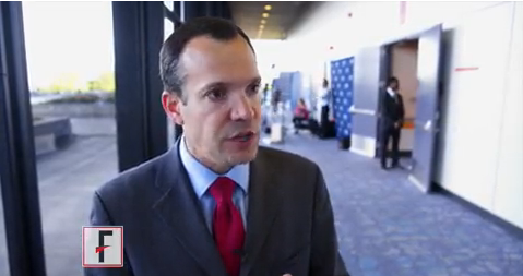
CHICAGO – Immune therapy shows promise for use in the treatment of melanoma brain metastases, especially for treatment-naive patients, judging from the findings of a new phase II randomized study.
For patients with asymptomatic brain metastases from melanoma who had not had prior treatment, nivolumab combined with ipilimumab produced a 50% intracranial response rate after at least 12 weeks of therapy. When nivolumab alone was given to untreated patients, the intracranial response rate was 21%, Georgina Long MD, PhD, co–medical director of the Melanoma Institute Australia , said during a video interview at the annual meeting of the American Society of Clinical Oncology.
“If you look at progression-free survival by response, none of our complete responders have progressed,” said Dr. Long. “And this is with a median follow-up of 16.4 months.” The partial responders have also done well, with little progression, she said. “Remember, these patients usually survive only a few weeks.”
The Anti-PD1 Brain Collaboration study, a phase II clinical trial, enrolled patients with melanoma brain metastases at least 5 mm but less than 40 mm in diameter who had not received previous anti-cytotoxic T-lymphocyte-associated protein 4 (anti-CTLA-4), anti-programmed cell death protein 1 (anti-PD-1), or anti-programmed death-ligand 1 (anti-PD-L1) therapies. Patients were permitted to have had previous BRAF and MEK inhibitor therapies. Asymptomatic patients who had no previous local brain therapy (i.e., radiation treatment or surgery) were randomized 1:1 to receive nivolumab alone, or nivolumab plus ipilimumab.
The nivolumab arm received 3 mg/kg by intravenous infusion every 2 weeks. The combination arm began with nivolumab 1 mg/kg and ipilimumab 3 mg/kg every 3 weeks for four doses. After this, they also received nivolumab 3 mg/kg monotherapy every 2 weeks.
The third cohort – a small group of 15 patients who received nivolumab alone – either had symptomatic brain metastases or leptomeningeal disease and could have had previous brain surgery or radiotherapy. Unlike the first two cohorts, they were also permitted to be on up to 10 mg/day of prednisone; these patients received nivolumab alone at 3 mg/kg every 2 weeks.
For all patients, immune therapy was given until the disease progressed, consent was withdrawn, or patients experienced unacceptable toxicity or they died.
“We were most interested in the randomized cohorts,” said Dr. Long. Interestingly, she said, ipilimumab became available in Australia when 27 patients were enrolled in the nivolumab arm and 26 to the combination arm. “So we stopped the monotherapy arm, and the rest of the 60 patients to be recruited all went into the combination arm,” she said. A total of 76 patients were recruited, 33 into the combination arm, 27 to the asymptomatic nivolumab monotherapy arm, and 16 to the symptomatic and/or previously treated arm.
Data analysis from the point of the data cut included 67 patients who were followed for a period ranging from 5 to 34 months. Intracranial disease was evaluated by gadolinium-enhanced MRI and modified Response Evaluation Criteria in Solid Tumors (RECIST) 1.1 criteria.
“The results of the trial were very interesting,” said Dr. Long. The nivolumab plus ipilimumab combo resulted in an overall 42% intracranial response rate, while nivolumab alone produced an overall intracranial response rate of 21%. However, patients in either arm who had prior BRAF or MEK inhibitor exposure “didn’t do too well on immunotherapy,” said Dr. Long, noting that the response rate was just 16% for these patients. These were, she said, “small numbers, but still, an interesting signal there.”
When comparing the secondary endpoint of extracranial response to intracranial response on a per-patient basis, Dr. Long and her collaborators could see that “the intracranial and extracranial results were mostly concordant.”
Analysis of the additional secondary endpoints of progression-free survival (PFS) and overall survival (OS) also showed an interesting pattern, said Dr. Long. After an initial drop-off period of about 5 months, the curves for patients in all arms have stabilized, so that patients who were responders are maintaining that response. The overall 6-month PFS rate for the combination cohort was 47%, with a durable response: “If you look at the curve, it’s flattened out since that stage, and we haven’t had any progression since that time,” said Dr. Long. The PFS rate was 29% for the cohort receiving nivolumab alone. “Activity is highest when nivolumab and ipilimumab are given upfront,” said Dr. Long.
For asymptomatic patients pretreated with BRAF or MEK inhibitors, “activity is low,” said Dr. Long, with an intracranial response rate of 16% in both cohorts.
Symptomatic patients who were more heavily pretreated fared even worse: “The activity of nivolumab monotherapy is low after multiple modality therapy or in leptomeningeal melanoma,” said Dr. Long. The intracranial response rate in the third cohort was just 6%.
The combination therapy cohort had the most treatment-related adverse events, with 96% of patients experiencing some adverse event. About half (12/26, 46%) had grade 3 or 4 events, and the same number had a serious adverse event. Seven patients (27%) discontinued therapy because of treatment-related adverse events in the combination study arm. However, said Dr. Long, this side effect profile is in keeping with what has been seen in other studies of combination therapy with nivolumab and ipilimumab. “There were not unexpected adverse events,” she said.
Dr. Long reported relationships with Bristol-Myers Squibb, Merck, and Roche.
The video associated with this article is no longer available on this site. Please view all of our videos on the MDedge YouTube channel
[email protected]
On Twitter @karioakes
AT ASCO 2017

New approval lights up gliomas to aid resection
The Food and Drug Administration has approved aminolevulinic acid hydrochloride (Gleolan) to help visualize gliomas during surgery and allow for more complete resection.
Aminolevulinic acid hydrochloride lights up the tumor so surgeons can distinguish it from healthy tissue. Patients take the drug orally – 20 mg/kg – approximately 3 hours before anesthesia. Glioma cells take it up and convert it to the fluorescent chemical protoporphyrin IX. When illuminated under blue light, protoporphyrin in the tumor glows an intense red, while normal brain tissue appears blue, enabling “the surgeon to see the tumor more clearly during brain surgery and to remove it more accurately, sparing healthy brain tissue,” according to information from NX Development Corp, which markets Gleolan.
In a phase III trial of 349 patients with suspected malignant glioma amenable to complete resection, a contrast-enhanced tumor was resected in 64% of patients in the aminolevulinic acid (ALA) arm, versus 38 % of patients in the control-group, who had conventional resection under white light (P less than .001); 20.5 % of ALA patients versus 11 % patients in the control arm were alive at 6 months without progression.
FDA officials noted that there’s a risk of false negatives and positives with ALA, and that “an increase in the extent of resection might increase the risk of serious neurologic deficits in the short term.”
Side effects in preapproval studies included fever, hypotension, nausea, and vomiting in more than 1% percent of patients within a week of surgery. Adverse events included chills, abnormal liver function tests, and diarrhea in less than 1% of patients within 6 weeks of surgery.
VIDEO: Routine genomic testing identifies actionable alterations in 52% of tumors
CHICAGO –
Molecular profiling, including genetic sequencing and copy number variation analysis, was performed in 1944 tumors from patients with advanced tumors enrolled in the profiLER study. Of the tumors screened, mutations deemed actionable were identified in 1,004 (52%), with 394 patients having two or more actionable targets, and the remainder having one identified targeted treatment. A molecular targeted treatment was recommended for 676 patients (35% of those tested).
“We showed that the patients who did receive the molecular targeted agents were doing better in terms of overall survival,” said Olivier Tredan, MD, PhD, the study’s lead investigator. Noting that these are trends as the trial was not randomized, he reported that the overall survival (OS) for those receiving targeted treatments was 53.7% at 3 years, compared with 46.1% for those who did not receive targeted treatment. The trend continued out to 5 years, with the OS for the targeted treatment group at 34.8%, compared with 28.1% OS for those who did not receive targeted treatment, he said at the annual meeting of the American Society of Clinical Oncology.
The video associated with this article is no longer available on this site. Please view all of our videos on the MDedge YouTube channel
Many patients either were too sick to receive the recommended treatment or died before they could be treated, Dr. Tredan said in a video interview.
Of the patients who did receive targeted treatment, over 60% received mTOR inhibitors. The next most common therapies were multitarget tyrosine kinase receptor (TKR)–inhibiting/antiangiogenic therapies, received by about one-third of patients. Fewer than one in five patients received any other therapies. Tumor types were colorectal, gynecological, breast, head and neck carcinomas, sarcomas, and brain tumors.
A new randomized clinical study, profiLER 2, is planned. The new study will pit a 315-gene commercial test against the 69-gene test used in profiLER 1, to see whether casting a wider net yields more targets for therapy.
Still, knowing that a treatment might help is useful only if the patient can actually receive the drug, said Dr. Tredan. “What we want is more molecular targeted agent initiation, so we need to have larger screening programs, but we need also to have access to novel targeted agents.”
Dr. Tredan reported financial relationships with Bayer, GlaxoSmithKline, and Novartis.
[email protected]
On Twitter @karioakes
AT ASCO 2017
VIDEO: TRK inhibitor shows 76% ORR across diverse cancers harboring TRK fusions
CHICAGO – An integrated analysis of three trials has shown that larotrectinib, an oral selective inhibitor of tropomyosin receptor kinase (TRK), has durable efficacy across diverse adult and pediatric cancers harboring TRK fusions, netting an impressive 76% overall response rate.
Lead study author David Hyman, MD, chief of early drug development at Memorial Sloan Kettering Cancer Center in New York, discussed highlights of the analysis, larotrectinib’s regulatory status, and implications for TRK fusion testing in clinical care at the annual meeting of the American Society of Clinical Oncology.
Dr. Hyman disclosed that he has a consulting or advisory role with Atara Biotherapeutics, Chugai Pharma, and CytomX Therapeutics, and that he receives research funding from AstraZeneca and Puma Biotechnology. The study was funded by Loxo Oncology.
The video associated with this article is no longer available on this site. Please view all of our videos on the MDedge YouTube channel
AT ASCO 2017
Modifying CAR-T with IL-15 improved activity in glioma models
MONTREAL – Adding an immunostimulatory cytokine to chimeric antigen receptor–T cells (CAR-T) improved the adaptive immunotherapy’s activity against aggressive pediatric brain malignancies both in vitro and in animal models, an investigator reported.
CAR-T cells engineered to express interleukin-15 (IL-15), an inducer of T-cell proliferation and survival, were associated with significantly longer progression-free survival (PFS) and overall survival in mouse models of glioma, compared with regular CAR-T cells, said Giedre Krenciute, PhD, of Baylor College of Medicine in Houston.

The improved T-cell persistence, however, still resulted in the eventual loss of both targeted and nontargeted tumor-associated antigens and tumor recurrence, Dr. Krenciute noted.
The results suggest that T-cell persistence is critical for antitumor activity, and that it will be necessary to perform antigen profiling of recurrent tumors in order to develop follow-on therapies targeting multiple tumor-associated antigens, she said.
Her team had previously reported on the development of a CAR-T cell directed specifically against the IL-13 receptor alpha-2, which is expressed at high frequency in diffuse intrinsic pontine glioma and glioblastoma tumors but not in normal brain tissues.
In preclinical models, the construct had potent antiglioblastoma activity. However, the T-cells had only limited persistence, and tumors positive for IL-13 receptor alpha-2 recurred.
To see whether they could improve on T-cell persistence, they took their CARs back to the shop and modified them with a retroviral vector to express IL-15 transgenes. They then tested the altered cells in vitro using standard assays and found that the addition of IL-15 did not change the T-cell phenotype or affect the cells’ cytotoxicity.
The addition of IL-15 significantly improved the persistence of the T cells when they were injected into the tumors of mice with human glioblastoma xenografts (P less than .05), and this persistence translated into a near-doubling of PFS, compared with regular CAR-T cells (media, 84 days vs. 49 days; P = .008), as well as improved overall survival (P = .02).
Of 10 mice that received the IL-15–expressing CAR-Ts, 4 were free of glioma through at least 80 days of follow-up.
Of five mice with recurring gliomas, three had down-regulated expression of the IL-13 receptor alpha-2 target, indicating immune escape, and all recurring tumors had reduced expression of the human epidermal growth factor receptor-2 antigen, which is associated with gliomas.
The investigators are currently test-driving a new CD20-targeted CAR, transduced to express IL-15, and have seen good expansion and persistence of the T cells for up to 15 days in culture, with in vivo tests in the planning stage, Dr. Krenciute said.
The work is supported by the National Institutes of Health, Alex’s Lemonade Stand Foundation, the James S. McDonnell Foundation, and the American Brain Tumor Association. The laboratory, the Center for Cell and Gene Therapy at Baylor, has or had research collaborations with Celgene, Bluebird Bio, and Tessa Therapeutics, and investigators at the center hold or have applied for patents in T-cell and gene-modified T-cell therapies for cancer.
MONTREAL – Adding an immunostimulatory cytokine to chimeric antigen receptor–T cells (CAR-T) improved the adaptive immunotherapy’s activity against aggressive pediatric brain malignancies both in vitro and in animal models, an investigator reported.
CAR-T cells engineered to express interleukin-15 (IL-15), an inducer of T-cell proliferation and survival, were associated with significantly longer progression-free survival (PFS) and overall survival in mouse models of glioma, compared with regular CAR-T cells, said Giedre Krenciute, PhD, of Baylor College of Medicine in Houston.
The improved T-cell persistence, however, still resulted in the eventual loss of both targeted and nontargeted tumor-associated antigens and tumor recurrence, Dr. Krenciute noted.
The results suggest that T-cell persistence is critical for antitumor activity, and that it will be necessary to perform antigen profiling of recurrent tumors in order to develop follow-on therapies targeting multiple tumor-associated antigens, she said.
Her team had previously reported on the development of a CAR-T cell directed specifically against the IL-13 receptor alpha-2, which is expressed at high frequency in diffuse intrinsic pontine glioma and glioblastoma tumors but not in normal brain tissues.
In preclinical models, the construct had potent antiglioblastoma activity. However, the T-cells had only limited persistence, and tumors positive for IL-13 receptor alpha-2 recurred.
To see whether they could improve on T-cell persistence, they took their CARs back to the shop and modified them with a retroviral vector to express IL-15 transgenes. They then tested the altered cells in vitro using standard assays and found that the addition of IL-15 did not change the T-cell phenotype or affect the cells’ cytotoxicity.
The addition of IL-15 significantly improved the persistence of the T cells when they were injected into the tumors of mice with human glioblastoma xenografts (P less than .05), and this persistence translated into a near-doubling of PFS, compared with regular CAR-T cells (media, 84 days vs. 49 days; P = .008), as well as improved overall survival (P = .02).
Of 10 mice that received the IL-15–expressing CAR-Ts, 4 were free of glioma through at least 80 days of follow-up.
Of five mice with recurring gliomas, three had down-regulated expression of the IL-13 receptor alpha-2 target, indicating immune escape, and all recurring tumors had reduced expression of the human epidermal growth factor receptor-2 antigen, which is associated with gliomas.
The investigators are currently test-driving a new CD20-targeted CAR, transduced to express IL-15, and have seen good expansion and persistence of the T cells for up to 15 days in culture, with in vivo tests in the planning stage, Dr. Krenciute said.
The work is supported by the National Institutes of Health, Alex’s Lemonade Stand Foundation, the James S. McDonnell Foundation, and the American Brain Tumor Association. The laboratory, the Center for Cell and Gene Therapy at Baylor, has or had research collaborations with Celgene, Bluebird Bio, and Tessa Therapeutics, and investigators at the center hold or have applied for patents in T-cell and gene-modified T-cell therapies for cancer.
FROM ASPHO
Key clinical point: CAR-T cells, modified to express IL-15, have improved activity against aggressive pediatric gliomas.
Major finding: IL-15 expressed T cells were associated with improved progression-free and overall survival in animal models of glioblastoma.
Data source: In vitro and in vivo experiments of CAR-T cells modified to improve T-cell persistence and clinical efficacy.
Disclosures: The work is supported by the National Institutes of Health, Alex’s Lemonade Stand Foundation, the James S. McDonnell Foundation, and the American Brain Tumor Association. The laboratory, the Center for Cell and Gene Therapy at Baylor, has or had research collaborations with Celgene, Bluebird Bio, and Tessa Therapeutics, and investigators at the center hold or have applied for patents in T-cell and gene-modified T-cell therapies for cancer.
Limit primary site radiation, but not craniospinal dose in medulloblastoma
BOSTON – For children with average-risk medulloblastoma, it is safe to limit the radiation boost to the posterior fossa, but reducing doses to the craniospinal axis results in worse outcomes among younger children, and is not advisable, according to results from the largest trial conducted in this population.
There were no significant differences in either 5-year event-free survival (EFS) or overall survival (OS) between children who received an involved field radiation therapy boost (boost to the tumor bed only) or a standard volume boost (to the whole posterior fossa) in a phase III randomized trial, reported lead author Jeff M. Michalski, MD, MBA, FASTRO, professor of radiation oncology at Washington University, St. Louis.
“We conclude that the survival rates and event-free survival rates following reduced boost volumes were comparable to standard radiation treatment volumes for the primary tumor site. This is the first trial to state definitively that there is no survival difference between these two approaches,” he said at a briefing following his presentation of the data in an oral abstract session at the annual meeting of the American Society for Radiation Oncology.
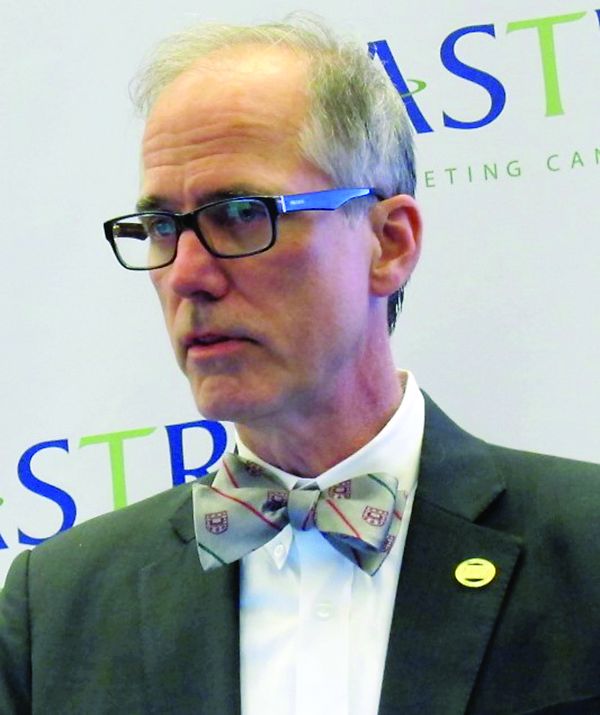
“However, the reduced craniospinal axis irradiation was associated with a higher event rate and worse survival. We believe that physicians can adopt smaller boost volumes to the posterior fossa in children with average-risk medulloblastoma, average risk being no evidence of spread at the time of diagnosis and near-complete or complete resection. But for all children, the standard of 23.4 Gy remains necessary to retain high-level tumor control,” he added.
Aggressive malignancy
Medulloblastoma, an aggressive tumor with the propensity to spread from the lower brain to the upper brain and spine, is the most common brain malignancy in children. The current standard of care for children with average-risk disease is surgical resection followed by systemic chemotherapy followed by irradiation to both the posterior fossa and to the craniospinal axis.
“Unfortunately, this strategy has significant negative consequences for the patients’ neurocognitive abilities, endocrine function, and hearing,” Dr. Michalski said.
The Children’s Oncology Group ACNS0331 trial was designed to determine whether reducing the volume of the boost from the whole posterior to the tumor bed only would compromise EFS and OS, and whether reducing the dose to the craniospinal axis from the current 23.4 Gy to 18 Gy in children aged 3-7 years would compromise survival measures.
The trial had two randomizations, both at the time of study enrollment. In the first, children from the ages of 3 to 7 years were randomly assigned to either low-dose craniospinal irradiation (18 Gy, 116 children) or to the standard dose (23.4 Gy, 110 children). All children aged 8 and older were assigned to receive the standard dose.
For the second randomization, all children were randomly allocated to either involved field RT boost or to standard volume boost to the whole posterior fossa.
Following radiation, all children were assigned to nine cycles of maintenance chemotherapy.
At a median follow-up of 6.6 years, 5-year EFS estimates for the primary site irradiation endpoint were 82.2% for 227 patients who received the involved-field radiation, compared with 80.8% for 237 patients who received whole posterior fossa irradiation.
Respective 5-year OS estimates were 84.1% and 85.2%. The upper limit of the hazard ratio confidence interval (CI) for the involved field therapy was lower than the prespecified limit, indicating that involved-field radiation was noninferior.
For the endpoint of low- vs. standard-dose craniospinal irradiation, the 5-year EFS estimates were 72.1% and 82.6%, respectively. The upper limit of the hazard ratio CI exceeded the prespecified limit, indicating that EFS was worse with the low-dose strategy.
Similarly, respective 5-year OS estimates were 78.1% and 85.9%, with the upper limit of the CI also higher than the prespecified upper limit.
When the investigators looked at the patterns of failure among the children in the two randomizations, they saw that there was no significant difference in the rate of isolated local failure between the involved-field or whole posterior fossa groups, or between the low-dose or high-dose craniospinal irradiation groups.
The finding that radiation volume to the primary site can be reduced is likely to be practice changing, said Geraldine Jacobson, MD, MPH, professor and chair of radiation oncology at West Virginia University, Morgantown. Dr. Jacobson moderated the briefing.
However, the worse survival with the low-dose craniospinal radiation “leaves the COG investigators with the challenge to explore other avenues for reducing toxicity of craniospinal irradiation,” she said.
BOSTON – For children with average-risk medulloblastoma, it is safe to limit the radiation boost to the posterior fossa, but reducing doses to the craniospinal axis results in worse outcomes among younger children, and is not advisable, according to results from the largest trial conducted in this population.
There were no significant differences in either 5-year event-free survival (EFS) or overall survival (OS) between children who received an involved field radiation therapy boost (boost to the tumor bed only) or a standard volume boost (to the whole posterior fossa) in a phase III randomized trial, reported lead author Jeff M. Michalski, MD, MBA, FASTRO, professor of radiation oncology at Washington University, St. Louis.
“We conclude that the survival rates and event-free survival rates following reduced boost volumes were comparable to standard radiation treatment volumes for the primary tumor site. This is the first trial to state definitively that there is no survival difference between these two approaches,” he said at a briefing following his presentation of the data in an oral abstract session at the annual meeting of the American Society for Radiation Oncology.
“However, the reduced craniospinal axis irradiation was associated with a higher event rate and worse survival. We believe that physicians can adopt smaller boost volumes to the posterior fossa in children with average-risk medulloblastoma, average risk being no evidence of spread at the time of diagnosis and near-complete or complete resection. But for all children, the standard of 23.4 Gy remains necessary to retain high-level tumor control,” he added.
Aggressive malignancy
Medulloblastoma, an aggressive tumor with the propensity to spread from the lower brain to the upper brain and spine, is the most common brain malignancy in children. The current standard of care for children with average-risk disease is surgical resection followed by systemic chemotherapy followed by irradiation to both the posterior fossa and to the craniospinal axis.
“Unfortunately, this strategy has significant negative consequences for the patients’ neurocognitive abilities, endocrine function, and hearing,” Dr. Michalski said.
The Children’s Oncology Group ACNS0331 trial was designed to determine whether reducing the volume of the boost from the whole posterior to the tumor bed only would compromise EFS and OS, and whether reducing the dose to the craniospinal axis from the current 23.4 Gy to 18 Gy in children aged 3-7 years would compromise survival measures.
The trial had two randomizations, both at the time of study enrollment. In the first, children from the ages of 3 to 7 years were randomly assigned to either low-dose craniospinal irradiation (18 Gy, 116 children) or to the standard dose (23.4 Gy, 110 children). All children aged 8 and older were assigned to receive the standard dose.
For the second randomization, all children were randomly allocated to either involved field RT boost or to standard volume boost to the whole posterior fossa.
Following radiation, all children were assigned to nine cycles of maintenance chemotherapy.
At a median follow-up of 6.6 years, 5-year EFS estimates for the primary site irradiation endpoint were 82.2% for 227 patients who received the involved-field radiation, compared with 80.8% for 237 patients who received whole posterior fossa irradiation.
Respective 5-year OS estimates were 84.1% and 85.2%. The upper limit of the hazard ratio confidence interval (CI) for the involved field therapy was lower than the prespecified limit, indicating that involved-field radiation was noninferior.
For the endpoint of low- vs. standard-dose craniospinal irradiation, the 5-year EFS estimates were 72.1% and 82.6%, respectively. The upper limit of the hazard ratio CI exceeded the prespecified limit, indicating that EFS was worse with the low-dose strategy.
Similarly, respective 5-year OS estimates were 78.1% and 85.9%, with the upper limit of the CI also higher than the prespecified upper limit.
When the investigators looked at the patterns of failure among the children in the two randomizations, they saw that there was no significant difference in the rate of isolated local failure between the involved-field or whole posterior fossa groups, or between the low-dose or high-dose craniospinal irradiation groups.
The finding that radiation volume to the primary site can be reduced is likely to be practice changing, said Geraldine Jacobson, MD, MPH, professor and chair of radiation oncology at West Virginia University, Morgantown. Dr. Jacobson moderated the briefing.
However, the worse survival with the low-dose craniospinal radiation “leaves the COG investigators with the challenge to explore other avenues for reducing toxicity of craniospinal irradiation,” she said.
Key clinical point: Primary site irradiation in children with medulloblastoma can be limited to the tumor bed rather than the whole posterior fossa.
Major finding: There were no differences in EFS or OS when radiation was limited to the tumor bed, but reduced dose to the craniospinal axis was associated with worse survival.
Data source: Randomized phase III trial of 690 children with average-risk medulloblastoma.
Disclosures: The National Cancer Institute funded the study. Dr. Michalski and Dr. Jacobson reported no relevant conflicts of interest.
Dexamethasone-associated posterior reversible encephalopathy syndrome
Less cognitive decline with stereotactic radiosurgery after brain metastases resection
BOSTON – For patients with resected brain metastases, stereotactic radiosurgery offers survival comparable with what’s seen with whole-brain radiotherapy, but with better quality of life and more effective preservation of cognitive function, investigators reported.
In the phase III N107C trial, there was no difference in overall survival between patients who were randomly assigned to undergo stereotactic radiosurgery (SRS) or whole-brain radiotherapy (WBRT), but patients who underwent WBRT had a twofold greater decline in cognitive function, compared with patients who underwent SRS, Paul D. Brown, MD, a radiation oncologist at the Mayo Clinic in Rochester, Minn., reported at the annual meeting of the American Society for Radiation Oncology.

In a similar prospective, randomized study, Anita Mahajan, MD, and colleagues from the University of Texas MD Anderson Cancer Center in Houston compared postoperative SRS after complete resection with observation alone in 128 patients, and found that although there was no difference in either distant brain metastases or overall survival, SRS was associated with significant improvements in local control.
“I think that going forward with the next patient I see with this scenario, I’m going to be a bit better informed and be able to inform my patient better of the trade-offs involved with regards to the decision of SRS vs. whole-brain radiotherapy,” commented George Rodrigues, MD, from the London (Ontario) Health Sciences Center in Canada. Dr. Rodrigues moderated a briefing during which Dr. Brown and Dr. Mahajan presented their data.
WBRT has been the standard of care for improving local control following surgical resection of brain metastases, but it does not offer a survival benefit and comes at a significant cost in side effects, including alopecia, fatigue, erythema, and, most distressing to patients, significant decline in cognitive function.

The precision of radiosurgery, on the other hand, allows the radiation dose to be concentrated on the surgical bed, limiting exposure of surrounding tissues and structures. For this reason, many centers have begun to adopt SRS for patients with resected brain metastases, but there is not level I evidence to back it up, Dr. Brown said.
WBRT vs. SRS
To rectify this situation, Dr. Brown and his colleagues at the Mayo Clinic and 47 other institutions conducted a clinical trial in 194 patients with one to four brain metastases.
Following surgical resection, the patients were stratified by age, duration of extracranial disease control, number of preoperative metastases, histology, maximum diameter of the resection cavity, and institution, and then randomly assigned to undergo either WBRT or SRS.
Patients were assessed for cognitive function (a coprimary endpoint with overall survival) at baseline and approximately every 3 months thereafter for up to 24 months. Other assessments included MRI scans, and FACT-Br (Functional Assessment of Cancer Therapy-Brain), a quality-of-life instrument.

After a median follow-up of 18.7 months, there was no difference in median overall survival, which was 11.5 months for WBRT and 11.8 months for SRS.
There was, however, a significant difference in cognitive deterioration–free survival, which was 2.8 months for WBRT vs. 3.3 months for SRS. The hazard ratio for WBRT was 2.05 (P = .0001). Cognitive deterioration–free survival rates at 6 months were 5.4% and 22.9%, respectively (P = .0012).
The declines in cognitive function were accounted for by significant differences in the Hopkins Verbal Learning Test (HVLT) domains of total and delayed recall and in the Trail Making Test (Part A).
Overall brain disease control was significantly better with WBRT than with SRS at 3 months (P = .003) and at 6 and 12 months (P less than .001 for each time point).
Surgical bed control was similar between the treatment groups at 6 and 12 months, but was significantly better with WBRT at 12 months, with surgical bed relapse occurring in 21.8% and 44.4% of patients, respectively.
Patients treated with SRS reported significantly better physical well being at 3 and 6 months (P = .002 and .014, respectively). There were 18 grade 3 or greater radiation-related adverse events among patients treated with WBRT, compared with 7 among patients treated with SRS.
SRS vs. observation
In the MD Anderson study, 45% of patients who underwent observation alone had local control of disease at 12 months, compared with 72% treated with SRS. The hazard ratio for SRS was 0.46 (P = .01). The median time to local recurrence was 7.6 months among patients on observation only, but no time point was reached for SRS-treated patients.
The evidence from the two trials suggests that “radiosurgery is a, but not the, standard of care following resection for brain metastasis,” said Vinai Gondi, MD, of the Northwestern Medicine Cancer Center in Warrenville, Ill., the invited discussant.
“While the MD Anderson trial clearly demonstrated that radiosurgery reduces the risk of surgical bed relapse, the N107C trial demonstrated a 44% risk of surgical bed relapse, a rate that is arguably too high in regards to the long survival of resected brain metastasis patients, and it also challenges and risks the resection of surgical bed relapse following radiosurgery,” he said.
The N107C trial was sponsored by the National Cancer Institute and the Alliance for Clinical Trials in Oncology. The MD Anderson trial was funded by a Cancer Center Grant. Dr. Brown, Dr. Mahajan, and Dr. Rodrigues reported no conflicts of interest.
BOSTON – For patients with resected brain metastases, stereotactic radiosurgery offers survival comparable with what’s seen with whole-brain radiotherapy, but with better quality of life and more effective preservation of cognitive function, investigators reported.
In the phase III N107C trial, there was no difference in overall survival between patients who were randomly assigned to undergo stereotactic radiosurgery (SRS) or whole-brain radiotherapy (WBRT), but patients who underwent WBRT had a twofold greater decline in cognitive function, compared with patients who underwent SRS, Paul D. Brown, MD, a radiation oncologist at the Mayo Clinic in Rochester, Minn., reported at the annual meeting of the American Society for Radiation Oncology.
In a similar prospective, randomized study, Anita Mahajan, MD, and colleagues from the University of Texas MD Anderson Cancer Center in Houston compared postoperative SRS after complete resection with observation alone in 128 patients, and found that although there was no difference in either distant brain metastases or overall survival, SRS was associated with significant improvements in local control.
“I think that going forward with the next patient I see with this scenario, I’m going to be a bit better informed and be able to inform my patient better of the trade-offs involved with regards to the decision of SRS vs. whole-brain radiotherapy,” commented George Rodrigues, MD, from the London (Ontario) Health Sciences Center in Canada. Dr. Rodrigues moderated a briefing during which Dr. Brown and Dr. Mahajan presented their data.
WBRT has been the standard of care for improving local control following surgical resection of brain metastases, but it does not offer a survival benefit and comes at a significant cost in side effects, including alopecia, fatigue, erythema, and, most distressing to patients, significant decline in cognitive function.
The precision of radiosurgery, on the other hand, allows the radiation dose to be concentrated on the surgical bed, limiting exposure of surrounding tissues and structures. For this reason, many centers have begun to adopt SRS for patients with resected brain metastases, but there is not level I evidence to back it up, Dr. Brown said.
WBRT vs. SRS
To rectify this situation, Dr. Brown and his colleagues at the Mayo Clinic and 47 other institutions conducted a clinical trial in 194 patients with one to four brain metastases.
Following surgical resection, the patients were stratified by age, duration of extracranial disease control, number of preoperative metastases, histology, maximum diameter of the resection cavity, and institution, and then randomly assigned to undergo either WBRT or SRS.
Patients were assessed for cognitive function (a coprimary endpoint with overall survival) at baseline and approximately every 3 months thereafter for up to 24 months. Other assessments included MRI scans, and FACT-Br (Functional Assessment of Cancer Therapy-Brain), a quality-of-life instrument.
After a median follow-up of 18.7 months, there was no difference in median overall survival, which was 11.5 months for WBRT and 11.8 months for SRS.
There was, however, a significant difference in cognitive deterioration–free survival, which was 2.8 months for WBRT vs. 3.3 months for SRS. The hazard ratio for WBRT was 2.05 (P = .0001). Cognitive deterioration–free survival rates at 6 months were 5.4% and 22.9%, respectively (P = .0012).
The declines in cognitive function were accounted for by significant differences in the Hopkins Verbal Learning Test (HVLT) domains of total and delayed recall and in the Trail Making Test (Part A).
Overall brain disease control was significantly better with WBRT than with SRS at 3 months (P = .003) and at 6 and 12 months (P less than .001 for each time point).
Surgical bed control was similar between the treatment groups at 6 and 12 months, but was significantly better with WBRT at 12 months, with surgical bed relapse occurring in 21.8% and 44.4% of patients, respectively.
Patients treated with SRS reported significantly better physical well being at 3 and 6 months (P = .002 and .014, respectively). There were 18 grade 3 or greater radiation-related adverse events among patients treated with WBRT, compared with 7 among patients treated with SRS.
SRS vs. observation
In the MD Anderson study, 45% of patients who underwent observation alone had local control of disease at 12 months, compared with 72% treated with SRS. The hazard ratio for SRS was 0.46 (P = .01). The median time to local recurrence was 7.6 months among patients on observation only, but no time point was reached for SRS-treated patients.
The evidence from the two trials suggests that “radiosurgery is a, but not the, standard of care following resection for brain metastasis,” said Vinai Gondi, MD, of the Northwestern Medicine Cancer Center in Warrenville, Ill., the invited discussant.
“While the MD Anderson trial clearly demonstrated that radiosurgery reduces the risk of surgical bed relapse, the N107C trial demonstrated a 44% risk of surgical bed relapse, a rate that is arguably too high in regards to the long survival of resected brain metastasis patients, and it also challenges and risks the resection of surgical bed relapse following radiosurgery,” he said.
The N107C trial was sponsored by the National Cancer Institute and the Alliance for Clinical Trials in Oncology. The MD Anderson trial was funded by a Cancer Center Grant. Dr. Brown, Dr. Mahajan, and Dr. Rodrigues reported no conflicts of interest.
AT ASTRO ANNUAL MEETING 2016
Key clinical point: Stereotactic radiosurgery following brain metastases resection was associated with similar survival but less toxicity than was whole-brain radiation therapy.
Major finding: WBRT was associated with a twofold greater risk for cognitive deterioration than SRS in one study, and SRS provided better local control than observation alone in another study.
Data source: A randomized, phase III trial in 194 patients from 48 centers in the United States and Canada, and a randomized trial in 128 patients from the MD Anderson Cancer Center.
Disclosures: The N107C trial was sponsored by the National Cancer Institute and the Alliance for Clinical Trials in Oncology. The MD Anderson trial was funded by a Cancer Center Grant. Dr. Brown, Dr. Mahajan, and Dr. Rodrigues reported no conflicts of interest.

FDA reports shortage of doxorubicin for injection, initiates importation
A critical shortage of doxorubicin hydrochloride 50 mg powder for injection has been reported to the Food and Drug Administration.
Doxorubicin is approved to treat acute lymphoblastic leukemia, acute myeloid leukemia, breast cancer, gastric cancer, ovarian cancer, neuroblastoma, and other cancer types.

To increase availability, the pharmaceutical company Hospira (a Pfizer company) is coordinating with the FDA to import the drug from Ahmedabad, India, where it is manufactured by Zydus Hospira Oncology Private Ltd. at an FDA-inspected facility that is in compliance with current good manufacturing practice requirements.
“It is important to note that there are substantive differences in the format and content of the labeling between the U.S.-approved doxorubicin hydrochloride for injection, USP and the Hospira Limited’s doxorubicin hydrochloride 50 mg powder for injection,” Hospira reported in a letter to health care providers.
To place an order or to get questions answered, contact Hospira directly by calling customer care at 1-877-946-7747 (Mondays-Fridays, 7 a.m.-6 p.m. Central time).
For clinical inquiries, contact Hospira Medical Communications at 1-800-615-0187 or email [email protected].
According to the letter, adverse events or quality problems associated with use of this product should be reported by calling Hospira Global Complaint Management by phone, 1-800-441-4100; by sending an email to [email protected]; or by submitting a report online to Medwatch.
On Twitter @jessnicolecraig
A critical shortage of doxorubicin hydrochloride 50 mg powder for injection has been reported to the Food and Drug Administration.
Doxorubicin is approved to treat acute lymphoblastic leukemia, acute myeloid leukemia, breast cancer, gastric cancer, ovarian cancer, neuroblastoma, and other cancer types.
To increase availability, the pharmaceutical company Hospira (a Pfizer company) is coordinating with the FDA to import the drug from Ahmedabad, India, where it is manufactured by Zydus Hospira Oncology Private Ltd. at an FDA-inspected facility that is in compliance with current good manufacturing practice requirements.
“It is important to note that there are substantive differences in the format and content of the labeling between the U.S.-approved doxorubicin hydrochloride for injection, USP and the Hospira Limited’s doxorubicin hydrochloride 50 mg powder for injection,” Hospira reported in a letter to health care providers.
To place an order or to get questions answered, contact Hospira directly by calling customer care at 1-877-946-7747 (Mondays-Fridays, 7 a.m.-6 p.m. Central time).
For clinical inquiries, contact Hospira Medical Communications at 1-800-615-0187 or email [email protected].
According to the letter, adverse events or quality problems associated with use of this product should be reported by calling Hospira Global Complaint Management by phone, 1-800-441-4100; by sending an email to [email protected]; or by submitting a report online to Medwatch.
On Twitter @jessnicolecraig

Heat shock protein peptide vaccine appears safe, effective for glioblastoma patients
Chicago – A newly developed heat shock protein peptide vaccination appears to be safe and effective in treating patients with newly diagnosed glioblastoma (GBM), according to the results of a phase II single arm study.
In adding the vaccine to standard therapy for 46 patients with newly diagnosed GBM, median overall survival was 23.8 months, and there were no grade 3 or 4 adverse events associated with the vaccine, lead author Dr. Orin Bloch of Northwestern University, Chicago, reported at the annual meeting of the American Society of Clinical Oncology.
Standard therapy typically results in a median survival of 16 months, he said.
“There is a lot of information out there right now regarding CNS and other solid organ tumors particularly in the area of checkpoint modulation and its ability to stimulate an innate immune response against a tumor. I think in GBM we are facing a bit of a different scenario, however, because the tumor exists in a very privileged area behind the blood brain barrier and doesn’t regularly metastasize beyond the CNS,” Dr. Bloch said.
Therefore, only modulating checkpoints without stimulating and educating the immune system may not be the most effective approach. Adaptive immunity through vaccination or some other form of stimulation might be more successful, Dr. Bloch said.
“As a way of inducing immune stimulation and education using tumor-autologous peptides, one can capitalize on the native system of heat shock stimulation. Heat shock proteins are chaperone proteins that are ubiquitously expressed in cells and they’re bound to any number of intracellular peptides at any one time including, in tumor cells, neoantigens. If you extract these heat shock proteins with their bound antigens and deliver them in a naked form into the systemic circulation, their uptake into antigen-presenting cells through the CD91 receptor [will result in] the peptide [being] cleaved and presented on MHC class one and two for stimulation of CD8- and CD4-positive T cell response,” he said.
Heat shock proteins also interact with toll-like receptors and stimulate the secretion of pro-inflammatory cytokines, “acting as their own adjuvant,” Dr. Bloch further explained. Utilizing heat shock proteins activates both the innate and adaptive immune responses.
“This is an ideal platform for developing an immunotherapy for glioblastoma,” Dr. Bloch said.
In this phase II study, 46 adult patients with GBM underwent surgical resection of their tumors followed by chemoradiotherapy. At least four 25-microgram doses of vaccine were generated from tissue obtained during surgery. Within 5 weeks of completing radiotherapy, patients began receiving weekly vaccinations in combination with adjuvant temozolomide. Patients continued receiving vaccines until depletion or until tumor progression.
Median progression-free survival was 17.8 months (95% confidence interval, 11.3-21.6) and median overall survival was 23.8 months (95% CI, 19.8-30.2).
PD-L1 expression on circulating monocytes was also measured from peripheral blood samples obtained during surgery. Patients were classified as having either high PD-L1 expression (54.5% or more of monocytes) or low PD-L1 expression. Among patients classified as having high PD-L1 expression, the median overall survival was 18.0 months (95% CI, 10.0-23.3). Patients who had low PD-L1 expression had a significantly longer median overall survival time of 44.7 months with a confidence interval not calculable (hazard ratio, 3.35; 95% CI, 1.36-8.23; P = .003).
Finally, a multivariate proportional hazards model showed the MGMT methylation status and PD-L1 expression were the two greatest independent predictors of survival.
“Survival among patients who received the HSPPV-96 was greater than expected compared to historical controls... These results certainly, we feel, provide rationale for a phase III trial of vaccine plus standard of care versus standard of care alone,” Dr. Bloch said.
“PD-L1 expression on circulating myeloid cells is independently predictive of clinical response to vaccination, and it suggests that the low PD-L1 expressing population will most benefit from this anti-tumor vaccination scheme, but it also suggests that high PD-L1 expressing patients may benefit from combined checkpoint inhibition. Systemic immunosuppression driven by peripheral monocyte expression of PD-L1 is a previously unidentified factor that may mitigate vaccine efficacy,” Dr. Bloch further commented.
This study was funded by the National Cancer Institute, the National Institute of Neurological Disorders and Stroke, the National Brain Tumor Society, the American Brain Tumor Association, and Accelerated Brain Cancer Cure. Dr. Bloch reporting having no relevant disclosures.
On Twitter @jessnicolecraig
AT THE 2016 ASCO ANNUAL MEETING
Key clinical point: A heat shock protein peptide vaccine appears safe and effective for patients with glioblastoma in an early stage trial.
Major finding: Median progression-free survival was 17.8 months (95% CI, 11.3-21.6). Median overall survival was 23.8 months (95% CI, 19.8-30.2).
Data source: A phase II single arm study of 46 adult patients with glioblastoma.
Disclosures: This study was funded by the National Cancer Institute, the National Institute of Neurological Disorders and Stroke, the National Brain Tumor Society, the American Brain Tumor Association, and Accelerated Brain Cancer Cure. Dr. Bloch reporting having no relevant disclosures.