User login
PULMONARY PERSPECTIVES®: Treatment of Lung Cancer in the High-Risk Patient
Division of Thoracic Surgery
Maimonides Medical Center
Brooklyn, NY
Lung cancer is currently the most common cause of adult cancer related to mortality in the United States. Surgical resection remains the gold standard therapy for resectable disease and offers the best chance for a cure. Unfortunately, age, poor lung function, and significant comorbidity preclude many patients with otherwise resectable lung cancer from surgical therapy. A conventional option for medically inoperable patients with lung cancer includes external beam radiation therapy. Long-term survival with this treatment modality is poor, with reported 5-year survival rates from 10% to 30% and 13% for stage I non-small cell lung cancer (NSCLC) (Sibley et al. Int J Radiat Oncol Biol Phys. 1998;40[1]:149).
The need to improve the treatment of this high-risk group of patients with lung cancer has prompted the development of newer treatment modalities as alternatives to conventional therapy. Radiofrequency ablation (RFA) and stereotactic radiosurgery (SRS) are two such alternative modalities that have emerged in the arena of lung cancer treatment in recent years. RFA has historically been used as an adjunct for treatment of tumors of solid abdominal viscera, and its use for pulmonary malignancy was first reported in 2000. Since that time, multiple case series have been published establishing the safety and efficacy of this treatment modality for pulmonary malignancies in high-risk patients.
SRS was a term originally coined by Leksell to describe a radiation delivery system in which multiple convergent beams of radiation could be delivered to a tumor, utilizing a three-dimensional imaging localization technique. This modality was originally applied to intracranial malignancies, and in 1994, was adapted to treat extracranial lesions as well (Song et al. Oncology. 2004;18[11]:1419). Several reports of its use for pulmonary malignancies began to emerge in the early 2000s; and since that time, refinement of imaging, tracking, and radiation delivery systems has given rise to several different commercially available SRS systems. One such system that has shown success in the treatment of pulmonary malignancy is the CyberKnife® System (Accuray, Sunnyvale, CA).
Radiofrequency Ablation
RFA utilizes heat-induced cellular necrosis and is administered by means of an alternating current applied via an electrode. The current is supplied by a radiofrequency generator and is transferred through the patient and completed via two grounding pads. The alternating current, when applied to tissue, results in agitation of water molecules and frictional release of thermal energy within the immediate area of the electrode. At 46°C cell death occurs within 60 minutes, at 50°C to 52°C irreversible cell death occurs at 4 to 6 minutes, and at 60°C there is instantaneous irreversible cell death.
The goal of RFA is to ablate the tumor with a 0.5- to 1.0-cm margin of surrounding lung tissue. The RFA electrode, with or without tines deployed, ablates a spherical target area of tissue. The target temperature during a conventional pulmonary RFA treatment protocol is 105°C. For an electrode with multiple tines, the temperature is typically averaged across all tines, which each provide accurate real-time temperature readings at their respective locations within the tissue.
Indications for pulmonary RFA include otherwise resectable pulmonary nodules in patients who either refuse surgery or are at high risk for surgical resection. This includes patients with poor pulmonary reserve and/or medically inoperable patients, such as those who have severe coronary/valvular disease, uncompensated congestive heart failure, or other severe comorbidities, who are candidates for RFA. Patients who have failed prior modalities, including surgical resection with or without chemoradiation, would also qualify for RFA treatment.
The only absolute contraindication to RFA is a central location of a nodule, defined as being within 3 cm of the hilum. Central nodules are close to large blood vessels, which function as heat sinks, limiting the therapeutic effect of RFA. Also, the presence of larger airways with corresponding pulmonary vessels near the hilum increases the risk of potentially lethal bronchovascular fistula formation from ablation. Relative contraindications include large nodules (>3 cm) that would require multiple ablations within the same nodule as well as multiple nodules. The latter may be cumbersome to treat and may not be technically feasible, although multiple staged treatments for bilateral pulmonary nodules are possible.
The procedure of RFA for pulmonary lesions with regard to electrode placement and treatment algorithm varies according to the protocols supplied by the individual RFA system manufacturers. Either general endotracheal anesthesia or local with intravenous sedation may be used. The latter generally decreases the risk of pneumothorax with the trade-off that the patient is more likely to move spontaneously during the procedure. Typically, the electrode is placed under image guidance into the center of the nodule after a small skin incision is made to accommodate the 14-gauge needle. Imaging should be repeated between every repositioning to confirm placement. In the event of a periprocedural pneumothorax, a pleural drainage catheter should be placed immediately to evacuate it, as this may cause the lung and nodule to fall away from the chest wall and impede adequate placement of the electrode. A completion CT scan is performed after treatment to visualize the adequacy of the ablation as well as to visualize delayed pneumothorax formation. Occasionally, fiber-optic bronchoscopy may be required to clear endobronchial secretions, which can be blood-tinged after treatment.
Postoperatively, the patient is typically admitted for observation overnight. Chest radiographs are obtained at 6 h postoperatively and the following morning to assess for delayed pneumothorax. If a chest pigtail was required during or after the procedure, a clamp trial is usually performed the following day prior to removal. Rarely, a patient will require a chest drain for a longer period of time, resulting in a prolonged hospital stay. A follow-up chest CT scan may be done the next morning to more accurately assess the post ablation lung but is not required. Patients are typically followed with CT/PET scans at 3- to 4-month intervals.
The most common complication of pulmonary RFA is pneumothorax, occurring in 59% of patients in a recent series. This is largely attributed to the size of the electrode used and is less frequent when positive pressure ventilation is avoided. Prolonged air leak (>5 days) occurred in 7% of patients. Other complications included hemoptysis requiring bronchoscopy, myocardial infarction, deep vein thrombosis, and respiratory failure, which occurred in 1% of patients. In addition, 3% of patients required subsequent drainage of pleural effusions. No intraoperative or in-hospital mortality was observed (Pennathur et al. Ann Thorac Surg. 2009[5];88:1601).
Local recurrence for stage I NSCLC after RFA was confirmed radiographically by CT scan, PET scan, or both after 31.5% of treatments (12/38). Two patients were successfully retreated for technical failures related to pneumothorax; three underwent radiotherapy with stable disease. Three patients died of metastatic disease; five died of pneumonia remote from treatment. The 2- and 4-year survivals were 78% and 47%, respectively. Median overall survival was 30 months. Tumors larger than 3 cm were more likely to recur locally (Pennathur et al. Ann Thorac Surg. 2009[5];88:1601).
CyberKnife® System
The rationale for SRS is based on the notion that higher doses of radiation improve local control and disease-related survival at the expense of increased toxicity in normal tissues (Sibley et al. Int J Radiat Oncol Biol Phys. 1998;40[1]:149). Initially described for the treatment of intracranial lesions, SRS utilized a rigid frame to immobilize a patient, stereotactically localize a lesion, and deliver a higher dose to a specific point with minimal dosing of surrounding normal tissue, using multiple convergent beams of radiation (Song et al. Oncology. 2004;18[11]:1419). As technology evolved, this technique was adapted for extracranial lesions. However, due to the presence of multiple critical structures in the thorax as well as the intrinsic movement of the lung, there was little interest in SRS for pulmonary lesions. Recent advances in SRS led to the development of CyberKnife, which overcame the limitations of conventional SRS in the thorax.
CyberKnife utilizes a frameless system in which the patient needs to not be immobilized. Instead, the CyberKnife System depends on tracking a tumor in real time and adjusting radiation beams accordingly, thus overcoming the limitations of respiratory movement and minimizing toxic exposure to nearby critical structures. It utilizes a 6-MV linear accelerator mounted on a robotic arm. Beams can be emitted in 12 directions from 110 arm positions. The CyberKnife System relies on internally placed radio-opaque fiducial markers that are implanted and allow tumor tracking based on internal rather than external reference points, thus eliminating the need for rigid immobilization (Pennathur et al. Ann Thorac Surg. 2007;83[5]:1820).
Indications for CyberKnife treatment are similar to those of RFA. Unlike those with RFA, patients with central nodules are also candidates for CyberKnife as there are no heat-sinking limitations.
There are no absolute contraindications to CyberKnife. Relative contraindications are similar to those of RFA. Multiple bulky nodules may be difficult to treat without surrounding radiation toxicity and may lead to treatment failure. Central lesions limit the total allowable dose.
The procedure for CyberKnife treatment begins with placement of one to four fiducials in and around the tumor. These are gold tumor markers that are 1 to 2 mm in size and allow for real-time tracking of the tumor. They are placed under image guidance, typically in an outpatient setting. Usually, a total of three fiducials (within, superior, and inferior to the tumor) will suffice. It is critical that the fiducials be placed within the lung parenchyma, as placement within the pleural space or in the fissures will allow migration, compromising tumor tracking.
A week after placement of fiducials, the patient is brought back for a contrast-enhanced CT scan of the chest and upper abdomen with 1.25-mm sections. The treatment plan is then jointly formulated by a thoracic surgeon and radiation oncologist. The tumor volume as well as a 0.5- to 1.0-cm margin of surrounding tissue is outlined using dosimetry software. The treatment area need not be spherical and in fact may be molded to encompass irregularly shaped nodules and to avoid neighboring critical structures. Precise doses at each point within and away from the planned treatment area may be calculated, thus avoiding reaching toxic thresholds in nearby critical structures.
During the treatment phase, the patient is typically positioned according to the previously formulated treatment plan. Fiducials are tracked in real time using two ceiling-mounted radiographic fluoroscopes, and these oblique dual images are combined with the CT scan data, using tracking software to direct or readjust the beams of radiation at frequent intervals. For peripheral lesions, 60 Gy is delivered in three fractions; and for central lesions, 48 Gy is delivered in four fractions to minimize toxicity to surrounding critical structures (Pennathur et al. Ann Thorac Surg. 2009;88[5]:1594).
Patients are followed at 3- to 4-month intervals with PET/CT scans. Nodules are assessed for response/progression based on size, mass quality (cavitation, replacement with scar, etc.), and PET avidity. In addition, treatment-related toxicity is assessed with each follow-up visit with pulmonary function testing and quality-of-life assessment.
Early complications of treatment are mainly related to placement of fiducials. In a recent series, 26% of patients developed a pneumothorax requiring tube thoracostomy. Late complications are mainly due to treatment-related toxicity. This is particularly true for central tumors (Pennathur et al. Ann Thorac Surg. 2009;88[5]:1594). In a phase II trial of 70 patients treated with 60 to 66 Gy in three fractions, only 54% of patients with central tumors were free from severe toxicity compared with 83% of patients with peripheral tumors at 2 years. In summary, 8.6% of patients died of treatment-related toxicity (Timmerman et al. J Clin Oncol. 2006[30];24:4833). Therefore, lower doses are required for central tumors, particularly those near larger central airways.
Using this modality, the overall 2-year survival for primary lung cancer (all stages) was 44%. The median overall survival was 22 months. In conclusion, 62% of patients had progression, which was observed at a median time of 9 months. Patients treated with 60-Gy doses (i.e., those with peripheral lesions) showed significantly improved survival and time to disease progression compared with those treated with 20-Gy doses (Pennathur et al. J Thorac Cardiovasc Surg. 2009;137[3]:597).
Conclusion
RFA and SRS each provide a minimally invasive alternative for high-risk patients with pulmonary malignancy. Studies are ongoing in their application to pulmonary metastases and as part of multimodality treatment protocols. As technology improves, RFA delivery probes will be smaller and can potentially be delivered endobronchially. Similarly, tumor-tracking technology in the spontaneously ventilating lung continues to improve, and fiducial placement may soon be unnecessary in SRS. As the technology evolves, the use of these modalities may expand beyond use only for the high-risk patients.
Dr. Brichkov is with the division of thoracic surgery at Maimonides Medical Center in Brooklyn, N.Y. He has disclosed that he has no significant relationships with the companies/organizations whose products or services are discussed within this Pulmonary Perspectives.
Stenting, then heart surgery best approach for carotid, coronary disease
Short-term outcomes after carotid artery stenting followed by open heart surgery were comparable with those after carotid endarterectomy and open heart surgery performed at the same time, in a retrospective study that compared three approaches with treating patients who had both severe carotid artery disease and coronary artery disease.
However, after 1 year, staged carotid artery stenting and open heart surgery (CAS-OHS) "appears to be a better choice," with a significantly lower risk in the primary composite endpoint of death, stroke, or myocardial infarction, reported Dr. Mehdi H. Shishehbor and his coauthors (J. Am. Coll. Cardiol. 2013 [doi:10.1016/j.jacc.2013.03.094]).
The primary composite endpoint after undergoing CAS-OHS or combined carotid endarterectomy and OHS (CEA-OHS) were similar in both groups.
The third approach studied was carotid endarterectomy (CEA) followed by open heart surgery (staged CEA-OHS), which had the least favorable outcomes of all three approaches, with a "substantial risk of interstage MI," they reported. This approach, therefore, "should be avoided if possible," they concluded in the studywhich was published online on July 31, in the Journal of the American College of Cardiology Cardiovascular Interventions. Dr. Shishehbor is director of endovascular services in the Miller Family Heart and Vascular Institute at the Cleveland Clinic.
The study evaluated outcomes among 350 patients with severe carotid artery stenosis and were candidates for OHS, who underwent carotid revascularization within 90 days of having open heart surgery, at the Cleveland Clinic from 1997 to 2009: 45 had staged CEA-OHS, 195 had combined CEA-OHS, and 110 has staged CAS-OHS. Most of the open heart surgeries were coronary artery bypass grafting procedures.
Based on their analyses, they determined that in the short term, the composite endpoint was similar between those in the staged CAS-OHS group and those in the combined CEA-OHS group – although those in the CAS-OHS group had more MIs, most of which were between the procedures, and those in the combined CEA-OHS group has more perioperative strokes.
Of all three approaches, short-term outcomes were worse in the staged CEA-OHS group, because of the significantly higher risk of interstage MIs.
After 1 year, those in the staged CAS-OHS group had a significantly lower risk of the composite outcomes, compared with the other two groups: a 65% lower risk, compared with those in the combined CEA-OHS group; and a 67% lower risk, compared with those in the staged CAS-OHS group. The risk in the composite outcomes after 1 year in the two CEA groups was similar. Mortality after 1 year was similar in the three groups.
"In choosing between staged CAS-OHS and combined CEA-OHS, the increased risk of interstage MI with the former and perioperative stroke with the latter are important considerations despite similar risks for the early composite endpoint," the authors noted.
"Our study shows that carotid stenting followed by open heart surgery should be the first line strategy for treating patients with severe carotid and coronary disease, if the three- to four-week wait between procedures is clinically acceptable," Dr. Shishehbor said in a statement issued by the Cleveland Clinic. Although there has never been a randomized trial to determine what the best approach is for the types of patients in the study, "the evidence in this study may be enough to change practice," he added.
In fact, as a result of the study findings, changes are being made to the way patients with severe carotid and coronary artery disease are being managed at the Cleveland Clinic, and "we are collaborating across disciplines to identify the lowest risk treatment option for each patient," he added.
In the United States, currently, only 3% of patients with severe carotid and coronary artery disease are treated with staged carotid stenting followed by open heart surgery – compared with 31% of the patients in this study, the statement points out.
Although it was retrospective, "this study provides clarity in the management of patients with carotid and coronary disease requiring OHS," Dr. Mahmud and Dr. Reeves wrote in an accompanying editorial (J. Am. Coll. Cardiol. 2013 [doi:10.1016/j.jacc.2013.07.011]).
Combined CEA-OHS is the optimum revascularization strategy for "patients presenting with an acute coronary syndrome requiring urgent coronary revascularization in whom waiting 3-4 weeks is not safe," although is it associated with more neurological ischemic events.
"However, for patients with a stable or an accelerating anginal syndrome who can wait 3-4 weeks to complete dual-antiplatelet therapy after carotid stenting, staged CAS followed by OHS leads to superior early and long term outcomes," they wrote.
Staged CEA followed by OHS should be avoided, as it "is associated with an increased short term (inter-stage myocardial infarction) and long term (mortality) hazard."
The study, they added, "suggests that the currently acceptable option of CEA prior to OHS actually endangers the patient leading to the highest ischemic event rate both early and late after OHS. These patients should either undergo combined CEA-OHS or be offered the option of CAS prior to OHS based on medical criteria, not reimbursement issues."
Dr. Ehtisham Mahmud and Dr. Ryan Reeves, of the division of cardiovascular medicine and the Sulpizio Cardiovascular Center, at the University of California, San Diego. Dr. Mahmud, chief of cardiovascular medicine at the center, disclosed potential conflicts of interest for Boston Scientific and Abbott Vascular (clinical trial research support), Cordis Corporation and Medicines Company (consulting), and Medtronic (speakers bureau). Dr. Reeves listed no disclosures.
Short-term outcomes after carotid artery stenting followed by open heart surgery were comparable with those after carotid endarterectomy and open heart surgery performed at the same time, in a retrospective study that compared three approaches with treating patients who had both severe carotid artery disease and coronary artery disease.
However, after 1 year, staged carotid artery stenting and open heart surgery (CAS-OHS) "appears to be a better choice," with a significantly lower risk in the primary composite endpoint of death, stroke, or myocardial infarction, reported Dr. Mehdi H. Shishehbor and his coauthors (J. Am. Coll. Cardiol. 2013 [doi:10.1016/j.jacc.2013.03.094]).
The primary composite endpoint after undergoing CAS-OHS or combined carotid endarterectomy and OHS (CEA-OHS) were similar in both groups.
The third approach studied was carotid endarterectomy (CEA) followed by open heart surgery (staged CEA-OHS), which had the least favorable outcomes of all three approaches, with a "substantial risk of interstage MI," they reported. This approach, therefore, "should be avoided if possible," they concluded in the studywhich was published online on July 31, in the Journal of the American College of Cardiology Cardiovascular Interventions. Dr. Shishehbor is director of endovascular services in the Miller Family Heart and Vascular Institute at the Cleveland Clinic.
The study evaluated outcomes among 350 patients with severe carotid artery stenosis and were candidates for OHS, who underwent carotid revascularization within 90 days of having open heart surgery, at the Cleveland Clinic from 1997 to 2009: 45 had staged CEA-OHS, 195 had combined CEA-OHS, and 110 has staged CAS-OHS. Most of the open heart surgeries were coronary artery bypass grafting procedures.
Based on their analyses, they determined that in the short term, the composite endpoint was similar between those in the staged CAS-OHS group and those in the combined CEA-OHS group – although those in the CAS-OHS group had more MIs, most of which were between the procedures, and those in the combined CEA-OHS group has more perioperative strokes.
Of all three approaches, short-term outcomes were worse in the staged CEA-OHS group, because of the significantly higher risk of interstage MIs.
After 1 year, those in the staged CAS-OHS group had a significantly lower risk of the composite outcomes, compared with the other two groups: a 65% lower risk, compared with those in the combined CEA-OHS group; and a 67% lower risk, compared with those in the staged CAS-OHS group. The risk in the composite outcomes after 1 year in the two CEA groups was similar. Mortality after 1 year was similar in the three groups.
"In choosing between staged CAS-OHS and combined CEA-OHS, the increased risk of interstage MI with the former and perioperative stroke with the latter are important considerations despite similar risks for the early composite endpoint," the authors noted.
"Our study shows that carotid stenting followed by open heart surgery should be the first line strategy for treating patients with severe carotid and coronary disease, if the three- to four-week wait between procedures is clinically acceptable," Dr. Shishehbor said in a statement issued by the Cleveland Clinic. Although there has never been a randomized trial to determine what the best approach is for the types of patients in the study, "the evidence in this study may be enough to change practice," he added.
In fact, as a result of the study findings, changes are being made to the way patients with severe carotid and coronary artery disease are being managed at the Cleveland Clinic, and "we are collaborating across disciplines to identify the lowest risk treatment option for each patient," he added.
In the United States, currently, only 3% of patients with severe carotid and coronary artery disease are treated with staged carotid stenting followed by open heart surgery – compared with 31% of the patients in this study, the statement points out.
FROM THE JOURNAL OF THE AMERICAN COLLEGE OF CARDIOLOGY
Major finding: Short-term outcomes among patients undergoing carotid revascularization and open heart surgery were comparable among those who had carotid artery stenting followed by open heart surgery and those who had the combined procedure of carotic endarterectomy and surgery at the same time. But after 1 year, the former approach was associated with more favorable outcomes.
Data source: A retrospective study that evaluated outcomes in 350 patients who had carotid artery stenting or carotid endarterectomy before open heart surgery, or carotid endarterectomy at the same time as open heart surgery.
Disclosures: Dr. Shishehbor is a speaker and consultant for Abbott Vascular, Medtronic, and GORE, but waived all compensations for this study. Another author disclosed serving as a consultant to Boston Scientific, GORE, Medtronic, Endologix, and Vessix Vascular. Nine authors had no disclosures. The REDCap project is supported by National Center for Research Resources/National Institutes of Health.
Postop pneumonia risk strong for subset of thoracic surgery patients
SAN DIEGO – For thoracic surgery patients, being on neoadjuvant chemotherapy, having chronic obstructive pulmonary disease, and a weight loss of greater than 10% were all associated with the development of postoperative pneumonia, results from a single-center study showed.
At the national conference of the American College of Surgeons/National Surgical Quality Improvement Program, Dr. Elisabeth Dexter noted that after the first ACS/NSQIP data harvest at the Roswell Park Cancer Institute in Buffalo, N.Y., the risk of postoperative pneumonia was found to be 4.4%, compared with a rate of 1.1% in all other NSQIP hospitals.
"Of particular note, the thoracic surgery service had a high incidence of 13.2%," said Dr. Dexter, an attending surgeon in the department of thoracic surgery at the Institute. "The high incidence of our postoperative pneumonia was likely [affected] by our thoracic surgery service because our thoracic surgery service had an increased percentage of the abstracted NSQIP data in our cohort, from 12% to 14%, compared with other NSQIP hospitals of similar academic size abstracting 2%. When we found this high postoperative pneumonia rate, we decided to query our NSQIP data and our tumor registry between July 1, 2011, and Oct. 8, 2012, to ask the question: Is there an increased incidence of postoperative pneumonia in thoracic surgery patients who received neoadjuvant chemotherapy compared with those who did not?"
Dr. Dexter and her associates cross-referenced ACS/NSQIP data on 1,723 patients at the cancer center with the tumor registry. Of the 1,723 patients, 1,645 had no postoperative pneumonia while 78 did. Compared with the non-pneumonia patients, those who had pneumonia tended to be older (a mean of 67 vs. 60 years, respectively; odds ratio, 1.05; P less than .001), more likely to be male (59% vs. 37%; OR, 2.48; P less than .001), have chronic obstructive pulmonary disease (35% vs. 9%; OR, 5.08; P less than .001), be a smoker (36% vs. 24%; OR, 1.75; P = .021), and had lost more than 10% of body weight (10% vs. 2.5%; OR, 4.47; P less than .001).
On univariate analysis, postoperative pneumonia was associated with being on neoadjuvant chemotherapy (4.2% vs. 14%; OR, 3.75; P less than .001).
In addition, certain surgical subspecialties at the Institute had a high incidence of postoperative pneumonia, including thoracic surgery (46%), GI surgery (21%), and gynecology (12%).
When the researchers included the entire cohort of patients, those who were on neoadjuvant therapy had an increased incidence of postoperative pneumonia, compared with those who were not on neoadjuvant chemotherapy (P = .001). When thoracic surgery patients were excluded from the analysis, non-thoracic surgery patients who were on neoadjuvant chemotherapy had no increased incidence of postoperative pneumonia, compared with the patients who were not on neoadjuvant chemotherapy (P = .681). On multivariate analysis, significant variables associated with postoperative pneumonia were being on neoadjuvant chemotherapy (P= .001), having chronic obstructive pulmonary disease (P less than .0001), and having weight loss of greater than 10% (P = .004).
"Institutions with disproportionately busy complex thoracic surgery programs may have rates of postoperative pneumonia skewed higher than predicted by NSQIP models," Dr. Dexter concluded. "Optimization of nutritional status and COPD treatment in neoadjuvant chemotherapy patients may reduce postoperative pneumonia risk and incidence. Balance of oncologic benefit of neoadjuvant chemotherapy versus risk and morbidity of postoperative chemotherapy warrants future study in thoracic surgery patients."
Dr. Dexter said that she had no relevant financial conflicts to make.
AT THE ACS NSQIP NATIONAL CONFERENCE
Major finding: On multivariate analysis, significant variables associated with postoperative pneumonia were being on neoadjuvant chemotherapy (P = .001), having COPD (P less than .0001), and having weight loss of greater than 10% (P = .004).
Data source: A study of 1,723 patients who underwent surgery at Roswell Park Cancer Institute in Buffalo, N.Y. Of the postoperative pneumonia cases that developed, 46% were from the thoracic surgery service.
Disclosures: Dr. Dexter said that she had no relevant financial disclosures to make.
A mitral valve replacement that may grow with the child
NEW YORK – Physicians at Boston Children’s Hospital have replaced the mitral valves of eight infants with irreparable mitral valve disease with a valve that offers the opportunity of sequential expansion as the child grows, according to Dr. Sitaram M. Emani, who described the results at the 2013 Mitral Valve Conclave.
"The Melody valve retains its competence if you expand it before putting it in. We asked whether the valve retains the ability to maintain competence even if expansion is performed after implantation as the patient grows," said Dr. Emani, a pediatric cardiac surgeon at Boston Children’s Hospital.
According to Dr. Emani, the current options for infants with damaged mitral valves that are beyond repair are replacement with mechanical or bioprosthetic valves or the Ross mitral procedure. Perhaps the main disadvantage of these options is the lack of a prosthetic valve small enough for an infant, one that is less than 12 mm in diameter. Another problem is the possibility of stenosis developing as the child grows, since the diameters of the prosthetics are fixed. Other drawbacks are that supra-annular fixation is generally associated with poor outcomes and that annular fixation limits the ability to upsize at reoperation.
The Melody valve is an externally stented bovine jugular vein graft that was designed for transcatheter pulmonary valve replacement. In this study, the valve was inserted surgically. The valve maintains competence over a range of sizes up to 22 mm. Although this valve is not approved for use for mitral valve replacement, the hope of using such a prosthetic is that it can be enlarged in the catheterization laboratory as the child grows.
Dr. Emani conducted a retrospective study of his experience with the Melody valve for mitral valve replacement in eight infants less than 12 months of age. The median age at implantation was 6 months (range, 1-9 months). Four infants had an atrioventricular canal (AVC) defect and four had congenital mitral valve stenosis. Most of the children had two prior operations for mitral valve repair. The longest follow-up to date has been 2 years.
At a median follow-up of 8 months, regurgitation on the echocardiogram was considered to be mild or less in all patients. The median gradient was 3 mm Hg (range, 2-7 mm Hg) on the immediate postoperative echocardiogram. Three patients developed a mild paravalvular leak; one of these patients had undergone aggressive stent resection, a modification Dr. Emani does not recommend. One patient developed left ventricular outflow tract obstruction (LVOTO), which Dr. Emani attributed to the lack of distal stent fixation in this patient. Another patient with an AVC defect developed complete heart block.
One patient who died 3 days postoperatively had heterotaxy, severe mitral regurgitation, and prior ventricular failure on extracorporeal membrane oxygenation support. That patient had undergone valve implantation as a last resort.
Three patients underwent sequential expansion about 6 months after implantation. After valve expansion, the median balloon size was 12 mm, ranging from 12 to 16 mm. None of the patients developed worsening valvular function and all had relief of obstruction. Transcatheter intervention was used to correct a paravalvular leak in one patient and to treat a left ventricular outflow tract problem in another. None of the patients developed endocarditis or a strut fracture, "although I worry about strut fracture if aggressive stent resection and manipulation is performed," Dr. Emani said at the meeting, which was sponsored by the American Association for Thoracic Surgery.
Dr. Emani offered some procedural tips. First, the Melody valve must be optimized for surgical implantation in infants. The length of the valve must be reduced by trimming it to reduce the chance of LVOTO or pulmonary vein obstruction. He recommends sizing the valves by echocardiogram and fixating the distal stent to the inferior free wall of the ventricle.
Although he has used both circumferential and four-point fixation, Dr. Emani has learned all that is really needed to prevent leakage is friction of the stent against the annulus. Early on he used a pericardial cuff to anchor to the annulus, particularly in patients who had undergone failed AVC repair. He tries to preserve at least part of the anterior leaflet to facilitate suture placement and create a "stand-off" from the LVOTO.
Dr. Emani also advised limiting intraoperative dilation to no more than 1 mm greater than the measured annulus. "Try not to overdilate at implantation to avoid heart block, LVOTO, and coronary compression. The nice thing is you don’t have to decide then and there what size you want. You can go back to the cath lab and, under direct visualization with the coronary view, you can dilate it under more controlled circumstances.
"The hope is that we will be able to dilate these valves as the patients grow into adolescence. If we can dilate them up to 22 mm, hopefully we will decrease the number of repeat replacements, delay the time to reoperation, and perhaps modify our thresholds for tolerating significant disease after unsuccessful repairs," he said. The investigators continue to accumulate experience with this device and hope to design a multicenter trial evaluating its safety and efficacy for this indication.
Dr. Emani had no relevant financial disclosures.
AT THE 2013 MITRAL VALVE CONCLAVE
Major finding: Eight infants with irreparable mitral valve disease underwent mitral valve replacement with the Melody valve, which offers the opportunity of sequential expansion as the child grows.
Data source: Small case series.
Disclosures: Dr. Emani had no relevant financial disclosures.
Tight glycemic control normalized thyroid function after pediatric heart surgery
SAN FRANCISCO – Tight glycemic control helps normalize thyroid hormone concentrations following cardiac surgery in children, according to results from a randomized trial presented at the Endocrine Society’s annual meeting.
That could be important because "it’s well known that cardiac surgical procedures can induce nonthyroidal illness in children" – a brief free-hormone surge followed by low T3 (triiodothyronine) and T4 (thyroxine) and increased rT3 (reverse triiodothyronine). It’s associated with longer recoveries, said lead investigator Dr. Carmen Soto-Rivera, an endocrinologist at Boston Children’s Hospital.
But it’s difficult to say what the clinical implications of her team’s findings are, she noted, because "we still don’t know if nonthyroidal illness is adaptive or maladaptive."
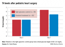
The study involved 191 children who underwent cardiac surgery with cardiopulmonary bypass. After surgery, her team randomized 99 children who were 0-36 months old to tight glycemic control, with a blood glucose target of 80-110 mg/dL; and 92 others to the standard treatment, permissive hyperglycemia; their median blood glucose hovered around 120 mg/dL.
Two days after surgery, 98% of the children had T4 and T3 levels below age-specific normal ranges, consistent with nonthyroidal illness. The thyroid hormone binding ratio (THBR) was normal in most of the patients, so it did not explain the drop in T4 and T3.
The groups had begun to separate by day 7. Tight-control patients had a higher median T4 than did patients on standard treatment (6.4 vs. 5.3 mcg/dL; P = .02) and a greater median T4 change from day 2 (2.4 vs. 1.3 mcg/dL, P = .02).
The trend continued among children still in the hospital on day 14. The 30 tight-control patients, compared with 37 on standard treatment, had a higher median T3 (104.5 vs. 74 ng/dL; P = .02) and a greater change from day 2 (26 vs. 5 ng/dL; P = .02). They had a higher T4 on day 14 too, but the difference was not significant.
"Interestingly, we found that [tight glucose control] resulted in lower [C-reactive protein] levels by day 7," about 3 mg/L, versus about 4 mg/L in the standard-treatment arm (P = .01). Perhaps "the faster increase in thyroid hormone levels may be mediated by decreased inhibition of deiodinases due to lowered inflammation," Dr. Soto-Rivera said.
The findings differ from previous reports of tight glucose control increasing peripheral inactivation of thyroid hormone, she said. "We believe that because our glucose targets were higher and we had [a low] rate of hypoglycemia in both arms, we potentially prevented a fasting response in these children," leading to decreased inhibition of the thyroid axis, she added.
"We believe that almost no one was treated with thyroid replacement during [the study], but we are in the process of confirming this," she said.
The project was a subgroup analysis of a larger trial that found no significant benefit for tight glycemic control on infection rates, mortality, length of stay, and organ failure following pediatric heart surgery (N. Engl. J. Med. 2012;367:1208-1219).
Dr. Soto-Rivera said she had no disclosures. The National Institutes of Health funded the project.
AT ENDO 2013
Major finding: Seven days after heart surgery, children on tight glycemic control had significantly higher median T4 levels than did children on standard treatment (6.4 vs. 5.3 mcg/dL).
Data source: Randomized trial of 191 children following heart surgery.
Disclosures: Dr. Soto-Rivera said she has no disclosures. The National Institutes of Health funded the project.
EVEREST II: Tight MR control improves MitraClip patients' survival
NEW YORK – In a group of 351 high-risk surgical patients with severe mitral regurgitation (grade 3 or 4) at baseline, more than 80% were alive 1 year after transcatheter treatment with the MitraClip device if their MR was well controlled (MR grade 1+ or 2+), compared with 58% survival for those with persistent severe MR.
These results from the EVEREST II High Surgical Risk cohort were presented by Dr. Paul A. Grayburn at the 2013 Mitral Valve Conclave.
The presentation was chosen as one of the Plenary "Top 10" Abstract Presentations at the meeting.
"Many high-risk surgical patients do not undergo mitral valve surgery and therefore have no therapeutic option for MR. Percutaneous repair with MitraClip is an option for selected patients with suitable anatomy who are too high of a risk for surgery. In this study, we found that the extent of clinical benefit observed corresponds with the degree of MR reduction achieved," said Dr. Grayburn, a cardiologist at the Baylor Heart and Vascular Institute in Dallas. "MR reduction to 1+ or 2+ results in significant clinical and symptomatic improvement."
The MitraClip is a catheter-based valve repair system intended to reduce MR. The device is inserted via the femoral vein using a guide catheter, and the MitraClip implant is attached directly to the mitral valve. On March 20, 2013, the FDA’s Circulatory System Devices Advisory Panel voted by a narrow margin (5-3) in favor of the device, finding that the benefits outweighed the risks.
Data were pooled from the EVEREST II High Risk Study and the EVEREST II REALISM Continued Access Study. The mean age of the 351 patients was 76 years, with 58% of the group over age 75. At baseline, patients had significant comorbidities, including 82% with coronary artery disease, 51% with a prior myocardial infarction, 69% with a history of atrial fibrillation, and 60% with previous cardiovascular surgery. Of the 98% with heart failure, 85% were New York Heart Association (NYHA) functional class III/IV. Aside from cardiac problems, 31% had moderate to severe renal disease, and 29% had chronic obstructive pulmonary disease. Functional MR was present in 70% of the group.
With these health problems in mind, the patients were considered to be at high risk for mitral valve surgery as indicated by a Society of Thoracic Surgeons (STS) risk calculator operative mortality of 12% or more.
Mortality at 1 year was 22.8%. Looking at freedom from mortality at 1 year after treatment, 80.9% of those discharged with MR greater than or equal to 1+ and 80.3% of those discharged with an MR grade of 2+ were alive. For those discharged with a grade of 3+ or 4+, 58.4% were alive (P = .0003).
"As you can see, it is not good for you to leave the operating room after MitraClip with an MR grade of 3+ or 4+," Dr. Grayburn said at the meeting, which was sponsored by the American Association for Thoracic Surgery.
Residual MR grade also influenced other measures. For instance, patients with low levels of MR showed better left ventricular remodeling, as measured by larger changes from baseline in left ventricular end diastolic volume (LVEDV) and left ventricular systolic volume. For LVEDV, there was a 15% reduction from baseline in the group with MR greater than or equal to 1+ and a 10% reduction in the 2+ group, but no statistically significant reduction in the 3+/4+ groups.
After treatment, significantly fewer patients with an MR grade greater than or equal to 1+ were considered NYHA functional class III/IV, compared with those with MR 3+ or 4+.
Other benefits of treatment included a 54% reduction in the annual rate of hospitalization due to congestive heart failure in the 12 months after the MitraClip procedure vs. the 12 months pre-MitraClip in patients with an MR grade of 2+ at discharge, compared with no change in hospitalization rate in those discharged with MR greater than 2+. Trends were noted between better MR control and scores on the Short Form-36 (SF-36) quality of life score and the mental component score.
Dr. Grayburn emphasized that the MitraClip procedure is not for everybody but offers an option for those considered at high risk. "If you can operate on these patients, you should; then you can get their MR to null. This device is really a palliative offering for those turned down by a mitral valve surgeon at an experienced mitral valve center. At our center, many patients referred to us for MitraClip actually end up getting surgery because they turn out to be surgical candidates." At his center, Dr. Grayburn says that for every patient who gets a MitraClip, two undergo surgery.
Nevertheless, Dr. Grayburn believes that some high-risk patients who might benefit from MitraClip are unaware of this alternative. "We haven’t figured out how to capture patients with severe symptomatic MR in the general internist’s or even cardiologist’s office."
Dr. Grayburn received research support from Abbott Vascular.
AT THE 2013 MITRAL VALVE CONCLAVE
Major finding: In a study of 351 high-risk patients with severe mitral regurgitation (MR) who underwent repair with the MitraClip device, survival was more than 80% for those with well-controlled MR, compared with 58% for those with persistent severe MR.
Data source: The prospective EVEREST II High Risk Study and the EVEREST II REALISM Continued Access Study.
Disclosures: Dr. Grayburn received research support from Abbott Vascular.
Aortic valve-sparing root surgery durable at 1 year
MINNEAPOLIS – Despite the complexity of aortic valve–sparing techniques, early outcomes and 1-year survival were similar to those achieved with valve replacing during root replacement surgery in patients with Marfan syndrome in a prospective registry study.
Major adverse valve-related events (MAVREs) at 1 year were also comparable, although there was an increase, of course, in aortic valve regurgitation with valve sparing (7% vs. 0%), Dr. Joseph Coselli said at the annual meeting of the American Association for Thoracic Surgery.
"Follow-up is needed for this particular incident because we don’t know exactly what’s going to happen to these 2-plus aortic regurgitations," he said. "It’s quite possible they may remain stable over a long period of time and don’t represent a failure of the concept."
In an initial report from the international registry, valve-sparing techniques were the most common, and provided comparable 30-day outcomes in 151 patients (J. Thorac. Cardiovasc. Surg. 2009;137:1124-32).
The current analysis involved 316 patients, aged 4-70 years, who underwent aortic valve–sparing (AVS) (n = 239) or aortic valve–replacing (AVR) (n = 63 mechanical and 14 tissue) root replacement surgery at 19 centers between March 2005 and November 2010. The type of operation was determined by clinical factors, and by surgeon and patient preference. AVR surgery was considered the only option in 17% of patients. At 1 year, clinical follow-up was complete in 98% and imaging follow-up in 93%.
"Interestingly, when we started out this particular collection of patients, over 30% were receiving aortic valve replacement, but toward the end of this observational study, late 2010, virtually almost all patients were receiving aortic valve sparing," said Dr. Coselli, chief of adult cardiac surgery at Baylor College of Medicine, Texas Heart Institute, Houston.
AVS patients were younger (33 vs. 39 years); had smaller sinuses of Valsalva (49 vs. 53 mm); and had less acute dissection (3% vs. 9%), chronic dissection (3% vs. 12%), and previous cardiovascular surgery (5% vs. 14%).
Aortic-sparing techniques required significantly longer cardiopulmonary bypass time (195 vs. 152 minutes) and aortic clamp time (156 vs. 115 minutes), but cut ICU time from 46 hours with AVR surgery to 26 hours, ventilator support time from 12 to 8 hours, and hospital length of stay from 7 to 6 days, Dr. Coselli reported.
At 30 days, MAVREs were reported in 6 patients in the AVR group and 15 in the AVS group (P = .4), with no differences in nonstructural dysfunction (1 vs. 7), embolism (1 vs. 3), or bleeding (2 vs. 3 events).
Two AVS patients required early reoperation: One required same-day reintervention because of coronary artery kinking after a Florida sleeve procedure, and the second needed reintervention because of a coronary pseudoaneurysm 6 days after a David-V procedure, he said. Two early deaths occurred, one in each group, but neither was valve related.
One-year survival rates were 98% after AVS surgery and 97% after AVR surgery (4 vs. 2 deaths; P = .06).
At 1 year, MAVREs occurred in 8 AVR and 35 AVS patients (P = .5). There was no significant difference between AVR and AVS in freedom from valve-related death (1 vs. 2), embolism (2 vs. 4 events), reintervention (0 vs. 1), endocarditis (1 vs. 0), valve thrombosis (0 both), or valve-related morbidity (7 vs. 28), Dr. Coselli said.
The AVS group had significantly more nonstructural dysfunction/structural valve deterioration (23 events vs. 1 event; P = .04), but significantly less bleeding (3 vs. 5 events; P = .01).
In a Cox regression analysis at 1 year, the type of surgery was not associated with overall survival, MAVREs, or any other valve-related outcome, he said.
Dr. Coselli reported research support from St. Jude Medical; an educational grant, consultancy, and royalties from Vascutek Terumo; and research support, speaking for, and steering committee membership with Medtronic. Two coauthors reported consultant/advisory board participation with Medtronic or Edwards Lifesciences.
AT THE AATS ANNUAL MEETING
Major finding: One-year survival rates were 98% after AVS surgery and 97% after AVR surgery (4 vs. 2 deaths; P = .06).
Data source: Prospective, international, observational registry study in 316 Marfan syndrome patients undergoing aortic root surgery.
Disclosures: Dr. Coselli reported research support from St. Jude Medical; an educational grant, consultancy, and royalties from Vascutek Terumo; and research support, speaking for, and steering committee membership with Medtronic. Two coauthors reported consultant/advisory board participation with Medtronic or Edwards Lifesciences.
Study IDs predictors of unplanned hospital readmission after CEA
SAN FRANCISCO – The 30-day unplanned readmission rate following carotid endarterectomy was 6.5% in a single-center study.
In addition, four variables were significantly associated with unplanned readmission: in-hospital postoperative congestive heart failure (CHF) exacerbation; in-hospital postoperative stroke; in-hospital postoperative hematoma; and prior coronary artery bypass graft (CABG).
"Whether these complications are completely avoidable is unknown, but we do identify a group of patients who would probably benefit from more comprehensive discharge planning and careful postdischarge care," Dr. Karen J. Ho said at the Society for Vascular Surgery annual meeting.
According to a study of Medicare claims data from 2003 to 2004, 20% of Medicare beneficiaries discharged from a hospital were rehospitalized within 30 days (N. Eng. J. Med. 2009;360:1418-28). The 30-day rehospitalization rate after vascular surgery was 24%, "the highest of all surgical specialties examined in the study," said Dr. Ho of the surgery department at Brigham and Women’s Hospital, Boston, who was not involved with the published study. "Medicare has started to decrease reimbursements for hospitals with excess readmissions after acute MI, heart failure, and pneumonia. Hip and knee replacements and chronic obstructive pulmonary disease will be added in 2014, and we anticipate that additional surgical procedures will be added thereafter," she said.
In an effort to determine the rate of 30-day unplanned readmission after carotid endarterectomy (CEA), Dr. Ho and her associates conducted a retrospective study of a prospectively collected vascular surgery database at Brigham and Women’s Hospital. The cohort included 896 consecutive CEAs performed between 2002 and 2011. Combined CABG/CEA procedures were excluded.
The primary endpoint was unplanned readmission within 30 days, defined as "any unanticipated, nonelective hospital readmission," she said. The secondary endpoint was 1-year survival.
The mean age of the patients was 70 years, 60% were male, and 95% were white. More than half (65%) had asymptomatic evidence of carotid artery disease.
Dr. Ho reported that the median postoperative length of stay was 1 day and that 9.9% of patients had at least one in-hospital complication. The most frequent in-hospital complication was bleeding/hematoma (4.1%), followed by arrhythmia (2.1%), dysphagia (1.7%), stroke (1.3%), and myocardial infarction (1.2%). Only 3% of patients required a reoperation, while most (94%) were discharged to home. The 30-day stroke rate was 1.7%, while the 30-day death rate was 0.6%.
The overall 30-day readmission rate was 8.6%, while the unplanned 30-day readmission rate was 6.5%. "Most of the overall readmissions (80%) occurred in the first 10 days, and the median time to unplanned readmission was 4 days," Dr. Ho said.
The most common reason for an unplanned readmission was a cardiac complication, followed by headache, bleeding/hematoma, stroke/transient ischemic attack/intracerebral hemorrhage, or other medical emergency. More than one-quarter of patients (27.5%) had more than one reason for an unplanned readmission, while 87.9% of patients had a CEA-related unplanned readmission.
When the researchers performed a univariate analysis followed by analysis with a multivariable Cox model for unplanned readmission, four variables were independently associated with unplanned readmission: in-hospital postoperative CHF exacerbation (hazard ratio, 15.1), in-hospital postoperative stroke (HR, 5.0), in-hospital postoperative hematoma (HR, 3.1), and prior CABG (HR, 2.0).
They also observed a significant difference in survival at 1 year between patients who had an unplanned readmission and those who did not (91% vs. 96%, respectively; P less than .01.) "It’s unclear whether these deaths in the unplanned readmission group were preventable or if they were related to carotid disease or to a procedure-related complication," Dr. Ho said. "Our guess is that the increased overall burden of comorbid disease in these patients, rather than the readmission itself, predicted decreased survival."
Limitations of the study included its retrospective design and the fact that it was conducted at a single center, she said, "but we do know that our unplanned readmission rate is comparable to estimates from recent Medicare data."
Dr. Ho said she had no relevant financial disclosures.
AT THE SVS ANNUAL MEETING
Major finding: Four variables were independently associated with unplanned readmission: in-hospital postoperative CHF exacerbation (hazard ratio, 15.1), in-hospital postoperative stroke (HR, 5.0), in-hospital postoperative hematoma (HR, 3.1), and prior CABG (HR, 2.0).
Data source: A study of 896 consecutive CEAs performed between 2002 and 2011 at Brigham and Women’s Hospital, Boston.
Disclosures: Dr. Ho said she had no relevant financial disclosures.
One-third of perioperative EVAR deaths occurred after discharge
SAN FRANCISCO – One-third of perioperative deaths and complications after elective endovascular repair of abdominal aortic aneurysms occur post discharge, results from a large analysis showed.
"Improved predischarge surveillance and close postdischarge follow-up of identified high-risk patients may further improve 30-day outcomes after EVAR," Dr. Prateek K. Gupta said at the Society for Vascular Surgery annual meeting.
Outcome improvement in the field of aortic surgery, specifically endovascular repair of abdominal aortic aneurysms, "has received much attention," said Dr. Gupta of the department of surgery at the University of Wisconsin Hospital and Clinics, Madison. "With EVAR, the index hospital stay after aortic surgery has decreased significantly, leaving a need for better understanding of postdischarge outcomes, which is necessary to improve quality and reduce readmission rates with implementation of targeted outpatient interventions."
In an effort to examine postdischarge 30-day outcomes after elective EVAR, Dr. Gupta and his associates identified 11,229 patients from the American College of Surgeons National Surgical Quality Improvement Program (NSQIP) database who underwent an elective EVAR for AAA between 2005 and 2010. The primary outcome of interest was postdischarge mortality, while the secondary outcome was postdischarge overall morbidity. The researchers performed univariate and multiple logistic regression analysis to assess factors associated with the primary and secondary study outcomes.
Of the 11,229 patient 83% were male and their mean age was 75 years. Dr. Gupta reported that 117 patients died within 30 days of EVAR, for a rate of 1%. Of these deaths, 31% occurred after hospital discharge, and the median time to death was 9 days. At the same time, 1,204 patients experienced complications within 30 days of EVAR, for a rate of 11%. Of these, 500 (40%) occurred post discharge, and the median time for a complication to occur was 3 days.
Only 20% of patients (7/36) who died post discharge experienced an in-hospital complication. Compared with patients who did not develop a postdischarge complication, those who had more than a sixfold likelihood of reoperation (20.4% vs. 3.1%, respectively; P less than .0001) and death (3.0% vs. 0.2%; P less than .0001) within 30 days of surgery.
Multivariable analysis revealed the following factors that were independently and significantly associated with postdischarge mortality: preoperative heart failure (adjusted odds ratio, 4.7), admission from a skilled nursing facility (AOR, 2.2), increase in age per year (AOR, 1.09), postdischarge renal failure requiring dialysis (AOR, 72.5), postdischarge cardiac arrest/MI (AOR, 46.6), and postdischarge pneumonia (AOR, 26.5).
Dr. Gupta reported that the 30-day postdischarge rate among patients admitted from a nursing facility or acute care was 2.5%. "In contrast to patients who survived after EVAR, patients who died post discharge were more likely to have been admitted from a nursing facility or acute care (13.9% vs. 1.8%; P less than .0001)," he said.
The 30-day post-discharge mortality was highest among patients who had postdischarge renal failure (27%),postdischarge MI (19%), and postdischarge pneumonia (15%).
The researchers also found that patients with a history of peripheral artery disease (PAD) had a significantly higher post-discharge complication rate after EVAR (7.1% vs. 4.3%; P = .001). This also correlated with a higher wound infection rate (3.2% vs. 1.7%; P = .01). A previous cardiac surgery also predisposed patients toward a higher overall postdischarge complication rate (5.3% vs. 4.2%; P = .007).
"Usually, patients undergoing EVAR are followed up at 2 weeks for wound evaluation, or at 1 month with a CT scan," Dr. Gupta said. "In the present study, the median occurrence for most of the postdischarge complications was within the first 10 days after surgery. The interquartile range was 11-22 days for the diagnosis of a wound infection after EVAR. These data suggest that earlier follow-up of high-risk patients may help identify and possibly prevent some of these complications and subsequently decrease readmissions. A standardized protocol for triage and surveillance of high-risk patients post EVAR is needed."
Limitations of the study include that fact that causality could not be determined because it was a retrospective analysis. "In addition, the timing of the operation is not specified in NSQIP," so it could either be a predischarge event or it could have occurred on readmission, Dr. Gupta said. "Data on readmission is not available from the 2005-2010 data sets."
Dr. Gupta said that he had no relevant financial disclosures to make.
Dr. Gupta and his colleagues have assessed postprocedure complications after elective EVAR based upon review of the NSQIP database, and concluded that earlier follow-up of high-risk patients might identify and prevent some of the complications. This study is limited by the database nature of the review. This study also does not provide us with data as to the size of the AAA in the high risk patients.
Most of the complications leading to increased risk of mortality were postoperative issues, such as renal failure or MI, which could not be identified at the time of procedure, or the time of discharge. The only identifiable preoperative risk factors for adverse outcomes were preoperative heart failure, admission from a skilled nursing facility and increasing age. While changing the timing of follow-up might be appropriate for the high-risk patients, other considerations would be changing to more percutaneous procedures , and use of other adjuncts, such as antibiotic irrigations or Prevena (negative pressure wound therapy for intact skin) to decrease the wound infection rates for those undergoing femoral exploration for EVAR. Further, any intervention on the elderly, especially nursing home patients, needs to be thoroughly considered, as EVAR is most often a preventive operation, assuming fitness and appropriate longevity remains for the patient.
The findings from this study are important, but mostly, should serve as a caution to properly assess patients to determine who will potentially benefit from EVAR, and which patients might be best managed by observation alone.
Dr. Linda Harris is the program director and division chief of vascular surgery at the State University of New York, Buffalo.
SAN FRANCISCO – One-third of perioperative deaths and complications after elective endovascular repair of abdominal aortic aneurysms occur post discharge, results from a large analysis showed.
"Improved predischarge surveillance and close postdischarge follow-up of identified high-risk patients may further improve 30-day outcomes after EVAR," Dr. Prateek K. Gupta said at the Society for Vascular Surgery annual meeting.
Outcome improvement in the field of aortic surgery, specifically endovascular repair of abdominal aortic aneurysms, "has received much attention," said Dr. Gupta of the department of surgery at the University of Wisconsin Hospital and Clinics, Madison. "With EVAR, the index hospital stay after aortic surgery has decreased significantly, leaving a need for better understanding of postdischarge outcomes, which is necessary to improve quality and reduce readmission rates with implementation of targeted outpatient interventions."
In an effort to examine postdischarge 30-day outcomes after elective EVAR, Dr. Gupta and his associates identified 11,229 patients from the American College of Surgeons National Surgical Quality Improvement Program (NSQIP) database who underwent an elective EVAR for AAA between 2005 and 2010. The primary outcome of interest was postdischarge mortality, while the secondary outcome was postdischarge overall morbidity. The researchers performed univariate and multiple logistic regression analysis to assess factors associated with the primary and secondary study outcomes.
Of the 11,229 patient 83% were male and their mean age was 75 years. Dr. Gupta reported that 117 patients died within 30 days of EVAR, for a rate of 1%. Of these deaths, 31% occurred after hospital discharge, and the median time to death was 9 days. At the same time, 1,204 patients experienced complications within 30 days of EVAR, for a rate of 11%. Of these, 500 (40%) occurred post discharge, and the median time for a complication to occur was 3 days.
Only 20% of patients (7/36) who died post discharge experienced an in-hospital complication. Compared with patients who did not develop a postdischarge complication, those who had more than a sixfold likelihood of reoperation (20.4% vs. 3.1%, respectively; P less than .0001) and death (3.0% vs. 0.2%; P less than .0001) within 30 days of surgery.
Multivariable analysis revealed the following factors that were independently and significantly associated with postdischarge mortality: preoperative heart failure (adjusted odds ratio, 4.7), admission from a skilled nursing facility (AOR, 2.2), increase in age per year (AOR, 1.09), postdischarge renal failure requiring dialysis (AOR, 72.5), postdischarge cardiac arrest/MI (AOR, 46.6), and postdischarge pneumonia (AOR, 26.5).
Dr. Gupta reported that the 30-day postdischarge rate among patients admitted from a nursing facility or acute care was 2.5%. "In contrast to patients who survived after EVAR, patients who died post discharge were more likely to have been admitted from a nursing facility or acute care (13.9% vs. 1.8%; P less than .0001)," he said.
The 30-day post-discharge mortality was highest among patients who had postdischarge renal failure (27%),postdischarge MI (19%), and postdischarge pneumonia (15%).
The researchers also found that patients with a history of peripheral artery disease (PAD) had a significantly higher post-discharge complication rate after EVAR (7.1% vs. 4.3%; P = .001). This also correlated with a higher wound infection rate (3.2% vs. 1.7%; P = .01). A previous cardiac surgery also predisposed patients toward a higher overall postdischarge complication rate (5.3% vs. 4.2%; P = .007).
"Usually, patients undergoing EVAR are followed up at 2 weeks for wound evaluation, or at 1 month with a CT scan," Dr. Gupta said. "In the present study, the median occurrence for most of the postdischarge complications was within the first 10 days after surgery. The interquartile range was 11-22 days for the diagnosis of a wound infection after EVAR. These data suggest that earlier follow-up of high-risk patients may help identify and possibly prevent some of these complications and subsequently decrease readmissions. A standardized protocol for triage and surveillance of high-risk patients post EVAR is needed."
Limitations of the study include that fact that causality could not be determined because it was a retrospective analysis. "In addition, the timing of the operation is not specified in NSQIP," so it could either be a predischarge event or it could have occurred on readmission, Dr. Gupta said. "Data on readmission is not available from the 2005-2010 data sets."
Dr. Gupta said that he had no relevant financial disclosures to make.
AT THE SVS ANNUAL MEETING
Major finding: Following endovascular repair of abdominal aortic aneurysms, 31% of deaths and 40% of complications occurred after hospital discharge.
Data source: An analysis of 11,229 patients from the American College of Surgeons National Surgical Quality Improvement Program (NSQIP) database who underwent an elective EVAR for AAA between 2005 and 2010.
Disclosures: Dr. Gupta said that he had no relevant financial conflicts to disclose.
Medicare-remunerated EVAR can mean negative operation margins
SAN FRANCISCO – Endovascular aneurysm repair (EVAR) is associated with negative operating margins among Medicare beneficiaries, and device costs account for more than 50% of the technical costs, results from a single-center study demonstrated.
"U.S. health care expenditures have steadily increased over several decades, with some projections now reaching 20% of gross domestic product by 2020," Dr. David H. Stone said at the annual meeting of the Society for Vascular Surgery Annual Meeting. "Accordingly, vigorous debate surrounding health care reform has ensued. In this setting physicians and health care system alike are placing a growing emphasis on both cost reduction and quality improvement, thus increasing the overall value of health care delivery. Endovascular aneurysm repair represents a high-value procedure, though it remains associated with significant cost. This places EVAR at odds with efforts to constrain procedure-associated health care dollars."
Dr. Stone, in the section of vascular surgery at Dartmouth-Hitchcock Medical Center, Lebanon, N.H., and his associates retrospectively examined the EVAR-associated technical costs, revenues, and resulting operating margins among 127 infrarenal EVARs performed at the center between April 2011 and March 2012. They excluded cases in which anatomy was deemed outside of conventional "Instructions for Use" guidelines, included cases treated only by a single vendor’s device, and restricted the payer source to Medicare-remunerated cases billed using the DRG 238 code. This left a cohort of 49 patients. The researchers then determined mean EVAR implant costs per procedure and used 2012 University HealthSystem Consortium data to benchmark their DRG 238 costs and length of stay – another major driver for cost.
"To our surprise, we initially determined that our section’s annual net operating margin for EVAR when billed using DRG 238 was substantially negative, approaching –$500,000 per year," Dr. Stone said. Specifically, mean technical costs among the 49 patients were $31,672, while technical revenue was $27,657, resulting in a negative technical operating margin of $4,015 per case. More specifically, stent grafts accounted for 52% of the technical costs while institutional overhead costs accounted for the remaining 48%.
Among the nonimplant costs the operating room accounted for the single greatest technical cost driver (17%). "By comparison, stent grafts account for roughly threefold more technical cost than [did] any nonimplant hospital costs," Dr. Stone said. "Interestingly, there is an apparent inequity between the stent graft costs when considered as a percentage of cost vs. a percentage of revenue. More specifically, stent grafts currently account for 52% of the technical costs but assume 60% of the DRG payment, thus contributing in part to our institution’s negative margin."
Given the substantial impact of graft costs to the procedure, the researchers also examined Dartmouth-Hitchcock’s current vendor market share for the medical center’s entire EVAR practice. The vendors were not named but rather described as vendors A, B, C, and D. "Though historically we have not routinely integrated costs into our case planning, we were somewhat surprised to learn that vendor D the highest-cost device derived the largest market share, while vendors A and B the two lowest-cost devices derived the smallest market shares, respectively," Dr. Stone said. "Surgeons were largely unaware of this cost disparity." He said that a "lack of transparency" of the device costs among institutions has also led in part to the sustainability of this practice pattern.
Dr. Stone acknowledged certain limitations of the study, including its single-center design, "thus graft pricing and institutional overhead will likely vary among hospitals," he said. "In addition, we did not analyze DRG 237–remunerated EVAR with major complications, where costs may be higher yet. However, we nevertheless believe that the adjudicated financial costs presented here may reflect a similar trend in many institutions throughout the country for Medicare-remunerated EVAR."
He concluded his remarks by noting that the negative operating margin for Medicare-remunerated EVAR "is likely unsustainable. Surgeon awareness of price differential among grafts may allow for competitive negotiated pricing. Accordingly, we believe that EVAR as a high-value procedure must undergo care delivery redesign, reflecting cost restructuring with viable remuneration schemes in order for current practice to remain sustainable."
Dr. Stone said that he had no relevant financial conflicts to disclose.
Dr. Stone and his colleagues at Dartmouth-Hitchcock have identified simple, uncomplicated EVAR cases as producing a negative contribution margin to the institution, primarily because of device costs. In today’s environment of cost containment, it is imperative that physicians partner with institutions to evaluate appropriate methods of cost containment, while maintaining excellence of care. These findings should not lead to the abandonment or restriction of EVAR, but rather further discussions between hospitals and physicians as to ways to decrease cost of the procedure. This will include negotiations with vendors to potentially decrease device costs and to provide, at the least, budget neutral interventions, as the current status is not sustainable.
As we move forward, these types of calculations will need to occur at all institutions for high-volume procedures to ensure viability of the institutions while maintaining excellence of medical care.
Dr. Linda Harris is the program director and division chief of vascular surgery at the State University of New York, Buffalo.
SAN FRANCISCO – Endovascular aneurysm repair (EVAR) is associated with negative operating margins among Medicare beneficiaries, and device costs account for more than 50% of the technical costs, results from a single-center study demonstrated.
"U.S. health care expenditures have steadily increased over several decades, with some projections now reaching 20% of gross domestic product by 2020," Dr. David H. Stone said at the annual meeting of the Society for Vascular Surgery Annual Meeting. "Accordingly, vigorous debate surrounding health care reform has ensued. In this setting physicians and health care system alike are placing a growing emphasis on both cost reduction and quality improvement, thus increasing the overall value of health care delivery. Endovascular aneurysm repair represents a high-value procedure, though it remains associated with significant cost. This places EVAR at odds with efforts to constrain procedure-associated health care dollars."
Dr. Stone, in the section of vascular surgery at Dartmouth-Hitchcock Medical Center, Lebanon, N.H., and his associates retrospectively examined the EVAR-associated technical costs, revenues, and resulting operating margins among 127 infrarenal EVARs performed at the center between April 2011 and March 2012. They excluded cases in which anatomy was deemed outside of conventional "Instructions for Use" guidelines, included cases treated only by a single vendor’s device, and restricted the payer source to Medicare-remunerated cases billed using the DRG 238 code. This left a cohort of 49 patients. The researchers then determined mean EVAR implant costs per procedure and used 2012 University HealthSystem Consortium data to benchmark their DRG 238 costs and length of stay – another major driver for cost.
"To our surprise, we initially determined that our section’s annual net operating margin for EVAR when billed using DRG 238 was substantially negative, approaching –$500,000 per year," Dr. Stone said. Specifically, mean technical costs among the 49 patients were $31,672, while technical revenue was $27,657, resulting in a negative technical operating margin of $4,015 per case. More specifically, stent grafts accounted for 52% of the technical costs while institutional overhead costs accounted for the remaining 48%.
Among the nonimplant costs the operating room accounted for the single greatest technical cost driver (17%). "By comparison, stent grafts account for roughly threefold more technical cost than [did] any nonimplant hospital costs," Dr. Stone said. "Interestingly, there is an apparent inequity between the stent graft costs when considered as a percentage of cost vs. a percentage of revenue. More specifically, stent grafts currently account for 52% of the technical costs but assume 60% of the DRG payment, thus contributing in part to our institution’s negative margin."
Given the substantial impact of graft costs to the procedure, the researchers also examined Dartmouth-Hitchcock’s current vendor market share for the medical center’s entire EVAR practice. The vendors were not named but rather described as vendors A, B, C, and D. "Though historically we have not routinely integrated costs into our case planning, we were somewhat surprised to learn that vendor D the highest-cost device derived the largest market share, while vendors A and B the two lowest-cost devices derived the smallest market shares, respectively," Dr. Stone said. "Surgeons were largely unaware of this cost disparity." He said that a "lack of transparency" of the device costs among institutions has also led in part to the sustainability of this practice pattern.
Dr. Stone acknowledged certain limitations of the study, including its single-center design, "thus graft pricing and institutional overhead will likely vary among hospitals," he said. "In addition, we did not analyze DRG 237–remunerated EVAR with major complications, where costs may be higher yet. However, we nevertheless believe that the adjudicated financial costs presented here may reflect a similar trend in many institutions throughout the country for Medicare-remunerated EVAR."
He concluded his remarks by noting that the negative operating margin for Medicare-remunerated EVAR "is likely unsustainable. Surgeon awareness of price differential among grafts may allow for competitive negotiated pricing. Accordingly, we believe that EVAR as a high-value procedure must undergo care delivery redesign, reflecting cost restructuring with viable remuneration schemes in order for current practice to remain sustainable."
Dr. Stone said that he had no relevant financial conflicts to disclose.
AT THE SVS ANNUAL MEETING
Major finding: In a study of DRG 238 remunerated EVAR, stent grafts accounted for 52% of the technical costs while institutional overhead costs accounted for the remaining 48%.
Data source: A retrospectively examination of the EVAR-associated technical costs, revenues, and resulting operating margins among 127 infrarenal EVARs performed at Dartmouth-Hitchcock Medical Center between April 2011 and March 2012.
Disclosures: Dr. Stone said that he had no relevant financial conflicts to disclose.
