User login
AAD: Tacrolimus Safe for Kids, Phase IV Study Finds
MIAMI – Long-term use of tacrolimus ointment in children with atopic dermatitis is associated with a low rate of serious adverse events, according to interim data from a prospective study.
The safety profile shown thus far is consistent with the established profile for tacrolimus ointment (Protopic), and it is hoped that these findings will encourage dermatologists to continue enrolling pediatric patients in this ongoing phase IV safety study, Dr. Joyce Rico said in a poster presentation at the annual meeting of the American Academy of Dermatology.
The interim report focused on 4,800 patients enrolled in APPLES (A Prospective Pediatric Longitudinal Evaluation Study) between May 2005 and September 2009. The investigators seek to enroll 8,000 children in the multinational, observational cohort study, said Dr. Rico, vice president of medical science at Astellas Pharma Inc., Chicago.
That is the number needed to accurately identify an increase in the risk of malignancy associated with tacrolimus treatment over the course of the 10-year study, but the investigators are also evaluating other serious adverse events.
Such events occurred in about 5% of the participants, who had a median age of 6 years at enrollment. Most of the serious events were asthma, occurring in 1% of patients, and infectionsincluding pneumonia, bronchitis, cellulitis, gastroenteritis, and tonsillitis – occurring in nearly 2% of patients. There was one report of a malignancy – a peripheral glioneuronal tumor – not typically associated with immunosuppression, Dr. Rico noted.
The malignancy rate in APPLES is half that seen in the general U.S. population, based on data from the Surveillance, Epidemiology, and End Results database, she said.
About 40% of children in APPLES had mild disease at baseline and about 60% reported moderate or severe disease. Treatment is either 0.03% ointment (the dosage approved for children) or off-label 0.1% ointment (the dosage approved for adults); most are being treated with 0.1% ointment. The median cumulative duration of time since initial treatment application is 2.8 years per patient.
“The serious adverse events profile we’re seeing is consistent with what was observed during the clinical development program,” Dr. Rico said, noting that, although the results are encouraging, enrollment of the full 8,000 patients is needed to adequately address the long-term safety of tacrolimus treatment in children.
“I encourage you and your colleagues to continue to enroll patients ... It’s going to take the dermatology community continuing to help us to reach that goal,” she concluded.
Dr. Rico is employed by Astellas, the maker of Protopic and sponsor of the study.
AAD: Novel Topical Shows Promise for Atopic Dermatitis
MIAMI — A substituted trans-stilbene derivative known as WBI-1001, which showed promise for the topical treatment of atopic dermatitis in preclinical studies, was safe and effective for mild to moderate disease in a small double-blind, randomized, vehicle-controlled study.
A total of 36 patients aged 18-65 years with chronic mild to moderate atopic dermatitis were randomized to apply either 0.5% WBI-1001 cream, 1.0% cream, or vehicle cream twice daily for 4 weeks.
At 1-week follow-up, the two treatment groups had significant reductions in Eczema Area and Severity Index (EASI) scores, compared with the vehicle group, with score reductions of 59.3% and 54.9% in the 0.5% and 1.0% groups, respectively, versus a 7.1% reduction in the vehicle group, Dr. Youwen Zhou reported during a poster discussion session at the annual meeting of the American Academy of Dermatology.
Similar improvements were seen in Severity Scoring of Atopic Dermatitis Severity Index (SCORAD) scores, Investigator Global Assessment (IGA) scores, body surface area (BSA) affected, and pruritis; in the 0.5% and 1.0% treatment groups vs. the vehicle group, respectively, SCORAD scores decreased 56.2% and 50.1% vs. 18.4%; IGA scores decreased by 38.9% and 45.8% vs. 5.6%; body surface area affected decreased by 64.4% and 57.7% vs. 1.08%; and pruritis scores decreased by 74% and 56% vs. 25%.
At 4-week follow-up, half of the patients in the treatment groups were clear or almost clear, compared with only 16.7% of those in the vehicle group, said Dr. Zhou of the University of British Columbia, Vancouver.
No treatment-related adverse events were seen in this study, no drug accumulation was reported, and no detectable drug was found in 76% of plasma samples, indicating that there was only minimal absorption into the blood system, Dr. Zhou said.
This is an early-stage clinical trial, but the findings suggest that the novel nonsteroidal anti-inflammatory WBI-1001 compound has promise for the treatment of mild to moderate atopic dermatitis, he concluded.
He noted that, except for pruritis, which occurred less often in the 0.5% treatment group, the 0.5% and 1.0% doses had similar outcomes that were significantly better than those seen in the vehicle arm.
The study was supported by Welichem Biotech Inc. Dr. Zhou is a paid consultant for Welichem Biotech.
AAD: Text Messages May Boost Atopic Dermatitis Treatment Compliance
Miami — Text daily educational and treatment reminder messages to patients with atopic dermatitis and you just might get a big “TY” message back.
That’s a “Thank You.”'
Reminder text messages were associated with improved treatment adherence, health maintenance behaviors, and possibly disease severity in a study of 20 patients aged 14 years and older, Venessa Pena-Robichaux reported in a poster at the annual meeting of the American Academy of Dermatology.
The patients, who had a mean age of 29.6 years, received daily texts for 6 weeks, which alternated between reminders (such as, “Please remember to use the medication or product you use to treat your atopic dermatitis today!”) and educational messages (such as, “People with atopic dermatitis have a lifelong tendency to develop skin infections.”). Eighty-five percent of participants said they found the medication reminder messages helpful, 90% said they found the educational messages helpful, 80% said they would like to keep receiving the messages, and 85% said they would recommend the texting program to a friend, said Ms. Pena-Robichaux, a fourth-year medical student at Harvard Medical School, Boston.
The texts were not only welcomed by the participants, they appeared to improve medication use: 80% of patients reported an increase in the number days each week that they were adherent to their medical treatment. The number of days of medication use increased from a prestudy mean of 3.7 days per week to a poststudy mean of 6.1 days. Additionally, 95% of patients reported an improvement in at least one health maintenance behavior during the study period, and the SCORing Atopic Dermatitis (SCORAD) index decreased in 70% of patients from pre- to post study by a mean of 7.3 points on the 100-point scale.
Given the popularity of cell phones and text messaging – and in particular the tech-savvy nature of younger patients these days (about half of the participants in this study were college students), the potential of this simple and cost-effective intervention for improving positive health behaviors deserves a second look, Ms. Pena-Robichaux said, noting that a larger randomized controlled study of the texting program is in the works and could be underway B 4 2 long.
Vitamin D Insufficiency May Be Linked to Allergies, Asthma in Kids
New Orleans — Approximately half of children with asthma were deficient in vitamin D in a study of 99 children aged 18 and younger. The findings were presented in a poster at the annual meeting of the American Academy of Allergy, Asthma and Immunology.
Previous studies have indicated that vitamin D insufficiency contributes to the pathophysiology of allergic disease, but data on vitamin D’s impact on children with allergies and asthma are limited, said Dr. Daniel Searing of National Jewish Health in Denver, Colo.
In this study, Dr. Searing and colleagues identified 99 children who had asthma, atopic dermatitis, and/or a food allergy. The researchers assessed vitamin D by measuring serum 25-hydroxyvitamin D levels.
Overall, 47% of the patients had insufficient levels of vitamin D (less than 30 ng/mL). The median vitamin D level was 31 ng/mL.
To assess the impact of vitamin D on inflammation, the researchers cultured peripheral blood mononuclear cells (PBMC) from 11 patients using either 10 nM vitamin D or a placebo medium for 24 hours, and supplemented them with either 10 or 100 nM of dexamethasone for the last 3 hours of culturing. Next, they measured mitogen-activated protein kinase phosphatase-1 (MKP-1) and interleukin-10 (IL-10.)
“Vitamin D enhances glucocorticoid induction of MKP-1 and IL-10 in asthmatic PBMC in vitro,” the researchers wrote. Vitamin D addition can enhance the activity of dexamethasone more than 10-fold, they added.
“The relationship between vitamin D and corticosteroid pathways, as well as its effect on the inflammatory response, is not fully understood,” the researchers emphasized. But the results suggest that vitamin D supplementation may enhance the anti-inflammatory function of corticosteroids in asthma patients, they noted.
Median vitamin D levels were significantly lower in children taking inhaled corticosteroids (29 ng/mL), oral corticosteroids (25 ng/mL) and long-acting beta-agonists (25 ng/mL), compared with children who were not taking inhaled corticosteroids, oral corticosteroids, or long-acting beta-agonists (35 ng/mL, 32 ng/mL, and 34 ng/mL, respectively)
In addition, median vitamin D levels were significantly lower in children with positive vs. negative aeroallergen sensitivity to dog dander (29 ng/mL vs. 35 ng/mL) and house dust mites (27 ng/mL vs. 31 ng/mL).
Dr. Searing had no financial conflicts to disclose. The study was supported in part by a grant from the National Institutes of Health.
Topical Antacid Therapy May Soothe Capsaicin-Induced Dermal Pain
Early treatment of capsaicin-induced dermal pain with the use of topical antacids can be effective in quickly and significantly reducing skin irritation and discomfort, according to a recent study.
"Dermal exposure of capsaicin affects cutaneous sensory neurons, inducing burning, redness, irritation, and pain that sometimes can be excruciating and last for hours to days after exposure," wrote Susan Y. Kim-Katz, Pharm.D., and her associates.
However, the results of their study, published online in the American Journal of Emergency Medicine, suggested the pain can be alleviated within 30 minutes if treated with an antacid containing calcium or magnesium.
Participants in the study were recruited from the California Poison Control System (CPCS) 24-hour hotline from a pool of callers who reported dermal exposure to capsaicin over the 15-month period between January 2001 and April 2002. In addition, study participants contacted the CPCS a median of 1 hour after exposure.
In the 64 subjects whose data were analyzed for outcomes, 45 (70%) reported a positive response to antacid treatment as a 33% reduction in pain within 30 minutes, noted Dr. Kim-Katz, a clinical pharmacist at the University of California, San Francisco.
A majority of participants, 36 (56%), were exposed to unrefined capsaicin through natural peppers (such as jalapeño, habañero, or red chili), while the remaining subjects were exposed to refined capsaicin (as contained in personal protection sprays such as pepper spray, animal repellents, creams, or ointments). They were advised by the CPCS to coat or soak the affected area liberally with a room-temperature antacid product that contains calcium or magnesium, such as Maalox, Mylanta, Milk of Magnesia, or Tums (crushed and mixed with water).
When asked to assess their pain on a scale of 0 (no pain) to 10 (worst pain ever experienced) during the initial phone call to CPCS, the median initial pain score was 7.5. During a follow-up call 2 hours after the initial CPCS consultation, the mean decrease in pain score after treatment was 4.2 points (doi:10.1016/j.ajcm.2009.02.007).
The study's authors discussed similar studies that have supported this outcome, including a 1998 study. In that study, seven patients had "severe dermal discomfort" after "pepper-mace" exposure. Within minutes after they applied liquid Maalox to the affected area, the patients were pain free (Am. J. Emerg. Med. 1998;16:613-4).
Topical antacids may provide pain relief by raising extracellular pH, which decreases skin pain receptors' sensitivity to capsaicin, the researchers said. In addition, the presence of positively charged calcium and magnesium atoms in the antacid may suppress capsaicin's actions, they noted.

Organic "red hot chili peppers" from Hutchins Farm in Concord, Mass. (Image courtesy "Arden" via flickr creative commons)
Dr. Kim-Katz and several of her colleagues reported that they are on staff at the California Poison Control System, San Francisco Division.
Early treatment of capsaicin-induced dermal pain with the use of topical antacids can be effective in quickly and significantly reducing skin irritation and discomfort, according to a recent study.
"Dermal exposure of capsaicin affects cutaneous sensory neurons, inducing burning, redness, irritation, and pain that sometimes can be excruciating and last for hours to days after exposure," wrote Susan Y. Kim-Katz, Pharm.D., and her associates.
However, the results of their study, published online in the American Journal of Emergency Medicine, suggested the pain can be alleviated within 30 minutes if treated with an antacid containing calcium or magnesium.
Participants in the study were recruited from the California Poison Control System (CPCS) 24-hour hotline from a pool of callers who reported dermal exposure to capsaicin over the 15-month period between January 2001 and April 2002. In addition, study participants contacted the CPCS a median of 1 hour after exposure.
In the 64 subjects whose data were analyzed for outcomes, 45 (70%) reported a positive response to antacid treatment as a 33% reduction in pain within 30 minutes, noted Dr. Kim-Katz, a clinical pharmacist at the University of California, San Francisco.
A majority of participants, 36 (56%), were exposed to unrefined capsaicin through natural peppers (such as jalapeño, habañero, or red chili), while the remaining subjects were exposed to refined capsaicin (as contained in personal protection sprays such as pepper spray, animal repellents, creams, or ointments). They were advised by the CPCS to coat or soak the affected area liberally with a room-temperature antacid product that contains calcium or magnesium, such as Maalox, Mylanta, Milk of Magnesia, or Tums (crushed and mixed with water).
When asked to assess their pain on a scale of 0 (no pain) to 10 (worst pain ever experienced) during the initial phone call to CPCS, the median initial pain score was 7.5. During a follow-up call 2 hours after the initial CPCS consultation, the mean decrease in pain score after treatment was 4.2 points (doi:10.1016/j.ajcm.2009.02.007).
The study's authors discussed similar studies that have supported this outcome, including a 1998 study. In that study, seven patients had "severe dermal discomfort" after "pepper-mace" exposure. Within minutes after they applied liquid Maalox to the affected area, the patients were pain free (Am. J. Emerg. Med. 1998;16:613-4).
Topical antacids may provide pain relief by raising extracellular pH, which decreases skin pain receptors' sensitivity to capsaicin, the researchers said. In addition, the presence of positively charged calcium and magnesium atoms in the antacid may suppress capsaicin's actions, they noted.
Organic "red hot chili peppers" from Hutchins Farm in Concord, Mass. (Image courtesy "Arden" via flickr creative commons)
Dr. Kim-Katz and several of her colleagues reported that they are on staff at the California Poison Control System, San Francisco Division.
Management of AD in Children Requires Broad Approach, Expert Says
The management of atopic dermatitis in children requires a comprehensive approach, as with any difficult chronic disease, according to Dr. Moise L. Levy.
"Atopic dermatitis [AD] is no different than any other chronic disease, including the psychosocial issues. It must be approached broadly. Whatever support the child and family need, you provide it," said Dr. Levy, chief of pediatric dermatology at Dell Children's Medical Center, Austin, Texas.
Ongoing education of the family about the importance of maintaining compliance with emollient and topical treatment is essential. Written "action plans" similar to those used with asthma patients can be helpful, Dr. Levy said at the annual Hawaii Dermatology Seminar sponsored by Skin Disease Education Foundation.
Recent evidence has highlighted the interaction between bacterial colonization - not active infection - and worsening of AD symptoms. Interleukins produced during active inflammation of AD appear to impair normal defenses against skin infection, he explained.
One study provides a rationale for the use of bleach baths in children with AD who are colonized with methicillin-resistant Staphylococcus aureus. Thirty-one children aged 6 months to 17 years with moderate to severe AD and clinical signs of bacterial infection received cephalexin for 2 weeks, then were randomized to intranasal mupirocin ointment and twice-weekly sodium hypochlorite baths (½ cup in 40 gallons) or a placebo of intranasal petrolatum plus plain water baths (Pediatrics 2009;123:e808-14).
Eczema Area and Severity Index (EASI) scores were improved at 1 and 3 months in the treatment arm, compared with the placebo group, in all submerged sites (not the head and neck), Dr. Levy reported.
Topical corticosteroids and calcineurin inhibitors can provide excellent control of moderate to severe AD flares, but data also suggest intermittent dosing may help prevent flares in the first place in patients with chronic relapsing disease.
In a two-phase study, 206 children aged 2-15 years with moderate to severe AD were randomized to aclometasone or tacrolimus for 4 days, and then received open-label tacrolimus twice daily for up to 16 weeks. After that, 105 children were randomized to application of either tacrolimus or a vehicle three times per week to clinically normal-appearing skin for up to 40 weeks.
The aclometasone group had better improvement during the acute phase, but there were no differences thereafter. In the long-term phase, the tacrolimus group had more disease-free days compared with the vehicle group, longer time to relapse, and fewer relapse days (Pediatrics 2008;122:e1210-8).
The use of systemic therapies should be reserved for children with the most severe AD, and considered only after aggressive topical therapy has been maximized and compliance with systemics can be ensured, Dr. Levy emphasized.
Currently, cyclosporine is the only one of these agents with sufficient pediatric efficacy and safety data to support its use in children with AD, and still must be used with caution. One pediatric study found that oral cyclosporine use in combination with topical corticosteroids was associated with greater loss of bone mass than was use of corticosteroids alone (Pediatr. Dermatol. 2007;24:613-20).
Although there is a large body of anecdotal evidence supporting the efficacy of other systemic agents such as mycophenolate mofetil, methotrexate, azathioprine, and thiopurine methyltransferase, at this time there are insufficient randomized, prospective clinical data in children with AD to support their use, he said.
The management of AD in children can be extremely challenging. At times, hospitalization and referral to a psychologist may be necessary to address the educational and psychosocial issues that prevent patients and families from maintaining long-term compliance.
One very helpful "treatment" is the American Academy of Dermatology's Camp Discovery (www.campdiscovery.org), where kids with chronic skin disorders can meet, share experiences, and feel "normal." All kids with AD should be offered the chance to go, Dr. Levy said.
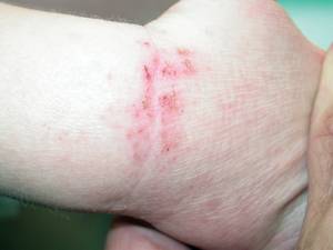
Wrist with erythema, lichenification, and crusting (Photo Courtesy: Dr. Moise L. Levy)
He disclosed being on the advisory board of SkinMedica Inc. SDEF and this news organization are owned by Elsevier.
The management of atopic dermatitis in children requires a comprehensive approach, as with any difficult chronic disease, according to Dr. Moise L. Levy.
"Atopic dermatitis [AD] is no different than any other chronic disease, including the psychosocial issues. It must be approached broadly. Whatever support the child and family need, you provide it," said Dr. Levy, chief of pediatric dermatology at Dell Children's Medical Center, Austin, Texas.
Ongoing education of the family about the importance of maintaining compliance with emollient and topical treatment is essential. Written "action plans" similar to those used with asthma patients can be helpful, Dr. Levy said at the annual Hawaii Dermatology Seminar sponsored by Skin Disease Education Foundation.
Recent evidence has highlighted the interaction between bacterial colonization - not active infection - and worsening of AD symptoms. Interleukins produced during active inflammation of AD appear to impair normal defenses against skin infection, he explained.
One study provides a rationale for the use of bleach baths in children with AD who are colonized with methicillin-resistant Staphylococcus aureus. Thirty-one children aged 6 months to 17 years with moderate to severe AD and clinical signs of bacterial infection received cephalexin for 2 weeks, then were randomized to intranasal mupirocin ointment and twice-weekly sodium hypochlorite baths (½ cup in 40 gallons) or a placebo of intranasal petrolatum plus plain water baths (Pediatrics 2009;123:e808-14).
Eczema Area and Severity Index (EASI) scores were improved at 1 and 3 months in the treatment arm, compared with the placebo group, in all submerged sites (not the head and neck), Dr. Levy reported.
Topical corticosteroids and calcineurin inhibitors can provide excellent control of moderate to severe AD flares, but data also suggest intermittent dosing may help prevent flares in the first place in patients with chronic relapsing disease.
In a two-phase study, 206 children aged 2-15 years with moderate to severe AD were randomized to aclometasone or tacrolimus for 4 days, and then received open-label tacrolimus twice daily for up to 16 weeks. After that, 105 children were randomized to application of either tacrolimus or a vehicle three times per week to clinically normal-appearing skin for up to 40 weeks.
The aclometasone group had better improvement during the acute phase, but there were no differences thereafter. In the long-term phase, the tacrolimus group had more disease-free days compared with the vehicle group, longer time to relapse, and fewer relapse days (Pediatrics 2008;122:e1210-8).
The use of systemic therapies should be reserved for children with the most severe AD, and considered only after aggressive topical therapy has been maximized and compliance with systemics can be ensured, Dr. Levy emphasized.
Currently, cyclosporine is the only one of these agents with sufficient pediatric efficacy and safety data to support its use in children with AD, and still must be used with caution. One pediatric study found that oral cyclosporine use in combination with topical corticosteroids was associated with greater loss of bone mass than was use of corticosteroids alone (Pediatr. Dermatol. 2007;24:613-20).
Although there is a large body of anecdotal evidence supporting the efficacy of other systemic agents such as mycophenolate mofetil, methotrexate, azathioprine, and thiopurine methyltransferase, at this time there are insufficient randomized, prospective clinical data in children with AD to support their use, he said.
The management of AD in children can be extremely challenging. At times, hospitalization and referral to a psychologist may be necessary to address the educational and psychosocial issues that prevent patients and families from maintaining long-term compliance.
One very helpful "treatment" is the American Academy of Dermatology's Camp Discovery (www.campdiscovery.org), where kids with chronic skin disorders can meet, share experiences, and feel "normal." All kids with AD should be offered the chance to go, Dr. Levy said.
Wrist with erythema, lichenification, and crusting (Photo Courtesy: Dr. Moise L. Levy)
He disclosed being on the advisory board of SkinMedica Inc. SDEF and this news organization are owned by Elsevier.
3.75% Imiquimod Formulation in the Works
WAIKOLOA, Hawaii — The next generation of imiquimod therapy for actinic keratoses will offer a simpler, more convenient regimen that is easier to tolerate than the available 5% cream, according to Dr. Brian Berman, professor of dermatology at the University of Miami.
A 3.75% topical formulation of imiquimod has been designed for once-daily treatment. The new formulation received marketing approval in Canada earlier this year but is investigational in the United States. Called Zyclara (Graceway Pharmaceuticals), it is used in cyclic fashion over a 6-week period: 2 weeks on, 2 weeks off, and 2 weeks on.
The 5% imiquimod formulation (Aldara, Graceway Pharmaceuticals) isn't supposed to be applied to an area greater than 25 cm2. The 3.75% formulation, however, can be used to treat the full face or balding scalp, he said at the annual Hawaii Dermatology Seminar sponsored by the Skin Disease Education Foundation.
As part of four clinical trials, a 2.5% and a 3.75% formulation of imiquimod cream were evaluated in two regimens. Reponses were measured in a total of 969 patients, each having 5-29 facial and scalp actinic keratoses (AKs) with up to 1 mm of hyperkeratosis.
Trial participants were randomized to receive either 2.5% or 3.75% imiquimod cream or placebo. Patients were further randomized to either a schedule of 3-weeks-on/3-weeks-off/3-weeks-on therapy or to the 2-2-2 regimen. Outcomes were best with 3.75% imiquimod on the 2-2-2 cycle.
After the first 2 weeks of treatment with the 3.75% cream given on the 2-2-2 regimen, subclinical AK lesions became apparent in 85% of patients. At 8 weeks post-treatment, their AK lesion count was reduced by nearly 82%. The placebo group had a 25% decrease in lesion count at 8 weeks.
An earlier study of 5% imiquimod cream, applied twice weekly for 16 weeks, resulted in an 83% median decrease in AKs.
The complete clearance rate was nearly 36% with 3.75% imiquimod on a 2-2-2 schedule, compared to 6.3% with placebo. The partial clearance rate, defined as at least 75% clearance of AKs, was slightly over 59% with 3.75% imiquimod and nearly 23% with placebo.
The efficacy of 3.75% imiquimod on the 3-3-3 cycle was comparable, but the rate of treatment-related adverse events was nearly twice as great, and the number of unscheduled rest periods was more than doubled.
Favorable outcomes also were impressively durable. Among patients who were completely cleared at 8 weeks post-treatment, the median number of AKs was zero at 6 months and 1 at 1 year.
Following cryotherapy, the 1-year sustained clearance rate is typically in the low single digits, he observed.
Audience members asked Dr. Berman's preferences for treating AKs in his own practice. He replied that his favorable clinical trial experience would make 3.75% imiquimod, if it is made available in the United States, his first-line treatment for AKs on the balding scalp, face, and ears. He has relied upon topical 5-FU for AKs on the arms, since these lesions tend to be quite hyperkeratotic. Given the good responses to 3.75% imiquimod in the numerous trial patients with mildly hyperkeratotic AKs, Dr. Berman said he would be equally likely to turn to topical 5-FU or imiquimod 2-2-2 cycle therapy for lesions on the arms.
He also discussed important new developments in the pathogenesis of AKs and squamous cell carcinomas--and how imiquimod counters this process.
Among its multiple immunomodulatory effects, topical imiquimod activates toll-like receptor-7, NFkappaB, and Th1 lymphocytes that have antiviral and antitumor activities.
Investigators at Dongguk University in Kyongju, Korea, recently identified a new major player in the development of AKs and squamous cell carcinoma: the Forkhead box p3 (Foxp3) positive cell. These cells infiltrate and surround AKs and squamous cell carcinomas, where they induce immune suppression and ultimately immune tolerance to the tumor. Foxp3-positive T regulatory cells do so through direct cell-to-cell contact and by elaborating the immunosuppressive cytokines interleukin-10 and transforming growth factor-beta (Yonsei Med.J. Dec. 31, 2008; 49:942-8).
"The good news is in vivo application of imiquimod to squamous cell carcinomas of the skin blocks these two immunosuppressive agents--IL-10 and TGF-beta--reversing the immune suppression. This obviously has a sanguine effect on removing AKs as well," Dr. Berman explained.
The Zyclara studies were funded by Graceway Pharmaceuticals. Dr. Berman, an investigator in the trials, is on the company's speakers' bureau and advisory board. He holds similar positions with 3M, Doak, PharmaDerm, Peplin, and Neutrogena.
SDEF and this news organization are owned by Elsevier.
Persistent Diaper Dermatitis Could Be Sign of More Serious Skin Condition
Persistent or unusual diaper dermatitis may be a sign of a serious skin disease or systemic illness in infant patients.
Because rare and uncommon skin eruptions in the diaper area can appear to be a conventional rash caused by prolonged skin exposure to wetness, dermatologists should closely examine each referral they receive for diaper dermatitis and look for the warning signs of a more serious skin condition, according to Dr. Ilona J. Frieden.
"When pediatricians ask a dermatologist to see a patient with a diaper rash, it is usually an unusual one. Diaper rashes are common and most never require referral. Thus, if asked, dermatologists should always say 'Yes' to these referrals," said Dr. Frieden, director of pediatric dermatology at the University of California San Francisco Children's Hospital.
Among the unusual eruptions she discussed in her presentation at the annual Hawaii Dermatology Seminar sponsored by Skin Disease Education Foundation were psoriasis, granular parakeratosis, and clear cell papulosis. Diaper rash can also be a manifestation of a systemic illness such as group A streptococcal infection, Langerhans cell histiocytosis, a zinc or other nutritional deficiency, or an asymmetric periflexural exanthem.
Group A streptococcal infection can cause a perianal rash, but may also occur at other intertriginous sites. It is typically characterized by intense, bright-red colors; satellite lesions are usually absent. Treatment requires oral antibiotics, and repeat courses are sometimes necessary, noted Dr. Frieden.
Langerhans cell histiocytosis may present as persistent diaper dermatitis that may also occur in other sites, particularly the scalp, ear canal, and oral mucosa. Be on the lookout for petechiae, atrophy, or deep ulcerations, said Dr. Frieden; a biopsy is necessary to make a diagnosis.
Diaper dermatitis due to zinc deficiency is most commonly seen in preterm breast fed infants. Metabolic disturbances and cystic fibrosis can cause similar eruptions.
Asymmetric periflexural exanthem typically starts in a flexure, often at the axilla, but it can also begin in the posterior of the thigh, leading to confusion with diaper rash. It is characterized by small red papules at the periphery with a slightly dusky, scaly center, said Dr. Frieden.
"This condition can last for several weeks and may be misdiagnosed as a contact dermatitis or other dermatologic condition," she said. "Eventually, many cases begin to become bilateral and more generalized." The cause of this exanthema is not known.
An obvious tip for preventing diaper dermatitis, or decreasing its prevalence, is frequent diaper changes to minimize skin exposure to urine and feces. Barrier creams can also be helpful as a preventative measure, she noted.
"It is also important to note that the differential diagnosis of diaper rash during the newborn period differs from that of older infants in that the rash is less likely to be caused by an irritant and more likely to represent an infection or other condition," said Dr. Frieden.

Photo Courtesy: Dr. Ilona J. Frieden
Dr. Frieden reported having no relevant conflicts of interest. SDEF and this news organization are owned by Elsevier.
Persistent or unusual diaper dermatitis may be a sign of a serious skin disease or systemic illness in infant patients.
Because rare and uncommon skin eruptions in the diaper area can appear to be a conventional rash caused by prolonged skin exposure to wetness, dermatologists should closely examine each referral they receive for diaper dermatitis and look for the warning signs of a more serious skin condition, according to Dr. Ilona J. Frieden.
"When pediatricians ask a dermatologist to see a patient with a diaper rash, it is usually an unusual one. Diaper rashes are common and most never require referral. Thus, if asked, dermatologists should always say 'Yes' to these referrals," said Dr. Frieden, director of pediatric dermatology at the University of California San Francisco Children's Hospital.
Among the unusual eruptions she discussed in her presentation at the annual Hawaii Dermatology Seminar sponsored by Skin Disease Education Foundation were psoriasis, granular parakeratosis, and clear cell papulosis. Diaper rash can also be a manifestation of a systemic illness such as group A streptococcal infection, Langerhans cell histiocytosis, a zinc or other nutritional deficiency, or an asymmetric periflexural exanthem.
Group A streptococcal infection can cause a perianal rash, but may also occur at other intertriginous sites. It is typically characterized by intense, bright-red colors; satellite lesions are usually absent. Treatment requires oral antibiotics, and repeat courses are sometimes necessary, noted Dr. Frieden.
Langerhans cell histiocytosis may present as persistent diaper dermatitis that may also occur in other sites, particularly the scalp, ear canal, and oral mucosa. Be on the lookout for petechiae, atrophy, or deep ulcerations, said Dr. Frieden; a biopsy is necessary to make a diagnosis.
Diaper dermatitis due to zinc deficiency is most commonly seen in preterm breast fed infants. Metabolic disturbances and cystic fibrosis can cause similar eruptions.
Asymmetric periflexural exanthem typically starts in a flexure, often at the axilla, but it can also begin in the posterior of the thigh, leading to confusion with diaper rash. It is characterized by small red papules at the periphery with a slightly dusky, scaly center, said Dr. Frieden.
"This condition can last for several weeks and may be misdiagnosed as a contact dermatitis or other dermatologic condition," she said. "Eventually, many cases begin to become bilateral and more generalized." The cause of this exanthema is not known.
An obvious tip for preventing diaper dermatitis, or decreasing its prevalence, is frequent diaper changes to minimize skin exposure to urine and feces. Barrier creams can also be helpful as a preventative measure, she noted.
"It is also important to note that the differential diagnosis of diaper rash during the newborn period differs from that of older infants in that the rash is less likely to be caused by an irritant and more likely to represent an infection or other condition," said Dr. Frieden.
Photo Courtesy: Dr. Ilona J. Frieden
Dr. Frieden reported having no relevant conflicts of interest. SDEF and this news organization are owned by Elsevier.
A Quick and Accurate Diagnosis Can Aid Photosensitivity Patients
While the diagnostic challenges of photodermatoses are multiple, one thing is certain, a proper and rapid diagnosis is essential to optimizing patient management and outcomes, according to Dr. Vincent DeLeo.
The diagnostic challenges of photodermatoses include increasing clinical suspicion to recognize the less common forms, ruling out dermatologic mimics in the differential diagnosis, and identifying etiology to guide therapy and speed patient recovery, Dr. DeLeo noted at the annual Hawaii Dermatology Seminar sponsored by Skin Disease Education Foundation.
Photodermatosis incidence varies from the relatively common to the relatively rare, he said. Specific presentations include porphyria, idiopathic photodermatitis, polymorphic light eruption, solar urticaria, chronic actinic dermatitis, exogenous chemical photosensitivity, and photocontact dermatitis.
Consider age of onset, timing of the skin eruption, and occupation, medication, or plant exposures when taking patient history. Ask about products used for personal care, particularly any sunscreens or colognes. Clinical workup should include a review of systems that focuses on cutaneous and neurologic effects and a complete physical examination, said Dr. DeLeo, chairman of the department of dermatology at St. Luke's-Roosevelt Hospital Center and Beth Israel Medical Center in New York.
Porphyrias present in two specific patterns - immediate with skin burning within a day of exposure or delayed with blistering, hirsutism, or hyperpigmentation. With an immediate form, such as erythropoietic protoporphyria (EPP), rule out solar urticaria and lupus erythematosus, Dr. DeLeo suggested.
With a delayed presentation, such as porphyria cutanea tarda (PCT), consider pseudo-PCT and epidermolysis bullosa acquisita in your differential diagnosis, he said. A blood test for plasma porphyrins is usually diagnostic in all porphyrias, but a red blood cell porphyrin assay will diagnose EPP and a 24-hour urine assay will diagnose PCT.
Among the sporadic presentations of PCT, it is important to check for a diagnosis of hemochromatosis early to prevent serious tissue damage, especially dysfunction of the liver, Dr. DeLeo said. A test for the culprit gene mutations can be ordered from any major diagnostic laboratory.
Idiopathic photodermatoses include polymorphous light eruption (PMLE), chronic actinic dermatitis, and solar urticaria. Dermatologists are most likely to encounter PMLE, the most common form of photosensitivity, Dr. DeLeo said.
PMLE tends to arise in young adults and in women more than men. Symptoms usually occur within days of exposure - during springtime or while on vacation, for example. Look for a symmetrical distribution on exposed areas only. The histologic differential with suspected PMLE includes lupus erythematosus, lymphocytic infiltrate of Jessner, and cutaneous lymphoid hyperplasia.
Solar urticaria also occurs more often in women than in men. A sudden onset of erythema, edema, urticaria, and pruritus in exposed areas is characteristic. Systemic involvement is rare, Dr. DeLeo said. Careful photo provocation can aid in the definitive diagnosis of this condition. Treatment options include H1 antihistamines and proper photoprotection.
Chronic actinic dermatitis, on the other hand, is more persistent and not related to a specific acute exposure. Patients typically present with patchy eczema and may have erythroderma or lymphoma-like plaques in sun-exposed areas.
This form of photosensitivity is much more common in men and tends to present in older patients, at a mean age of 65 years, Dr. DeLeo said. Rigorous photoprotection is warranted. Consider patch testing and photopatch testing to rule out underlying contact or photocontact allergy. PUVA or immunosuppressants can be used in severe cases.
Some patients can experience photoirritant contact dermatitis from plants. A number of plants can cause this, including celery, dill, lemon, lime, and St. John's wort, he concluded.

Dermatologists are most likely to encounter PMLE (shown above). It tends to arise in young adults and in women more than men. Symptoms usually occur within days of exposure. (Photo Courtesy: Dr. Vincent DeLeo)
Dr. DeLeo disclosed being a consultant for Neutrogena, Schering-Plough, L'Oreal, Pfizer, Estée Lauder, Orfagen, Goodyear, and Limited Brands. SDEF and this news organization are owned by Elsevier.
While the diagnostic challenges of photodermatoses are multiple, one thing is certain, a proper and rapid diagnosis is essential to optimizing patient management and outcomes, according to Dr. Vincent DeLeo.
The diagnostic challenges of photodermatoses include increasing clinical suspicion to recognize the less common forms, ruling out dermatologic mimics in the differential diagnosis, and identifying etiology to guide therapy and speed patient recovery, Dr. DeLeo noted at the annual Hawaii Dermatology Seminar sponsored by Skin Disease Education Foundation.
Photodermatosis incidence varies from the relatively common to the relatively rare, he said. Specific presentations include porphyria, idiopathic photodermatitis, polymorphic light eruption, solar urticaria, chronic actinic dermatitis, exogenous chemical photosensitivity, and photocontact dermatitis.
Consider age of onset, timing of the skin eruption, and occupation, medication, or plant exposures when taking patient history. Ask about products used for personal care, particularly any sunscreens or colognes. Clinical workup should include a review of systems that focuses on cutaneous and neurologic effects and a complete physical examination, said Dr. DeLeo, chairman of the department of dermatology at St. Luke's-Roosevelt Hospital Center and Beth Israel Medical Center in New York.
Porphyrias present in two specific patterns - immediate with skin burning within a day of exposure or delayed with blistering, hirsutism, or hyperpigmentation. With an immediate form, such as erythropoietic protoporphyria (EPP), rule out solar urticaria and lupus erythematosus, Dr. DeLeo suggested.
With a delayed presentation, such as porphyria cutanea tarda (PCT), consider pseudo-PCT and epidermolysis bullosa acquisita in your differential diagnosis, he said. A blood test for plasma porphyrins is usually diagnostic in all porphyrias, but a red blood cell porphyrin assay will diagnose EPP and a 24-hour urine assay will diagnose PCT.
Among the sporadic presentations of PCT, it is important to check for a diagnosis of hemochromatosis early to prevent serious tissue damage, especially dysfunction of the liver, Dr. DeLeo said. A test for the culprit gene mutations can be ordered from any major diagnostic laboratory.
Idiopathic photodermatoses include polymorphous light eruption (PMLE), chronic actinic dermatitis, and solar urticaria. Dermatologists are most likely to encounter PMLE, the most common form of photosensitivity, Dr. DeLeo said.
PMLE tends to arise in young adults and in women more than men. Symptoms usually occur within days of exposure - during springtime or while on vacation, for example. Look for a symmetrical distribution on exposed areas only. The histologic differential with suspected PMLE includes lupus erythematosus, lymphocytic infiltrate of Jessner, and cutaneous lymphoid hyperplasia.
Solar urticaria also occurs more often in women than in men. A sudden onset of erythema, edema, urticaria, and pruritus in exposed areas is characteristic. Systemic involvement is rare, Dr. DeLeo said. Careful photo provocation can aid in the definitive diagnosis of this condition. Treatment options include H1 antihistamines and proper photoprotection.
Chronic actinic dermatitis, on the other hand, is more persistent and not related to a specific acute exposure. Patients typically present with patchy eczema and may have erythroderma or lymphoma-like plaques in sun-exposed areas.
This form of photosensitivity is much more common in men and tends to present in older patients, at a mean age of 65 years, Dr. DeLeo said. Rigorous photoprotection is warranted. Consider patch testing and photopatch testing to rule out underlying contact or photocontact allergy. PUVA or immunosuppressants can be used in severe cases.
Some patients can experience photoirritant contact dermatitis from plants. A number of plants can cause this, including celery, dill, lemon, lime, and St. John's wort, he concluded.
Dermatologists are most likely to encounter PMLE (shown above). It tends to arise in young adults and in women more than men. Symptoms usually occur within days of exposure. (Photo Courtesy: Dr. Vincent DeLeo)
Dr. DeLeo disclosed being a consultant for Neutrogena, Schering-Plough, L'Oreal, Pfizer, Estée Lauder, Orfagen, Goodyear, and Limited Brands. SDEF and this news organization are owned by Elsevier.