User login
Bringing you the latest news, research and reviews, exclusive interviews, podcasts, quizzes, and more.
div[contains(@class, 'header__large-screen')]
div[contains(@class, 'read-next-article')]
div[contains(@class, 'nav-primary')]
nav[contains(@class, 'nav-primary')]
section[contains(@class, 'footer-nav-section-wrapper')]
footer[@id='footer']
div[contains(@class, 'main-prefix')]
section[contains(@class, 'nav-hidden')]
div[contains(@class, 'ce-card-content')]
nav[contains(@class, 'nav-ce-stack')]
This brain surgery was BYOS: Bring your own saxophone
Tumor vs. saxophone: The surgical grudge match
Brain surgery is a notoriously difficult task. There’s a reason we say, “Well, at least it’s not brain surgery” when we’re trying to convince someone that a task isn’t that tough. Make one wrong incision, cut the wrong neuron, and it’s goodbye higher cognitive function. And most people appreciate thinking. Crazy, right?
One would imagine that the act of brain surgery would become even more difficult when the patient brings his saxophone and plays it randomly throughout the operation. It’s a hospital, after all, not a jazz club. Patients don’t get to play musical instruments during other surgeries. Why should brain surgery patients get special treatment?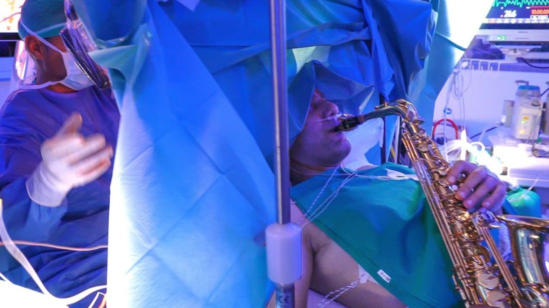
As it turns out, the musical performance was actually quite helpful. A man in Italy had a brain tumor in a particularly complex area, and he’s left-handed, which apparently makes the brain’s neural pathways much more complicated. Plus, he insisted that he retain his musical ability after the surgery. So he and his medical team had a crazy thought: Why not play the saxophone throughout the surgery? After all, according to head surgeon Christian Brogna, MD, playing an instrument means you understand music, which tests many higher cognitive functions such as coordination, mathematics, and memory.
And so, at various points throughout the 9-hour surgery, the patient played his saxophone for his doctors. Doing so allowed the surgeons to map the patient’s brain in a more complete and personalized fashion. With that extra knowledge, they were able to successfully remove the tumor while maintaining the patient’s musical ability, and the patient was discharged on Oct. 13, just 3 days after his operation.
While we’re happy the patient recovered, we do have to question his choice of music. During the surgery, he played the theme to the 1970 movie “Love Story” and the Italian national anthem. Perfectly fine pieces, no doubt, but the saxophone solo in “Jungleland” exists. And we could listen to that for 9 hours straight. In fact, we do that every Friday in the LOTME office.
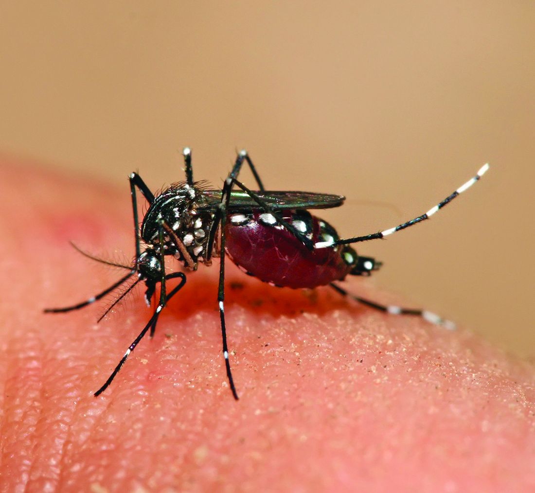
Basketball has the Big Dance. Mosquitoes get the Big Sniff
In this week’s installment of our seemingly never-ending series, “Mosquitoes and the scientists who love them,” we visit The Rockefeller University in New York, where the olfactory capabilities of Aedes Aegypti – the primary vector species for Zika, dengue, yellow fever, and chikungunya – became the subject of a round robin–style tournament.
First things first, though. If you’re going to test mosquito noses, you have to give them something to smell. The researchers enrolled eight humans who were willing to wear nylon stockings on their forearms for 6 hours a day for multiple days. “Over the next few years, the researchers tested the nylons against each other in all possible pairings,” Leslie B. Vosshall, PhD, and associates said in a statement from the university. In other words, mosquito March Madness.
Nylons from different participants were hooked up in pairs to an olfactometer assay consisting of a plexiglass chamber divided into two tubes, each ending in a box that held a stocking. The mosquitoes were placed in the main chamber and observed as they flew down the tubes toward one stocking or the other.
Eventually, the “winner” of the “tournament” was Subject 33. And no, we don’t know why there was a Subject 33 since the study involved only eight participants. We do know that the nylons worn by Subject 33 were “four times more attractive to the mosquitoes than the next most-attractive study participant, and an astonishing 100 times more appealing than the least attractive, Subject 19,” according to the written statement.
Chemical analysis identified 50 molecular compounds that were elevated in the sebum of the high-attracting participants, and eventually the investigators discovered that mosquito magnets produced carboxylic acids at much higher levels than the less-attractive volunteers.
We could go on about the research team genetically engineering mosquitoes without odor receptors, but we have to save something for later. Tune in again next week for another exciting episode of “Mosquitoes and the scientists who love them.”
Are women better with words?
Men vs. Women is probably the oldest argument in the book, but there may now be movement. Researchers have been able not only to shift the advantage toward women, but also to use that knowledge to medical advantage.

When it comes to the matter of words and remembering them, women apparently have men beat. The margin is small, said lead author Marco Hirnstein, PhD, of the University of Bergen, Norway, but, after performing a meta-analysis of 168 published studies and PhD theses involving more than 350,000 participants, it’s pretty clear. The research supports women’s advantage over men in recall, verbal fluency (categorical and phonemic), and recognition.
So how is this information useful from a medical standpoint?
Dr. Hirnstein and colleagues suggested that this information can help in interpreting diagnostic assessment results. The example given was dementia diagnosis. Since women are underdiagnosed because their baseline exceeds average while men are overdiagnosed, taking gender and performance into account could clear up or catch cases that might otherwise slip through the cracks.
Now, let’s just put this part of the debate to rest and take this not only as a win for women but for science as well.
Tumor vs. saxophone: The surgical grudge match
Brain surgery is a notoriously difficult task. There’s a reason we say, “Well, at least it’s not brain surgery” when we’re trying to convince someone that a task isn’t that tough. Make one wrong incision, cut the wrong neuron, and it’s goodbye higher cognitive function. And most people appreciate thinking. Crazy, right?
One would imagine that the act of brain surgery would become even more difficult when the patient brings his saxophone and plays it randomly throughout the operation. It’s a hospital, after all, not a jazz club. Patients don’t get to play musical instruments during other surgeries. Why should brain surgery patients get special treatment?
As it turns out, the musical performance was actually quite helpful. A man in Italy had a brain tumor in a particularly complex area, and he’s left-handed, which apparently makes the brain’s neural pathways much more complicated. Plus, he insisted that he retain his musical ability after the surgery. So he and his medical team had a crazy thought: Why not play the saxophone throughout the surgery? After all, according to head surgeon Christian Brogna, MD, playing an instrument means you understand music, which tests many higher cognitive functions such as coordination, mathematics, and memory.
And so, at various points throughout the 9-hour surgery, the patient played his saxophone for his doctors. Doing so allowed the surgeons to map the patient’s brain in a more complete and personalized fashion. With that extra knowledge, they were able to successfully remove the tumor while maintaining the patient’s musical ability, and the patient was discharged on Oct. 13, just 3 days after his operation.
While we’re happy the patient recovered, we do have to question his choice of music. During the surgery, he played the theme to the 1970 movie “Love Story” and the Italian national anthem. Perfectly fine pieces, no doubt, but the saxophone solo in “Jungleland” exists. And we could listen to that for 9 hours straight. In fact, we do that every Friday in the LOTME office.
Basketball has the Big Dance. Mosquitoes get the Big Sniff
In this week’s installment of our seemingly never-ending series, “Mosquitoes and the scientists who love them,” we visit The Rockefeller University in New York, where the olfactory capabilities of Aedes Aegypti – the primary vector species for Zika, dengue, yellow fever, and chikungunya – became the subject of a round robin–style tournament.
First things first, though. If you’re going to test mosquito noses, you have to give them something to smell. The researchers enrolled eight humans who were willing to wear nylon stockings on their forearms for 6 hours a day for multiple days. “Over the next few years, the researchers tested the nylons against each other in all possible pairings,” Leslie B. Vosshall, PhD, and associates said in a statement from the university. In other words, mosquito March Madness.
Nylons from different participants were hooked up in pairs to an olfactometer assay consisting of a plexiglass chamber divided into two tubes, each ending in a box that held a stocking. The mosquitoes were placed in the main chamber and observed as they flew down the tubes toward one stocking or the other.
Eventually, the “winner” of the “tournament” was Subject 33. And no, we don’t know why there was a Subject 33 since the study involved only eight participants. We do know that the nylons worn by Subject 33 were “four times more attractive to the mosquitoes than the next most-attractive study participant, and an astonishing 100 times more appealing than the least attractive, Subject 19,” according to the written statement.
Chemical analysis identified 50 molecular compounds that were elevated in the sebum of the high-attracting participants, and eventually the investigators discovered that mosquito magnets produced carboxylic acids at much higher levels than the less-attractive volunteers.
We could go on about the research team genetically engineering mosquitoes without odor receptors, but we have to save something for later. Tune in again next week for another exciting episode of “Mosquitoes and the scientists who love them.”
Are women better with words?
Men vs. Women is probably the oldest argument in the book, but there may now be movement. Researchers have been able not only to shift the advantage toward women, but also to use that knowledge to medical advantage.
When it comes to the matter of words and remembering them, women apparently have men beat. The margin is small, said lead author Marco Hirnstein, PhD, of the University of Bergen, Norway, but, after performing a meta-analysis of 168 published studies and PhD theses involving more than 350,000 participants, it’s pretty clear. The research supports women’s advantage over men in recall, verbal fluency (categorical and phonemic), and recognition.
So how is this information useful from a medical standpoint?
Dr. Hirnstein and colleagues suggested that this information can help in interpreting diagnostic assessment results. The example given was dementia diagnosis. Since women are underdiagnosed because their baseline exceeds average while men are overdiagnosed, taking gender and performance into account could clear up or catch cases that might otherwise slip through the cracks.
Now, let’s just put this part of the debate to rest and take this not only as a win for women but for science as well.
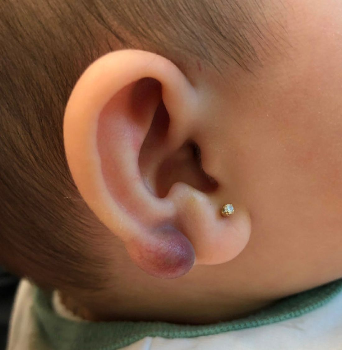
An infant with a tender bump on her ear
A biopsy of the lesion was performed that showed a well-defined nodulocystic tumor composed of nests of basaloid cells that are undergoing trichilemmal keratinization. Shadow cells are seen as well as small areas of calcification. There is also a histiocytic infiltrate with multinucleated giant cells. The histologic diagnosis is of a pilomatrixoma.
Pilomatrixoma, also known as calcifying epithelioma of Malherbe, was first described in 1880, as a tumor of sebaceous gland origin. Later, in 1961, Robert Forbis Jr, MD, and Elson B. Helwig, MD, coined the term pilomatrixoma to describe the hair follicle matrix as the source of the tumor. Pilomatrixomas are commonly seen in the pediatric population, usually in children between 8 and 13 years of age. Our patient is one of the youngest described. The lesions are commonly seen on the face and neck in about 70% of the cases followed by the upper extremities, back, and legs. Clinically, the lesions appear as a firm dermal papule or nodule, which moves freely and may have associated erythema on the skin surface or a blueish gray hue on the underlying skin.

Most pilomatrixomas that have been studied have shown a mutation in Exon 3 of the beta-catenin gene (CTNNB1). The beta-catenin molecule is a subunit of the cadherin protein, which is part of an important pathway in the terminal hair follicle differentiation. Beta-catenin also plays an important role in the Wnt pathway, which regulates cell fate as well as early embryonic patterning. Beta-catenin is responsible for forming adhesion junctions among cells. There have also been immunohistochemical studies that have shown a BCL2 proto-oncogene overexpression to pilomatrixoma.
There are several genetic syndromes that have been associated with the presence of pilomatrixomas: Turner syndrome (XO chromosome abnormality associated with short stature and cardiac defects), Gardner syndrome (polyposis coli and colon and rectal cancer), myotonic dystrophy, Rubinstein-Taybi syndrome (characterized by broad thumbs and toes, short stature, distinctive facial features, and varying degrees of intellectual disability), and trisomy 9. On physical examination our patient didn’t present with any of the typical features or history that could suggest any of these syndromes. A close follow-up and evaluation by a geneticist was recommended because after the initial visit she developed a second lesion on the forehead.
The differential diagnosis for this lesion includes other cysts that may occur on the ear such as epidermal inclusion cyst or dermoid cysts, though these lesions do not tend to be as firm as pilomatrixomas are, which can help with the diagnosis. Dermoid cysts are made of dermal and epidermal components. They are usually present at birth and are commonly seen on the scalp and the periorbital face.
Keloids are rubbery nodules of scar tissue that can form on sites of trauma, and although the lesion occurred after she had her ears pierced, the consistency and rapid growth of the lesion as well as the pathological description made this benign fibrous growth less likely.
When pilomatrixomas are inflamed they can be confused with vascular growths: in this particular case, a hemangioma or another vascular tumor such as a tufted angioma or kaposiform hemangioendothelioma. An ultrasound of the lesion could have helped in the differential diagnosis of the lesion.
Pilomatrixomas can grow significantly and in some cases get inflamed or infected. Surgical management of pilomatrixomas is often required because the lesions do not regress spontaneously.
Dr. Matiz is a pediatric dermatologist at Southern California Permanente Medical Group, San Diego.
References
Forbis R Jr and Helwig EB. Arch Dermatol 1961;83:606-18.
Schwarz Y et al. Int J Pediatr Otorhinolaryngol. 2016 Jun;85:148-53.
A biopsy of the lesion was performed that showed a well-defined nodulocystic tumor composed of nests of basaloid cells that are undergoing trichilemmal keratinization. Shadow cells are seen as well as small areas of calcification. There is also a histiocytic infiltrate with multinucleated giant cells. The histologic diagnosis is of a pilomatrixoma.
Pilomatrixoma, also known as calcifying epithelioma of Malherbe, was first described in 1880, as a tumor of sebaceous gland origin. Later, in 1961, Robert Forbis Jr, MD, and Elson B. Helwig, MD, coined the term pilomatrixoma to describe the hair follicle matrix as the source of the tumor. Pilomatrixomas are commonly seen in the pediatric population, usually in children between 8 and 13 years of age. Our patient is one of the youngest described. The lesions are commonly seen on the face and neck in about 70% of the cases followed by the upper extremities, back, and legs. Clinically, the lesions appear as a firm dermal papule or nodule, which moves freely and may have associated erythema on the skin surface or a blueish gray hue on the underlying skin.
Most pilomatrixomas that have been studied have shown a mutation in Exon 3 of the beta-catenin gene (CTNNB1). The beta-catenin molecule is a subunit of the cadherin protein, which is part of an important pathway in the terminal hair follicle differentiation. Beta-catenin also plays an important role in the Wnt pathway, which regulates cell fate as well as early embryonic patterning. Beta-catenin is responsible for forming adhesion junctions among cells. There have also been immunohistochemical studies that have shown a BCL2 proto-oncogene overexpression to pilomatrixoma.
There are several genetic syndromes that have been associated with the presence of pilomatrixomas: Turner syndrome (XO chromosome abnormality associated with short stature and cardiac defects), Gardner syndrome (polyposis coli and colon and rectal cancer), myotonic dystrophy, Rubinstein-Taybi syndrome (characterized by broad thumbs and toes, short stature, distinctive facial features, and varying degrees of intellectual disability), and trisomy 9. On physical examination our patient didn’t present with any of the typical features or history that could suggest any of these syndromes. A close follow-up and evaluation by a geneticist was recommended because after the initial visit she developed a second lesion on the forehead.
The differential diagnosis for this lesion includes other cysts that may occur on the ear such as epidermal inclusion cyst or dermoid cysts, though these lesions do not tend to be as firm as pilomatrixomas are, which can help with the diagnosis. Dermoid cysts are made of dermal and epidermal components. They are usually present at birth and are commonly seen on the scalp and the periorbital face.
Keloids are rubbery nodules of scar tissue that can form on sites of trauma, and although the lesion occurred after she had her ears pierced, the consistency and rapid growth of the lesion as well as the pathological description made this benign fibrous growth less likely.
When pilomatrixomas are inflamed they can be confused with vascular growths: in this particular case, a hemangioma or another vascular tumor such as a tufted angioma or kaposiform hemangioendothelioma. An ultrasound of the lesion could have helped in the differential diagnosis of the lesion.
Pilomatrixomas can grow significantly and in some cases get inflamed or infected. Surgical management of pilomatrixomas is often required because the lesions do not regress spontaneously.
Dr. Matiz is a pediatric dermatologist at Southern California Permanente Medical Group, San Diego.
References
Forbis R Jr and Helwig EB. Arch Dermatol 1961;83:606-18.
Schwarz Y et al. Int J Pediatr Otorhinolaryngol. 2016 Jun;85:148-53.
A 4-month-old female was referred to our clinic for evaluation of a bump on the right ear. The lesion was first noted at 2 months of age as a little pimple. She was evaluated by her pediatrician and was treated with topical and oral antibiotics without resolution of the lesion. The bump continued to grow and seemed tender to palpation, so she was referred to dermatology for evaluation.
She was born via normal vaginal delivery at 40 weeks. Her mother has no medical conditions and the pregnancy was uneventful. She has been growing and developing well. She takes vitamin D and is currently breast fed.
There have been no other family members with similar lesions. She had her ears pierced at a month of age without any complications.
On skin examination she has a firm red nodule on the right ear that appears slightly tender to touch. She has no other skin lesions of concern. She has normal muscle tone and there are no other abnormalities noted on the physical exam. She has no hepatomegaly, splenomegaly, or lymphadenopathy.
Bipolar risk and parental age: What’s the relationship?
VIENNA –
Results from a meta-analysis of more than 210,000 patients with bipolar disorder and over 13 million healthy individuals showed that children of mothers younger than 20 years had a 23% increased risk for bipolar disorder vs. those whose parents were aged 25-29 years. For participants whose mothers were aged 35-39 years, there was a 10% increased risk for bipolar disorder, which rose to 20% if the mother was aged 40 or older.
Having a father younger than 20 years conferred a 29% increased risk for bipolar disorder, which was the same increase in risk found in individuals whose fathers were aged 45 years or older.
These findings, which are an update of data published in the journal European Pharmacology, were presented at the 35th European College of Neuropsychopharmacology (ECNP) Congress.
Fourteen studies included
Previous studies have suggested that parental age at birth is a risk factor for several psychiatric disorders in offspring, including bipolar disorder, and that advanced parental age, specifically, is associated with earlier onset schizophrenia.
To investigate further, the current researchers conducted a systematic review and meta-analysis, searching the PubMed/MEDLINE, EMBASE, Scopus, and PsychINFO databases for relevant studies published to Dec. 1, 2021.
From 712 studies initially identified, 16 met all the inclusion criteria and 14 were included in the quantitative analysis.
Five studies reported only paternal age and risk for bipolar disorder in their offspring, one included just maternal age, and eight reported both maternal and paternal age in relation to the risk for offspring bipolar disorder.
Individuals with a history of any psychiatric disorders were excluded, leaving a total of 13.4 million individuals without bipolar disorder and 217,089 who had received a diagnosis for the disorder.
The investigators also corrected for both socioeconomic status and, when assessing the impact of maternal or paternal age at birth, corrected for the age of the other parent. However, they were unable to correct for the number of children in a family.
Results after stratifying maternal and paternal age showed that, compared with those born to parents aged 25-29 years, there was an increased risk for bipolar disorder in the offspring of both fathers and mothers younger than 20 years of age, with adjusted odds ratios of 1.29 (95% confidence interval, 1.13-1.48) and 1.23 (95% CI, 1.14-1.33), respectively.
Compared with those aged 25-29 years, there was also an increased risk for bipolar disorder in children born to mothers aged 35-39 years (adjusted OR, 1.1; 95% CI, 1.01-1.19) and aged 40 or older (OR, 1.2; 95% CI, 1.02-1.40).
Among fathers, there was increased risk for offspring bipolar disorder in those aged 45 or older vs. those aged 25-29 years (adjusted OR, 1.29; 95% CI, 1.15-1.46).
Several hypotheses
There are several hypotheses that could explain the results, lead study author Giovanna Fico, MD, bipolar and depressive disorders unit, Hospital Clínic Barcelona, told this news organization.
In older age, it may be “more related to genetic or epigenetic modification, especially in fathers,” Dr. Fico said. “Some studies have shown that there are de novo mutations in the germ lines, which increase the risk of several diseases, including schizophrenia.”
In younger individuals, there could be a “mixed effect between sociocultural factors, such as substance abuse, low educational status,” and other issues, Dr. Fico noted.
Moreover, as bipolar disorder onset can be as late as 30 years of age, the younger group could include “undiagnosed patients with bipolar disorder, which would increase the risk” of the disease in their offspring, she added.
Dr. Fico noted the investigators are now planning on studying the impact of environmental factors such as pollution, climate change, and urbanization on risk for bipolar disorder, with the aim of being better able to inform parents or to develop prevention strategies.
Psychoeducation is “very common for infertility, birth defects, and Down syndrome, but it’s not so common for psychiatric disorders because we need more data. But I think it’s important that parents know they have an increased risk,” she said.
Nevertheless, “We must stress that this risk is moderate, and it must be kept in perspective,” Dr. Fico said in a news release.
‘Exciting’ questions raised
The study “raises several exciting research questions, including the possibility of early prevention and intervention,” Maj Vinberg, MD, PhD, clinical professor, department of clinical medicine, University of Copenhagen, said in the release.
She said she agrees there are likely to be different factors at play at different ages, with the risk for bipolar disorder associated with younger-age parenthood more likely to be related to socioeconomic status.
For older parents, “there has been a lot of speculation around the father’s age especially, which everybody thought didn’t matter,” said Dr. Vinberg, who was not involved with the research.
“But you might have some epigenetic changes as you grow older that might transfer into the next generation,” given that there is 20 years of additional exposure to potential epigenetic changes between a man aged 25 years and one aged 45 years, she noted.
Dr. Vinberg also highlighted that there could be cases of undiagnosed bipolar disorder among the younger parents, and she noted that “men with bipolar disorder tend to have more children,” particularly during manic phases.
She explained that if someone were to get divorced at 35 years of age, then have a new manic episode at 45 “and have a new wife and children, I don’t know whether it’s possible to correct for that.”
The research is supported by a fellowship from “la Caixa” Foundation. The investigators have reported no relevant financial relationships. Dr. Vinberg reported having relationships with Lundbeck and Janssen.
A version of this article first appeared on Medscape.com.
AT ECNP CONGRESS 2022
Rapid point-of-care test could help avoid inappropriate antibiotic prescribing
The fingerstick test, FebriDx, works by detecting myxovirus resistance protein A, which the body generates in response to viral infections, and C-reactive protein (CRP), which is associated with systemic bacterial or viral infection.
In a study of 520 adults and children with symptoms of acute respiratory illness who were treated in outpatient settings, the test correctly classified bacterial infections 93.2% of the time (95% confidence interval [CI], 84.9-97.0). The negative predictive value (NPV), or probability that a person with a negative test result was truly free of a bacterial infection, was 98.7% (95% CI, 96.9-99.4).
The findings of the study, which was sponsored by the test’s manufacturer, were published in JAMA Network Open).
The ability to rule out a bacterial cause “may provide clinicians with reassurance to withhold antibiotics when supported by the clinical assessment,” the researchers wrote.
They added that the ability to identify infections that may benefit from antibiotics and confidently rule out those that will not “is essential to optimizing clinical management and addressing global antimicrobial resistance.”
FDA concerned about false negative viral infection results
FebriDx has been cleared for sale in the United Kingdom, Europe, Canada, United Arab Emirates, Brazil, and Australia, according to the manufacturer, Australia-based Lumos Diagnostics.
However, the product is not available in the United States, where the Food and Drug Administration denied marketing clearance in July. In a news release, Lumos said the FDA determined that FebriDx did not demonstrate “substantial equivalence” to a predicate device and expressed concern that false negative viral infection results could lead to missed cases of COVID-19.
In the newly published study, FebriDx identified individuals with viral infections 70.3% of the time (95% CI, 64.8-75.2). The probability that a person who tested negative for a viral infection was truly negative was 66.7% (95%CI, 60.8-72.1).
The study included patients with respiratory symptoms and recent fever who were enrolled from October 2019 to April 2021 at nine emergency departments, six urgent care clinics, and five primary care clinics in the United States. All patients were tested with FebriDx and underwent separate laboratory testing to determine a final diagnosis.
In addition, researchers recruited a control group of 120 individuals without symptoms.
Among 496 symptomatic individuals who had a final diagnosis, 73 (14.7%) were classified as having a response associated with a bacterial infection, 296 (59.7%) as having a viral-associated response, and 127 (25.6%) as negative.
FebriDx correctly ruled out a bacterial infection 88.4% of the time (95% CI, 85.0-91.1). The probability that a patient with a positive result for bacterial infection actually had a bacterial infection was 58.1% (95%CI, 49.1-66.7).
The findings bolster those of a previous study on the same test. This research included 220 patients who reported having a fever within the prior 3 days or had a measurable fever at the time of enrollment. In that study, the test correctly identified bacterial infections 85% of the time and correctly ruled out bacterial infection 93% of the time, with a NPV of 97%.
Too early to say test will be useful in practice
The idea of a test to guide the prescribing of antibiotics isn’t new, according to an expert who was not involved in FebriDx research.
Noah Ivers, MD, PhD, a family physician and associate professor at the University of Toronto who studies strategies to optimize primary care delivery, said, “many such point-of-care tests have been tried” to detect biomarkers such as CRP or procalcitonin, which is associated with bacterial infections.

Such tests have looked good in initial studies, he said, but when trialed in urgent care clinics, primary care clinics, or emergency departments, “they tend run into implementation challenges or simply lack of effects, or both.
“So, while I am happy at the news of this result, it’s too early to say with any certainty that it will prove useful in practice,” he added.
Meanwhile, Dr. Ivers said it’s “crucial that people understand that most illnesses are likely to be viral” and therefore not helped by antibiotics. When antibiotics are needed for outpatients, he said, “5 days is usually ample.”
The study was funded by Lumos Diagnostics. Among the 15 study authors, 6 had conflicts of interest disclosures, reporting ties to Inflammatix, Medical College of Wisconsin, Siemens, Technomics Research, and Lumos Diagnostics. Dr. Ivers reported no relevant financial interests.
The fingerstick test, FebriDx, works by detecting myxovirus resistance protein A, which the body generates in response to viral infections, and C-reactive protein (CRP), which is associated with systemic bacterial or viral infection.
In a study of 520 adults and children with symptoms of acute respiratory illness who were treated in outpatient settings, the test correctly classified bacterial infections 93.2% of the time (95% confidence interval [CI], 84.9-97.0). The negative predictive value (NPV), or probability that a person with a negative test result was truly free of a bacterial infection, was 98.7% (95% CI, 96.9-99.4).
The findings of the study, which was sponsored by the test’s manufacturer, were published in JAMA Network Open).
The ability to rule out a bacterial cause “may provide clinicians with reassurance to withhold antibiotics when supported by the clinical assessment,” the researchers wrote.
They added that the ability to identify infections that may benefit from antibiotics and confidently rule out those that will not “is essential to optimizing clinical management and addressing global antimicrobial resistance.”
FDA concerned about false negative viral infection results
FebriDx has been cleared for sale in the United Kingdom, Europe, Canada, United Arab Emirates, Brazil, and Australia, according to the manufacturer, Australia-based Lumos Diagnostics.
However, the product is not available in the United States, where the Food and Drug Administration denied marketing clearance in July. In a news release, Lumos said the FDA determined that FebriDx did not demonstrate “substantial equivalence” to a predicate device and expressed concern that false negative viral infection results could lead to missed cases of COVID-19.
In the newly published study, FebriDx identified individuals with viral infections 70.3% of the time (95% CI, 64.8-75.2). The probability that a person who tested negative for a viral infection was truly negative was 66.7% (95%CI, 60.8-72.1).
The study included patients with respiratory symptoms and recent fever who were enrolled from October 2019 to April 2021 at nine emergency departments, six urgent care clinics, and five primary care clinics in the United States. All patients were tested with FebriDx and underwent separate laboratory testing to determine a final diagnosis.
In addition, researchers recruited a control group of 120 individuals without symptoms.
Among 496 symptomatic individuals who had a final diagnosis, 73 (14.7%) were classified as having a response associated with a bacterial infection, 296 (59.7%) as having a viral-associated response, and 127 (25.6%) as negative.
FebriDx correctly ruled out a bacterial infection 88.4% of the time (95% CI, 85.0-91.1). The probability that a patient with a positive result for bacterial infection actually had a bacterial infection was 58.1% (95%CI, 49.1-66.7).
The findings bolster those of a previous study on the same test. This research included 220 patients who reported having a fever within the prior 3 days or had a measurable fever at the time of enrollment. In that study, the test correctly identified bacterial infections 85% of the time and correctly ruled out bacterial infection 93% of the time, with a NPV of 97%.
Too early to say test will be useful in practice
The idea of a test to guide the prescribing of antibiotics isn’t new, according to an expert who was not involved in FebriDx research.
Noah Ivers, MD, PhD, a family physician and associate professor at the University of Toronto who studies strategies to optimize primary care delivery, said, “many such point-of-care tests have been tried” to detect biomarkers such as CRP or procalcitonin, which is associated with bacterial infections.
Such tests have looked good in initial studies, he said, but when trialed in urgent care clinics, primary care clinics, or emergency departments, “they tend run into implementation challenges or simply lack of effects, or both.
“So, while I am happy at the news of this result, it’s too early to say with any certainty that it will prove useful in practice,” he added.
Meanwhile, Dr. Ivers said it’s “crucial that people understand that most illnesses are likely to be viral” and therefore not helped by antibiotics. When antibiotics are needed for outpatients, he said, “5 days is usually ample.”
The study was funded by Lumos Diagnostics. Among the 15 study authors, 6 had conflicts of interest disclosures, reporting ties to Inflammatix, Medical College of Wisconsin, Siemens, Technomics Research, and Lumos Diagnostics. Dr. Ivers reported no relevant financial interests.
FROM JAMA NETWORK OPEN
Chest reconstruction surgeries up nearly fourfold among adolescents
The number of chest reconstruction surgeries performed for adolescents rose nearly fourfold between 2016 and 2019, researchers report in a study published in JAMA Pediatrics.
“To our knowledge, this study is the largest investigation to date of gender-affirming chest reconstruction in a pediatric population. The results demonstrate substantial increases in gender-affirming chest reconstruction for adolescents,” the authors report.
The researchers, from Vanderbilt University School of Medicine, Nashville, Tenn., used the Nationwide Ambulatory Surgery Sample to identify youth with gender dysphoria who underwent top surgery to remove, or, in rare cases, to add breasts.
The authors identified 829 chest surgeries. They adjusted the number to a weighted figure of 1,130 patients who underwent chest reconstruction during the study period. Of those, 98.6% underwent masculinizing surgery to remove breasts, and 1.4% underwent feminizing surgery. Roughly 100 individuals received gender-affirming chest surgeries in 2016. In 2019, the number had risen to 489 – a 389% increase, the authors reported.
Approximately 44% of the patients in the study were aged 17 years at the time of surgery, while 5.5% were younger than 14.
Around 78% of the individuals who underwent chest surgeries in 2019 were White, 2.7% were Black, 12.2% were Hispanic, and 2.5% were Asian or Pacific Islander. Half of the patients who underwent surgery had a household income of $82,000 or more, according to the researchers.
“Most transgender adolescents had either public or private health insurance coverage for these procedures, contrasting with the predominance of self-payers reported in earlier studies on transgender adults,” write the researchers, citing a 2018 study of trends in transgender surgery.
Masculinizing chest reconstruction, such as mastectomy, and feminizing chest reconstruction, such as augmentation mammaplasty, can be performed as outpatient procedures or as ambulatory surgeries, according to another study .
The study was supported by a grant from the National Center for Advancing Translational Sciences Clinical and Translational Science Awards Program. One author has reported receiving grant funding from Merck.
A version of this article first appeared on Medscape.com.
FROM JAMA PEDIATRICS
Screening gaps miss childhood heart problems
People with a rare genetic condition that causes extremely elevated levels of low-density lipoprotein cholesterol (LDL-C) may miss out on decades of treatment because of a lack of lipid screening in childhood, researchers reported at the annual meeting of the American Academy of Pediatrics.
The condition, homozygous familial hypercholesterolemia (FH), raises the risk for atherosclerotic cardiovascular disease (ASCVD) as early as the first decade of life.
Routine screening for FH is uncommon, however, the researchers said. Lack of familiarity with guidelines and limited access to lipid specialists have been cited as possible reasons for inconsistent screening practices.
“These findings and recent improvement in lipid lowering therapies make a compelling case for rigorous compliance with AAP’s guidelines on lipid screening for children with a family history of FH or ASCVD at age 2,” study coauthor Mary P. McGowan, MD, chief medical officer of the Family Heart Foundation, said in a statement about the new study.
Early consequences
To characterize patients with homozygous FH, Dr. McGowan and her colleagues examined data from 67 participants in the CASCADE-FH registry. The Family Heart Foundation created the registry in 2013, and 40 medical centers in the United States contribute data to the repository. The researchers had access to data about patients with homozygous FH from 20 centers in the registry.
Dr. McGowan’s group compared 16 patients with homozygous FH who enrolled in the registry when they were children and 51 patients who were adults at the time of their enrollment.
Patients enrolled as children had a median age at diagnosis of 2 years (interquartile range [IQR], 2-3.5), whereas patients enrolled as adults had a median age at diagnosis of 12.6 years (IQR, 4.1-26.5).
The median untreated level of LDL-C in those enrolled as children was 776 mg/dL (IQR, 704-892). Among those enrolled as adults, it was 533 mg/dL (IQR, 467-702).
Approximately 19% of those enrolled as children had evidence of aortic valve stenosis, and 43.8% had evidence of ASCVD. The median age at onset of ASCVD was 8.9 years. One child was diagnosed with ASCVD at age 2 years and underwent liver transplant at age 4 years. Another was diagnosed with the condition at age 3 years and underwent liver transplant at age 8 years. Two children underwent coronary artery bypass grafting at ages 6 years and 14 years. Five participants underwent liver transplant before age 18 years.
About 56% of participants who enrolled as children had xanthomas, or fat deposits in tendons, and none had corneal arcus — a gray-white line of fat deposits around the edge of the cornea, both of which can indicate homozygous FH in children.
Treatment reduced LDL-C substantially, but only 25% of children achieved goal levels of cholesterol, the researchers reported. Patients who received more lipid-lowering therapies had a better chance of reaching their target levels, they found.
The data raise “the possibility that only children with the most severe phenotypes are diagnosed before adulthood,” the researchers said.
Clinical diagnosis of homozygous FH can be based on LDL-C levels, family history, and the presence of xanthomas, the researchers noted. Many children do not have physical findings, however, and a lipid panel or genetic testing may be necessary.
“There is a clear need to implement universal screening” to identify all children with homozygous FH and heterozygous FH, a less severe and more common form of FH, Dr. McGowan said.
Possible missed cases
As many as 1 in 250 people may have heterozygous FH, and 1 in 300,000 people may have homozygous FH, according to estimates. Patients with homozygous FH have two FH genes, one from each parent. In patients with homozygous FH, levels of LDL-C levels typically range between 400 and 1,000 mg/dL without treatment, which is four to 10 times higher than normal concentrations of the blood fat, according to the Family Heart Foundation.
“This study adds to a growing body of literature – including our own work – demonstrating that recommended universal screening occurs in barely 1 in 5 children. This means some patients are not being recognized as having treatable diseases,” said Justin H. Berger, MD, PhD, a pediatric cardiologist at Children’s Hospital of Philadelphia.
Even among children who are at the highest risk for early onset adult-type heart disease, only a quarter to two-thirds receive recommended screening, said Dr. Berger, who was not a member of the study team.
While Dr. Berger advocates universal lipid screening, improving screening rates in practice probably isn’t as simple as telling clinicians to screen more, he said. “Increasing testing will increase health care spending and the burden on busy primary care providers without addressing who will subsequently evaluate and manage children with abnormal lipid screening results,” Dr. Berger said.
Instead, clinicians may want to focus on screening patients who are at risk, which “could have dramatic benefits for their life-long cardiovascular health,” he said.
Dr. McGowan disclosed ties to Abbott and Regeneron, and her coauthors disclosed ties to Esperion Therapeutics and research funding from Regeneron and REGENXBIO. Dr. Berger disclosed no relevant financial relationships.
A version of this article first appeared on Medscape.com.
FROM AAP 2022
Sleep kits help foster children manage effects of trauma
A stuffed animal, aromatherapy, a night light. A kit containing these and other items can help children in foster care who have experienced trauma sleep more soundly, a critical step in helping them cope with their emotional distress.
In a new study, researchers at the Children’s Hospital of Philadelphia reported that sleep kits specially tailored to foster children appeared to be helpful in most cases. The kits can be distributed by pediatricians in the office or clinic setting.
“Children who have experienced trauma can have issues with behavior, it can impact their school, and they have difficulties sleeping,” said Kristine Fortin, MD, MPH, director of the fostering health program at Safe Place: Center for Child Protection and Health at CHOP. “I thought, what could a pediatrician do in the office in one visit to help children with sleep?”
Dr. Fortin and colleagues designed sleep kits for both younger children and adolescents.
The version for teenagers contained a sound machine, aromatherapy spray, and a sleep mask. The kits for younger children contained matching stuffed toys to share with someone they felt connected to, and a rechargeable night-light. All kits included written materials about sleep hygiene, a journal, and directions for downloading a free age-appropriate relaxation app for belly breathing or a PTSD Coach app from the Department of Veterans Affairs to manage symptoms of trauma.
In a pilot study presented at the annual meeting of the American Academy of Pediatrics, Dr. Fortin and colleagues surveyed caregivers in foster homes about their use of the kits.
Of the 20 foster parents who responded to the survey, 11 said the kits helped “very much,” 5 others reported they helped “somewhat,” another 2 reported no improvements in sleep, and 2 said they didn’t know the effect. The children for whom results were unknown moved from the home without the sleep kit or had difficulties communicating with the foster parents, resulting in incomplete assessment, according to the researchers.
Night-lights were used most in the kits, followed by the stuffed toys, sound machines, and sleep journals.
Dr. Fortin said existing resources like sleep therapy or medication can be costly or difficult to find, and many pediatricians don’t have enough time during wellness visits to address symptoms like sleep deprivation. She said the sleep kits could be an alternative to other forms of sleep therapy. “If these sleep kits were effective, and could really help them sleep, then maybe less children would need something like medication.”
Dr. Fortin said the kits her group has designed are tailored specifically for children with symptoms of trauma, or with difficult emotions associated with foster care.
“We’ve tried to design something that can be really practical and easy to use in a pediatric visit, where there’s a lot of written information that can be discussed with the child and their family,” Dr. Fortin said.
She added that she would like to see clinicians give out the sleep kits during in-office visits.
“These sleep issues are common in foster children,” she said. “We felt it was important to do an intervention.”
Kristina Lenker, PhD, a sleep psychologist at Penn State Health, Hershey, said children in foster care often struggle with falling or staying asleep, an inability to sleep alone, nightmares, and bed wetting.
“Sleep kits can be particularly helpful for these children, given how they can help caregivers to provide a safe sleep environment and predictable routine, and send messages of safety and comfort at bedtime, with tangible objects, and enable children to feel a sense of control,” she said.
Charitable organizations like Pajama Program and Sleeping Children Around the World provide sleep kits to children from underserved backgrounds. But Candice Alfano, PhD, director of the University of Houston’s Sleep and Anxiety Center of Houston, said the CHOP kits are the first to specifically target sleeping difficulties in foster children.
“Sleep is a largely neglected yet essential area of health, development, and well-being in this highly-vulnerable population of youth, so I am very excited to see this work being done,” Dr. Alfano said in an interview.
Dr. Fortin and Dr. Lenker reported no relevant financial relationships.
A version of this article first appeared on Medscape.com.
FROM AAP 2022
Connected: Preterm infant program makes progress
Martha Welch, MD, spent the better part of three decades in private practice treating children with emotional, behavioral, and developmental disorders before accepting a job on the faculty of Columbia University, New York, in 1997.
She took the position, she said, with a mission: to find evidence to support what she’d observed in her practice – that parents could, by making stronger emotional connections, change the trajectory of development for preemie infants.
With that understanding, Dr. Welch created Family Nurture Intervention (FNI), which has been shown to improve the development of premature babies.
“We saw that no matter what happened to the baby, no matter how avoidant the baby might be, we’re able to overcome this with emotional expression,” Dr. Welch said.
Over the course of the intervention, families work with a specialist who helps bring mother and baby together – both physically and emotionally – until both are calm, which can initially take several hours and over time, minutes.
FNI appears to help families – especially mothers – re-establish an emotional connection often interrupted by their babies’ stressful and uncertain stay in a neonatal intensive care unit (NICU). In turn, both the infant and maternal nervous systems become better regulated, according to researchers.
Early challenges
Babies born preterm can face a range of short-term and long-term challenges, such as breathing problems due to an underdeveloped respiratory system, an increased risk of infection from an underdeveloped immune system, and learning difficulties, according to the Mayo Clinic.
Many aspects of FNI are not new: The neonatal intensive care unit has long incorporated activities such as scent cloth exchanges, talking to the baby, and skin-to-skin contact. But the approach Dr. Welch and her colleagues advocate emphasizes building a bond between the mother and the infant.
Mounting evidence shows that FNI can improve a wide range of outcomes for premature babies. In a 2021 study, for example, Dr. Welch’s group showed that FNI was associated with lower heart rates among preemies in the NICU. A 2016 study linked the intervention to reduced depression and anxiety symptoms in mothers of preterm infants. And a 2015 randomized controlled trial showed FNI improved development and behavioral outcomes in infants up to 18 months.
A new study published in Science Translational Medicine showed that the intervention led to a greater likelihood that babies had improved cognitive development later on, narrowing the developmental gap between healthy, full-term babies.
Dr. Welch and her colleagues tested to see if FNI measurably changed brain development in preterm infants who were born at 26-34 weeks of a pregnancy.
“We were blown away by the strength of the effect,” said Pauliina Yrjölä, MSc, a doctoral student and medical physicist at the University of Helsinki, who led the study on which Dr. Welch is a co-author.
Mothers in the intervention group made as much eye contact with the infants as possible and spoke with infants about their feelings.
Intimate sensory interactions between mothers and infants physically altered infants’ cortical networks in the brain and was later correlated to improved neurocognitive performance, according to the researchers.
“I was convinced there were physiological changes; I knew that from my clinical work,” Dr. Welch said. “I wanted to show it in this concrete, scientific way.”
Preterm babies face many hurdles
“If we can prevent problems in brain network organization to the extent that’s shown in this study and improve their outcomes, this is worth millions of dollars in terms of cost to society, schooling, health care, especially education, and families,” said Ruth Grunau, PhD, a professor in the Division of Neonatology in the department of pediatrics at the University of British Columbia, Vancouver, who was not involved with the most recent study but has worked with Dr. Welch previously.
Babies born too early, especially before 32 weeks, have higher rates of death and disability, according to the Centers for Disease Control and Prevention.
And preterm babies overall may experience breathing problems and feeding difficulties almost immediately following birth. They may also experience long-term problems such as developmental delays, vision problems, and hearing problems.
Dr. Grunau said that while many other programs and interventions have been used in the neonatal intensive care unit to help infants and mothers, the results from FNI stand out.
Ms. Yrjölä said she was surprised by the strength of the correlation as the infants continued to develop. The infants receiving the Family Nurture Intervention showed brain development close to the control group, which was infants born at full-term.
“Emotional connection is a state, not a trait – and a state can be changed,” said Dr. Welch. “And in this case, it can be changed by the parent through emotional expression.”
Steps clinicians can take
Dr. Welch said the approach is highly scalable, and two NICUs that participated in the FNI studies have implemented the program as standard care.
The approach is also gaining interest outside of the clinical setting, as preschool partners have expressed interest in implementing some of the methods to promote development.
Parents, family members, and teachers can use many of the FNI techniques – such as eye contact and emotional expression – to continue to develop and strengthen connection.
For clinicians who want to implement parts of the intervention on their own, Dr. Welch said doctors can observe if the baby looks at or turns toward their mother.
Clinicians can encourage mothers to express deep, emotional feelings toward the infant. Dr. Welch stressed that feelings don’t have to be positive, as many mothers with babies in the NICU have a hard time expressing positive emotions. Crying or talking about the difficulties of their childbirth experience count as expressing emotion. The important part is that the baby hears emotion, of any kind, in the mother’s voice, Dr. Welch said.
As the connection develops, it will eventually take less time for the mother and the baby to form a bond, and eventually the pair will become autonomically regulated.
“This is what gives us hope,” she said. “We affect each other in our autonomic nervous systems. It’s why this treatment works.”
The study was funded by the Finnish Pediatric Foundation, The Finnish Academy, the Juselius Foundation, Aivosäätiö, Neuroscience Center at University of Helsinki and Helsinki University Central Hospital, gifts from the Einhorn Family Charitable Trust, the Fleur Fairman Family, M. D. Stephenson, and The National Health and Medical Research Council of Australia. The authors have disclosed no relevant financial relationships.
A version of this article first appeared on Medscape.com.
Are doctors savers or spenders?
In a poll that ran from August 30 to Sept. 21, conducted by Medscape, physicians were asked if they lived within their means. They were asked whether they pay their bills on time, save at least 20% of their monthly income toward retirement, pay down student loan debt, and contribute to their kids’ college savings or a rainy-day emergency fund.
Medscape polled 468 U.S. physicians and 159 living outside of the United States. Eighty-nine percent of U.S. respondents report living within their means, while only 11% said they don’t.
Medscape’s Physician Wealth & Debt Report 2022 similarly reported that of 13,000 physicians in more than 29 specialties, 94% said they live at or below their means.
For example, over half of physicians have a net worth above $1 million. In contrast, according to Credit Suisse’s Global Wealth Report, less than 7% of the general population has a seven-figure net worth.
So just how do physicians stack up financially?
Habits of physician super savers
Physicians who consider themselves savers likely have money habits that correlate. They buy things on sale, are DIYers for home projects and maintenance, and wait to buy luxury or large expenses when the timing is right, an item is on sale, or they’ve saved for it.
For example, when it comes to life’s luxuries like buying a new car or dining out, overall, physicians seem to be more frugal, as 43% of those who buy cars said they only buy a new car every 10 years; 30% said they buy a new vehicle every 6-7 years, and 22% said every 4-5 years.
When asked about weekly dine-out or delivery habits, 82% of those polled who said they dine out, or order takeout, do so a nominal 1-2 times per week. That’s on par with the Centers for Disease Control, which reports that 3 in 5 Americans eat out once weekly. Another 14% of polled physicians said they dine out 3-5 nights per week. Only 4% revealed they eat out or grab to-go food more than 5 nights a week.
When hiring for essential home maintenance, like house cleaning and pool or lawn service, almost a third of physicians we polled who require such maintenance employ a service for these tasks, and 23% hire out often while 21% hire out only sometimes. However, 14% say they rarely hire out for home maintenance, and 11% never do.
Since physicians are typically tight on time, they tend to favor outsourcing things like housecleaning, lawn service, landscaping, maintenance, and even cooking. So, the fact that a quarter of physicians polled rarely or never hire out for household help is somewhat surprising.
Most physicians also prioritize saving. When asked how important it is to save money consistently, 93% think it’s either extremely or very important, while only 6% think it’s somewhat important.
Barriers to wealth
When asked what barriers prevent them from saving at least 20% of their monthly income, physician respondents who said they live within their means and encountered barriers reported that family necessities (35%), student loan debt (19%), and mortgage sizes (18%) were the top reasons. The average doctor earns five times as much as the average American, according to the Global Wealth Report.
“What prevents me from saving is holding too much debt, responsibilities at home, bills, being unprepared for what is coming, and making excuses to spend even when it’s not necessary,” says Sean Ormond, MD, a dual board-certified physician in Anesthesiology and Pain Management in Phoenix.
When physician respondents who said they didn’t live within their means were asked about the barriers preventing them from saving at least 20% of their monthly income, they cited the cost of family necessities (49%), the size of their mortgage (47%), credit card debt (30%), student loan debt (21%), other loans (15%), and car lease/loan (13%).
“My most significant financial splurge is vacation, since I always choose the best, and the best comes at an extra cost,” says Dr. Ormond.
What’s your financial grade?
Finally, physicians were asked who they considered better at saving money, themselves or their spouse/domestic partner. Forty-four percent think they are the better saver, whereas 41% said that both they and their partner were equally good at saving. Thirteen percent credited their partner with better saving habits, and 2% said neither themselves nor their partner were good at saving money.
More than half (63%) of physicians polled pay off their credit card balance monthly, but 18% carry a $1,000-$5,000 balance, 10% have $5,000-$10,000 in credit card debt, and 6% hold more than $10,000 of credit card debt.
“I would grade myself with a B, because however much I love having the best, I still have a budget, and I always ensure that I follow it to the dot,” says Dr. Ormond.
A version of this article first appeared on Medscape.com.
New COVID variant gaining traction in U.S.
, according to the CDC’s latest data.
Just 1 month ago, the variant accounted for less than 1% of cases.
“When you get variants like that, you look at what their rate of increase is as a relative proportion of the variants, and this has a pretty troublesome doubling time,” Anthony Fauci, MD, said in an interview with CBS News. Dr. Fauci is the director of the National Institute of Allergy and Infectious Diseases and also the chief medical adviser to President Joe Biden.
There are also concerning features of the BQ.1 variant, which include mutations that could potentially escape vaccines and treatments for COVID-19.
Currently, the most widespread variant in the U.S. is the Omicron subvariant known as BA.5, which accounts for 68% of all infections. One of the go-to treatments for BA.5 infections is monoclonal antibodies, which may not be as effective when fighting the up-and-coming strains of BQ.1 and its descendant BQ.1.1, according to experts.
“That’s the reason why people are concerned about BQ.1.1, for the double reason of its doubling time and the fact that it seems to elude important monoclonal antibodies,” Dr. Fauci told CBS News.
Currently, BQ.1 and BQ.1.1 appear most widespread in the New York and New Jersey region, accounting for nearly 20% of infections there, according to the CDC.
But because the new variant is a descendant of Omicron, Dr. Fauci said the currently available booster shots are still the best first line of protection against this up-and-coming threat.
“The bad news is that there’s a new variant that’s emerging and that has qualities or characteristics that could evade some of the interventions we have. But, the somewhat encouraging news is that it’s a BA.5 sub-lineage, so there is almost certainly going to be some cross-protection that you can boost up,” he said.
A version of this article first appeared on WebMD.com.