User login
Study characterizes injury risk in cervical myelopathy patients
SAN DIEGO – Compared with age-matched controls, patients with cervical spondylotic myelopathy had a significantly increased incidence of falls, hip fractures, and other injuries, preliminary results from a study of Medicare data suggest.
“Cervical myelopathy is the most common cause of spinal cord dysfunction in patients over age 55,” Dr. Daniel J. Blizzard said at the annual meeting of the Cervical Spine Research Society. “In general, it’s cord compression secondary to their ossification of posterior latitudinal ligament, congenital stenosis, and/or degenerative changes to vertebral bodies, discs, and facet joints. These create an upper motor neuron lesion, which causes gait disturbances, imbalance, loss of manual dexterity and coordination, and sensory changes and weakness.”

Dr. Blizzard, an orthopedic surgery resident at Duke University, Durham, N.C., noted that myelopathy gait is the most common presenting symptom in cervical spondylotic myelopathy (CSM), affecting almost 30% of patients. “It’s present in three-quarters of CSM patients undergoing decompression,” he said. “Cord compression can lead to impaired proprioception, spasticity, and stiffness. We know that this gait dysfunction is multifactorial. Imbalance and unsteadiness lead to compensatory broad-based arrhythmic shuffling and clumsy-appearing gait to maintain balance.”
An estimated one-third of people over age 65 fall at least once per year and this may lead to significant morbidity, including institutionalization, loss of independence, and mortality, Dr. Blizzard continued. “We know that gait dysfunction is a significant risk factor for falls,” he said. “This can be CSM, lower extremity osteoarthritis, deconditioning, or poor vision. The primary cause of a gait disturbance may not be accurately identified, especially if a more obvious cause is already known.”
The researchers set out to determine the fall and injury risk of patients with CSM, “with the goal of guiding attention to what we thought might be a potentially underestimated disease with regard to morbidity, and to provide data to consider when determining the type and timing of CSM treatment,” Dr. Blizzard said. They used the PearlDiver database to search the Medicare sample during 2005-2012, and used ICD-9 codes to identify patients with CSM. They also identified a subpopulation of CSM patients that underwent decompression, “not for the purpose of comparing the effect of decompression, but to identify a population with more severe disease,” he explained. They included a control population with no CSM, vestibular disease, or Parkinson’s disease.
Dr. Blizzard reported preliminary results from a total of 601,390 patients with CSM, 77,346 patients with CSM plus decompression, and 49,550,651 controls. They looked at the incidence of falls, head injuries, skull fractures, subdural hematomas, and other orthopedic injuries including fractures of the hip, femur, leg, ankle, pelvis, and lower extremity sprains. The researchers found that when compared with controls, patients with CSM had a statistically significant increased incidence of all injuries, including hip fracture (risk ratio, 2.62), head injury (RR, 7.34), and fall (RR, 8.08). The incidence of hip fracture, head injury, and fall was also increased among the subset of CSM patients who had undergone decompression (RR of 2.25, 8.34, and 9.62, respectively).
Dr. Blizzard acknowledged certain limitations of the study, including its retrospective design. “Statistical and clinical significance are two very different things,” he emphasized. “When we get numbers this big, everything will become statistically significant, but whether things are clinically significant is up to interpretation. The presence of disease and complications is contingent upon proper coding and recognition by providers. We have no measures of severity, extent, or chronicity of disease.”
Despite such limitations, he concluded that the findings suggest that impact of CSM on morbidity “is probably underestimated by many. Symptoms of CSM can be insidious or masked. Patients can often attribute these to normal effects of aging, and often primary care physicians will not recognize these initial symptoms, especially if there is another confounding presenting complaint.”
Conservative interventions for CSM patients, he said, include gait training/physical therapy, assistive aids, hip pads, exercise programs with balance training, and an assessment of hazards in the home environment. From a surgical standpoint, the findings raise the possibility that surgeons may want to “be more aggressive” in their decision to operate on patients with CSM. “This dataset is in no way able to address this question, but I think it provides interesting information regarding the true morbidity of the disease,” Dr. Blizzard said. “There is clear risk and morbidity with cervical compression. Studies show improvement in patients regardless of age, severity, and chronicity.”
Dr. Blizzard reported having no financial disclosures.
SAN DIEGO – Compared with age-matched controls, patients with cervical spondylotic myelopathy had a significantly increased incidence of falls, hip fractures, and other injuries, preliminary results from a study of Medicare data suggest.
“Cervical myelopathy is the most common cause of spinal cord dysfunction in patients over age 55,” Dr. Daniel J. Blizzard said at the annual meeting of the Cervical Spine Research Society. “In general, it’s cord compression secondary to their ossification of posterior latitudinal ligament, congenital stenosis, and/or degenerative changes to vertebral bodies, discs, and facet joints. These create an upper motor neuron lesion, which causes gait disturbances, imbalance, loss of manual dexterity and coordination, and sensory changes and weakness.”
Dr. Blizzard, an orthopedic surgery resident at Duke University, Durham, N.C., noted that myelopathy gait is the most common presenting symptom in cervical spondylotic myelopathy (CSM), affecting almost 30% of patients. “It’s present in three-quarters of CSM patients undergoing decompression,” he said. “Cord compression can lead to impaired proprioception, spasticity, and stiffness. We know that this gait dysfunction is multifactorial. Imbalance and unsteadiness lead to compensatory broad-based arrhythmic shuffling and clumsy-appearing gait to maintain balance.”
An estimated one-third of people over age 65 fall at least once per year and this may lead to significant morbidity, including institutionalization, loss of independence, and mortality, Dr. Blizzard continued. “We know that gait dysfunction is a significant risk factor for falls,” he said. “This can be CSM, lower extremity osteoarthritis, deconditioning, or poor vision. The primary cause of a gait disturbance may not be accurately identified, especially if a more obvious cause is already known.”
The researchers set out to determine the fall and injury risk of patients with CSM, “with the goal of guiding attention to what we thought might be a potentially underestimated disease with regard to morbidity, and to provide data to consider when determining the type and timing of CSM treatment,” Dr. Blizzard said. They used the PearlDiver database to search the Medicare sample during 2005-2012, and used ICD-9 codes to identify patients with CSM. They also identified a subpopulation of CSM patients that underwent decompression, “not for the purpose of comparing the effect of decompression, but to identify a population with more severe disease,” he explained. They included a control population with no CSM, vestibular disease, or Parkinson’s disease.
Dr. Blizzard reported preliminary results from a total of 601,390 patients with CSM, 77,346 patients with CSM plus decompression, and 49,550,651 controls. They looked at the incidence of falls, head injuries, skull fractures, subdural hematomas, and other orthopedic injuries including fractures of the hip, femur, leg, ankle, pelvis, and lower extremity sprains. The researchers found that when compared with controls, patients with CSM had a statistically significant increased incidence of all injuries, including hip fracture (risk ratio, 2.62), head injury (RR, 7.34), and fall (RR, 8.08). The incidence of hip fracture, head injury, and fall was also increased among the subset of CSM patients who had undergone decompression (RR of 2.25, 8.34, and 9.62, respectively).
Dr. Blizzard acknowledged certain limitations of the study, including its retrospective design. “Statistical and clinical significance are two very different things,” he emphasized. “When we get numbers this big, everything will become statistically significant, but whether things are clinically significant is up to interpretation. The presence of disease and complications is contingent upon proper coding and recognition by providers. We have no measures of severity, extent, or chronicity of disease.”
Despite such limitations, he concluded that the findings suggest that impact of CSM on morbidity “is probably underestimated by many. Symptoms of CSM can be insidious or masked. Patients can often attribute these to normal effects of aging, and often primary care physicians will not recognize these initial symptoms, especially if there is another confounding presenting complaint.”
Conservative interventions for CSM patients, he said, include gait training/physical therapy, assistive aids, hip pads, exercise programs with balance training, and an assessment of hazards in the home environment. From a surgical standpoint, the findings raise the possibility that surgeons may want to “be more aggressive” in their decision to operate on patients with CSM. “This dataset is in no way able to address this question, but I think it provides interesting information regarding the true morbidity of the disease,” Dr. Blizzard said. “There is clear risk and morbidity with cervical compression. Studies show improvement in patients regardless of age, severity, and chronicity.”
Dr. Blizzard reported having no financial disclosures.
AT CSRS 2015
Key clinical point: Medicare patients with cervical spondylotic myelopathy face an increased risk of falls and fractures.
Major finding: Compared with controls, patients with CSM had a statistically significant increased incidence of all injuries, including hip fracture (risk ratio, 2.62), head injury (RR, 7.34), and fall (RR, 8.08).
Data source: A retrospective analysis of Medicare patients during 2005-2012, including 601,390 patients with CSM, 77,346 patients with CSM plus decompression, and 49,550,651 controls.
Disclosures: Dr. Blizzard reported having no financial disclosures.

Group recommends adding rituximab to ALL therapy

Photo courtesy of ASH
ORLANDO, FL—Investigators from the Group for Research on Adult Lymphoblastic Leukemia (GRAALL) recommend integrating rituximab into the treatment of adult patients with acute lymphoblastic leukemia (ALL) based on results of the GRAALL-R 2005 study.
Patients who received rituximab as part of their therapy had a median event-free survival (EFS) at 2 years of 65%, compared to 52% for patients who did not receive rituximab. After censoring for stem cell transplant in first complete remission, the benefit was even greater.
Sébastien Maury, MD, PhD, of Hȏpital Hénri Mondor in Creteil, France, presented the results during the plenary session of the 2015 ASH Annual Meeting as abstract 1.
Dr Maury said GRAALL-R 2005 is the first phase 3, randomized study to evaluate the role of rituximab in the treatment of B-cell precursor (BCP) ALL.
Only one previous study, he said, suggested a potential benefit of adding rituximab compared to historic controls of chemotherapy alone.
He explained that, because the CD20 antigen is expressed at diagnosis in 30% to 40% of patients with BCP-ALL, investigators undertook to evaluate whether adding the anti-CD20 monoclonal antibody rituximab to the ALL treatment regimen could be beneficial for newly diagnosed Ph-negative BCP-ALL patients.
Study design & population
Investigators randomized 105 patients to receive the pediatric-inspired GRAALL protocol plus rituximab and 104 patients to the same regimen without rituximab.
Patients had to have 20% or more CD20-positive leukemic blasts.
Patients in the rituximab arm received 375 mg/m2 during induction on days 1 and 7, during salvage reinduction (if needed) on days 1 and 7, during consolidation blocks (6 infusions), during late intensification on days 1 and 7, and during the first year of maintenance (6 infusions), for a total of 16 to 18 infusions.
“In this trial, allogeneic transplantation was offered in first remission to high-risk patients who were those patients with at least one of these baseline or response-related criteria,” Dr Maury said.
Investigators defined high-risk at baseline as having a white blood cell count of 30 x 109/L or higher, CNS involvement, CD10-negative disease, or unfavorable cytogenetics.
And response-related criteria for high-risk disease included poor peripheral blast clearance after the 1-week steroid pre-phase, poor bone marrow blast clearance after the first week of chemotherapy, or no hematologic complete response after the first induction course.
Patient characteristics were well balanced between the arms, with a median age for the entire group of 40.2 years. Rituximab-treated patients had 61% CD20-positive blasts, and the no-rituximab arm had 69%.
More patients in the rituximab arm had a better ECOG performance status, although the difference was not significant. Thirteen percent were assessed as being grade 2 or higher in the rituximab arm, compared with 18% in the no-rituximab arm (P=0.06).
“The proportion of high-risk patients was comparable in both arms,” Dr Maury said, “representing around two-thirds of the study population.”
In the rituximab arm, 70% were considered high-risk, compared with 64% in the no-rituximab arm (P=0.46).
“However, despite this,” he said, “a significantly higher proportion of patients received allo transplant at first remission in the rituximab arm, 34% versus 20%. And since this was not explained by a different proportion of high-risk patients, this was probably due to differences in donor availability.”
Dr Maury noted that compliance to treatment was “quite good.”
Efficacy
The median follow-up was 30 months, and the primary endpoint was EFS.
The EFS rate for rituximab-treated patients at 2 years was 65%, compared with 52% for the non-rituximab patients (hazard ratio=0.66, P=0.038).
EFS was also significantly better with rituximab when patients were censored at allogeneic transplant, with a hazard ratio of 0.59 and a significance of 0.021.
However, there were no significant differences in early complete response rates, minimal residual disease (MRD) after induction, and MRD after consolidation.
“[O]nly 40% of patients could be centrally analyzed [for MRD],” Dr Maury explained, “which may be the reason why we could not detect any impact of rituximab on MRD.”
The cumulative incidence of relapse at 2 years was 18% in the rituximab arm and 32% in the no-rituximab arm (hazard ratio=0.52, P=0.017). And after censoring for stem cell transplant in first complete remission, the hazard ratio was 0.49 in favor of rituximab (P=0.018).
Overall survival (OS) was not significantly different between the arms. Rituximab-treated patients had an OS rate of 71%, compared with 64% in the no-rituximab arm (P=0.095).
“However, this difference became significant when censoring patients at time of allo-transplant,” Dr Maury said.
There was a 12% cumulative incidence of death in first complete remission at 2 years in each arm.
Investigators performed multivariate analysis and found that treatment with rituximab (P=0.020), age (P=0.022), white blood cell count of 30 x 109/L or higher (P=0.005), and CNS involvement all significantly impacted EFS.
When they introduced stem cell transplant in first remission as a covariable, the same factors remained significant. Allogeneic stem cell transplant in first remission did not make a significant difference on EFS (P=0.62).
Safety
One hundred twenty-four patients reported 246 severe adverse events, the most frequent of which was infection—71 in the rituximab arm and 55 in the no-rituximab arm, a difference that was not significant (P=0.16).
Severe allergic events were significantly different between the arms, with 2 severe allergic events reported in the rituximab arm and 14 in the no-rituximab arm (P=0.002). Of these 16 events, all but one were due to asparaginase.
“We believe that this may reflect the protective effect of rituximab that might inhibit B-cell protection of antibodies against asparaginase,” Dr Maury said, although the investigators did not actually measure the antibodies.
Severe lab abnormalities, neurologic and pulmonary events, coagulopathy, cardiologic and gastrointestinal events were not significantly different between the arms.
Dr Maury emphasized that the addition of rituximab to standard intensive chemotherapy is well tolerated, significantly improves EFS, and prolongs OS in patients not receiving allogeneic transplant in first remission.
While the optimal dose schedule of rituximab still remains to be determined, the GRAALL investigators believe that “the addition of rituximab should be the new standard of care for these patients,” Dr Maury declared. ![]()
FDA approves treatment for chemotherapy ODs, life-threatening toxicities
Uridine triacetate, a pyrimidine analogue, has been approved for the emergency treatment of fluorouracil or capecitabine overdoses in adults and children, and for patients who develop “certain severe or life-threatening toxicities within 4 days of receiving” these treatments, the Food and Drug Administration announced on Dec. 11.

“Today’s approval is a first-of-its-kind therapy that can potentially save lives following overdose or life-threatening toxicity from these chemotherapy agents,” Dr. Richard Pazdur, director of the office of hematology and oncology products in the FDA’s Center for Drug Evaluation and Research, said in the FDA statement. It will be marketed as Vistogard by Wellstat Therapeutics.
Uridine comes in an oral granule formulation that can be mixed into soft foods or, when necessary, administered via a nasogastric or gastrostomy tube, the prescribing information states. The indication is for use after an overdose “regardless of the presence of symptoms,” and for treating “early-onset, severe, or life-threatening toxicity affecting the cardiac or central nervous system, and/or early-onset, unusually severe adverse reactions (e.g., gastrointestinal toxicity and/or neutropenia) within 96 hours following the end of fluorouracil or capecitabine administration,” according to the prescribing information.
Uridine blocks cell damage and cell death caused by fluorouracil chemotherapy, according to the statement, which adds that it is up to the patient’s health care provider to “determine when he or she should return to the prescribed chemotherapy after treatment with Vistogard.”
Uridine was evaluated in two studies of 135 adults and children with cancer, treated with uridine for a fluorouracil or capecitabine overdose, or for early-onset, unusually severe or life-threatening toxicities within 96 hours after receiving fluorouracil (not because of an overdose). Among those treated for an overdose, 97% were alive 30 days after treatment, and among those treated for early-onset severe or life-threatening toxicity, 89% were alive 30 days after treatment. In addition, 33% of the patients resumed chemotherapy within 30 days, according to the FDA statement. Diarrhea, vomiting, and nausea were the most common adverse events associated with treatment.
Uridine was granted orphan drug, priority review, and fast track designations.
Uridine triacetate, a pyrimidine analogue, has been approved for the emergency treatment of fluorouracil or capecitabine overdoses in adults and children, and for patients who develop “certain severe or life-threatening toxicities within 4 days of receiving” these treatments, the Food and Drug Administration announced on Dec. 11.
“Today’s approval is a first-of-its-kind therapy that can potentially save lives following overdose or life-threatening toxicity from these chemotherapy agents,” Dr. Richard Pazdur, director of the office of hematology and oncology products in the FDA’s Center for Drug Evaluation and Research, said in the FDA statement. It will be marketed as Vistogard by Wellstat Therapeutics.
Uridine comes in an oral granule formulation that can be mixed into soft foods or, when necessary, administered via a nasogastric or gastrostomy tube, the prescribing information states. The indication is for use after an overdose “regardless of the presence of symptoms,” and for treating “early-onset, severe, or life-threatening toxicity affecting the cardiac or central nervous system, and/or early-onset, unusually severe adverse reactions (e.g., gastrointestinal toxicity and/or neutropenia) within 96 hours following the end of fluorouracil or capecitabine administration,” according to the prescribing information.
Uridine blocks cell damage and cell death caused by fluorouracil chemotherapy, according to the statement, which adds that it is up to the patient’s health care provider to “determine when he or she should return to the prescribed chemotherapy after treatment with Vistogard.”
Uridine was evaluated in two studies of 135 adults and children with cancer, treated with uridine for a fluorouracil or capecitabine overdose, or for early-onset, unusually severe or life-threatening toxicities within 96 hours after receiving fluorouracil (not because of an overdose). Among those treated for an overdose, 97% were alive 30 days after treatment, and among those treated for early-onset severe or life-threatening toxicity, 89% were alive 30 days after treatment. In addition, 33% of the patients resumed chemotherapy within 30 days, according to the FDA statement. Diarrhea, vomiting, and nausea were the most common adverse events associated with treatment.
Uridine was granted orphan drug, priority review, and fast track designations.
Oral contraception and medical liability
Question: Oral contraceptives are prescription drugs sold with highly specific manufacturer instructions on how and when to take them, because the sequence of pill ingestion is critical to their anovulatory efficacy.
Suppose a manufacturing mishap resulted in improper labeling and sequencing of the pills, and some women, relying on the product, became pregnant. In a lawsuit against the manufacturer, which of the following choices is best?
A. This is a case of product liability.
B. Affected plaintiffs should consider filing a class-action lawsuit.
C. Mothers can sue for wrongful pregnancy.
D. Children can sue for wrongful life.
E. All are possible legal causes of action.
Answer: E. This hypothetical is adapted from a recent report that the use of mispackaged oral contraceptives had resulted in more than 100 women becoming pregnant. The prescription drugs, available in blister packs, were erroneously sequenced such that the daily use of active or inactive drug was asynchronous with the woman’s ovulatory cycle, thus foiling the drug’s pregnancy prevention efficacy.
Typically, each packet of oral contraceptives comes with 28 days’ worth of color-coded pills, with the first 21 containing the active principle to inhibit ovulation, followed by 7 inert pills. Each monthly pack begins with the same strict pill sequence.
In 2011, the manufacturer of several brands of oral contraceptives recalled half a million such packs when it was discovered that some of them had the pill sequence reversed. Foreseeably, this debacle resulted in a number of unplanned pregnancies – and live births. Legal action soon followed.
▶ Product liability: A simple negligence lawsuit would typically cover a situation in which a wrongdoer has breached the requisite standard of care, as appears to be the case here. However, when a product such as a prescription drug leads to “harm,” an injured party, using the law of product liability, can sue the manufacturer that had placed it into the stream of commerce. This allows the plaintiff to rely on legal theories other than negligence, including breach of warranty and strict liability.
Under the latter legal theory, there is no need to prove fault or contractual breach, and the significant part of the complaint is whether the product is both defective and unreasonably dangerous. “Defective” is usually defined as product quality that is less than what a reasonable consumer expects, and “unreasonably dangerous” is a conclusion that the risks that result from its condition outweigh the product’s advantages.
Although the medication itself in this case is not defective or unreasonably dangerous, the assembly and labeling fiasco would suffice to keep the lawsuit within the product liability category. According to Section 102(2) of the Uniform Product Liability Act, product liability includes “all claims or action brought for personal injury, death, or property damage caused by the manufacture, design, formula, preparation, assembly, installation, testing, warnings, instructions, marketing, packaging, or labeling of any product.”
▶ Class action: A class action lawsuit, governed by Rule 23 of the Federal Rules of Civil Procedure, describes a legal cause of action where a representative plaintiff asserts claims on behalf of a large class of similarly injured members, who then give up their rights to pursue an individual lawsuit. It confers several advantages upon the plaintiffs, including the potential of higher damages.
However, four prerequisites must be present before a lawsuit can be certified a class action: numerosity, commonality, typicality, and adequacy.
Although there is the possibility of going forward with a class action suit, a federal judge in Georgia refused to certify class action status in the 2011 recall case. The judge stated that only 53 of the half-million recalled blister packs had the pills arranged in reverse order, and each woman’s case should be individually adjudicated given the controlling laws in her state, the need to prove use of the product, and whether she became pregnant and carried the pregnancy to term.
▶ Wrongful life: Strictly speaking, tort issues in this case can be divided into two categories: wrongful pregnancy (sometimes confusingly referred to as wrongful birth) alleged by the mother, and wrongful life by the child. Unfortunately, these claims are frequently lumped together under the rubric of wrongful life.
The women affected by this mix-up are reportedly seeking damages for lost income, medical costs, and, in some cases, the cost of raising their children, including the cost of college. However, the common law has traditionally barred a wrongful life action, although state laws have evolved over the years. So, court decisions and statutes in each state should be carefully consulted for any individual case.
The prime reason for disallowing a wrongful life action is that life, even if imperfect, is always preferable to non-life. Besides, it will be impossible to assess the quantum of damages, because this necessarily requires placing a monetary worth on human existence.
The seminal case is the 1967 New Jersey decision of Gleitman v. Cosgrove (227 A.2d 689 [N.J. 1967]), but the state’s position has since changed. In Berman v. Allan (404 A.2d 8 [N.J. 1979]), the court allowed damages for maternal emotional distress, though not for medical and other expenses of raising the child.
Overall, the law of wrongful life appears to be increasingly willing to award damages to the mother for the physical, emotional, and financial costs of pregnancy and delivery, but not the cost associated with the normal rearing of a healthy child.
The legal situation is quite different for a lawsuit filed by the child, who in essence is arguing that he/she should not have been born at all. Courts continue to refuse a claim brought by a healthy infant for wrongful life, adopting the reasoning in Berman that the infant has not suffered any damage cognizable at law by being brought into existence. Even an infant with birth disabilities will not prevail in the majority of jurisdictions, with California being a notable exception.
Dr. Tan is emeritus professor of medicine and former adjunct professor of law at the University of Hawaii, and currently directs the St. Francis International Center for Healthcare Ethics in Honolulu. This article is meant to be educational and does not constitute medical, ethical, or legal advice. Some of the articles in this series are adapted from the author’s 2006 book, “Medical Malpractice: Understanding the Law, Managing the Risk,” and his 2012 Halsbury treatise, “Medical Negligence and Professional Misconduct.” For additional information, readers may contact the author at [email protected].

Documentation for Mohs Surgery
In 2013, the Centers for Medicare and Medicaid Services (CMS) issued a guidance to reduce reimbursement issues for Mohs micrographic surgery (MMS).1 One crucial question that remains is when and if these documentation guidelines will be formally implemented. The guidelines outlined by the CMS currently are regarded as suggestions until Medicare contractors adopt them into the local coverage determinations (LCDs).
Key Documentation Guidelines
To reduce MMS reimbursement issues, documentation in the patient’s medical record should support the medical necessity of the procedure and reflect the number and anatomic locations of specimens taken and the reason for the procedure should be clearly communicated. The specific tumor type also should be approved for treatment with MMS in the respective LCD.
Nonphysician providers are not authorized by Medicare to perform MMS. To ensure proper coding, both surgery and pathology must be performed by a single physician and should be supported by documentation in the patient’s medical record (eg, relevant chart notes should be made under the provider’s signature). These documentation guidelines are not new but are included in the CMS guidance to reiterate their importance in reducing MMS reimbursement issues.
Per customary clinical practice, the CMS guidance specifies that MMS documentation should include gross description of the tissue removed, including the location, number, and size of the lesions, as well as how many specimens were removed for each stage. However, the guidance diverges from routine MMS documentation requirements in its emphasis on providing a histologic description of the tissue removed. The guidance suggests that the depth of tumor invasion, pathologic pattern, cell morphology (which is not typically specified for skin cancers), and, if present, the existence of perineural invasion or scar tissue should be documented. If these features are constant across stages, they only need to be noted for the first stage.
Adapting Guidelines for Clinical Practice
The CMS guidance may create some conundrums for physicians regarding MMS documentation; for instance, if a tumor is cleared in one stage, as is often the case, no tumor will be seen on glass slides prepared to assess tissue margins during the procedure and therefore documentation of characteristics like depth and pattern will be impossible. Similarly, cell morphology is not a feature that usually is relevant for most squamous and basal cell carcinomas, although it may be useful in certain unusual instances, such as in cases of rare tumors with particular histologic features that may influence management and/or prognosis. When in doubt regarding the appropriate documentation method for MMS, the surgeon should use his or her best judgment based on clinical experience rather than simply following guidelines that may not be applicable.
Final Thoughts
The CMS guidance serves as a reminder of the documentation requirements for MMS and extends current practice by suggesting a detailed microscopic description of the removed tissue. The American Academy of Dermatology has developed a guide to help Mohs surgeons provide the necessary documentation without creating cumbersome chart notes.2 Mohs surgeons should consult the most recent version of the LCD that applies to their geographic area to determine if the new documentation guidelines have been adopted.
- Centers for Medicare and Medicaid Services. Guidance to Reduce Mohs Surgery Reimbursement Issues. Bethesda, MD: Centers for Medicare and Medicaid Services, US Department of Health and Human Services; 2013. MLN Matters SE1318.
- Position statement on documentation of frozen section specimens during Mohs micrographic surgery. American Academy of Dermatology Web site. https://www.aad.org/forms/policies/Uploads/PS/PS%20-%20Documentation%20of%20Frozen%20Section%20Specimens%20during%20Mohs%20Micrographic%20Surgery.pdf. Accessed November 30, 2015.
In 2013, the Centers for Medicare and Medicaid Services (CMS) issued a guidance to reduce reimbursement issues for Mohs micrographic surgery (MMS).1 One crucial question that remains is when and if these documentation guidelines will be formally implemented. The guidelines outlined by the CMS currently are regarded as suggestions until Medicare contractors adopt them into the local coverage determinations (LCDs).
Key Documentation Guidelines
To reduce MMS reimbursement issues, documentation in the patient’s medical record should support the medical necessity of the procedure and reflect the number and anatomic locations of specimens taken and the reason for the procedure should be clearly communicated. The specific tumor type also should be approved for treatment with MMS in the respective LCD.
Nonphysician providers are not authorized by Medicare to perform MMS. To ensure proper coding, both surgery and pathology must be performed by a single physician and should be supported by documentation in the patient’s medical record (eg, relevant chart notes should be made under the provider’s signature). These documentation guidelines are not new but are included in the CMS guidance to reiterate their importance in reducing MMS reimbursement issues.
Per customary clinical practice, the CMS guidance specifies that MMS documentation should include gross description of the tissue removed, including the location, number, and size of the lesions, as well as how many specimens were removed for each stage. However, the guidance diverges from routine MMS documentation requirements in its emphasis on providing a histologic description of the tissue removed. The guidance suggests that the depth of tumor invasion, pathologic pattern, cell morphology (which is not typically specified for skin cancers), and, if present, the existence of perineural invasion or scar tissue should be documented. If these features are constant across stages, they only need to be noted for the first stage.
Adapting Guidelines for Clinical Practice
The CMS guidance may create some conundrums for physicians regarding MMS documentation; for instance, if a tumor is cleared in one stage, as is often the case, no tumor will be seen on glass slides prepared to assess tissue margins during the procedure and therefore documentation of characteristics like depth and pattern will be impossible. Similarly, cell morphology is not a feature that usually is relevant for most squamous and basal cell carcinomas, although it may be useful in certain unusual instances, such as in cases of rare tumors with particular histologic features that may influence management and/or prognosis. When in doubt regarding the appropriate documentation method for MMS, the surgeon should use his or her best judgment based on clinical experience rather than simply following guidelines that may not be applicable.
Final Thoughts
The CMS guidance serves as a reminder of the documentation requirements for MMS and extends current practice by suggesting a detailed microscopic description of the removed tissue. The American Academy of Dermatology has developed a guide to help Mohs surgeons provide the necessary documentation without creating cumbersome chart notes.2 Mohs surgeons should consult the most recent version of the LCD that applies to their geographic area to determine if the new documentation guidelines have been adopted.
- Centers for Medicare and Medicaid Services. Guidance to Reduce Mohs Surgery Reimbursement Issues. Bethesda, MD: Centers for Medicare and Medicaid Services, US Department of Health and Human Services; 2013. MLN Matters SE1318.
- Position statement on documentation of frozen section specimens during Mohs micrographic surgery. American Academy of Dermatology Web site. https://www.aad.org/forms/policies/Uploads/PS/PS%20-%20Documentation%20of%20Frozen%20Section%20Specimens%20during%20Mohs%20Micrographic%20Surgery.pdf. Accessed November 30, 2015.

Hormone treatment associated with better kidney function
SAN DIEGO – The use of hormone therapy was associated with a lower urine albumin-to-creatinine ratio and a decreased risk of albuminuria, results from a cross-sectional study suggest.
“This may be useful information for providers taking care of menopausal women who are considering the use of hormone therapy for vasomotor symptoms and are worried about the systemic effects of these medications,” lead study author Dr. Andrea G. Kattah said in an interview after the annual meeting of the American Society of Nephrology. “Though our data only show an association and not cause and effect, hormone therapy is associated with better kidney function in this study.”

Results from many animal studies suggest that estrogen can have beneficial effects on the kidneys, noted Dr. Kattah, who conducted the research with Dr. Vesna D. Garovic and colleagues in the division of hypertension and nephrology at the Mayo Clinic, Rochester, Minn.
“In addition, in human studies of chronic kidney disease, premenopausal women tend to have slower progression of kidney disease than men,” she said. “Studies on the effects of hormone therapy on kidney function in women have had variable results. We wanted to look at the association of hormone therapy and renal function in a large, multiethnic cohort with well-defined health conditions that may confound the relationship between hormone therapy and renal disease.”
Study participants included 2,217 women enrolled in the Family Blood Pressure Program, a multinetwork effort to study the genetics of hypertension. During a study visit between 2000 and 2004, the women completed questionnaires about medical history, menopausal status, and use of hormone therapy (HT) in the past month. Clinicians also took their blood pressure, measured their body mass index, and drew blood to determine levels of serum creatinine and urine albumin-to-creatinine ratio (UACR).
Of the 2,217 women, 673 were on HT and 1,544 were not, and their mean ages were 60 years and 63 years, respectively.
In unadjusted analysis, Dr. Kattah and her associates found that UACR was significantly lower in those on HT, compared with those who were not (3.5 mg/g creatinine vs. 5.2 mg/g creatinine, respectively, P less than .001), as was the number of women with an estimated glomerular filtration rate (eGFR) of less than 60 mL/min per 1.73 m2 (7% vs. 10%, P = .003).
After adjusting for renal and cardiovascular risk factors including age, race, smoking, diabetes, hypertension, and family history of hypertension, the use of HT was still significantly associated with a lower UACR and decreased risk of microalbuminuria (odds ratio, 0.61). The association between HT and eGFR of less than 60 mL/min per 1.73 m2 was no longer significant after adjustment, but there was a trend toward higher eGFR and fewer women with an eGFR of less than 60 mL/min per 1.73 m2 among those on HT.
“Not surprisingly, the women taking hormone therapy were different than those who were not, and they generally had fewer health problems, such as diabetes and hyperlipidemia,” Dr. Kattah said. “However, after taking these differences into account in our models, we still found a significant decrease in the risk of having microalbuminuria in those on hormone therapy.”
Dr. Kattah acknowledged certain limitations of the study, including the fact that its design is “cross-sectional and cannot answer the question of whether or not hormone therapy can improve kidney function.
In addition, “we do not have data on how long women were taking hormone therapy, which other studies have suggested is an important factor.”
The researchers reported having no financial disclosures.
SAN DIEGO – The use of hormone therapy was associated with a lower urine albumin-to-creatinine ratio and a decreased risk of albuminuria, results from a cross-sectional study suggest.
“This may be useful information for providers taking care of menopausal women who are considering the use of hormone therapy for vasomotor symptoms and are worried about the systemic effects of these medications,” lead study author Dr. Andrea G. Kattah said in an interview after the annual meeting of the American Society of Nephrology. “Though our data only show an association and not cause and effect, hormone therapy is associated with better kidney function in this study.”
Results from many animal studies suggest that estrogen can have beneficial effects on the kidneys, noted Dr. Kattah, who conducted the research with Dr. Vesna D. Garovic and colleagues in the division of hypertension and nephrology at the Mayo Clinic, Rochester, Minn.
“In addition, in human studies of chronic kidney disease, premenopausal women tend to have slower progression of kidney disease than men,” she said. “Studies on the effects of hormone therapy on kidney function in women have had variable results. We wanted to look at the association of hormone therapy and renal function in a large, multiethnic cohort with well-defined health conditions that may confound the relationship between hormone therapy and renal disease.”
Study participants included 2,217 women enrolled in the Family Blood Pressure Program, a multinetwork effort to study the genetics of hypertension. During a study visit between 2000 and 2004, the women completed questionnaires about medical history, menopausal status, and use of hormone therapy (HT) in the past month. Clinicians also took their blood pressure, measured their body mass index, and drew blood to determine levels of serum creatinine and urine albumin-to-creatinine ratio (UACR).
Of the 2,217 women, 673 were on HT and 1,544 were not, and their mean ages were 60 years and 63 years, respectively.
In unadjusted analysis, Dr. Kattah and her associates found that UACR was significantly lower in those on HT, compared with those who were not (3.5 mg/g creatinine vs. 5.2 mg/g creatinine, respectively, P less than .001), as was the number of women with an estimated glomerular filtration rate (eGFR) of less than 60 mL/min per 1.73 m2 (7% vs. 10%, P = .003).
After adjusting for renal and cardiovascular risk factors including age, race, smoking, diabetes, hypertension, and family history of hypertension, the use of HT was still significantly associated with a lower UACR and decreased risk of microalbuminuria (odds ratio, 0.61). The association between HT and eGFR of less than 60 mL/min per 1.73 m2 was no longer significant after adjustment, but there was a trend toward higher eGFR and fewer women with an eGFR of less than 60 mL/min per 1.73 m2 among those on HT.
“Not surprisingly, the women taking hormone therapy were different than those who were not, and they generally had fewer health problems, such as diabetes and hyperlipidemia,” Dr. Kattah said. “However, after taking these differences into account in our models, we still found a significant decrease in the risk of having microalbuminuria in those on hormone therapy.”
Dr. Kattah acknowledged certain limitations of the study, including the fact that its design is “cross-sectional and cannot answer the question of whether or not hormone therapy can improve kidney function.
In addition, “we do not have data on how long women were taking hormone therapy, which other studies have suggested is an important factor.”
The researchers reported having no financial disclosures.
AT KIDNEY WEEK 2015
Key clinical point: Women using hormone therapy had a significantly lower urine albumin-to-creatinine ratio and decreased risk of microalbuminuria, compared with those who did not.
Major finding: After adjusting for renal and cardiovascular risk factors, hormone therapy was significantly associated with a lower urine albumin-to-creatinine ratio and a decreased risk of microalbuminuria (OR, 0.61).
Data source: An analysis of 2,217 women enrolled in the Family Blood Pressure Program, a multinetwork effort to study the genetics of hypertension.
Disclosures: The researchers reported having no financial disclosures.

Leadership Academy ‘Hits Nail on the Head’

The Leadership Academy, presented by SHM, was held in October in Austin, Texas. Three courses were offered: Leadership Foundations and two Advanced Leadership courses, Influential Management and Mastering Teamwork. These sessions are designed to offer training in leadership skills specifically for those working in the field of hospital medicine, as well as to provide career development and extensive networking among colleagues. I had the opportunity to attend the Mastering Teamwork course this year as one of a handful of pediatric hospitalists.
A large majority of attendees at the conference were hospitalists in internal medicine, but many other areas were represented as well, including pediatrics, family medicine, pulmonary/critical care, palliative care, and hospital administration. Although I was in the minority as a pediatric hospitalist, I found that all the information discussed was applicable to my own practice setting.
The Mastering Teamwork session began with a presentation given by Mark Williams, MD, FACP, MHM, and Amit Prachand, MEng, on secrets and misconceptions of teamwork. They discussed ways to improve organizational health by developing trust within the team, addressing and nurturing healthy conflict, obtaining commitment through clarity and buy-in, encouraging accountability, and focusing on team results. They also shared practical ideas for making meetings more valuable, with suggestions for preparation, the meeting itself, and follow-up.
Day two of Mastering Teamwork consisted of lectures and discussion on meta-leadership (defined as “overarching leadership that strategically links the work of different agencies”) within and throughout one’s sphere of influence. Leonard Marcus, PhD, a national leader in the fields of healthcare negotiation, conflict resolution, and emergency preparedness, led this session.
A new course offered to all levels of the Leadership Academy this year, on physician burnout, was led by Kay Cannon, MBA, MCC, a nationally recognized executive coach. This session, which certainly hit close to home for many attendees, was very well attended and well received. Causes include poor job fit and work demands exceeding resources. Signs of burnout were discussed, including fatigue, anxiety, depression, health problems, and breakdown. Burnout can have significant effects in the work place, from early retirements to poor morale to poor patient care. Cannon presented ways to improve burnout, with the idea of achieving a better balance between demands and resources.
Cannon also presented in the Mastering Teamwork course. Her topic, “Investing in Yourself,” consisted of a discussion about career paths and planning. Course members were encouraged to engage in self-assessment and a situational assessment of their positions and organizations. Attendees began to delineate personal and career goals and to develop action plans based on these goals.
To close the course, Jeffrey Wiese, MD, MHM, spoke on advanced communications. This broad session offered practical advice in multiple areas. Specific topics included building teams through mission and purpose, encouraging loyalty, and motivating performance and happiness, while touching on burnout and feedback as well. He also discussed leading a team through change, dealing with politics within an organization, and making decisions.
“Dr. Wiese was amazing; his first day hit the nail on the head in forming and keeping a team. He was blunt and to the point, and it was so helpful,” said Kayce Morton, DO, who attended the Mastering Teamwork session. “Dr. Marcus accentuated leadership at its best through his stories. Kay Cannon was great at discussing topics that affect our leadership every day—it was like a therapy session. This was a very stimulating conference.”
The Leadership Academy, and, specifically, the Mastering Teamwork course, was very valuable. The lessons learned were highly practical, regardless of one’s area of practice and level of leadership. Each of the speakers was engaging and well received. TH
Dr. Galloway is a pediatric hospitalist at Sanford Children’s Hospital in Sioux Falls, S.D., assistant professor of pediatrics at the University of South Dakota Sanford School of Medicine, and vice chief of the division of hospital pediatrics at USD SSOM and Sanford Children’s Hospital.
Core needle biopsy proves sensitive for first-line thyroid nodule diagnosis
LAKE BUENA VISTA, FLA. – The nondiagnostic result rate was significantly lower with core needle biopsy than with fine needle aspiration as a first-line biopsy method for newly detected thyroid nodules in a comparative study.
In 631 pairs of initially detected thyroid nodules that were matched based on propensity score analysis, the nondiagnostic result rate was 1.4% when core needle biopsy (CNB) was used, compared with 8.1% with ultrasound-guided fine-needle aspiration. The indeterminate result rate was 5.1% vs. 8.1% with the two approaches, respectively, and the differences between the groups were statistically significant, Dr. Hyun Kyung Lim of Soonchunhyang University Seoul Hospital, Korea, reported at the International Thyroid Congress.
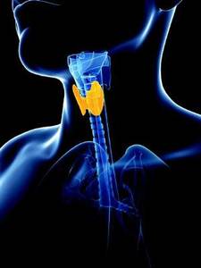
The nondiagnostic rate with CNB was also significantly lower than with fine-needle aspiration for nodules with calcifications, posterior location, or diameter less than 1 cm, Dr. Lim said at the meeting at the meeting held by the American Thyroid Association, Asia-Oceania Thyroid Association, European Thyroid Association, and Latin American Thyroid Society.
No difference was seen between the groups with respect to diagnostic performance based on degree of clinician experience.
The complication rate was higher with CNB than with fine -needle aspiration (3.6% vs. 1.6%), but complications were minor, Dr. Lim said.
Core needle biopsy has been suggested as a complementary tool for the diagnosis of thyroid nodules when the results of fine-needle aspiration are inconclusive, and the approach has been shown to be both safe and accurate for biopsy. However, its role as a first-line approach for thyroid nodule biopsy has been controversial and few studies have evaluated it as a first-line tool, she noted.
The current findings suggest that CNB is a safe and highly sensitive first-line biopsy method for such nodules, she concluded.
Dr. Lim reported having no disclosures.
LAKE BUENA VISTA, FLA. – The nondiagnostic result rate was significantly lower with core needle biopsy than with fine needle aspiration as a first-line biopsy method for newly detected thyroid nodules in a comparative study.
In 631 pairs of initially detected thyroid nodules that were matched based on propensity score analysis, the nondiagnostic result rate was 1.4% when core needle biopsy (CNB) was used, compared with 8.1% with ultrasound-guided fine-needle aspiration. The indeterminate result rate was 5.1% vs. 8.1% with the two approaches, respectively, and the differences between the groups were statistically significant, Dr. Hyun Kyung Lim of Soonchunhyang University Seoul Hospital, Korea, reported at the International Thyroid Congress.
The nondiagnostic rate with CNB was also significantly lower than with fine-needle aspiration for nodules with calcifications, posterior location, or diameter less than 1 cm, Dr. Lim said at the meeting at the meeting held by the American Thyroid Association, Asia-Oceania Thyroid Association, European Thyroid Association, and Latin American Thyroid Society.
No difference was seen between the groups with respect to diagnostic performance based on degree of clinician experience.
The complication rate was higher with CNB than with fine -needle aspiration (3.6% vs. 1.6%), but complications were minor, Dr. Lim said.
Core needle biopsy has been suggested as a complementary tool for the diagnosis of thyroid nodules when the results of fine-needle aspiration are inconclusive, and the approach has been shown to be both safe and accurate for biopsy. However, its role as a first-line approach for thyroid nodule biopsy has been controversial and few studies have evaluated it as a first-line tool, she noted.
The current findings suggest that CNB is a safe and highly sensitive first-line biopsy method for such nodules, she concluded.
Dr. Lim reported having no disclosures.
AT THE INTERNATIONAL THYROID CONGRESS
Key clinical point: The nondiagnostic result rate was significantly lower with core needle biopsy than with fine-needle aspiration as a first-line biopsy method for newly detected thyroid nodules in a comparative study.
Major finding: The nondiagnostic result rate was 1.4% with core needle biopsy vs. 8.1% with fine-needle aspiration.
Data source: A comparative study in 631 propensity score–matched pairs of thyroid nodules.
Disclosures: Dr. Lim reported having no disclosures.

Ophthalmic drugs in pregnancy and lactation
A number of drugs are available for ophthalmic use. This review focuses on single drug products and, although combination drug products are not discussed, these formulations typically include the drugs reviewed here.
Since it appears that ophthalmic medications are commonly used for a wide range of conditions and ages, one would expect to see numerous reports of their use in the eyes of pregnant or breastfeeding patients. Unfortunately, the opposite is true. The majority of the drugs have no human pregnancy or lactation data. When there are human data, it invariably involves the systemic use of the drug for other indications, rather than its ophthalmic use. Moreover, the animal reproduction data are usually not relevant because they involve systemic drugs (e.g., IV or oral). Consequently, determining the level of risk an ophthalmic drug presents to an embryo and/or fetus is primarily based on time and dose, the two cardinal principles of teratology.

Avoiding exposure during organogenesis – the period when a drug can cause developmental toxicity (altered growth, structural anomalies, functional and/or behavioral deficits, or death) – is usually best, but may not be possible in some cases, including glaucoma, eye infections, and eye surgery. Fortunately, the systemic concentrations of drugs applied topically to the eye are typically thought to be low, even though the levels of most drugs have not been studied. Thus, the risk to the embryo and/or fetus in most cases can be considered low and the drug classified as compatible in pregnancy and breastfeeding.
If a topical eye drug must be used during pregnancy or lactation, teach the patient how to decrease the amount of drug reaching the systemic circulation. This involves placing pressure over the tear duct in the corner of the eye for 1 minute or more, then removing any excess solution with absorbent tissue.
In the sections below, drugs are shown by indication or by pharmacologic class. The term “human eye data” refers to the use of the drug in pregnancy and/or lactation.
Glaucoma
If you are caring for a pregnant patient who is being treated for glaucoma, two recent reviews may be helpful: Surv Ophthalmol. 2011 Jul-Aug;56(4):324-35 and Curr Opin Ophthalmol. 2014 Mar;25(2):93-7.
Sympathomimetics (alpha-adrenergic agonists) include apraclonidine (Iopidine), which has no human eye data, and brimonidine (Alphagan P), which has one case report in pregnancy and breastfeeding showing no fetal or nursing infant harm.
Four of the five beta-adrenergic blockers have no human eye data: betaxolol (Betoptic), carteolol (Ocupress), levobunolol (Betagan), and metipranolol, but there are two case reports for timolol (Betimol, Istalol, Timoptic, Timoptic-XE) showing no fetal harm in one newborn and growth restriction in the other.
Among the miotics, there are limited human eye data for pilocarpine (Isopto Carpine) and no fetal harm was observed. For echothiophate iodide (Phospholine Iodide), there is one case report of a normal full-term infant whose mother was treated with the agent up to 32 weeks’ gestation and then with pilocarpine for 8 weeks (Arch Ophthalmol. 1968 Mar;79[3]:283-5).

Carbonic anhydrase inhibitors include brinzolamide (Azopt), which has no human eye data, and dorzolamide (Trusopt), which has growth restriction in one case treated with fixed combination of dorzolamide and timolol. There are no human eye data for unoprostone isopropyl (Rescula), a synthetic docosanoid.
Of the four prostaglandin analogs, three have no human eye data, bimatoprost (Lumigan), tafluprost (Zioptan), and travoprost (Travatan Z). There were 11 pregnancies exposed to latanoprost (Xalatan). The outcomes of these cases were one lost to follow-up, one miscarriage, and nine infants without congenital anomalies (Am J Ophthalmol. 2004 Aug;138[2]:305-6).
Mitomycin (Mitosol) is an antimetabolite that is given topically to the surgical site of glaucoma filtration surgery. No reports of its use in pregnant humans have been located. According to the manufacturer, clinically significant systemic concentrations are not expected.
Antiseptics
Povidone-iodine (Betadine) is indicated for prepping of the periocular region. There does not appear to be any risk to the embryo-fetus or nursing infant from this indication.
Antihistamines
The four ophthalmic agents in this class are alcaftadine (Lastacaft), azelastine (Optivar), emedastine (Emadine), and epinastine (Elestat). There are no human eye data for these agents but, like antihistamines given systemically, they are probably compatible in pregnancy and lactation.
Antihistamine-mast cell stabilizers
There are no human eye data for bepotastine (Bepreve), ketotifen (Alaway), and olopatadine (Pataday, Patanol). Peak plasma concentrations of bepotastine were 5.1-7.3 ng/mL for 1-2 hours after instillation and were less than 2 ng/mL at 24 hours. It appears that these drugs can also be classified as compatible in pregnancy and lactation.
Anti-infectives
Ten anti-infectives are available for topical treatment of eye infections (systemic concentrations if known): besifloxacin (Besivance) (0.43 ng/mL), ciprofloxacin (less than 2.5 ng/mL), gentamicin, gatifloxacin (Zymaxid) (less than 5 ng/mL), levofloxacin (Iquix) (10.9 ng/mL), moxifloxacin (Vigamox) (2.7 ng/mL), ofloxacin (1.9 ng/mL), sulfacetamide (Isopto Cetamide), and tobramycin. The tenth agent, natamycin (Natacyn), is an antifungal. According to the manufacturer, systemic absorption is not expected.
None of these agents have human eye data, but they are usually considered compatible in pregnancy and breastfeeding when systemic formulations are used, so they should be compatible when used in the eye.
Antivirals
Ganciclovir (Zirgan) has no human eye data but, according to the manufacturer, the daily ophthalmic dose is about 0.04% and 0.1% of the oral and IV doses, respectively. Thus, minimal systemic exposure is expected. Trifluridine (Viroptic) also has no eye human data. As reported in the product information, detectable blood concentrations of the drug were not found in healthy normal subjects indicating that systemic absorption was negligible.
Corticosteroids
Among the nine corticosteroid products, six are suspensions or ointments, one is an injection, and two are implants. In most nonpregnant patients receiving the dexamethasone (Ozurdex) intravitreal implant, plasma dexamethasone concentrations were undetectable (less than 50 pg/mL) but, in some, ranged from 52 pg/mL to 102 pg/mL. There is only one case report describing the use of topical dexamethasone suspension (Maxidex) in pregnancy. In that case, dexamethasone and clindamycin were given in the first and second trimesters and the woman eventually gave birth to a normal full term infant (Int Ophthalmol. 1998-1999;22[2]:85-8).
For the fluocinolone (Retisert) ocular implant, systemic absorption of detectable amounts of the drug have not been observed. In a second report, a patient who had type 1 diabetes and was 6 months pregnant received a 2-mg intravitreal injection of triamcinolone (Triesence) in both eyes. No adverse effects in the mother or fetus were noted (Clin Ophthalmol. 2011;5:439-41).
There are no human eye data for five topical corticosteroids: difluprednate (Durezol), fluorometholone (Flarex, Fluor-OP, FML), loteprednol (Alrex, Lotemax), prednisolone (Econopred), and rimexolone (Vexol). Only two of the five drugs had information about systemic absorption. For difluprednate, blood levels were below the quantification limit (50 ng/mL). Extremely low levels were detected after the use of rimexolone with a mean serum concentration of 130 pg/mL (range less than 80-470 pg/mL).
Cycloplegics-mydriatics
This class includes four anticholinergic agents: atropine, cyclopentolate (Cyclogyl), homatropine, and tropicamide (Mydriacyl). Systemic absorption has not been studied for these drugs and there are no reports of their use in pregnancy or lactation.
Cystine-depleting agents
There are no reports describing the use of cysteamine (Cystaran) in human pregnancy or lactation. Since the drug is given as one drop in each eye every waking hour, transfer to the systemic circulation should be expected, but the amount, if it occurs, has not been reported.
Immunologics
There are no reports on the use of cyclosporine (Restasis) eye drops in human pregnancy or during breastfeeding. However, data for the systemic use of the drug during these conditions has shown it to be low risk. After long term ophthalmic use, blood concentrations of the drug were below the quantitation limit of 0.1 ng/mL.
Local anesthetics
The three drugs in this class are lidocaine, proparacaine (Alcaine), and tetracaine (Altacaine, Tetravisc). There is no information regarding the use of these agents in pregnancy or lactation. Since they are used for brief periods, the risk to an embryo or nursing infant appears to be nil.
Mast cell stabilizers
There are three drugs in this class that can be used topically in the eye: cromolyn, lodoxamide (Alomide), and nedocromil (Alocril). There are no human eye data for these agents. Systemic concentrations of lodoxamide were below the detection limit of 2.5 ng/mL.
Miotics
The two miotics are acetylcholine (Miochol E) and carbachol (Carbastat, Miostat). Both are used immediately before eye surgery. The amount, if any, in the systemic circulation is unknown. No reports of human eye use in pregnancy or lactation have been found.
NSAIDs
The five drugs in this class are bromfenac (Xibrom), diclofenac (Voltaren), flurbiprofen (Ocufen), ketorolac, and nepafenac (Nevanac). Although it has not been studied, the estimated plasma level for bromfenac is less than 50 ng/mL. For diclofenac, the plasma concentration was below the limit of quantification (10 ng/mL). In a study conducted with ketorolac, only 5 of 26 subjects had detectable plasma concentrations (10.7 to 22.5 ng/mL). These levels were about 2% of the peak plasma levels after oral dosing. For nepafenac, the peak plasma concentrations of the parent drug and active metabolite were 0.3 and 0.4 ng/mL, respectively. These data suggest that the risk to the embryo-fetus or a nursing infant are nil.
Photodynamic therapy
Verteporfin (Visudyne) is given intravenously. Three reports have described its use in three pregnancies. In all cases, the infants were healthy at birth and had normal growth (Acta Ophthalmol Scand. 2004 Oct;82[5]:623-4, Eye [Lond]. 2009 Jun;23[6]:1479, and Aust N Z J Obstet Gynaecol. 2009 Apr;49[2]:236-7).
Proteolytic enzymes
No human eye data were found for ocriplasmin (Jetrea), an agent given as an intravitreal injection. Although it was not studied, detectable levels of the drug in the systemic circulation are not expected, according to the manufacturer.
Selective vascular endothelial growth factor antagonists
There are three agents in this class: aflibercept (Eylea), pegaptanib (Macugen), and ranibizumab (Lucentis). There are no human eye data for these drugs. All are given as intravitreal injection. The systemic concentrations of the three drugs were a mean 0.02 mcg/mL; 80 ng/mL (after a dose of 10 times the recommended dose); and an estimated minimum 0.22 ng/mL, respectively. These data suggest that the risk to the embryo-fetus or a nursing infant is nil.
Sympathomimetics (decongestants)
There are four ophthalmic agents in this class and none have human eye data. Naphazoline (Naphcon, Vasocon) is the only one of the four that requires a prescription. No reports describing the systemic absorption, if any, have been found. The other three agents are available over the counter. They are oxymetazoline (Visine L.R.), phenylephrine, and tetrahydrozoline (Opti-Clear). Although the amount reaching the systemic circulation was not provided by the manufacturers for these three agents, a caution was placed on phenylephrine stating that systemic absorption, although rare, might cause alpha-adrenergic effects, such as a rise in blood pressure and reflex atropine-sensitive bradycardia.
The risk to the embryo-fetus or nursing infant from ophthalmic drugs appears to be low, with the possible exception of phenylephrine. Nevertheless, if any of these agents are used in pregnancy or during breastfeeding, careful assessment of the embryo-fetus and nursing infant should be conducted. Moreover, research on the potential effects of ophthalmic drugs on the embryo-fetus and nursing infant are desperately needed.
Mr. Briggs is clinical professor of pharmacy at the University of California, San Francisco; and adjunct professor of pharmacy at the University of Southern California, Los Angeles, and Washington State University, Spokane. He is coauthor of “Drugs in Pregnancy and Lactation,” and coeditor of “Diseases, Complications, and Drug Therapy in Obstetrics.” He has no relevant financial disclosures.
A number of drugs are available for ophthalmic use. This review focuses on single drug products and, although combination drug products are not discussed, these formulations typically include the drugs reviewed here.
Since it appears that ophthalmic medications are commonly used for a wide range of conditions and ages, one would expect to see numerous reports of their use in the eyes of pregnant or breastfeeding patients. Unfortunately, the opposite is true. The majority of the drugs have no human pregnancy or lactation data. When there are human data, it invariably involves the systemic use of the drug for other indications, rather than its ophthalmic use. Moreover, the animal reproduction data are usually not relevant because they involve systemic drugs (e.g., IV or oral). Consequently, determining the level of risk an ophthalmic drug presents to an embryo and/or fetus is primarily based on time and dose, the two cardinal principles of teratology.
Avoiding exposure during organogenesis – the period when a drug can cause developmental toxicity (altered growth, structural anomalies, functional and/or behavioral deficits, or death) – is usually best, but may not be possible in some cases, including glaucoma, eye infections, and eye surgery. Fortunately, the systemic concentrations of drugs applied topically to the eye are typically thought to be low, even though the levels of most drugs have not been studied. Thus, the risk to the embryo and/or fetus in most cases can be considered low and the drug classified as compatible in pregnancy and breastfeeding.
If a topical eye drug must be used during pregnancy or lactation, teach the patient how to decrease the amount of drug reaching the systemic circulation. This involves placing pressure over the tear duct in the corner of the eye for 1 minute or more, then removing any excess solution with absorbent tissue.
In the sections below, drugs are shown by indication or by pharmacologic class. The term “human eye data” refers to the use of the drug in pregnancy and/or lactation.
Glaucoma
If you are caring for a pregnant patient who is being treated for glaucoma, two recent reviews may be helpful: Surv Ophthalmol. 2011 Jul-Aug;56(4):324-35 and Curr Opin Ophthalmol. 2014 Mar;25(2):93-7.
Sympathomimetics (alpha-adrenergic agonists) include apraclonidine (Iopidine), which has no human eye data, and brimonidine (Alphagan P), which has one case report in pregnancy and breastfeeding showing no fetal or nursing infant harm.
Four of the five beta-adrenergic blockers have no human eye data: betaxolol (Betoptic), carteolol (Ocupress), levobunolol (Betagan), and metipranolol, but there are two case reports for timolol (Betimol, Istalol, Timoptic, Timoptic-XE) showing no fetal harm in one newborn and growth restriction in the other.
Among the miotics, there are limited human eye data for pilocarpine (Isopto Carpine) and no fetal harm was observed. For echothiophate iodide (Phospholine Iodide), there is one case report of a normal full-term infant whose mother was treated with the agent up to 32 weeks’ gestation and then with pilocarpine for 8 weeks (Arch Ophthalmol. 1968 Mar;79[3]:283-5).
Carbonic anhydrase inhibitors include brinzolamide (Azopt), which has no human eye data, and dorzolamide (Trusopt), which has growth restriction in one case treated with fixed combination of dorzolamide and timolol. There are no human eye data for unoprostone isopropyl (Rescula), a synthetic docosanoid.
Of the four prostaglandin analogs, three have no human eye data, bimatoprost (Lumigan), tafluprost (Zioptan), and travoprost (Travatan Z). There were 11 pregnancies exposed to latanoprost (Xalatan). The outcomes of these cases were one lost to follow-up, one miscarriage, and nine infants without congenital anomalies (Am J Ophthalmol. 2004 Aug;138[2]:305-6).
Mitomycin (Mitosol) is an antimetabolite that is given topically to the surgical site of glaucoma filtration surgery. No reports of its use in pregnant humans have been located. According to the manufacturer, clinically significant systemic concentrations are not expected.
Antiseptics
Povidone-iodine (Betadine) is indicated for prepping of the periocular region. There does not appear to be any risk to the embryo-fetus or nursing infant from this indication.
Antihistamines
The four ophthalmic agents in this class are alcaftadine (Lastacaft), azelastine (Optivar), emedastine (Emadine), and epinastine (Elestat). There are no human eye data for these agents but, like antihistamines given systemically, they are probably compatible in pregnancy and lactation.
Antihistamine-mast cell stabilizers
There are no human eye data for bepotastine (Bepreve), ketotifen (Alaway), and olopatadine (Pataday, Patanol). Peak plasma concentrations of bepotastine were 5.1-7.3 ng/mL for 1-2 hours after instillation and were less than 2 ng/mL at 24 hours. It appears that these drugs can also be classified as compatible in pregnancy and lactation.
Anti-infectives
Ten anti-infectives are available for topical treatment of eye infections (systemic concentrations if known): besifloxacin (Besivance) (0.43 ng/mL), ciprofloxacin (less than 2.5 ng/mL), gentamicin, gatifloxacin (Zymaxid) (less than 5 ng/mL), levofloxacin (Iquix) (10.9 ng/mL), moxifloxacin (Vigamox) (2.7 ng/mL), ofloxacin (1.9 ng/mL), sulfacetamide (Isopto Cetamide), and tobramycin. The tenth agent, natamycin (Natacyn), is an antifungal. According to the manufacturer, systemic absorption is not expected.
None of these agents have human eye data, but they are usually considered compatible in pregnancy and breastfeeding when systemic formulations are used, so they should be compatible when used in the eye.
Antivirals
Ganciclovir (Zirgan) has no human eye data but, according to the manufacturer, the daily ophthalmic dose is about 0.04% and 0.1% of the oral and IV doses, respectively. Thus, minimal systemic exposure is expected. Trifluridine (Viroptic) also has no eye human data. As reported in the product information, detectable blood concentrations of the drug were not found in healthy normal subjects indicating that systemic absorption was negligible.
Corticosteroids
Among the nine corticosteroid products, six are suspensions or ointments, one is an injection, and two are implants. In most nonpregnant patients receiving the dexamethasone (Ozurdex) intravitreal implant, plasma dexamethasone concentrations were undetectable (less than 50 pg/mL) but, in some, ranged from 52 pg/mL to 102 pg/mL. There is only one case report describing the use of topical dexamethasone suspension (Maxidex) in pregnancy. In that case, dexamethasone and clindamycin were given in the first and second trimesters and the woman eventually gave birth to a normal full term infant (Int Ophthalmol. 1998-1999;22[2]:85-8).
For the fluocinolone (Retisert) ocular implant, systemic absorption of detectable amounts of the drug have not been observed. In a second report, a patient who had type 1 diabetes and was 6 months pregnant received a 2-mg intravitreal injection of triamcinolone (Triesence) in both eyes. No adverse effects in the mother or fetus were noted (Clin Ophthalmol. 2011;5:439-41).
There are no human eye data for five topical corticosteroids: difluprednate (Durezol), fluorometholone (Flarex, Fluor-OP, FML), loteprednol (Alrex, Lotemax), prednisolone (Econopred), and rimexolone (Vexol). Only two of the five drugs had information about systemic absorption. For difluprednate, blood levels were below the quantification limit (50 ng/mL). Extremely low levels were detected after the use of rimexolone with a mean serum concentration of 130 pg/mL (range less than 80-470 pg/mL).
Cycloplegics-mydriatics
This class includes four anticholinergic agents: atropine, cyclopentolate (Cyclogyl), homatropine, and tropicamide (Mydriacyl). Systemic absorption has not been studied for these drugs and there are no reports of their use in pregnancy or lactation.
Cystine-depleting agents
There are no reports describing the use of cysteamine (Cystaran) in human pregnancy or lactation. Since the drug is given as one drop in each eye every waking hour, transfer to the systemic circulation should be expected, but the amount, if it occurs, has not been reported.
Immunologics
There are no reports on the use of cyclosporine (Restasis) eye drops in human pregnancy or during breastfeeding. However, data for the systemic use of the drug during these conditions has shown it to be low risk. After long term ophthalmic use, blood concentrations of the drug were below the quantitation limit of 0.1 ng/mL.
Local anesthetics
The three drugs in this class are lidocaine, proparacaine (Alcaine), and tetracaine (Altacaine, Tetravisc). There is no information regarding the use of these agents in pregnancy or lactation. Since they are used for brief periods, the risk to an embryo or nursing infant appears to be nil.
Mast cell stabilizers
There are three drugs in this class that can be used topically in the eye: cromolyn, lodoxamide (Alomide), and nedocromil (Alocril). There are no human eye data for these agents. Systemic concentrations of lodoxamide were below the detection limit of 2.5 ng/mL.
Miotics
The two miotics are acetylcholine (Miochol E) and carbachol (Carbastat, Miostat). Both are used immediately before eye surgery. The amount, if any, in the systemic circulation is unknown. No reports of human eye use in pregnancy or lactation have been found.
NSAIDs
The five drugs in this class are bromfenac (Xibrom), diclofenac (Voltaren), flurbiprofen (Ocufen), ketorolac, and nepafenac (Nevanac). Although it has not been studied, the estimated plasma level for bromfenac is less than 50 ng/mL. For diclofenac, the plasma concentration was below the limit of quantification (10 ng/mL). In a study conducted with ketorolac, only 5 of 26 subjects had detectable plasma concentrations (10.7 to 22.5 ng/mL). These levels were about 2% of the peak plasma levels after oral dosing. For nepafenac, the peak plasma concentrations of the parent drug and active metabolite were 0.3 and 0.4 ng/mL, respectively. These data suggest that the risk to the embryo-fetus or a nursing infant are nil.
Photodynamic therapy
Verteporfin (Visudyne) is given intravenously. Three reports have described its use in three pregnancies. In all cases, the infants were healthy at birth and had normal growth (Acta Ophthalmol Scand. 2004 Oct;82[5]:623-4, Eye [Lond]. 2009 Jun;23[6]:1479, and Aust N Z J Obstet Gynaecol. 2009 Apr;49[2]:236-7).
Proteolytic enzymes
No human eye data were found for ocriplasmin (Jetrea), an agent given as an intravitreal injection. Although it was not studied, detectable levels of the drug in the systemic circulation are not expected, according to the manufacturer.
Selective vascular endothelial growth factor antagonists
There are three agents in this class: aflibercept (Eylea), pegaptanib (Macugen), and ranibizumab (Lucentis). There are no human eye data for these drugs. All are given as intravitreal injection. The systemic concentrations of the three drugs were a mean 0.02 mcg/mL; 80 ng/mL (after a dose of 10 times the recommended dose); and an estimated minimum 0.22 ng/mL, respectively. These data suggest that the risk to the embryo-fetus or a nursing infant is nil.
Sympathomimetics (decongestants)
There are four ophthalmic agents in this class and none have human eye data. Naphazoline (Naphcon, Vasocon) is the only one of the four that requires a prescription. No reports describing the systemic absorption, if any, have been found. The other three agents are available over the counter. They are oxymetazoline (Visine L.R.), phenylephrine, and tetrahydrozoline (Opti-Clear). Although the amount reaching the systemic circulation was not provided by the manufacturers for these three agents, a caution was placed on phenylephrine stating that systemic absorption, although rare, might cause alpha-adrenergic effects, such as a rise in blood pressure and reflex atropine-sensitive bradycardia.
The risk to the embryo-fetus or nursing infant from ophthalmic drugs appears to be low, with the possible exception of phenylephrine. Nevertheless, if any of these agents are used in pregnancy or during breastfeeding, careful assessment of the embryo-fetus and nursing infant should be conducted. Moreover, research on the potential effects of ophthalmic drugs on the embryo-fetus and nursing infant are desperately needed.
Mr. Briggs is clinical professor of pharmacy at the University of California, San Francisco; and adjunct professor of pharmacy at the University of Southern California, Los Angeles, and Washington State University, Spokane. He is coauthor of “Drugs in Pregnancy and Lactation,” and coeditor of “Diseases, Complications, and Drug Therapy in Obstetrics.” He has no relevant financial disclosures.
Self-reported poor functional status predicts perioperative morbidity
SAN DIEGO – Among patients with pulmonary hypertension presenting for elective surgery, self-reported poor functional status is associated with multiple comorbidities and is independently predictive of longer hospital length of stay, results from an ongoing single-center study suggest.
“Patients with pulmonary hypertension (PHTN) presenting for elective surgery are at significantly higher risk for adverse perioperative outcomes, including increased hospital length of stay, right ventricular failure, cardiac arrhythmia, persistent postoperative hypoxemia, coronary ischemia and death,” researchers led by Dr. Aalap C. Shah wrote in an abstract presented at the at the annual meeting of the American Society of Anesthesiologists. “The diagnosis of PHTN is based on costly echocardiographic examination and right heart catheterization and should be reserved for high-risk patients. No studies have assessed the role of self-reported functional classification on PHTN severity stratification, and few studies have achieved a sufficiently large patient sample size.”

In an effort to evaluate the predictive value of self-reported exercise tolerance on echocardiogram findings, outcomes, and length of stay (LOS) after noncardiac, nonobstetric surgery, the researchers queried the University of Washington database for all PHTN seen in preoperative anesthesia clinic for noncardiac, nonobstetric procedures from April 2007 through September 2013. Inclusion criteria required an echocardiogram less than 1 year prior to the procedure and available patient-reported functional status, which was defined as less than four metabolic equivalents (METS) in exercise testing or four METS or greater. Dr. Shah, formerly a resident in the University of Washington’s department of anesthesiology and pain medicine, and his associates used univariate analyses to compare functional status with echocardiographic findings, complication rates, and length of stay (LOS). At the meeting he presented results from 294 patients evaluated to date: 143 with normal functional status and 151 with poor functional status. Their mean age was 62 years, and 51% of patients were female.
Compared with their counterparts with normal functional status, patients with poor functional status trended toward a higher complication rate at hospital discharge (14.6% vs. 7%, respectively; P = .041) and had a higher cumulative rate of complications (33 vs. 15; P = .035). However, no association between functional status and complications was observed 30 days postoperatively.
Patients with poor functional status had a significantly longer average LOS, compared with patients with normal functional status (7.21 vs. 4.73 days; P = .047). Open surgical approach was also an independent predictor of increased LOS (odds ratio 2.39; P = .005). No significant independent predictors of complications were observed at discharge or 30 days postoperatively.
“Going forward, the goal is to use these data to create a risk stratification algorithm to figure out: Does a patient with good functional status and pulmonary hypertension undergoing toe surgery, for example, really need an echocardiogram before getting surgery?” said Dr. Shah said, who is now an anesthesiology fellow at Boston Children’s Hospital. “Hopefully we can show that using these risk stratification algorithms can decrease the costs and decrease the time to actually getting surgery.”
The researchers reported having no financial disclosures.
SAN DIEGO – Among patients with pulmonary hypertension presenting for elective surgery, self-reported poor functional status is associated with multiple comorbidities and is independently predictive of longer hospital length of stay, results from an ongoing single-center study suggest.
“Patients with pulmonary hypertension (PHTN) presenting for elective surgery are at significantly higher risk for adverse perioperative outcomes, including increased hospital length of stay, right ventricular failure, cardiac arrhythmia, persistent postoperative hypoxemia, coronary ischemia and death,” researchers led by Dr. Aalap C. Shah wrote in an abstract presented at the at the annual meeting of the American Society of Anesthesiologists. “The diagnosis of PHTN is based on costly echocardiographic examination and right heart catheterization and should be reserved for high-risk patients. No studies have assessed the role of self-reported functional classification on PHTN severity stratification, and few studies have achieved a sufficiently large patient sample size.”
In an effort to evaluate the predictive value of self-reported exercise tolerance on echocardiogram findings, outcomes, and length of stay (LOS) after noncardiac, nonobstetric surgery, the researchers queried the University of Washington database for all PHTN seen in preoperative anesthesia clinic for noncardiac, nonobstetric procedures from April 2007 through September 2013. Inclusion criteria required an echocardiogram less than 1 year prior to the procedure and available patient-reported functional status, which was defined as less than four metabolic equivalents (METS) in exercise testing or four METS or greater. Dr. Shah, formerly a resident in the University of Washington’s department of anesthesiology and pain medicine, and his associates used univariate analyses to compare functional status with echocardiographic findings, complication rates, and length of stay (LOS). At the meeting he presented results from 294 patients evaluated to date: 143 with normal functional status and 151 with poor functional status. Their mean age was 62 years, and 51% of patients were female.
Compared with their counterparts with normal functional status, patients with poor functional status trended toward a higher complication rate at hospital discharge (14.6% vs. 7%, respectively; P = .041) and had a higher cumulative rate of complications (33 vs. 15; P = .035). However, no association between functional status and complications was observed 30 days postoperatively.
Patients with poor functional status had a significantly longer average LOS, compared with patients with normal functional status (7.21 vs. 4.73 days; P = .047). Open surgical approach was also an independent predictor of increased LOS (odds ratio 2.39; P = .005). No significant independent predictors of complications were observed at discharge or 30 days postoperatively.
“Going forward, the goal is to use these data to create a risk stratification algorithm to figure out: Does a patient with good functional status and pulmonary hypertension undergoing toe surgery, for example, really need an echocardiogram before getting surgery?” said Dr. Shah said, who is now an anesthesiology fellow at Boston Children’s Hospital. “Hopefully we can show that using these risk stratification algorithms can decrease the costs and decrease the time to actually getting surgery.”
The researchers reported having no financial disclosures.
AT THE ASA ANNUAL MEETING
Key clinical point:Poor self-reported exercise tolerance by patients with pulmonary hypertension is associated with multiple comorbidities and increased hospital length of stay.
Major finding: Compared with their counterparts with normal functional status, patients with poor functional status trended toward a higher complication rate at hospital discharge (14.6% vs. 7%, respectively; P = .041) and had a higher cumulative rate of complications (33 vs. 15; P = .035).
Data source: A study 294 PHTN patients seen in preoperative anesthesia clinic at the University of Washington for non-cardiac, nonobstetric procedures from April 2007 through September 2013.
Disclosures: The researchers reported having no financial disclosures.