User login
Session highlights controversies in diverticulitis management
SAN DIEGO – During the annual clinical congress of the American College of Surgeons, a panel of experts discussed a wide range of evolving controversies in the management of complicated diverticulitis.
John Migaly, MD, kicked off the session by exploring the current evidence for laparoscopic lavage versus primary resection for Hinchey III diverticulitis. “Over the past two decades there has been a growing utilization of primary resection and reanastomoses with or without the use of ileostomy,” said Dr. Migaly, director of the general surgery residency program at Duke University, Durham, N.C. “And most recently over the last 5 or 10 years there’s been growing enthusiasm for laparoscopic lavage for Hinchey III diverticulitis. The enthusiasm for this procedure is based on [its accessibility] to all of us who operate on the colon in the acute setting.”

The DILALA trial makes the best case for laparoscopic lavage, “but it doesn’t make a very good one,” he commented. The trial was conducted in nine surgical departments in Sweden and Denmark (Ann Surg. 2016;263[1]:117-22). Hinchey III patients were randomized 1:1 to laparoscopic lavage or the Hartmann procedure. The primary outcome was reoperations within 12 months. An early analysis of the short-term outcomes in 83 patients found similar 30-day and 90-day mortality and morbidity. Its authors concluded that laparoscopic lavage had equivalent morbidity and mortality compared with radical resection, shorter operative time, shorter time in the recovery unit, a shorter hospital stay, but no difference in the rate of reoperation.
“It was found to be safe and feasible,” Dr. Migaly said. One year later, the researchers presented their 12-month outcomes and came to the same conclusions. Limitations of the data are that it was conducted in nine centers “but they enrolled only 83 patients, so it seems underpowered,” he said. “And there was no mention of the incidence of abdominal abscess requiring percutaneous drainage or episodes of diverticulitis. The data seem a little less granular than the LADIES trial.”
The SCANDIV trial is the largest study on the topic to date, a randomized clinical superiority trial conducted at 21 centers in Sweden and Norway (JAMA 2015;314[13]:1364-75). Of the 509 patients screened, 415 were eligible and 199 were enrolled: 101 to laparoscopic lavage, 98 to colon resection. The primary endpoint was severe postoperative complications within 90 days, defined as a Clavien-Dindo score of over 3. The researchers found no difference in major complications nor in 90-day mortality between the two groups. The rate of reoperation was significantly higher in the lavage group, compared with the resection group (20.3% vs. 5.7%, respectively). Four sigmoid cancers were missed in the lavage group and, while the length of operation was significantly shorter in the lavage group, there were no differences between the two groups in hospital length of stay or quality of life.
A meta-analysis of the three randomized controlled trials that Dr. Migaly reviewed concluded that laparoscopic lavage, compared with resection, for Hinchey III diverticulitis increased the rate of total reoperations, the rate of reoperation for infection, and the rate of subsequent percutaneous drainage (J Gastrointest Surg 2017;21[9]:1491-99). A larger, more recent meta-analysis of 589 patients, including the three randomized controlled trials that Dr. Migaly discussed, concluded that laparoscopic lavage patients, compared with resection patients, had three times the risk of persistent peritonitis, intra-abdominal abscess, and emergency reoperative surgery. “Therefore, a reasonable conclusion would be that data at this point does not support the use of laparoscopic lavage,” he said.

The initial procedure of choice for most patients is abscess drainage via CT-guided percutaneous drainage. “There are some patients who are not candidates for percutaneous drainage, [such as] if the abscess is not accessible to the radiologist, if the patient is anticoagulated, and if the patient requires emergent surgical intervention irrespective of the abscess,” said Dr. Maron, a colorectal surgeon who practices at Cleveland Clinic Florida, Weston. “There is also a question of cavity size. Most authors in the literature use a cut-off of 3-4 cm.”
The goal is complete drainage of the abscess, and sometimes multiple catheters will be required. One study found that predictors of successful abscess drainage included having a well-defined, unilocular abscess. The success rate fell to 63% for patients who presented with more complex abscesses, including those that were loculated, poorly defined, associated with a fistula, and contained feces or semisolid material (Dis Colon Rectum. 1997;40:1009-13).
The 2014 ASCRS Practice Parameters includes the recommendation that elective colectomy should typically be considered after the patient recovers from an episode of complicated diverticulitis, “but some of the data may be calling that into question,” Dr. Maron said. In one series of 18 patients with an abscess treated percutaneously, 11 refused surgery and 7 had significant comorbidity (Dis Colon Rectum. 2014;57:331-6). Three patients died of a pre-existing condition and 7 of the 15 surviving patients had recurrent diverticulitis. Three underwent surgery and four were treated medically. The authors found no association between long-term failure and abscess location or previous episodes of diverticulitis.
In a larger study, researchers identified 218 patients who were initially treated with intravenous antibiotics and percutaneous drain (Dis Colon Rectum. 2013;56:622-6). About 10% of the patients required an urgent operation, while most of the other patients underwent elective resection, but 15% of patients did not undergo a subsequent colectomy. “Most of these patients were medically unfit to undergo surgery,” he noted. Abscess location was more commonly paracolic than pelvic. The mean abscess size was 4.2 cm, and the drain was left in for a median of 20 days. The recurrence rate in this series was only 30%, but none of the recurrences required surgery. The authors found that abscesses greater than 5 cm in size were associated with a greater risk of recurrence (P = .003). They concluded that observation after percutaneous drainage is safe in selected patients.
Based on results from this and other more recent studies, Dr. Maron said that it remains unclear whether surgeons should wait and watch or operate after successful percutaneous drainage of diverticular abscess. The data are “not as robust as we’d like … most of these are retrospective studies,” he said. “There’s quite a bit of inherent selection bias, and there is no standardization with regard to length of time of percutaneous drain, rationale for nonoperative management versus elective colectomy. What we do know is that there are some patients who can be managed safely without surgery. Unfortunately, there is no good algorithm I can offer you: Perhaps larger abscesses can portend a higher recurrence. I don’t think we’ll have a good answer to this until we perform a prospective randomized trial. However, we may learn some data from patients managed by peritoneal lavage and drain placement.”

Next, consider how to perform the procedure: laparoscopic or open? “But again, you’re going to look at how stable your patient is, what your skill set is, what equipment is available, and if the patient has had previous abdominal surgeries,” she advised. “In the traditional Hartmann procedure, you resect the perforated segment. You try not to open any tissue planes that you don’t have to. You do just enough so you can bring up a colostomy; you close the rectum or you make a mucous fistula. The problem with this operation is that up to 80% of these patients have their colostomy closed. That is why there is all this controversy. If there is any question, this [procedure] is always the safest option; there’s no anastomosis.”
What about a performing resection and a colorectal anastomosis? This is usually done more commonly in the elective situation, “when things are perfect, when you have healthy tissue,” Dr. Hull said. “If you do it in the emergent situation you have to think to yourself, ‘Could my patient tolerate a leak?’ You won’t want to do this operation on a 72-year-old who’s on steroids for chronic pulmonary disease and has coronary artery disease, because if they had a leak, they’d probably die.”
The third procedural option is to resect bowel (usually sigmoid) with colorectal anastomosis and diversion (loop ileostomy). “This is always my preferred choice,” Dr. Hull said. “I always am thinking, ‘Why can’t I do this?’ The reason is, closure of ileostomy is much easier than a Hartmann reversal, and 90% of these patients get reversed.” In a multicenter trial conducted by Swedish researchers, 62 patients were randomized to Hartmann’s procedure versus primary anastomosis with diverting ileostomy for perforated left-sided diverticulitis (Ann Surg. 2012;256[5]:819-27). The mortality and complications were similar, but the number of patients who got their stoma reversed was significantly less in the Hartmann’s group, compared with the primary anastomosis group (57% vs. 90%, respectively), and the serious complications were much higher in the Hartmann’s group (20% vs. 0). She cited an article from the World Journal of Emergency Surgery as one of the most comprehensive reviews of the subject.

So what is a surgeon to do? For an anastomosis after resection, “consider patient factors: Are they stable?” Dr. Hull said. “What are their comorbid conditions? You have to think, ‘What is my preference? Am I comfortable putting this back together?’ But a primary anastomosis is feasible, even in the acute setting with or without diverting ileostomy. The laparoscopic approach is typically preferred, but if that’s not in your armamentarium, the open [approach] is just fine. You should always perform a leak test. You can also do on table colonic lavage if feasible, especially if you have a large stool burden.”
None of the speakers reported having financial disclosures.
SAN DIEGO – During the annual clinical congress of the American College of Surgeons, a panel of experts discussed a wide range of evolving controversies in the management of complicated diverticulitis.
John Migaly, MD, kicked off the session by exploring the current evidence for laparoscopic lavage versus primary resection for Hinchey III diverticulitis. “Over the past two decades there has been a growing utilization of primary resection and reanastomoses with or without the use of ileostomy,” said Dr. Migaly, director of the general surgery residency program at Duke University, Durham, N.C. “And most recently over the last 5 or 10 years there’s been growing enthusiasm for laparoscopic lavage for Hinchey III diverticulitis. The enthusiasm for this procedure is based on [its accessibility] to all of us who operate on the colon in the acute setting.”
The DILALA trial makes the best case for laparoscopic lavage, “but it doesn’t make a very good one,” he commented. The trial was conducted in nine surgical departments in Sweden and Denmark (Ann Surg. 2016;263[1]:117-22). Hinchey III patients were randomized 1:1 to laparoscopic lavage or the Hartmann procedure. The primary outcome was reoperations within 12 months. An early analysis of the short-term outcomes in 83 patients found similar 30-day and 90-day mortality and morbidity. Its authors concluded that laparoscopic lavage had equivalent morbidity and mortality compared with radical resection, shorter operative time, shorter time in the recovery unit, a shorter hospital stay, but no difference in the rate of reoperation.
“It was found to be safe and feasible,” Dr. Migaly said. One year later, the researchers presented their 12-month outcomes and came to the same conclusions. Limitations of the data are that it was conducted in nine centers “but they enrolled only 83 patients, so it seems underpowered,” he said. “And there was no mention of the incidence of abdominal abscess requiring percutaneous drainage or episodes of diverticulitis. The data seem a little less granular than the LADIES trial.”
The SCANDIV trial is the largest study on the topic to date, a randomized clinical superiority trial conducted at 21 centers in Sweden and Norway (JAMA 2015;314[13]:1364-75). Of the 509 patients screened, 415 were eligible and 199 were enrolled: 101 to laparoscopic lavage, 98 to colon resection. The primary endpoint was severe postoperative complications within 90 days, defined as a Clavien-Dindo score of over 3. The researchers found no difference in major complications nor in 90-day mortality between the two groups. The rate of reoperation was significantly higher in the lavage group, compared with the resection group (20.3% vs. 5.7%, respectively). Four sigmoid cancers were missed in the lavage group and, while the length of operation was significantly shorter in the lavage group, there were no differences between the two groups in hospital length of stay or quality of life.
A meta-analysis of the three randomized controlled trials that Dr. Migaly reviewed concluded that laparoscopic lavage, compared with resection, for Hinchey III diverticulitis increased the rate of total reoperations, the rate of reoperation for infection, and the rate of subsequent percutaneous drainage (J Gastrointest Surg 2017;21[9]:1491-99). A larger, more recent meta-analysis of 589 patients, including the three randomized controlled trials that Dr. Migaly discussed, concluded that laparoscopic lavage patients, compared with resection patients, had three times the risk of persistent peritonitis, intra-abdominal abscess, and emergency reoperative surgery. “Therefore, a reasonable conclusion would be that data at this point does not support the use of laparoscopic lavage,” he said.
The initial procedure of choice for most patients is abscess drainage via CT-guided percutaneous drainage. “There are some patients who are not candidates for percutaneous drainage, [such as] if the abscess is not accessible to the radiologist, if the patient is anticoagulated, and if the patient requires emergent surgical intervention irrespective of the abscess,” said Dr. Maron, a colorectal surgeon who practices at Cleveland Clinic Florida, Weston. “There is also a question of cavity size. Most authors in the literature use a cut-off of 3-4 cm.”
The goal is complete drainage of the abscess, and sometimes multiple catheters will be required. One study found that predictors of successful abscess drainage included having a well-defined, unilocular abscess. The success rate fell to 63% for patients who presented with more complex abscesses, including those that were loculated, poorly defined, associated with a fistula, and contained feces or semisolid material (Dis Colon Rectum. 1997;40:1009-13).
The 2014 ASCRS Practice Parameters includes the recommendation that elective colectomy should typically be considered after the patient recovers from an episode of complicated diverticulitis, “but some of the data may be calling that into question,” Dr. Maron said. In one series of 18 patients with an abscess treated percutaneously, 11 refused surgery and 7 had significant comorbidity (Dis Colon Rectum. 2014;57:331-6). Three patients died of a pre-existing condition and 7 of the 15 surviving patients had recurrent diverticulitis. Three underwent surgery and four were treated medically. The authors found no association between long-term failure and abscess location or previous episodes of diverticulitis.
In a larger study, researchers identified 218 patients who were initially treated with intravenous antibiotics and percutaneous drain (Dis Colon Rectum. 2013;56:622-6). About 10% of the patients required an urgent operation, while most of the other patients underwent elective resection, but 15% of patients did not undergo a subsequent colectomy. “Most of these patients were medically unfit to undergo surgery,” he noted. Abscess location was more commonly paracolic than pelvic. The mean abscess size was 4.2 cm, and the drain was left in for a median of 20 days. The recurrence rate in this series was only 30%, but none of the recurrences required surgery. The authors found that abscesses greater than 5 cm in size were associated with a greater risk of recurrence (P = .003). They concluded that observation after percutaneous drainage is safe in selected patients.
Based on results from this and other more recent studies, Dr. Maron said that it remains unclear whether surgeons should wait and watch or operate after successful percutaneous drainage of diverticular abscess. The data are “not as robust as we’d like … most of these are retrospective studies,” he said. “There’s quite a bit of inherent selection bias, and there is no standardization with regard to length of time of percutaneous drain, rationale for nonoperative management versus elective colectomy. What we do know is that there are some patients who can be managed safely without surgery. Unfortunately, there is no good algorithm I can offer you: Perhaps larger abscesses can portend a higher recurrence. I don’t think we’ll have a good answer to this until we perform a prospective randomized trial. However, we may learn some data from patients managed by peritoneal lavage and drain placement.”
Next, consider how to perform the procedure: laparoscopic or open? “But again, you’re going to look at how stable your patient is, what your skill set is, what equipment is available, and if the patient has had previous abdominal surgeries,” she advised. “In the traditional Hartmann procedure, you resect the perforated segment. You try not to open any tissue planes that you don’t have to. You do just enough so you can bring up a colostomy; you close the rectum or you make a mucous fistula. The problem with this operation is that up to 80% of these patients have their colostomy closed. That is why there is all this controversy. If there is any question, this [procedure] is always the safest option; there’s no anastomosis.”
What about a performing resection and a colorectal anastomosis? This is usually done more commonly in the elective situation, “when things are perfect, when you have healthy tissue,” Dr. Hull said. “If you do it in the emergent situation you have to think to yourself, ‘Could my patient tolerate a leak?’ You won’t want to do this operation on a 72-year-old who’s on steroids for chronic pulmonary disease and has coronary artery disease, because if they had a leak, they’d probably die.”
The third procedural option is to resect bowel (usually sigmoid) with colorectal anastomosis and diversion (loop ileostomy). “This is always my preferred choice,” Dr. Hull said. “I always am thinking, ‘Why can’t I do this?’ The reason is, closure of ileostomy is much easier than a Hartmann reversal, and 90% of these patients get reversed.” In a multicenter trial conducted by Swedish researchers, 62 patients were randomized to Hartmann’s procedure versus primary anastomosis with diverting ileostomy for perforated left-sided diverticulitis (Ann Surg. 2012;256[5]:819-27). The mortality and complications were similar, but the number of patients who got their stoma reversed was significantly less in the Hartmann’s group, compared with the primary anastomosis group (57% vs. 90%, respectively), and the serious complications were much higher in the Hartmann’s group (20% vs. 0). She cited an article from the World Journal of Emergency Surgery as one of the most comprehensive reviews of the subject.
So what is a surgeon to do? For an anastomosis after resection, “consider patient factors: Are they stable?” Dr. Hull said. “What are their comorbid conditions? You have to think, ‘What is my preference? Am I comfortable putting this back together?’ But a primary anastomosis is feasible, even in the acute setting with or without diverting ileostomy. The laparoscopic approach is typically preferred, but if that’s not in your armamentarium, the open [approach] is just fine. You should always perform a leak test. You can also do on table colonic lavage if feasible, especially if you have a large stool burden.”
None of the speakers reported having financial disclosures.
SAN DIEGO – During the annual clinical congress of the American College of Surgeons, a panel of experts discussed a wide range of evolving controversies in the management of complicated diverticulitis.
John Migaly, MD, kicked off the session by exploring the current evidence for laparoscopic lavage versus primary resection for Hinchey III diverticulitis. “Over the past two decades there has been a growing utilization of primary resection and reanastomoses with or without the use of ileostomy,” said Dr. Migaly, director of the general surgery residency program at Duke University, Durham, N.C. “And most recently over the last 5 or 10 years there’s been growing enthusiasm for laparoscopic lavage for Hinchey III diverticulitis. The enthusiasm for this procedure is based on [its accessibility] to all of us who operate on the colon in the acute setting.”
The DILALA trial makes the best case for laparoscopic lavage, “but it doesn’t make a very good one,” he commented. The trial was conducted in nine surgical departments in Sweden and Denmark (Ann Surg. 2016;263[1]:117-22). Hinchey III patients were randomized 1:1 to laparoscopic lavage or the Hartmann procedure. The primary outcome was reoperations within 12 months. An early analysis of the short-term outcomes in 83 patients found similar 30-day and 90-day mortality and morbidity. Its authors concluded that laparoscopic lavage had equivalent morbidity and mortality compared with radical resection, shorter operative time, shorter time in the recovery unit, a shorter hospital stay, but no difference in the rate of reoperation.
“It was found to be safe and feasible,” Dr. Migaly said. One year later, the researchers presented their 12-month outcomes and came to the same conclusions. Limitations of the data are that it was conducted in nine centers “but they enrolled only 83 patients, so it seems underpowered,” he said. “And there was no mention of the incidence of abdominal abscess requiring percutaneous drainage or episodes of diverticulitis. The data seem a little less granular than the LADIES trial.”
The SCANDIV trial is the largest study on the topic to date, a randomized clinical superiority trial conducted at 21 centers in Sweden and Norway (JAMA 2015;314[13]:1364-75). Of the 509 patients screened, 415 were eligible and 199 were enrolled: 101 to laparoscopic lavage, 98 to colon resection. The primary endpoint was severe postoperative complications within 90 days, defined as a Clavien-Dindo score of over 3. The researchers found no difference in major complications nor in 90-day mortality between the two groups. The rate of reoperation was significantly higher in the lavage group, compared with the resection group (20.3% vs. 5.7%, respectively). Four sigmoid cancers were missed in the lavage group and, while the length of operation was significantly shorter in the lavage group, there were no differences between the two groups in hospital length of stay or quality of life.
A meta-analysis of the three randomized controlled trials that Dr. Migaly reviewed concluded that laparoscopic lavage, compared with resection, for Hinchey III diverticulitis increased the rate of total reoperations, the rate of reoperation for infection, and the rate of subsequent percutaneous drainage (J Gastrointest Surg 2017;21[9]:1491-99). A larger, more recent meta-analysis of 589 patients, including the three randomized controlled trials that Dr. Migaly discussed, concluded that laparoscopic lavage patients, compared with resection patients, had three times the risk of persistent peritonitis, intra-abdominal abscess, and emergency reoperative surgery. “Therefore, a reasonable conclusion would be that data at this point does not support the use of laparoscopic lavage,” he said.
The initial procedure of choice for most patients is abscess drainage via CT-guided percutaneous drainage. “There are some patients who are not candidates for percutaneous drainage, [such as] if the abscess is not accessible to the radiologist, if the patient is anticoagulated, and if the patient requires emergent surgical intervention irrespective of the abscess,” said Dr. Maron, a colorectal surgeon who practices at Cleveland Clinic Florida, Weston. “There is also a question of cavity size. Most authors in the literature use a cut-off of 3-4 cm.”
The goal is complete drainage of the abscess, and sometimes multiple catheters will be required. One study found that predictors of successful abscess drainage included having a well-defined, unilocular abscess. The success rate fell to 63% for patients who presented with more complex abscesses, including those that were loculated, poorly defined, associated with a fistula, and contained feces or semisolid material (Dis Colon Rectum. 1997;40:1009-13).
The 2014 ASCRS Practice Parameters includes the recommendation that elective colectomy should typically be considered after the patient recovers from an episode of complicated diverticulitis, “but some of the data may be calling that into question,” Dr. Maron said. In one series of 18 patients with an abscess treated percutaneously, 11 refused surgery and 7 had significant comorbidity (Dis Colon Rectum. 2014;57:331-6). Three patients died of a pre-existing condition and 7 of the 15 surviving patients had recurrent diverticulitis. Three underwent surgery and four were treated medically. The authors found no association between long-term failure and abscess location or previous episodes of diverticulitis.
In a larger study, researchers identified 218 patients who were initially treated with intravenous antibiotics and percutaneous drain (Dis Colon Rectum. 2013;56:622-6). About 10% of the patients required an urgent operation, while most of the other patients underwent elective resection, but 15% of patients did not undergo a subsequent colectomy. “Most of these patients were medically unfit to undergo surgery,” he noted. Abscess location was more commonly paracolic than pelvic. The mean abscess size was 4.2 cm, and the drain was left in for a median of 20 days. The recurrence rate in this series was only 30%, but none of the recurrences required surgery. The authors found that abscesses greater than 5 cm in size were associated with a greater risk of recurrence (P = .003). They concluded that observation after percutaneous drainage is safe in selected patients.
Based on results from this and other more recent studies, Dr. Maron said that it remains unclear whether surgeons should wait and watch or operate after successful percutaneous drainage of diverticular abscess. The data are “not as robust as we’d like … most of these are retrospective studies,” he said. “There’s quite a bit of inherent selection bias, and there is no standardization with regard to length of time of percutaneous drain, rationale for nonoperative management versus elective colectomy. What we do know is that there are some patients who can be managed safely without surgery. Unfortunately, there is no good algorithm I can offer you: Perhaps larger abscesses can portend a higher recurrence. I don’t think we’ll have a good answer to this until we perform a prospective randomized trial. However, we may learn some data from patients managed by peritoneal lavage and drain placement.”
Next, consider how to perform the procedure: laparoscopic or open? “But again, you’re going to look at how stable your patient is, what your skill set is, what equipment is available, and if the patient has had previous abdominal surgeries,” she advised. “In the traditional Hartmann procedure, you resect the perforated segment. You try not to open any tissue planes that you don’t have to. You do just enough so you can bring up a colostomy; you close the rectum or you make a mucous fistula. The problem with this operation is that up to 80% of these patients have their colostomy closed. That is why there is all this controversy. If there is any question, this [procedure] is always the safest option; there’s no anastomosis.”
What about a performing resection and a colorectal anastomosis? This is usually done more commonly in the elective situation, “when things are perfect, when you have healthy tissue,” Dr. Hull said. “If you do it in the emergent situation you have to think to yourself, ‘Could my patient tolerate a leak?’ You won’t want to do this operation on a 72-year-old who’s on steroids for chronic pulmonary disease and has coronary artery disease, because if they had a leak, they’d probably die.”
The third procedural option is to resect bowel (usually sigmoid) with colorectal anastomosis and diversion (loop ileostomy). “This is always my preferred choice,” Dr. Hull said. “I always am thinking, ‘Why can’t I do this?’ The reason is, closure of ileostomy is much easier than a Hartmann reversal, and 90% of these patients get reversed.” In a multicenter trial conducted by Swedish researchers, 62 patients were randomized to Hartmann’s procedure versus primary anastomosis with diverting ileostomy for perforated left-sided diverticulitis (Ann Surg. 2012;256[5]:819-27). The mortality and complications were similar, but the number of patients who got their stoma reversed was significantly less in the Hartmann’s group, compared with the primary anastomosis group (57% vs. 90%, respectively), and the serious complications were much higher in the Hartmann’s group (20% vs. 0). She cited an article from the World Journal of Emergency Surgery as one of the most comprehensive reviews of the subject.
So what is a surgeon to do? For an anastomosis after resection, “consider patient factors: Are they stable?” Dr. Hull said. “What are their comorbid conditions? You have to think, ‘What is my preference? Am I comfortable putting this back together?’ But a primary anastomosis is feasible, even in the acute setting with or without diverting ileostomy. The laparoscopic approach is typically preferred, but if that’s not in your armamentarium, the open [approach] is just fine. You should always perform a leak test. You can also do on table colonic lavage if feasible, especially if you have a large stool burden.”
None of the speakers reported having financial disclosures.
AT THE ACS CLINICAL CONGRESS
Federal judge skeptical of claims that dropping subsidies hurts consumers
SAN FRANCISCO – A federal judge Monday expressed skepticism that President Donald Trump’s decision to halt certain health law insurance subsidies would cause consumers immediate harm, as California and many other states claim in a lawsuit.
U.S. District Judge Vince Chhabria said he would issue a ruling in the case Tuesday.
Earlier this month, Trump announced that the administration would stop payments that compensate insurers for discounts given to low-income consumers to help cover their out-of-pocket expenses under policies sold on the Affordable Care Act’s insurance marketplaces. These subsidies are different from the tax credits many consumers get, depending on their income, to pay Obamacare premiums.
The lawsuit was filed by 18 states and the District of Columbia, led by California Attorney General Xavier Becerra. It seeks an emergency restraining order compelling the Trump administration to resume the Obamacare payments. Nationwide, cost-sharing payments were expected to total $7 billion this year.
Since assuming office in January, Trump has repeatedly threatened to stop the subsidies, known as cost-sharing reduction payments. But he held off while Republicans in Congress were working to replace the ACA. Republicans have argued that the subsidies are illegal because they have not been approved by Congress and that they amount to a bailout for insurers.
Responding to the uncertainty, a number of states have allowed insurers to raise their premiums. California earlier this month ordered insurers to add a surcharge to some policies next year, to offset the potential loss in federal funding and keep the individual insurance market stable. The 12.4% surcharge was added to silver plans only, the second-least-expensive tier.
“California is doing a really good job in responding to the termination of [cost-sharing reduction] payments in a way that is avoiding harm for people and actually benefiting people,” said Judge Chhabria.
He said that the vast majority of states have “seen the writing on the wall” and chosen to respond by increasing premiums for silver plans. That, in turn, will force the federal government to give higher tax credits to most consumers, so they won’t feel any financial pinch.
Under intense questioning by the judge, California Deputy Attorney General Gregory Brown acknowledged that California has done a lot to mitigate the harm to consumers. But he said the administration’s actions are destabilizing the exchanges and the individual insurance market, and causing chaos for states and consumers just eight days before enrollment begins Nov. 1.
Some experts and states are concerned jumpy insurers will bolt from the market and leave some regions with minimal or no choices for coverage. However, a bipartisan bill in Congress would restore the cost-sharing subsidies and aims to stabilize the insurance markets. But it’s not clear the bill will muster the support it needs to pass both the Senate and House or whether Trump would sign it.
In California, 1.4 million people buy their own coverage through the state marketplace, and 90 percent receive federal subsidies that reduce what they pay.
During the hearing, Chhabria read from a Covered California press release that predicts how the changes will affect consumers in 2018. It notes that even though silver plan premiums will rise as a result of the surcharge, the federal tax credits will also increase to cover the rise in premiums. That would leave 4 out of 5 consumers with monthly premiums that stay the same or decrease.
The judge also said ruling in favor of the restraining order would mean insurance companies could essentially “double collect” – benefiting from both the premium increases from the surcharge on silver plans and the cost-sharing subsidies.
Brown said a restraining order to resume the cost-sharing payments would bring back the status quo. If insurance companies double collect, the state would compensate by reducing rates down the line, he said.
“We’re not looking to give insurance companies a windfall ... but the stability is important to insurance companies,” he said.
This story was produced by Kaiser Health News, which publishes California Healthline, an editorially independent service of the California Health Care Foundation.
SAN FRANCISCO – A federal judge Monday expressed skepticism that President Donald Trump’s decision to halt certain health law insurance subsidies would cause consumers immediate harm, as California and many other states claim in a lawsuit.
U.S. District Judge Vince Chhabria said he would issue a ruling in the case Tuesday.
Earlier this month, Trump announced that the administration would stop payments that compensate insurers for discounts given to low-income consumers to help cover their out-of-pocket expenses under policies sold on the Affordable Care Act’s insurance marketplaces. These subsidies are different from the tax credits many consumers get, depending on their income, to pay Obamacare premiums.
The lawsuit was filed by 18 states and the District of Columbia, led by California Attorney General Xavier Becerra. It seeks an emergency restraining order compelling the Trump administration to resume the Obamacare payments. Nationwide, cost-sharing payments were expected to total $7 billion this year.
Since assuming office in January, Trump has repeatedly threatened to stop the subsidies, known as cost-sharing reduction payments. But he held off while Republicans in Congress were working to replace the ACA. Republicans have argued that the subsidies are illegal because they have not been approved by Congress and that they amount to a bailout for insurers.
Responding to the uncertainty, a number of states have allowed insurers to raise their premiums. California earlier this month ordered insurers to add a surcharge to some policies next year, to offset the potential loss in federal funding and keep the individual insurance market stable. The 12.4% surcharge was added to silver plans only, the second-least-expensive tier.
“California is doing a really good job in responding to the termination of [cost-sharing reduction] payments in a way that is avoiding harm for people and actually benefiting people,” said Judge Chhabria.
He said that the vast majority of states have “seen the writing on the wall” and chosen to respond by increasing premiums for silver plans. That, in turn, will force the federal government to give higher tax credits to most consumers, so they won’t feel any financial pinch.
Under intense questioning by the judge, California Deputy Attorney General Gregory Brown acknowledged that California has done a lot to mitigate the harm to consumers. But he said the administration’s actions are destabilizing the exchanges and the individual insurance market, and causing chaos for states and consumers just eight days before enrollment begins Nov. 1.
Some experts and states are concerned jumpy insurers will bolt from the market and leave some regions with minimal or no choices for coverage. However, a bipartisan bill in Congress would restore the cost-sharing subsidies and aims to stabilize the insurance markets. But it’s not clear the bill will muster the support it needs to pass both the Senate and House or whether Trump would sign it.
In California, 1.4 million people buy their own coverage through the state marketplace, and 90 percent receive federal subsidies that reduce what they pay.
During the hearing, Chhabria read from a Covered California press release that predicts how the changes will affect consumers in 2018. It notes that even though silver plan premiums will rise as a result of the surcharge, the federal tax credits will also increase to cover the rise in premiums. That would leave 4 out of 5 consumers with monthly premiums that stay the same or decrease.
The judge also said ruling in favor of the restraining order would mean insurance companies could essentially “double collect” – benefiting from both the premium increases from the surcharge on silver plans and the cost-sharing subsidies.
Brown said a restraining order to resume the cost-sharing payments would bring back the status quo. If insurance companies double collect, the state would compensate by reducing rates down the line, he said.
“We’re not looking to give insurance companies a windfall ... but the stability is important to insurance companies,” he said.
This story was produced by Kaiser Health News, which publishes California Healthline, an editorially independent service of the California Health Care Foundation.
SAN FRANCISCO – A federal judge Monday expressed skepticism that President Donald Trump’s decision to halt certain health law insurance subsidies would cause consumers immediate harm, as California and many other states claim in a lawsuit.
U.S. District Judge Vince Chhabria said he would issue a ruling in the case Tuesday.
Earlier this month, Trump announced that the administration would stop payments that compensate insurers for discounts given to low-income consumers to help cover their out-of-pocket expenses under policies sold on the Affordable Care Act’s insurance marketplaces. These subsidies are different from the tax credits many consumers get, depending on their income, to pay Obamacare premiums.
The lawsuit was filed by 18 states and the District of Columbia, led by California Attorney General Xavier Becerra. It seeks an emergency restraining order compelling the Trump administration to resume the Obamacare payments. Nationwide, cost-sharing payments were expected to total $7 billion this year.
Since assuming office in January, Trump has repeatedly threatened to stop the subsidies, known as cost-sharing reduction payments. But he held off while Republicans in Congress were working to replace the ACA. Republicans have argued that the subsidies are illegal because they have not been approved by Congress and that they amount to a bailout for insurers.
Responding to the uncertainty, a number of states have allowed insurers to raise their premiums. California earlier this month ordered insurers to add a surcharge to some policies next year, to offset the potential loss in federal funding and keep the individual insurance market stable. The 12.4% surcharge was added to silver plans only, the second-least-expensive tier.
“California is doing a really good job in responding to the termination of [cost-sharing reduction] payments in a way that is avoiding harm for people and actually benefiting people,” said Judge Chhabria.
He said that the vast majority of states have “seen the writing on the wall” and chosen to respond by increasing premiums for silver plans. That, in turn, will force the federal government to give higher tax credits to most consumers, so they won’t feel any financial pinch.
Under intense questioning by the judge, California Deputy Attorney General Gregory Brown acknowledged that California has done a lot to mitigate the harm to consumers. But he said the administration’s actions are destabilizing the exchanges and the individual insurance market, and causing chaos for states and consumers just eight days before enrollment begins Nov. 1.
Some experts and states are concerned jumpy insurers will bolt from the market and leave some regions with minimal or no choices for coverage. However, a bipartisan bill in Congress would restore the cost-sharing subsidies and aims to stabilize the insurance markets. But it’s not clear the bill will muster the support it needs to pass both the Senate and House or whether Trump would sign it.
In California, 1.4 million people buy their own coverage through the state marketplace, and 90 percent receive federal subsidies that reduce what they pay.
During the hearing, Chhabria read from a Covered California press release that predicts how the changes will affect consumers in 2018. It notes that even though silver plan premiums will rise as a result of the surcharge, the federal tax credits will also increase to cover the rise in premiums. That would leave 4 out of 5 consumers with monthly premiums that stay the same or decrease.
The judge also said ruling in favor of the restraining order would mean insurance companies could essentially “double collect” – benefiting from both the premium increases from the surcharge on silver plans and the cost-sharing subsidies.
Brown said a restraining order to resume the cost-sharing payments would bring back the status quo. If insurance companies double collect, the state would compensate by reducing rates down the line, he said.
“We’re not looking to give insurance companies a windfall ... but the stability is important to insurance companies,” he said.
This story was produced by Kaiser Health News, which publishes California Healthline, an editorially independent service of the California Health Care Foundation.
VIDEO: Huge database analysis affirms genes associated with NAFLD
WASHINGTON – A genome-wide association study of the Million Veteran Program confirmed three specific genes associated with nonalcoholic fatty liver disease, underscoring the robustness of those loci as well as the clinical phenotyping in the program.
Marina Serper, MD, of the Cpl. Michael J. Crescenz Veterans Affairs Medical Center, and University of Pennsylvania, both in Philadelphia, and her colleagues looked at patients with NAFLD in the Million Veterans Program (MVP), a project of the federal Precision Medicine Initiative designed to leverage the data and experience associated with the Veterans Health Care Administration, Dr. Serper said at the annual meeting of the American Association for the Study of Liver Diseases. Currently, more than 600,000 veterans have been enrolled at over 50 sites across the United States, with a goal of 1 million participants by 2020.
About one-third (108,458) of 352,953 MVP enrollees whose DNA has been analyzed met the study definition of NAFLD. In their study, Dr. Serper and her associates defined the clinical phenotype of NAFLD as patients having abnormal alanine aminotransferase levels (greater than 30 U/L for men and greater than 20 U/L for women) detected twice in a 2-year period, plus at least 1 metabolic risk factor, such as body mass index of 30 kg/m2 or greater, type 2 diabetes or prediabetes, hypertension, or dyslipidemia. Further, included patients did not have alcohol misuse disorders or viral hepatitis.
Most patients were male (90%) and white (72%), with a median age of 64 years. More than half (56%) had a BMI of 30 or greater, 30% were diagnosed with type 2 diabetes, and 71% with dyslipidemia – aligning the cohort closely with rest of the MVP population, Dr. Serper said.
Logistic regression analysis adjusted for age, sex, and principal components stratified by ancestry (European, African American, and Hispanic). On initial analysis, 21 genetic loci met the criteria for genome-wide significant association; specifically, investigators successfully replicated three key variants that have been previously seen associated with NAFLD – PNPLA3, ERLIN1, and TRIB1.
“We were able to use clinical VA data to come up with a robust and clinically relevant definition and validate that definition because the genes we found associated with our definition of NAFLD have previously been shown by others who used biopsy data and imaging data for steatosis,” Dr. Serper said in a video interview. “This is important because the diagnosis of fatty liver disease is really a clinical diagnosis.”
Panel moderator Elizabeth K. Speliotes, MD, of the University of Michigan, Ann Arbor, said, “Really what makes us unique is our genetics and our exposures and the environment, and if we can capture that better, then we can use that to more precisely tailor diagnoses and treatments for patients. That’s really the hope of the next generation.”
The study was supported by the VA Office of Research and Development award 1I01BX003362. Dr. Serper disclosed no relevant conflicts of interest.
Watch this video interview with Dr. Serper and Dr. Chang for more information on the Million Veteran Program.
[email protected]
On Twitter @denisefulton
WASHINGTON – A genome-wide association study of the Million Veteran Program confirmed three specific genes associated with nonalcoholic fatty liver disease, underscoring the robustness of those loci as well as the clinical phenotyping in the program.
Marina Serper, MD, of the Cpl. Michael J. Crescenz Veterans Affairs Medical Center, and University of Pennsylvania, both in Philadelphia, and her colleagues looked at patients with NAFLD in the Million Veterans Program (MVP), a project of the federal Precision Medicine Initiative designed to leverage the data and experience associated with the Veterans Health Care Administration, Dr. Serper said at the annual meeting of the American Association for the Study of Liver Diseases. Currently, more than 600,000 veterans have been enrolled at over 50 sites across the United States, with a goal of 1 million participants by 2020.
About one-third (108,458) of 352,953 MVP enrollees whose DNA has been analyzed met the study definition of NAFLD. In their study, Dr. Serper and her associates defined the clinical phenotype of NAFLD as patients having abnormal alanine aminotransferase levels (greater than 30 U/L for men and greater than 20 U/L for women) detected twice in a 2-year period, plus at least 1 metabolic risk factor, such as body mass index of 30 kg/m2 or greater, type 2 diabetes or prediabetes, hypertension, or dyslipidemia. Further, included patients did not have alcohol misuse disorders or viral hepatitis.
Most patients were male (90%) and white (72%), with a median age of 64 years. More than half (56%) had a BMI of 30 or greater, 30% were diagnosed with type 2 diabetes, and 71% with dyslipidemia – aligning the cohort closely with rest of the MVP population, Dr. Serper said.
Logistic regression analysis adjusted for age, sex, and principal components stratified by ancestry (European, African American, and Hispanic). On initial analysis, 21 genetic loci met the criteria for genome-wide significant association; specifically, investigators successfully replicated three key variants that have been previously seen associated with NAFLD – PNPLA3, ERLIN1, and TRIB1.
“We were able to use clinical VA data to come up with a robust and clinically relevant definition and validate that definition because the genes we found associated with our definition of NAFLD have previously been shown by others who used biopsy data and imaging data for steatosis,” Dr. Serper said in a video interview. “This is important because the diagnosis of fatty liver disease is really a clinical diagnosis.”
Panel moderator Elizabeth K. Speliotes, MD, of the University of Michigan, Ann Arbor, said, “Really what makes us unique is our genetics and our exposures and the environment, and if we can capture that better, then we can use that to more precisely tailor diagnoses and treatments for patients. That’s really the hope of the next generation.”
The study was supported by the VA Office of Research and Development award 1I01BX003362. Dr. Serper disclosed no relevant conflicts of interest.
Watch this video interview with Dr. Serper and Dr. Chang for more information on the Million Veteran Program.
[email protected]
On Twitter @denisefulton
WASHINGTON – A genome-wide association study of the Million Veteran Program confirmed three specific genes associated with nonalcoholic fatty liver disease, underscoring the robustness of those loci as well as the clinical phenotyping in the program.
Marina Serper, MD, of the Cpl. Michael J. Crescenz Veterans Affairs Medical Center, and University of Pennsylvania, both in Philadelphia, and her colleagues looked at patients with NAFLD in the Million Veterans Program (MVP), a project of the federal Precision Medicine Initiative designed to leverage the data and experience associated with the Veterans Health Care Administration, Dr. Serper said at the annual meeting of the American Association for the Study of Liver Diseases. Currently, more than 600,000 veterans have been enrolled at over 50 sites across the United States, with a goal of 1 million participants by 2020.
About one-third (108,458) of 352,953 MVP enrollees whose DNA has been analyzed met the study definition of NAFLD. In their study, Dr. Serper and her associates defined the clinical phenotype of NAFLD as patients having abnormal alanine aminotransferase levels (greater than 30 U/L for men and greater than 20 U/L for women) detected twice in a 2-year period, plus at least 1 metabolic risk factor, such as body mass index of 30 kg/m2 or greater, type 2 diabetes or prediabetes, hypertension, or dyslipidemia. Further, included patients did not have alcohol misuse disorders or viral hepatitis.
Most patients were male (90%) and white (72%), with a median age of 64 years. More than half (56%) had a BMI of 30 or greater, 30% were diagnosed with type 2 diabetes, and 71% with dyslipidemia – aligning the cohort closely with rest of the MVP population, Dr. Serper said.
Logistic regression analysis adjusted for age, sex, and principal components stratified by ancestry (European, African American, and Hispanic). On initial analysis, 21 genetic loci met the criteria for genome-wide significant association; specifically, investigators successfully replicated three key variants that have been previously seen associated with NAFLD – PNPLA3, ERLIN1, and TRIB1.
“We were able to use clinical VA data to come up with a robust and clinically relevant definition and validate that definition because the genes we found associated with our definition of NAFLD have previously been shown by others who used biopsy data and imaging data for steatosis,” Dr. Serper said in a video interview. “This is important because the diagnosis of fatty liver disease is really a clinical diagnosis.”
Panel moderator Elizabeth K. Speliotes, MD, of the University of Michigan, Ann Arbor, said, “Really what makes us unique is our genetics and our exposures and the environment, and if we can capture that better, then we can use that to more precisely tailor diagnoses and treatments for patients. That’s really the hope of the next generation.”
The study was supported by the VA Office of Research and Development award 1I01BX003362. Dr. Serper disclosed no relevant conflicts of interest.
Watch this video interview with Dr. Serper and Dr. Chang for more information on the Million Veteran Program.
[email protected]
On Twitter @denisefulton
AT THE LIVER MEETING 2017
Key clinical point:
Major finding: About one-third (108,458) of 352,953 Million Veteran Program enrollees whose DNA has been analyzed met the study definition of NAFLD.
Data source: Genome-wide association study of more than 100,000 patients.
Disclosures: The study was supported by the Veterans Administration Office of Research and Development award 1I01BX003362. Dr. Serper disclosed no relevant conflicts of interest.

Survey colonoscopy outpatients to flag high cancer risk
Point-of-care surveys on family history of colorectal cancer for individuals undergoing colonoscopy can identify people who would benefit from genetic evaluation, a study showed.
The feasibility and performance of two survey methods – one paper and one electronic – were evaluated to identify individuals at high genetic risk of colorectal cancer.
“Multiple studies have shown that family history assessments performed in primary care and in oncology and gastroenterology clinical settings are incomplete or inaccurate,” wrote Tannaz Guivatchian, MD, of the department of internal medicine at the University of Michigan Hospital, Ann Arbor, and coauthors. “There remains a need for targeted family history assessments to screen patients for hereditary cancer syndromes at point-of-care cancer screenings, such as colonoscopy.”
In the first cohort of the current study, a five-question paper survey was given to 600 patients after they had checked in for their colonoscopy, and the results were immediately given to the endoscopist performing the procedure. The second cohort of 100 patients took the paper survey and a more comprehensive tablet-based electronic survey (Gastrointest Endosc. 2017;86[4]:684-91).
The paper survey alone identified 60 colonoscopy patients (10%) as high risk because they met at least 1 of the 10 genetic referral criteria. The retrospective chart review 60 days after the procedure showed that 32 patients (5.3%) were referred for genetic evaluation, 31 of whom met at least 1 of the 10 criteria for referral.
Of the patients picked up by the paper survey, 21 (35%) had documentation of a genetic evaluation. Seven of these had germline mutations that predisposed them to cancer, 10 had undergone genetic testing that did not find any pathogenic mutations, and 4 did not undergo genetic testing; 3 were lost to follow-up and 1 was in hospice.
The research team also sought feedback from 21 endoscopists about the paper survey. The majority (85%) found the risk assessment tool helpful, with nearly three-quarters of them (71%) saying that it influenced their surveillance recommendations and 28.5% saying it prompted them to refer the patient for genetic evaluation.
In the second cohort, 9 of the 100 patients were found to be high risk but meeting at least 1 of the 10 criteria on the paper survey and/or achieving a PREMM1,2,6 score – a tool for assessing the likelihood of mutations associated with Lynch syndrome – of 5% or higher.
Of these nine patients, six were flagged for genetic evaluation based on the results of the paper survey, and three were picked up by the electronic survey. Three were referred for genetic evaluation.
An additional patient was also flagged for genetic evaluation after a review of the patient’s electronic medical record picked up information that the patient did not provide in either the paper or electronic survey.
In this second phase of the study, researchers found that only 73% of the patients approached were able to successfully complete the electronic survey before their procedure. The team had also mailed letters to 500 patients who invited them to complete the electronic survey at home before their colonoscopy appointment, but only two patients did so.
“Although several family history surveys and CRC [colorectal cancer] risk assessment tools have been published in the literature, operationalizing cancer risk assessments in busy clinical settings has been a consistent barrier to implementation,” the authors wrote. “Our results using both electronic and paper-based tools demonstrate that collection and review of family history information is feasible in the outpatient colonoscopy setting and provides physicians with information for CRC risk assessment that is immediately relevant to patient care.”
The authors stressed that while the short paper survey could be filled out quickly, and had a near 100% completion rate, the more comprehensive electronic survey provided a more complete family cancer history, which would help clinicians identify patients needing genetic evaluation. They also pointed out that each survey method identified patients not picked up by the other method.
The study was supported by the National Cancer Institute. No conflicts of interest were declared.
Point-of-care surveys on family history of colorectal cancer for individuals undergoing colonoscopy can identify people who would benefit from genetic evaluation, a study showed.
The feasibility and performance of two survey methods – one paper and one electronic – were evaluated to identify individuals at high genetic risk of colorectal cancer.
“Multiple studies have shown that family history assessments performed in primary care and in oncology and gastroenterology clinical settings are incomplete or inaccurate,” wrote Tannaz Guivatchian, MD, of the department of internal medicine at the University of Michigan Hospital, Ann Arbor, and coauthors. “There remains a need for targeted family history assessments to screen patients for hereditary cancer syndromes at point-of-care cancer screenings, such as colonoscopy.”
In the first cohort of the current study, a five-question paper survey was given to 600 patients after they had checked in for their colonoscopy, and the results were immediately given to the endoscopist performing the procedure. The second cohort of 100 patients took the paper survey and a more comprehensive tablet-based electronic survey (Gastrointest Endosc. 2017;86[4]:684-91).
The paper survey alone identified 60 colonoscopy patients (10%) as high risk because they met at least 1 of the 10 genetic referral criteria. The retrospective chart review 60 days after the procedure showed that 32 patients (5.3%) were referred for genetic evaluation, 31 of whom met at least 1 of the 10 criteria for referral.
Of the patients picked up by the paper survey, 21 (35%) had documentation of a genetic evaluation. Seven of these had germline mutations that predisposed them to cancer, 10 had undergone genetic testing that did not find any pathogenic mutations, and 4 did not undergo genetic testing; 3 were lost to follow-up and 1 was in hospice.
The research team also sought feedback from 21 endoscopists about the paper survey. The majority (85%) found the risk assessment tool helpful, with nearly three-quarters of them (71%) saying that it influenced their surveillance recommendations and 28.5% saying it prompted them to refer the patient for genetic evaluation.
In the second cohort, 9 of the 100 patients were found to be high risk but meeting at least 1 of the 10 criteria on the paper survey and/or achieving a PREMM1,2,6 score – a tool for assessing the likelihood of mutations associated with Lynch syndrome – of 5% or higher.
Of these nine patients, six were flagged for genetic evaluation based on the results of the paper survey, and three were picked up by the electronic survey. Three were referred for genetic evaluation.
An additional patient was also flagged for genetic evaluation after a review of the patient’s electronic medical record picked up information that the patient did not provide in either the paper or electronic survey.
In this second phase of the study, researchers found that only 73% of the patients approached were able to successfully complete the electronic survey before their procedure. The team had also mailed letters to 500 patients who invited them to complete the electronic survey at home before their colonoscopy appointment, but only two patients did so.
“Although several family history surveys and CRC [colorectal cancer] risk assessment tools have been published in the literature, operationalizing cancer risk assessments in busy clinical settings has been a consistent barrier to implementation,” the authors wrote. “Our results using both electronic and paper-based tools demonstrate that collection and review of family history information is feasible in the outpatient colonoscopy setting and provides physicians with information for CRC risk assessment that is immediately relevant to patient care.”
The authors stressed that while the short paper survey could be filled out quickly, and had a near 100% completion rate, the more comprehensive electronic survey provided a more complete family cancer history, which would help clinicians identify patients needing genetic evaluation. They also pointed out that each survey method identified patients not picked up by the other method.
The study was supported by the National Cancer Institute. No conflicts of interest were declared.
Point-of-care surveys on family history of colorectal cancer for individuals undergoing colonoscopy can identify people who would benefit from genetic evaluation, a study showed.
The feasibility and performance of two survey methods – one paper and one electronic – were evaluated to identify individuals at high genetic risk of colorectal cancer.
“Multiple studies have shown that family history assessments performed in primary care and in oncology and gastroenterology clinical settings are incomplete or inaccurate,” wrote Tannaz Guivatchian, MD, of the department of internal medicine at the University of Michigan Hospital, Ann Arbor, and coauthors. “There remains a need for targeted family history assessments to screen patients for hereditary cancer syndromes at point-of-care cancer screenings, such as colonoscopy.”
In the first cohort of the current study, a five-question paper survey was given to 600 patients after they had checked in for their colonoscopy, and the results were immediately given to the endoscopist performing the procedure. The second cohort of 100 patients took the paper survey and a more comprehensive tablet-based electronic survey (Gastrointest Endosc. 2017;86[4]:684-91).
The paper survey alone identified 60 colonoscopy patients (10%) as high risk because they met at least 1 of the 10 genetic referral criteria. The retrospective chart review 60 days after the procedure showed that 32 patients (5.3%) were referred for genetic evaluation, 31 of whom met at least 1 of the 10 criteria for referral.
Of the patients picked up by the paper survey, 21 (35%) had documentation of a genetic evaluation. Seven of these had germline mutations that predisposed them to cancer, 10 had undergone genetic testing that did not find any pathogenic mutations, and 4 did not undergo genetic testing; 3 were lost to follow-up and 1 was in hospice.
The research team also sought feedback from 21 endoscopists about the paper survey. The majority (85%) found the risk assessment tool helpful, with nearly three-quarters of them (71%) saying that it influenced their surveillance recommendations and 28.5% saying it prompted them to refer the patient for genetic evaluation.
In the second cohort, 9 of the 100 patients were found to be high risk but meeting at least 1 of the 10 criteria on the paper survey and/or achieving a PREMM1,2,6 score – a tool for assessing the likelihood of mutations associated with Lynch syndrome – of 5% or higher.
Of these nine patients, six were flagged for genetic evaluation based on the results of the paper survey, and three were picked up by the electronic survey. Three were referred for genetic evaluation.
An additional patient was also flagged for genetic evaluation after a review of the patient’s electronic medical record picked up information that the patient did not provide in either the paper or electronic survey.
In this second phase of the study, researchers found that only 73% of the patients approached were able to successfully complete the electronic survey before their procedure. The team had also mailed letters to 500 patients who invited them to complete the electronic survey at home before their colonoscopy appointment, but only two patients did so.
“Although several family history surveys and CRC [colorectal cancer] risk assessment tools have been published in the literature, operationalizing cancer risk assessments in busy clinical settings has been a consistent barrier to implementation,” the authors wrote. “Our results using both electronic and paper-based tools demonstrate that collection and review of family history information is feasible in the outpatient colonoscopy setting and provides physicians with information for CRC risk assessment that is immediately relevant to patient care.”
The authors stressed that while the short paper survey could be filled out quickly, and had a near 100% completion rate, the more comprehensive electronic survey provided a more complete family cancer history, which would help clinicians identify patients needing genetic evaluation. They also pointed out that each survey method identified patients not picked up by the other method.
The study was supported by the National Cancer Institute. No conflicts of interest were declared.
FROM GASTROINTESTINAL ENDOSCOPY
Key clinical point: Surveying colonoscopy patients about their family history before they undergo the procedure can help identify those who would benefit from genetic evaluation for colorectal cancer risk.
Major finding: A paper and electronic survey, administered at point of care, identified several patients at high familial risk of colorectal cancer.
Data source: A prospective study of two survey methods in 700 patients undergoing colonoscopies.
Disclosures: The study was supported by the National Cancer Institute. No conflicts of interest were declared.
Reminder calls to patients improve fecal test response
Automated and live phone calls were shown to improve patient return of fecal test samples for both English and Spanish speakers, based on the results of a pilot study.
Colorectal cancer (CRC) is the second deadliest cancer in the United States. Screening has been shown to be a very effective tool in decreasing the mortality and incidence of CRC, but screening rates are low with 63% of adults adhering to recommended screening schedules. This problem has been addressed by direct-mail fecal immunochemical testing (FIT) kits with associated reminders, but few studies have evaluated effectiveness of follow-up techniques on FIT return rates until this pilot study.
“While many direct-mail fecal testing programs have delivered patient reminders, ours is the first study to rigorously test the effectiveness of these reminders in a community health center population, and among patients with differing language preferences,” wrote Gloria Coronado, PhD, of the Center for Health Research at Kaiser Permanente and her colleagues.
The trial had two groups, one randomized and the other nonrandomized. Nonrandomized patients had active patient portals and received email reminders through the portal. The randomized group was sorted into seven intervention groups: Four of the groups used a unimodal contact method, and three groups used a multimodal contact method. The unimodal contact methods were letter reminders, automated call reminders, text reminders, and live call reminders. The multimodal contact methods were a reminder letter plus live call reminders, automated calls plus live call reminders, and text message reminders plus live call reminders. All written materials to contact patients were developed in English and later translated into Spanish and Russian. Phone call scripts were also developed in English and later translated into Spanish but not Russian because of a lack of Russian-speaking outreach workers.
After combining early-return FIT samples, those in the nonrandomized patient portal group, and the randomized samples, the overall return rate was 32.7%.
The method of contact for patients strongly influenced return rates for patients. Patients who received live phone calls were 50% more likely to return their FIT kit, compared with those who simply received a reminder email. Both English and Spanish speakers were much more likely to return their FIT kits if they were contacted with live or automated phone calls with odds ratios of 2.17 and 3.45, respectively. All other reminder techniques that did not include a phone call had similar completion rates to that of a reminder letter.
“Automated phone calls and text messages are the least costly options to implement, yet live reminders may allow staff to address or triage other patient health care needs,” they wrote.
Dr. Coronado was a coinvestigator for a study funded by Epigenomics. All other authors had no conflicts of interest to report.
AGA Resource
AGA offers education materials to help your patients better understand their colorectal cancer screening options. Learn more here.
Automated and live phone calls were shown to improve patient return of fecal test samples for both English and Spanish speakers, based on the results of a pilot study.
Colorectal cancer (CRC) is the second deadliest cancer in the United States. Screening has been shown to be a very effective tool in decreasing the mortality and incidence of CRC, but screening rates are low with 63% of adults adhering to recommended screening schedules. This problem has been addressed by direct-mail fecal immunochemical testing (FIT) kits with associated reminders, but few studies have evaluated effectiveness of follow-up techniques on FIT return rates until this pilot study.
“While many direct-mail fecal testing programs have delivered patient reminders, ours is the first study to rigorously test the effectiveness of these reminders in a community health center population, and among patients with differing language preferences,” wrote Gloria Coronado, PhD, of the Center for Health Research at Kaiser Permanente and her colleagues.
The trial had two groups, one randomized and the other nonrandomized. Nonrandomized patients had active patient portals and received email reminders through the portal. The randomized group was sorted into seven intervention groups: Four of the groups used a unimodal contact method, and three groups used a multimodal contact method. The unimodal contact methods were letter reminders, automated call reminders, text reminders, and live call reminders. The multimodal contact methods were a reminder letter plus live call reminders, automated calls plus live call reminders, and text message reminders plus live call reminders. All written materials to contact patients were developed in English and later translated into Spanish and Russian. Phone call scripts were also developed in English and later translated into Spanish but not Russian because of a lack of Russian-speaking outreach workers.
After combining early-return FIT samples, those in the nonrandomized patient portal group, and the randomized samples, the overall return rate was 32.7%.
The method of contact for patients strongly influenced return rates for patients. Patients who received live phone calls were 50% more likely to return their FIT kit, compared with those who simply received a reminder email. Both English and Spanish speakers were much more likely to return their FIT kits if they were contacted with live or automated phone calls with odds ratios of 2.17 and 3.45, respectively. All other reminder techniques that did not include a phone call had similar completion rates to that of a reminder letter.
“Automated phone calls and text messages are the least costly options to implement, yet live reminders may allow staff to address or triage other patient health care needs,” they wrote.
Dr. Coronado was a coinvestigator for a study funded by Epigenomics. All other authors had no conflicts of interest to report.
AGA Resource
AGA offers education materials to help your patients better understand their colorectal cancer screening options. Learn more here.
Automated and live phone calls were shown to improve patient return of fecal test samples for both English and Spanish speakers, based on the results of a pilot study.
Colorectal cancer (CRC) is the second deadliest cancer in the United States. Screening has been shown to be a very effective tool in decreasing the mortality and incidence of CRC, but screening rates are low with 63% of adults adhering to recommended screening schedules. This problem has been addressed by direct-mail fecal immunochemical testing (FIT) kits with associated reminders, but few studies have evaluated effectiveness of follow-up techniques on FIT return rates until this pilot study.
“While many direct-mail fecal testing programs have delivered patient reminders, ours is the first study to rigorously test the effectiveness of these reminders in a community health center population, and among patients with differing language preferences,” wrote Gloria Coronado, PhD, of the Center for Health Research at Kaiser Permanente and her colleagues.
The trial had two groups, one randomized and the other nonrandomized. Nonrandomized patients had active patient portals and received email reminders through the portal. The randomized group was sorted into seven intervention groups: Four of the groups used a unimodal contact method, and three groups used a multimodal contact method. The unimodal contact methods were letter reminders, automated call reminders, text reminders, and live call reminders. The multimodal contact methods were a reminder letter plus live call reminders, automated calls plus live call reminders, and text message reminders plus live call reminders. All written materials to contact patients were developed in English and later translated into Spanish and Russian. Phone call scripts were also developed in English and later translated into Spanish but not Russian because of a lack of Russian-speaking outreach workers.
After combining early-return FIT samples, those in the nonrandomized patient portal group, and the randomized samples, the overall return rate was 32.7%.
The method of contact for patients strongly influenced return rates for patients. Patients who received live phone calls were 50% more likely to return their FIT kit, compared with those who simply received a reminder email. Both English and Spanish speakers were much more likely to return their FIT kits if they were contacted with live or automated phone calls with odds ratios of 2.17 and 3.45, respectively. All other reminder techniques that did not include a phone call had similar completion rates to that of a reminder letter.
“Automated phone calls and text messages are the least costly options to implement, yet live reminders may allow staff to address or triage other patient health care needs,” they wrote.
Dr. Coronado was a coinvestigator for a study funded by Epigenomics. All other authors had no conflicts of interest to report.
AGA Resource
AGA offers education materials to help your patients better understand their colorectal cancer screening options. Learn more here.
FROM THE JOURNAL OF GENERAL INTERNAL MEDICINE
Ribaxamase reduced new CDI by 71%
SAN DIEGO – , results from a phase 2b study showed.
At an annual scientific meeting on infectious diseases, lead investigator John F. Kokai-Kun, PhD, said that the finding represents a paradigm shift in the use of intravenous beta-lactam antibiotics to prevent opportunistic infections. “We currently treat Clostridium difficile infection (CDI) with antibiotics, which attack the vegetative cells,” said Dr. Kokai-Kun, vice president of nonclinical affairs for Rockville, Md.–based Synthetic Biologics, which is developing ribaxamase. “Since C. diff. is primarily a toxin-mediated disease, certain products seem to neutralize the toxin. There’s also been work with probiotics and prebiotics to try to strengthen and repair the dysbiotic colon. Fecal replacement therapy has been shown to be fairly effective for treatment of recurrent C. diff. infection. What if we could simply block the initial insult that leads to this cascade? That’s the damage caused to the gut microbiome by the antibiotic that’s excreted to the intestine.”
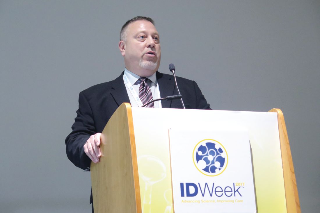
That’s where ribaxamase comes in, he said at the combined annual meetings of the Infectious Diseases Society of America, the Society for Healthcare Epidemiology of America, the HIV Medicine Association, and the Pediatric Infectious Diseases Society. Ribaxamase is an orally administered beta-lac-tamase designed to degrade penicillin and cephalosporins in the intestinal lumen. It’s formulated for release in the proximal small intestine and is expected to be given during or a short time after administration of IV beta-lactam antibiotics such as ceftriaxone. “This is expected to degrade the excess antibiotics that are excreted into the small intestine via the bile,” Dr. Kokai-Kun explained. “It’s designed to prevent disruption of the gut microbiome and thus protect from opportunistic GI infections like CDI.”
Early-stage clinical studies demonstrated that ribaxamase was well tolerated and that it is not systemically absorbed, while phase 2 studies showed that ribaxamase degrades ceftriaxone in the intestine to below the level of detection while not affecting the pharmacokinetics of ceftriaxone in the plasma.
For the current study, 412 patients were enrolled at 84 multinational clinical sites. These patients were admitted to the hospital for treatment of a lower respiratory tract infection and were randomized 1:1 to receive ceftriaxone plus 150 mg ribaxamase or ceftriaxone plus placebo. Patients in both groups could also receive an oral macrolide at the discretion of the clinical investigator.
The researchers also obtained fecal samples at screening, 72 hours post antibiotic treatment, and at the end of a 4-week follow-up visit, to determine colonization by opportunistic pathogens and to examine changes in the gut microbiome.
Patients were monitored for 6 weeks for diarrhea and CDI. Diarrhea was defined as three or more loose or watery stools in a 24-hour period. “If that occurred, then we collected a sample, which was sent to the local lab to determine the presence of C. difficile toxins,” Dr. Kokai-Kun said.
The average age of study participants was 70 years, and about one-third in each arm received oral macrolides. The number of adverse events and serious adverse events were similar between active and placebo arms, and there was no trend associated with ribaxamase use. The lower respiratory tract infection cure rate to the ceftriaxone treatment was about 99% in both arms at 72 hours post treatment and at 2 weeks post treatment.
To analyze changes in the gut microbiome, the researchers conducted 16S rRNA sequencing of DNA extracted from fecal samples. In all, 652 samples were sequenced from 229 patients. Results from that analysis suggests that ribaxamase “appears to protect the gut microbiome from the onslaught of the ceftriaxone,” he said.
Ribaxamase reduced the incidence of new-onset CDI by 71%, compared with placebo (P = .045). “It apparently did this by protecting the integrity of the gut microbiome,” Dr. Kokai-Kun said. “There was also a significant reduction of new colonization by vancomycin-resistant enterococci at 72 hours and 4 weeks (P = .0001 and P = .0002, respectively) which is an opportunistic pathogen that is known to be able to inhabit gut microbiome when there is dysbiosis.”
The study was sponsored by Synthetic Biologics. Dr. Kokai-Kun is an employee of the company.
AGA Resource
Help your patients better understand C. difficile by using AGA’s patient education materials available here.
[email protected]
SAN DIEGO – , results from a phase 2b study showed.
At an annual scientific meeting on infectious diseases, lead investigator John F. Kokai-Kun, PhD, said that the finding represents a paradigm shift in the use of intravenous beta-lactam antibiotics to prevent opportunistic infections. “We currently treat Clostridium difficile infection (CDI) with antibiotics, which attack the vegetative cells,” said Dr. Kokai-Kun, vice president of nonclinical affairs for Rockville, Md.–based Synthetic Biologics, which is developing ribaxamase. “Since C. diff. is primarily a toxin-mediated disease, certain products seem to neutralize the toxin. There’s also been work with probiotics and prebiotics to try to strengthen and repair the dysbiotic colon. Fecal replacement therapy has been shown to be fairly effective for treatment of recurrent C. diff. infection. What if we could simply block the initial insult that leads to this cascade? That’s the damage caused to the gut microbiome by the antibiotic that’s excreted to the intestine.”
That’s where ribaxamase comes in, he said at the combined annual meetings of the Infectious Diseases Society of America, the Society for Healthcare Epidemiology of America, the HIV Medicine Association, and the Pediatric Infectious Diseases Society. Ribaxamase is an orally administered beta-lac-tamase designed to degrade penicillin and cephalosporins in the intestinal lumen. It’s formulated for release in the proximal small intestine and is expected to be given during or a short time after administration of IV beta-lactam antibiotics such as ceftriaxone. “This is expected to degrade the excess antibiotics that are excreted into the small intestine via the bile,” Dr. Kokai-Kun explained. “It’s designed to prevent disruption of the gut microbiome and thus protect from opportunistic GI infections like CDI.”
Early-stage clinical studies demonstrated that ribaxamase was well tolerated and that it is not systemically absorbed, while phase 2 studies showed that ribaxamase degrades ceftriaxone in the intestine to below the level of detection while not affecting the pharmacokinetics of ceftriaxone in the plasma.
For the current study, 412 patients were enrolled at 84 multinational clinical sites. These patients were admitted to the hospital for treatment of a lower respiratory tract infection and were randomized 1:1 to receive ceftriaxone plus 150 mg ribaxamase or ceftriaxone plus placebo. Patients in both groups could also receive an oral macrolide at the discretion of the clinical investigator.
The researchers also obtained fecal samples at screening, 72 hours post antibiotic treatment, and at the end of a 4-week follow-up visit, to determine colonization by opportunistic pathogens and to examine changes in the gut microbiome.
Patients were monitored for 6 weeks for diarrhea and CDI. Diarrhea was defined as three or more loose or watery stools in a 24-hour period. “If that occurred, then we collected a sample, which was sent to the local lab to determine the presence of C. difficile toxins,” Dr. Kokai-Kun said.
The average age of study participants was 70 years, and about one-third in each arm received oral macrolides. The number of adverse events and serious adverse events were similar between active and placebo arms, and there was no trend associated with ribaxamase use. The lower respiratory tract infection cure rate to the ceftriaxone treatment was about 99% in both arms at 72 hours post treatment and at 2 weeks post treatment.
To analyze changes in the gut microbiome, the researchers conducted 16S rRNA sequencing of DNA extracted from fecal samples. In all, 652 samples were sequenced from 229 patients. Results from that analysis suggests that ribaxamase “appears to protect the gut microbiome from the onslaught of the ceftriaxone,” he said.
Ribaxamase reduced the incidence of new-onset CDI by 71%, compared with placebo (P = .045). “It apparently did this by protecting the integrity of the gut microbiome,” Dr. Kokai-Kun said. “There was also a significant reduction of new colonization by vancomycin-resistant enterococci at 72 hours and 4 weeks (P = .0001 and P = .0002, respectively) which is an opportunistic pathogen that is known to be able to inhabit gut microbiome when there is dysbiosis.”
The study was sponsored by Synthetic Biologics. Dr. Kokai-Kun is an employee of the company.
AGA Resource
Help your patients better understand C. difficile by using AGA’s patient education materials available here.
[email protected]
SAN DIEGO – , results from a phase 2b study showed.
At an annual scientific meeting on infectious diseases, lead investigator John F. Kokai-Kun, PhD, said that the finding represents a paradigm shift in the use of intravenous beta-lactam antibiotics to prevent opportunistic infections. “We currently treat Clostridium difficile infection (CDI) with antibiotics, which attack the vegetative cells,” said Dr. Kokai-Kun, vice president of nonclinical affairs for Rockville, Md.–based Synthetic Biologics, which is developing ribaxamase. “Since C. diff. is primarily a toxin-mediated disease, certain products seem to neutralize the toxin. There’s also been work with probiotics and prebiotics to try to strengthen and repair the dysbiotic colon. Fecal replacement therapy has been shown to be fairly effective for treatment of recurrent C. diff. infection. What if we could simply block the initial insult that leads to this cascade? That’s the damage caused to the gut microbiome by the antibiotic that’s excreted to the intestine.”
That’s where ribaxamase comes in, he said at the combined annual meetings of the Infectious Diseases Society of America, the Society for Healthcare Epidemiology of America, the HIV Medicine Association, and the Pediatric Infectious Diseases Society. Ribaxamase is an orally administered beta-lac-tamase designed to degrade penicillin and cephalosporins in the intestinal lumen. It’s formulated for release in the proximal small intestine and is expected to be given during or a short time after administration of IV beta-lactam antibiotics such as ceftriaxone. “This is expected to degrade the excess antibiotics that are excreted into the small intestine via the bile,” Dr. Kokai-Kun explained. “It’s designed to prevent disruption of the gut microbiome and thus protect from opportunistic GI infections like CDI.”
Early-stage clinical studies demonstrated that ribaxamase was well tolerated and that it is not systemically absorbed, while phase 2 studies showed that ribaxamase degrades ceftriaxone in the intestine to below the level of detection while not affecting the pharmacokinetics of ceftriaxone in the plasma.
For the current study, 412 patients were enrolled at 84 multinational clinical sites. These patients were admitted to the hospital for treatment of a lower respiratory tract infection and were randomized 1:1 to receive ceftriaxone plus 150 mg ribaxamase or ceftriaxone plus placebo. Patients in both groups could also receive an oral macrolide at the discretion of the clinical investigator.
The researchers also obtained fecal samples at screening, 72 hours post antibiotic treatment, and at the end of a 4-week follow-up visit, to determine colonization by opportunistic pathogens and to examine changes in the gut microbiome.
Patients were monitored for 6 weeks for diarrhea and CDI. Diarrhea was defined as three or more loose or watery stools in a 24-hour period. “If that occurred, then we collected a sample, which was sent to the local lab to determine the presence of C. difficile toxins,” Dr. Kokai-Kun said.
The average age of study participants was 70 years, and about one-third in each arm received oral macrolides. The number of adverse events and serious adverse events were similar between active and placebo arms, and there was no trend associated with ribaxamase use. The lower respiratory tract infection cure rate to the ceftriaxone treatment was about 99% in both arms at 72 hours post treatment and at 2 weeks post treatment.
To analyze changes in the gut microbiome, the researchers conducted 16S rRNA sequencing of DNA extracted from fecal samples. In all, 652 samples were sequenced from 229 patients. Results from that analysis suggests that ribaxamase “appears to protect the gut microbiome from the onslaught of the ceftriaxone,” he said.
Ribaxamase reduced the incidence of new-onset CDI by 71%, compared with placebo (P = .045). “It apparently did this by protecting the integrity of the gut microbiome,” Dr. Kokai-Kun said. “There was also a significant reduction of new colonization by vancomycin-resistant enterococci at 72 hours and 4 weeks (P = .0001 and P = .0002, respectively) which is an opportunistic pathogen that is known to be able to inhabit gut microbiome when there is dysbiosis.”
The study was sponsored by Synthetic Biologics. Dr. Kokai-Kun is an employee of the company.
AGA Resource
Help your patients better understand C. difficile by using AGA’s patient education materials available here.
[email protected]
Golimumab earns new FDA approvals
The U.S. Food and Drug Administration has approved golimumab (Simponi Aria) for use in adults with active psoriatic arthritis (PsA) or active ankylosing spondylitis (AS).
Simponi Aria is an intravenous formulation of golimumab that is already approved for moderate to severe rheumatoid arthritis. The subcutaneous injection formulation of golimumab, Simponi, is already approved for RA, PsA, AS, and ulcerative colitis. Golimumab is a fully human anti–tumor necrosis factor-alpha therapy, and the intravenous formulation is designed for use as a 30-minute infusion.

“In the study for the treatment of active PsA, patients experienced improvement in joint symptoms and inhibition of structural damage. In the study for treatment of active AS, results showed improvement in measures of disease activity,” according to an Oct. 20 announcement from the manufacturer of golimumab, Janssen Biotech.
Read the revised prescribing information for Simponi Aria here.
The U.S. Food and Drug Administration has approved golimumab (Simponi Aria) for use in adults with active psoriatic arthritis (PsA) or active ankylosing spondylitis (AS).
Simponi Aria is an intravenous formulation of golimumab that is already approved for moderate to severe rheumatoid arthritis. The subcutaneous injection formulation of golimumab, Simponi, is already approved for RA, PsA, AS, and ulcerative colitis. Golimumab is a fully human anti–tumor necrosis factor-alpha therapy, and the intravenous formulation is designed for use as a 30-minute infusion.
“In the study for the treatment of active PsA, patients experienced improvement in joint symptoms and inhibition of structural damage. In the study for treatment of active AS, results showed improvement in measures of disease activity,” according to an Oct. 20 announcement from the manufacturer of golimumab, Janssen Biotech.
Read the revised prescribing information for Simponi Aria here.
The U.S. Food and Drug Administration has approved golimumab (Simponi Aria) for use in adults with active psoriatic arthritis (PsA) or active ankylosing spondylitis (AS).
Simponi Aria is an intravenous formulation of golimumab that is already approved for moderate to severe rheumatoid arthritis. The subcutaneous injection formulation of golimumab, Simponi, is already approved for RA, PsA, AS, and ulcerative colitis. Golimumab is a fully human anti–tumor necrosis factor-alpha therapy, and the intravenous formulation is designed for use as a 30-minute infusion.
“In the study for the treatment of active PsA, patients experienced improvement in joint symptoms and inhibition of structural damage. In the study for treatment of active AS, results showed improvement in measures of disease activity,” according to an Oct. 20 announcement from the manufacturer of golimumab, Janssen Biotech.
Read the revised prescribing information for Simponi Aria here.
Carvedilol fails to reduce variceal bleeds in acute-on-chronic liver failure
WASHINGTON – Treatment with carvedilol reduced the incidence of sepsis and acute kidney injury and improved survival at 28 days but did not significantly reduce the progression of esophageal varices in patients with acute-on-chronic liver failure.
A total of 136 patients with acute-on-chronic liver failure with small or no esophageal varices and a hepatic venous pressure gradient (HVPG) of 12 mm Hg or greater were enrolled in a single center, prospective, open-label, randomized controlled trial: 66 were randomized to carvedilol and 70 to placebo, according to Sumeet Kainth, MD, of the Institute of Liver and Biliary Sciences in New Delhi.

More than 90% of patients were men with a mean age of 44 years, and composition of the treatment and placebo groups was similar. About 70% in each group had alcoholic hepatitis (the reason for acute liver failure in most). Mean Model for End-Stage Liver Disease (MELD) scores were about 25. Hemodynamic parameters also were comparable, with a mean HVPG of about 19, Dr. Kainth said at the annual meeting of the American Association for the Study of Liver Diseases.
Patients in the treatment group received a median maximum tolerated dose of carvedilol of 12.5 mg, with a range of 3.13 mg to 25 mg.
Morbidity and mortality were high, as is expected with acute-on-chronic liver failure, he noted. A total of 36 patients died before the end of the 90-day study period. Another 23 experienced adverse events and 2 progressed to liver transplant.
HVPG at 90 days decreased significantly in both groups. In the carvedilol group, 90-day HVPG was 16 mm Hg, compared with 19.7 mm Hg at baseline (P less than .01). For placebo patients, 90-day HVPG spontaneously improved to 14.8 mm Hg, compared with a baseline of 17.2 mm Hg (P less than .01).
Carvedilol did not significantly slow the development or growth of varices, however, Dr. Kainth said. At 90 days, varices had progressed in 9 of 40 patients (22.5%) of patients on carvedilol and 8 of 31 (25.8%) of placebo patients.
Significantly fewer patients in the carvedilol group developed acute kidney injury at 28 days (14% vs. 38% on placebo) and sepsis (5% vs. 20%). Mortality also was reduced significantly at 28 days (11% vs. 24%), he reported.
Treatment with carvedilol did not achieve significant reductions in variceal bleeding, “possibly due to the low number of bleeds seen in the study [because of] the exclusion of patients with large varices,” Dr. Kainth said.
The study was sponsored by Institute of Liver and Biliary Sciences. Dr. Kainth reported no relevant conflicts of interest.
[email protected]
On Twitter @denisefulton
WASHINGTON – Treatment with carvedilol reduced the incidence of sepsis and acute kidney injury and improved survival at 28 days but did not significantly reduce the progression of esophageal varices in patients with acute-on-chronic liver failure.
A total of 136 patients with acute-on-chronic liver failure with small or no esophageal varices and a hepatic venous pressure gradient (HVPG) of 12 mm Hg or greater were enrolled in a single center, prospective, open-label, randomized controlled trial: 66 were randomized to carvedilol and 70 to placebo, according to Sumeet Kainth, MD, of the Institute of Liver and Biliary Sciences in New Delhi.
More than 90% of patients were men with a mean age of 44 years, and composition of the treatment and placebo groups was similar. About 70% in each group had alcoholic hepatitis (the reason for acute liver failure in most). Mean Model for End-Stage Liver Disease (MELD) scores were about 25. Hemodynamic parameters also were comparable, with a mean HVPG of about 19, Dr. Kainth said at the annual meeting of the American Association for the Study of Liver Diseases.
Patients in the treatment group received a median maximum tolerated dose of carvedilol of 12.5 mg, with a range of 3.13 mg to 25 mg.
Morbidity and mortality were high, as is expected with acute-on-chronic liver failure, he noted. A total of 36 patients died before the end of the 90-day study period. Another 23 experienced adverse events and 2 progressed to liver transplant.
HVPG at 90 days decreased significantly in both groups. In the carvedilol group, 90-day HVPG was 16 mm Hg, compared with 19.7 mm Hg at baseline (P less than .01). For placebo patients, 90-day HVPG spontaneously improved to 14.8 mm Hg, compared with a baseline of 17.2 mm Hg (P less than .01).
Carvedilol did not significantly slow the development or growth of varices, however, Dr. Kainth said. At 90 days, varices had progressed in 9 of 40 patients (22.5%) of patients on carvedilol and 8 of 31 (25.8%) of placebo patients.
Significantly fewer patients in the carvedilol group developed acute kidney injury at 28 days (14% vs. 38% on placebo) and sepsis (5% vs. 20%). Mortality also was reduced significantly at 28 days (11% vs. 24%), he reported.
Treatment with carvedilol did not achieve significant reductions in variceal bleeding, “possibly due to the low number of bleeds seen in the study [because of] the exclusion of patients with large varices,” Dr. Kainth said.
The study was sponsored by Institute of Liver and Biliary Sciences. Dr. Kainth reported no relevant conflicts of interest.
[email protected]
On Twitter @denisefulton
WASHINGTON – Treatment with carvedilol reduced the incidence of sepsis and acute kidney injury and improved survival at 28 days but did not significantly reduce the progression of esophageal varices in patients with acute-on-chronic liver failure.
A total of 136 patients with acute-on-chronic liver failure with small or no esophageal varices and a hepatic venous pressure gradient (HVPG) of 12 mm Hg or greater were enrolled in a single center, prospective, open-label, randomized controlled trial: 66 were randomized to carvedilol and 70 to placebo, according to Sumeet Kainth, MD, of the Institute of Liver and Biliary Sciences in New Delhi.
More than 90% of patients were men with a mean age of 44 years, and composition of the treatment and placebo groups was similar. About 70% in each group had alcoholic hepatitis (the reason for acute liver failure in most). Mean Model for End-Stage Liver Disease (MELD) scores were about 25. Hemodynamic parameters also were comparable, with a mean HVPG of about 19, Dr. Kainth said at the annual meeting of the American Association for the Study of Liver Diseases.
Patients in the treatment group received a median maximum tolerated dose of carvedilol of 12.5 mg, with a range of 3.13 mg to 25 mg.
Morbidity and mortality were high, as is expected with acute-on-chronic liver failure, he noted. A total of 36 patients died before the end of the 90-day study period. Another 23 experienced adverse events and 2 progressed to liver transplant.
HVPG at 90 days decreased significantly in both groups. In the carvedilol group, 90-day HVPG was 16 mm Hg, compared with 19.7 mm Hg at baseline (P less than .01). For placebo patients, 90-day HVPG spontaneously improved to 14.8 mm Hg, compared with a baseline of 17.2 mm Hg (P less than .01).
Carvedilol did not significantly slow the development or growth of varices, however, Dr. Kainth said. At 90 days, varices had progressed in 9 of 40 patients (22.5%) of patients on carvedilol and 8 of 31 (25.8%) of placebo patients.
Significantly fewer patients in the carvedilol group developed acute kidney injury at 28 days (14% vs. 38% on placebo) and sepsis (5% vs. 20%). Mortality also was reduced significantly at 28 days (11% vs. 24%), he reported.
Treatment with carvedilol did not achieve significant reductions in variceal bleeding, “possibly due to the low number of bleeds seen in the study [because of] the exclusion of patients with large varices,” Dr. Kainth said.
The study was sponsored by Institute of Liver and Biliary Sciences. Dr. Kainth reported no relevant conflicts of interest.
[email protected]
On Twitter @denisefulton
AT THE LIVER MEETING 2017
Key clinical point:
Major finding: At 90 days, varices had progressed in 9 of 40 (22.5%) patients on carvedilol vs. 8 of 31 (25.8%) of placebo patients.
Data source: A single-center, prospective, open-label, randomized controlled trial of 136 patients with acute-on-chronic liver failure.
Disclosures: The study was sponsored by the Institute of Liver and Biliary Sciences. Dr. Kainth reported no relevant conflicts of interest.
In high-risk patients, methylation strikes genes before psychosis hits
BERLIN – Researchers are honing in on several sets of genes that, when altered by as-yet-unknown factors, may signal conversion to full-blown psychosis in people at ultrahigh risk for the disorder.
If confirmed, these candidate markers might have potential as blood-based biomarkers to predict conversion risk and assist in clinical staging, Marie-Odile Krebs, MD, PhD, said at the meeting of the World Psychiatric Association.
The genes modulate three biologic pathways that also have been implicated in schizophrenia: glutathione metabolism, axonal targeting, and inflammation, said Dr. Krebs of Saint-Anne Hospital, Paris. “Knowing this may even help us to target some drugs that work in those pathways,” she said.

Several blood-based analyte screens have been investigated with mixed results, Dr. Krebs noted.
In 2015, researchers at the University of North Carolina at Chapel Hill, and Harvard Medical School, Boston, created a 15-analyte plasma panel that performed well in the North American Prodrome Longitudinal Study (NAPL-S) cohort. The project is a multisite endeavor that aims to better understand predictors and mechanisms for the development of psychosis. The panel separated 35 unaffected controls from 32 with high-risk symptoms who converted to psychosis and from 40 who did not, with an area under the curve (AUC) of 0.91 (Schizophr Bull. 2015 Mar;41[2]:419-28).
Selected from an initial group of 185 analytes, the candidate markers were inflammatory cytokines, proteins modulating blood-brain barrier inflammation, and hormones related to the hypothalamic-pituitary axes. Several also were involved in reacting to oxidative stress.
Earlier this year, members of that same group identified a set of nine microRNAs related to cortical thinning in patients who converted to psychosis. These microRNAs also have been implicated in brain development, synaptic plasticity, immune function, and schizophrenia (Neuropsychopharmacology. 2017 Feb 10. doi: 10.1038/npp.2017.34).
Although these studies are helpful signposts, Dr. Krebs said they do not reflect the dynamic interaction of disease risk, which includes not only the intrinsic factors of genetics, enzymes, and proteins, but the extrinsic risks imposed by other factors: stress, trauma, cannabis use, and other completely individual experiences. “This is a dynamic process, and we need a dynamic assessment,” she said.
To that end, Dr. Krebs and her colleagues decided to look at methylomic changes in a small group of 39 patients at ultrahigh risk for psychosis conversion. All of these patients (mean age, 22 years) were seen at Saint-Anne Hospital from 2009 to 2013. Using whole blood, Dr. Krebs performed a genomewide DNA methylation study to determine what genes – if any – were differently methylated between the converters and nonconverters. The mean follow-up was 1 year (Mol Psychiatry. 2017 Apr;22[4]:512-8).
Although no significant difference was found in global methylation associated with conversion, Dr. Krebs did find longitudinal changes associated with conversion in three regions.
A cluster of five genes in the glutathione S-transferase family was differently methylated between the converters and nonconverters. Two were related to the GSTM5 promoter gene, which encodes for cytosolic and membrane-bound glutathione S-transferase – an important antioxidant enzyme, the downregulation of which has been implicated in schizophrenia. These two regions appeared to be stable over time, suggesting that methylation occurred before conversion, Dr. Krebs said.
Oxidative stress has been implicated in schizophrenia, and GSTM5 is expressed in the brain, Dr. Krebs noted. Some researchers suggest the gene is involved in dopamine metabolism. It’s also underexpressed in the prefrontal cortex of schizophrenia patients.
Three other regions in the GST family changed with conversion: two on the glutathione S-transferase theta 1 gene and one on the glutathione S-transferase P gene. Since all of these have to do with production of the innate antioxidant glutathione, “these findings suggest a potential use for antioxidant drugs,” Dr. Krebs said.
She found two other differently methylated regions as well.
One was a cluster of eight genes that are all involved in axon guidance – the process by which axons branch out to their correct targets. The second cluster comprised seven genes, all of which are involved in regulating interleukin-17 signaling. This cytokine has been implicated in autoimmune disorders.
Finally, Dr. Krebs performed a transcriptome analysis looking at the brain-expressed messenger RNA in the samples. “The methylome seemed less dynamic than the transcriptome,” she said. “Some methylomic changes may have occurred several months before the conversion, whereas transcriptomic analysis may reflect more rapid changes.”
There was only a 22% concordance between the two analyses. However, the GSTM5 gene and the neuropilin 1 gene – one of those involved in axon guidance – were both methylated and downregulated in the converters. The transcriptome analysis also found significantly decreased expression (although not methylation) of another gene, carnitine palmitoyltransferase 1A. This is a key enzyme in oxidizing long-chain fatty acids and transporting them into the mitochondria.
Adapting these observed differences in gene expression into a useful clinical tool will be challenging, Dr. Krebs said. In addition to large-group validation, any risk prediction model would have to take into account the many other factors that influence psychosis conversion: cerebral and sexual maturation during adolescence, cannabis use, and stress and other completely individual life experiences.
Nevertheless, she concluded, “longitudinal ‘multi-omics’ may be a step toward a future of personalized molecular psychiatry.”
Dr. Krebs had no relevant financial disclosures.
[email protected]
On Twitter @alz_gal
BERLIN – Researchers are honing in on several sets of genes that, when altered by as-yet-unknown factors, may signal conversion to full-blown psychosis in people at ultrahigh risk for the disorder.
If confirmed, these candidate markers might have potential as blood-based biomarkers to predict conversion risk and assist in clinical staging, Marie-Odile Krebs, MD, PhD, said at the meeting of the World Psychiatric Association.
The genes modulate three biologic pathways that also have been implicated in schizophrenia: glutathione metabolism, axonal targeting, and inflammation, said Dr. Krebs of Saint-Anne Hospital, Paris. “Knowing this may even help us to target some drugs that work in those pathways,” she said.
Several blood-based analyte screens have been investigated with mixed results, Dr. Krebs noted.
In 2015, researchers at the University of North Carolina at Chapel Hill, and Harvard Medical School, Boston, created a 15-analyte plasma panel that performed well in the North American Prodrome Longitudinal Study (NAPL-S) cohort. The project is a multisite endeavor that aims to better understand predictors and mechanisms for the development of psychosis. The panel separated 35 unaffected controls from 32 with high-risk symptoms who converted to psychosis and from 40 who did not, with an area under the curve (AUC) of 0.91 (Schizophr Bull. 2015 Mar;41[2]:419-28).
Selected from an initial group of 185 analytes, the candidate markers were inflammatory cytokines, proteins modulating blood-brain barrier inflammation, and hormones related to the hypothalamic-pituitary axes. Several also were involved in reacting to oxidative stress.
Earlier this year, members of that same group identified a set of nine microRNAs related to cortical thinning in patients who converted to psychosis. These microRNAs also have been implicated in brain development, synaptic plasticity, immune function, and schizophrenia (Neuropsychopharmacology. 2017 Feb 10. doi: 10.1038/npp.2017.34).
Although these studies are helpful signposts, Dr. Krebs said they do not reflect the dynamic interaction of disease risk, which includes not only the intrinsic factors of genetics, enzymes, and proteins, but the extrinsic risks imposed by other factors: stress, trauma, cannabis use, and other completely individual experiences. “This is a dynamic process, and we need a dynamic assessment,” she said.
To that end, Dr. Krebs and her colleagues decided to look at methylomic changes in a small group of 39 patients at ultrahigh risk for psychosis conversion. All of these patients (mean age, 22 years) were seen at Saint-Anne Hospital from 2009 to 2013. Using whole blood, Dr. Krebs performed a genomewide DNA methylation study to determine what genes – if any – were differently methylated between the converters and nonconverters. The mean follow-up was 1 year (Mol Psychiatry. 2017 Apr;22[4]:512-8).
Although no significant difference was found in global methylation associated with conversion, Dr. Krebs did find longitudinal changes associated with conversion in three regions.
A cluster of five genes in the glutathione S-transferase family was differently methylated between the converters and nonconverters. Two were related to the GSTM5 promoter gene, which encodes for cytosolic and membrane-bound glutathione S-transferase – an important antioxidant enzyme, the downregulation of which has been implicated in schizophrenia. These two regions appeared to be stable over time, suggesting that methylation occurred before conversion, Dr. Krebs said.
Oxidative stress has been implicated in schizophrenia, and GSTM5 is expressed in the brain, Dr. Krebs noted. Some researchers suggest the gene is involved in dopamine metabolism. It’s also underexpressed in the prefrontal cortex of schizophrenia patients.
Three other regions in the GST family changed with conversion: two on the glutathione S-transferase theta 1 gene and one on the glutathione S-transferase P gene. Since all of these have to do with production of the innate antioxidant glutathione, “these findings suggest a potential use for antioxidant drugs,” Dr. Krebs said.
She found two other differently methylated regions as well.
One was a cluster of eight genes that are all involved in axon guidance – the process by which axons branch out to their correct targets. The second cluster comprised seven genes, all of which are involved in regulating interleukin-17 signaling. This cytokine has been implicated in autoimmune disorders.
Finally, Dr. Krebs performed a transcriptome analysis looking at the brain-expressed messenger RNA in the samples. “The methylome seemed less dynamic than the transcriptome,” she said. “Some methylomic changes may have occurred several months before the conversion, whereas transcriptomic analysis may reflect more rapid changes.”
There was only a 22% concordance between the two analyses. However, the GSTM5 gene and the neuropilin 1 gene – one of those involved in axon guidance – were both methylated and downregulated in the converters. The transcriptome analysis also found significantly decreased expression (although not methylation) of another gene, carnitine palmitoyltransferase 1A. This is a key enzyme in oxidizing long-chain fatty acids and transporting them into the mitochondria.
Adapting these observed differences in gene expression into a useful clinical tool will be challenging, Dr. Krebs said. In addition to large-group validation, any risk prediction model would have to take into account the many other factors that influence psychosis conversion: cerebral and sexual maturation during adolescence, cannabis use, and stress and other completely individual life experiences.
Nevertheless, she concluded, “longitudinal ‘multi-omics’ may be a step toward a future of personalized molecular psychiatry.”
Dr. Krebs had no relevant financial disclosures.
[email protected]
On Twitter @alz_gal
BERLIN – Researchers are honing in on several sets of genes that, when altered by as-yet-unknown factors, may signal conversion to full-blown psychosis in people at ultrahigh risk for the disorder.
If confirmed, these candidate markers might have potential as blood-based biomarkers to predict conversion risk and assist in clinical staging, Marie-Odile Krebs, MD, PhD, said at the meeting of the World Psychiatric Association.
The genes modulate three biologic pathways that also have been implicated in schizophrenia: glutathione metabolism, axonal targeting, and inflammation, said Dr. Krebs of Saint-Anne Hospital, Paris. “Knowing this may even help us to target some drugs that work in those pathways,” she said.
Several blood-based analyte screens have been investigated with mixed results, Dr. Krebs noted.
In 2015, researchers at the University of North Carolina at Chapel Hill, and Harvard Medical School, Boston, created a 15-analyte plasma panel that performed well in the North American Prodrome Longitudinal Study (NAPL-S) cohort. The project is a multisite endeavor that aims to better understand predictors and mechanisms for the development of psychosis. The panel separated 35 unaffected controls from 32 with high-risk symptoms who converted to psychosis and from 40 who did not, with an area under the curve (AUC) of 0.91 (Schizophr Bull. 2015 Mar;41[2]:419-28).
Selected from an initial group of 185 analytes, the candidate markers were inflammatory cytokines, proteins modulating blood-brain barrier inflammation, and hormones related to the hypothalamic-pituitary axes. Several also were involved in reacting to oxidative stress.
Earlier this year, members of that same group identified a set of nine microRNAs related to cortical thinning in patients who converted to psychosis. These microRNAs also have been implicated in brain development, synaptic plasticity, immune function, and schizophrenia (Neuropsychopharmacology. 2017 Feb 10. doi: 10.1038/npp.2017.34).
Although these studies are helpful signposts, Dr. Krebs said they do not reflect the dynamic interaction of disease risk, which includes not only the intrinsic factors of genetics, enzymes, and proteins, but the extrinsic risks imposed by other factors: stress, trauma, cannabis use, and other completely individual experiences. “This is a dynamic process, and we need a dynamic assessment,” she said.
To that end, Dr. Krebs and her colleagues decided to look at methylomic changes in a small group of 39 patients at ultrahigh risk for psychosis conversion. All of these patients (mean age, 22 years) were seen at Saint-Anne Hospital from 2009 to 2013. Using whole blood, Dr. Krebs performed a genomewide DNA methylation study to determine what genes – if any – were differently methylated between the converters and nonconverters. The mean follow-up was 1 year (Mol Psychiatry. 2017 Apr;22[4]:512-8).
Although no significant difference was found in global methylation associated with conversion, Dr. Krebs did find longitudinal changes associated with conversion in three regions.
A cluster of five genes in the glutathione S-transferase family was differently methylated between the converters and nonconverters. Two were related to the GSTM5 promoter gene, which encodes for cytosolic and membrane-bound glutathione S-transferase – an important antioxidant enzyme, the downregulation of which has been implicated in schizophrenia. These two regions appeared to be stable over time, suggesting that methylation occurred before conversion, Dr. Krebs said.
Oxidative stress has been implicated in schizophrenia, and GSTM5 is expressed in the brain, Dr. Krebs noted. Some researchers suggest the gene is involved in dopamine metabolism. It’s also underexpressed in the prefrontal cortex of schizophrenia patients.
Three other regions in the GST family changed with conversion: two on the glutathione S-transferase theta 1 gene and one on the glutathione S-transferase P gene. Since all of these have to do with production of the innate antioxidant glutathione, “these findings suggest a potential use for antioxidant drugs,” Dr. Krebs said.
She found two other differently methylated regions as well.
One was a cluster of eight genes that are all involved in axon guidance – the process by which axons branch out to their correct targets. The second cluster comprised seven genes, all of which are involved in regulating interleukin-17 signaling. This cytokine has been implicated in autoimmune disorders.
Finally, Dr. Krebs performed a transcriptome analysis looking at the brain-expressed messenger RNA in the samples. “The methylome seemed less dynamic than the transcriptome,” she said. “Some methylomic changes may have occurred several months before the conversion, whereas transcriptomic analysis may reflect more rapid changes.”
There was only a 22% concordance between the two analyses. However, the GSTM5 gene and the neuropilin 1 gene – one of those involved in axon guidance – were both methylated and downregulated in the converters. The transcriptome analysis also found significantly decreased expression (although not methylation) of another gene, carnitine palmitoyltransferase 1A. This is a key enzyme in oxidizing long-chain fatty acids and transporting them into the mitochondria.
Adapting these observed differences in gene expression into a useful clinical tool will be challenging, Dr. Krebs said. In addition to large-group validation, any risk prediction model would have to take into account the many other factors that influence psychosis conversion: cerebral and sexual maturation during adolescence, cannabis use, and stress and other completely individual life experiences.
Nevertheless, she concluded, “longitudinal ‘multi-omics’ may be a step toward a future of personalized molecular psychiatry.”
Dr. Krebs had no relevant financial disclosures.
[email protected]
On Twitter @alz_gal
EXPERT ANALYSIS FROM WPA 2017
Genetic analysis indicates ovarian cancer may originate in fallopian tubes
Many of the most severe ovarian cancer cases may originate in the fallopian tube (FT), based on data from an analysis of nine patients published online in Nature Communications.
“Our data suggest that FT neoplasia is the origin of ovarian serous carcinogenesis, and can directly lead to cancer of the ovaries and of other sites,” wrote Sana Intidhar Labidi-Galy, MD, of Dana-Farber Cancer Institute, Boston, and her colleagues (Nature Commun. 2017 Oct 23. doi: 10.1038/s41467-017-00962-1).
Preliminary evidence suggests that fallopian tube cancers may develop into high-grade serous ovarian carcinoma (HGSOC), but evolutionary evidence is limited, the researchers said.
They conducted genetic sequencing on 37 tumor samples from five adult patients with HGSOC. They identified changes in the TP53 tumor suppressor gene in all cases of HGSOC. They also studied serous tubal intraepithelial carcinomas from four patients.
“As expected, we identified sequence changes in the TP53 tumor suppressor gene, a well-known driver gene in HGSOC, in all cases,” the researchers wrote.
“The TP53 alterations were identical in all samples analyzed for each patient including in the p53 signatures, the [serous tubal intraepithelial carcinoma] lesions, and other carcinomas,” Dr. Labidi-Galy and her associates said. Although TP53 was the only gene analyzed in this study, the researchers also noted changes in areas of several known ovarian cancer genes, including BRCA1 and BRCA2.
The study findings were limited by the small size of the tumor samples and small number of cells, the researchers noted.
The results, however, suggest an avenue for further research to help guide early detection and treatment of ovarian cancer, such as the potential removal of fallopian tubes rather than the ovaries in some cases, they concluded.
The research was supported by multiple foundations and organizations, including the National Institutes of Health. One of the investigators is a founder of Personal Genome Diagnostics and a member of its scientific advisory board and board of directors. The other researchers had no financial conflicts to disclose.
Many of the most severe ovarian cancer cases may originate in the fallopian tube (FT), based on data from an analysis of nine patients published online in Nature Communications.
“Our data suggest that FT neoplasia is the origin of ovarian serous carcinogenesis, and can directly lead to cancer of the ovaries and of other sites,” wrote Sana Intidhar Labidi-Galy, MD, of Dana-Farber Cancer Institute, Boston, and her colleagues (Nature Commun. 2017 Oct 23. doi: 10.1038/s41467-017-00962-1).
Preliminary evidence suggests that fallopian tube cancers may develop into high-grade serous ovarian carcinoma (HGSOC), but evolutionary evidence is limited, the researchers said.
They conducted genetic sequencing on 37 tumor samples from five adult patients with HGSOC. They identified changes in the TP53 tumor suppressor gene in all cases of HGSOC. They also studied serous tubal intraepithelial carcinomas from four patients.
“As expected, we identified sequence changes in the TP53 tumor suppressor gene, a well-known driver gene in HGSOC, in all cases,” the researchers wrote.
“The TP53 alterations were identical in all samples analyzed for each patient including in the p53 signatures, the [serous tubal intraepithelial carcinoma] lesions, and other carcinomas,” Dr. Labidi-Galy and her associates said. Although TP53 was the only gene analyzed in this study, the researchers also noted changes in areas of several known ovarian cancer genes, including BRCA1 and BRCA2.
The study findings were limited by the small size of the tumor samples and small number of cells, the researchers noted.
The results, however, suggest an avenue for further research to help guide early detection and treatment of ovarian cancer, such as the potential removal of fallopian tubes rather than the ovaries in some cases, they concluded.
The research was supported by multiple foundations and organizations, including the National Institutes of Health. One of the investigators is a founder of Personal Genome Diagnostics and a member of its scientific advisory board and board of directors. The other researchers had no financial conflicts to disclose.
Many of the most severe ovarian cancer cases may originate in the fallopian tube (FT), based on data from an analysis of nine patients published online in Nature Communications.
“Our data suggest that FT neoplasia is the origin of ovarian serous carcinogenesis, and can directly lead to cancer of the ovaries and of other sites,” wrote Sana Intidhar Labidi-Galy, MD, of Dana-Farber Cancer Institute, Boston, and her colleagues (Nature Commun. 2017 Oct 23. doi: 10.1038/s41467-017-00962-1).
Preliminary evidence suggests that fallopian tube cancers may develop into high-grade serous ovarian carcinoma (HGSOC), but evolutionary evidence is limited, the researchers said.
They conducted genetic sequencing on 37 tumor samples from five adult patients with HGSOC. They identified changes in the TP53 tumor suppressor gene in all cases of HGSOC. They also studied serous tubal intraepithelial carcinomas from four patients.
“As expected, we identified sequence changes in the TP53 tumor suppressor gene, a well-known driver gene in HGSOC, in all cases,” the researchers wrote.
“The TP53 alterations were identical in all samples analyzed for each patient including in the p53 signatures, the [serous tubal intraepithelial carcinoma] lesions, and other carcinomas,” Dr. Labidi-Galy and her associates said. Although TP53 was the only gene analyzed in this study, the researchers also noted changes in areas of several known ovarian cancer genes, including BRCA1 and BRCA2.
The study findings were limited by the small size of the tumor samples and small number of cells, the researchers noted.
The results, however, suggest an avenue for further research to help guide early detection and treatment of ovarian cancer, such as the potential removal of fallopian tubes rather than the ovaries in some cases, they concluded.
The research was supported by multiple foundations and organizations, including the National Institutes of Health. One of the investigators is a founder of Personal Genome Diagnostics and a member of its scientific advisory board and board of directors. The other researchers had no financial conflicts to disclose.
FROM NATURE COMMUNICATIONS