User login
Meta-analysis confirms probiotics’ pediatric safety and efficacy
ORLANDO – Probiotics are safe and effective for treating functional abdominal pain in children, based on findings from a meta-analysis of 11 randomized studies with a total of 790 patients.
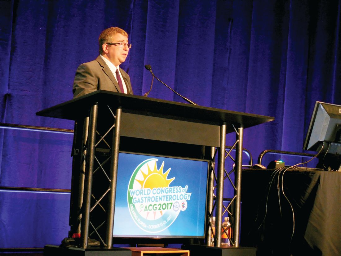
“We think there is pretty strong evidence” for the efficacy of probiotics, and “by any analysis you can throw at them probiotics are safe,” Gordon Morris, MD, said at the World Congress of Gastroenterology at ACG 2017. “The evidence is of moderate and high quality,” added Dr. Morris, a pediatric gastroenterologist at the University of Central Lancashire in Preston, England.
The most widely studied probiotic in the analysis was Lactobacillus reuteri, used in six of the studies with a total of 405 randomized patients. The next most commonly studied agent was Lactobacillus rhamnosus GG, the focus of four studies and tested in a total of 270 randomized patients. Both microbes showed statistically significant and clinically meaningful levels of pain reduction when compared with placebo in subgroup analyses, said Dr. Morris, who performed the meta-analysis as a Cochrane Review Groups systematic review.
“The pain score reductions we saw [with these two strains] could certainly have an impact. I think it matters clinically,” he explained. “Severity of pain is most important to patients.”
Both L. reuteri and L. rhamnosus GG have received “generally regarded as safe” designations from the Food and Drug Administration.
Based on these findings, “I don’t think we can justify, especially with these two main strains, any further basic efficacy studies,” Dr. Morris said. The primary focus for future clinical assessments of these probiotics should be long-term efficacy and safety and whether patients have rebound pain on withdrawal from probiotic use, he added.
The meta-analysis used studies that compared probiotics against placebo in children aged 4-18 years who received treatment for 4-16 weeks. The full analysis showed an average 0.57-unit reduction in pain scores across all 11 studies included, with an average 0.61-unit reduction using L. reuteri and an average 0.75-unit reduction using L. rhamnosus GG. All three between-group differences were statistically significant. Safety data came from eight of the included studies, and they collectively showed absolutely no safety difference between actively treated and control patients.
Dr. Morris noted that the mechanism by which probiotic bacilli relieve abdominal pain remains unclear, but suggested that both prokinetic and anti-inflammatory effects might be involved.
[email protected]
On Twitter @mitchelzoler
ORLANDO – Probiotics are safe and effective for treating functional abdominal pain in children, based on findings from a meta-analysis of 11 randomized studies with a total of 790 patients.
“We think there is pretty strong evidence” for the efficacy of probiotics, and “by any analysis you can throw at them probiotics are safe,” Gordon Morris, MD, said at the World Congress of Gastroenterology at ACG 2017. “The evidence is of moderate and high quality,” added Dr. Morris, a pediatric gastroenterologist at the University of Central Lancashire in Preston, England.
The most widely studied probiotic in the analysis was Lactobacillus reuteri, used in six of the studies with a total of 405 randomized patients. The next most commonly studied agent was Lactobacillus rhamnosus GG, the focus of four studies and tested in a total of 270 randomized patients. Both microbes showed statistically significant and clinically meaningful levels of pain reduction when compared with placebo in subgroup analyses, said Dr. Morris, who performed the meta-analysis as a Cochrane Review Groups systematic review.
“The pain score reductions we saw [with these two strains] could certainly have an impact. I think it matters clinically,” he explained. “Severity of pain is most important to patients.”
Both L. reuteri and L. rhamnosus GG have received “generally regarded as safe” designations from the Food and Drug Administration.
Based on these findings, “I don’t think we can justify, especially with these two main strains, any further basic efficacy studies,” Dr. Morris said. The primary focus for future clinical assessments of these probiotics should be long-term efficacy and safety and whether patients have rebound pain on withdrawal from probiotic use, he added.
The meta-analysis used studies that compared probiotics against placebo in children aged 4-18 years who received treatment for 4-16 weeks. The full analysis showed an average 0.57-unit reduction in pain scores across all 11 studies included, with an average 0.61-unit reduction using L. reuteri and an average 0.75-unit reduction using L. rhamnosus GG. All three between-group differences were statistically significant. Safety data came from eight of the included studies, and they collectively showed absolutely no safety difference between actively treated and control patients.
Dr. Morris noted that the mechanism by which probiotic bacilli relieve abdominal pain remains unclear, but suggested that both prokinetic and anti-inflammatory effects might be involved.
[email protected]
On Twitter @mitchelzoler
ORLANDO – Probiotics are safe and effective for treating functional abdominal pain in children, based on findings from a meta-analysis of 11 randomized studies with a total of 790 patients.
“We think there is pretty strong evidence” for the efficacy of probiotics, and “by any analysis you can throw at them probiotics are safe,” Gordon Morris, MD, said at the World Congress of Gastroenterology at ACG 2017. “The evidence is of moderate and high quality,” added Dr. Morris, a pediatric gastroenterologist at the University of Central Lancashire in Preston, England.
The most widely studied probiotic in the analysis was Lactobacillus reuteri, used in six of the studies with a total of 405 randomized patients. The next most commonly studied agent was Lactobacillus rhamnosus GG, the focus of four studies and tested in a total of 270 randomized patients. Both microbes showed statistically significant and clinically meaningful levels of pain reduction when compared with placebo in subgroup analyses, said Dr. Morris, who performed the meta-analysis as a Cochrane Review Groups systematic review.
“The pain score reductions we saw [with these two strains] could certainly have an impact. I think it matters clinically,” he explained. “Severity of pain is most important to patients.”
Both L. reuteri and L. rhamnosus GG have received “generally regarded as safe” designations from the Food and Drug Administration.
Based on these findings, “I don’t think we can justify, especially with these two main strains, any further basic efficacy studies,” Dr. Morris said. The primary focus for future clinical assessments of these probiotics should be long-term efficacy and safety and whether patients have rebound pain on withdrawal from probiotic use, he added.
The meta-analysis used studies that compared probiotics against placebo in children aged 4-18 years who received treatment for 4-16 weeks. The full analysis showed an average 0.57-unit reduction in pain scores across all 11 studies included, with an average 0.61-unit reduction using L. reuteri and an average 0.75-unit reduction using L. rhamnosus GG. All three between-group differences were statistically significant. Safety data came from eight of the included studies, and they collectively showed absolutely no safety difference between actively treated and control patients.
Dr. Morris noted that the mechanism by which probiotic bacilli relieve abdominal pain remains unclear, but suggested that both prokinetic and anti-inflammatory effects might be involved.
[email protected]
On Twitter @mitchelzoler
AT THE WORLD CONGRESS OF GASTROENTEROLOGY
Key clinical point:
Major finding: Probiotic treatment led to an average 0.57-unit reduction in pain intensity compared with placebo controls.
Data source: A Cochrane Group meta-analysis of 11 studies with 790 patients.
Disclosures: Dr. Morris had no disclosures.
Product Update: Natera’s Panorama NIPT; Solosec from Symbiomix Therapeutics
NIPT SCREENING FOR TWIN ZYGOSITY AND GENETIC RISK FACTORS

Building on its history as the only NIPT that can differentiate between the mother’s and the baby’s DNA, Panorama is now also the only NIPT to distinguish between each twin’s DNA, says Natera. Panorama’s unique technology enables it to determine zygosity and each twin’s gender while also allowing it to identify risk for more genetic conditions in twin pregnancies than other NIPTs, including monosomy X, sex chromosome trisomies, and 22q11.2 deletion syndrome.
FOR MORE INFORMATION, VISIT: http://www.panoramatest.com/panorama-test/
NEW DRUG FOR TREATING BACTERIAL VAGINOSIS

Symbiomix Therapeutics reported that FDA approval was supported by comprehensive studies that found the single-dose secnidazole 2g was efficacious. All treatment-emergent adverse effects were mild or moderate in intensity; no serious adverse events were reported. Solosec will be available to patients in the first quarter of 2018.
FOR MORE INFORMATION, VISIT: https://symbiomix.com/sym-1219/
Share your thoughts! Send your Letter to the Editor to [email protected]. Please include your name and the city and state in which you practice.
NIPT SCREENING FOR TWIN ZYGOSITY AND GENETIC RISK FACTORS
Building on its history as the only NIPT that can differentiate between the mother’s and the baby’s DNA, Panorama is now also the only NIPT to distinguish between each twin’s DNA, says Natera. Panorama’s unique technology enables it to determine zygosity and each twin’s gender while also allowing it to identify risk for more genetic conditions in twin pregnancies than other NIPTs, including monosomy X, sex chromosome trisomies, and 22q11.2 deletion syndrome.
FOR MORE INFORMATION, VISIT: http://www.panoramatest.com/panorama-test/
NEW DRUG FOR TREATING BACTERIAL VAGINOSIS
Symbiomix Therapeutics reported that FDA approval was supported by comprehensive studies that found the single-dose secnidazole 2g was efficacious. All treatment-emergent adverse effects were mild or moderate in intensity; no serious adverse events were reported. Solosec will be available to patients in the first quarter of 2018.
FOR MORE INFORMATION, VISIT: https://symbiomix.com/sym-1219/
Share your thoughts! Send your Letter to the Editor to [email protected]. Please include your name and the city and state in which you practice.
NIPT SCREENING FOR TWIN ZYGOSITY AND GENETIC RISK FACTORS
Building on its history as the only NIPT that can differentiate between the mother’s and the baby’s DNA, Panorama is now also the only NIPT to distinguish between each twin’s DNA, says Natera. Panorama’s unique technology enables it to determine zygosity and each twin’s gender while also allowing it to identify risk for more genetic conditions in twin pregnancies than other NIPTs, including monosomy X, sex chromosome trisomies, and 22q11.2 deletion syndrome.
FOR MORE INFORMATION, VISIT: http://www.panoramatest.com/panorama-test/
NEW DRUG FOR TREATING BACTERIAL VAGINOSIS
Symbiomix Therapeutics reported that FDA approval was supported by comprehensive studies that found the single-dose secnidazole 2g was efficacious. All treatment-emergent adverse effects were mild or moderate in intensity; no serious adverse events were reported. Solosec will be available to patients in the first quarter of 2018.
FOR MORE INFORMATION, VISIT: https://symbiomix.com/sym-1219/
Share your thoughts! Send your Letter to the Editor to [email protected]. Please include your name and the city and state in which you practice.
VIDEO: Back off on screening colonoscopy after nonadvanced adenomas
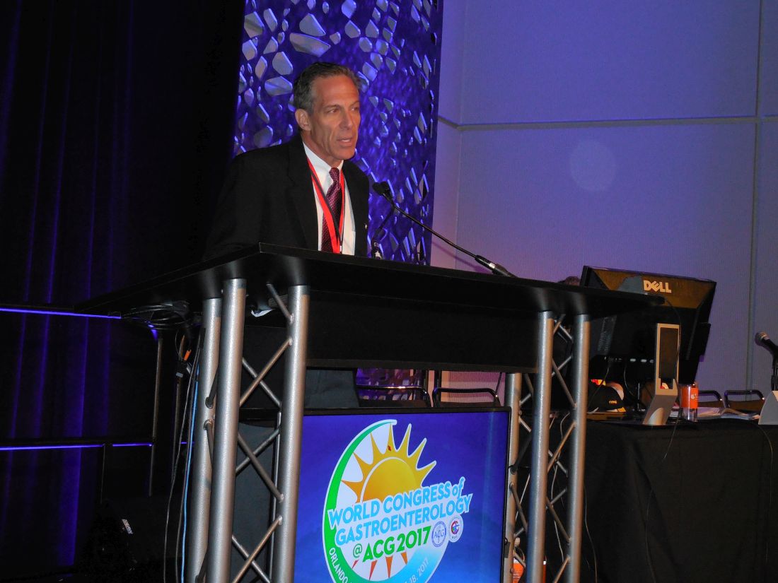
ORLANDO – Evidence supports “backing off” from screening colonoscopies every 5 years for patients who had one or two nonadvanced adenomas removed during a prior colonoscopy, Thomas F. Imperiale, MD, AGAF, said at the World Congress of Gastroenterology at ACG 2017.
He reported findings from more than 66,000 U.S. veterans followed at any one of 13 Veterans Affairs medical centers for an average of more than 7 years. The 10,220 patients who underwent a second screening colonoscopy after an index colonoscopy that led to removal of one or two nonadvanced adenomas had 0.16% colorectal cancer mortality, compared with 0.13% among 8,718 patients with a similar history who did not receive follow-up colonoscopy. The rate of colorectal cancer death was 0.12% among 47,629 control veterans who had no adenomas removed during their index colonoscopy.
In current U.S. practice, many gastroenterologists perform follow-up colonoscopy about 5 years after removing one or two nonadvanced adenomas during a screening colonoscopy, Dr. Imperiale said during a video interview. Deferring follow-up colonoscopy in the absence of any clinical indication seems advisable, he said, especially for older patients with two or more comorbidities who had a high-quality index colonoscopy with good preparation and good colonic visibility.
“We just can’t do colonoscopy for surveillance on this subgroup continuously; it doesn’t make sense,” he said.
No randomized trial results have documented the need for stepped up colonoscopies in patients with a history of one or two nonadvanced adenomas, and these new observational findings are consistent with prior observational reports.
“These data need to be integrated with common sense,” he said. An extended interval before repeat surveillance seems particularly appropriate for patients with a higher risk for adverse effects from the colonoscopy preparation and for patients more likely to die from a cause other than colorectal cancer.
Backing off on repeat colonoscopy “minimizes the harm from surveillance. As patients get older they don’t tolerate the prep as well. It grows more onerous, and the returns diminish,” Dr. Imperiale said.
The patients included in the review had their index colonoscopy performed during 2002-2009, when they averaged about 61 years old, and about 95% were men. Their average Charlson comorbidity index was about 1.3. The incidence of colorectal cancer during follow-up after the index colonoscopy was 0.18% in patients with one or two nonadvanced adenomas in their index examination and no follow-up colonoscopy, 0.71% in those with nonadvanced adenomas who had one or more subsequent colonoscopies, and 0.31% in the people with no adenomas removed during the index procedure.
The rates of all-cause death during follow-up of the three subgroups were notably different: 34% in those with nonadvanced adenomas and no repeat colonoscopy, 13% in patients with nonadvanced adenomas and repeat colonoscopy, and 21% in those without nonadvanced adenomas. Dr. Imperiale discounted the significance of comparing rates of all-cause mortality, stressing that the most relevant primary endpoint is colorectal cancer mortality.
Dr. Imperiale reported having no disclosures.
[email protected]
On Twitter @mitchelzoler
The video associated with this article is no longer available on this site. Please view all of our videos on the MDedge YouTube channel
ORLANDO – Evidence supports “backing off” from screening colonoscopies every 5 years for patients who had one or two nonadvanced adenomas removed during a prior colonoscopy, Thomas F. Imperiale, MD, AGAF, said at the World Congress of Gastroenterology at ACG 2017.
He reported findings from more than 66,000 U.S. veterans followed at any one of 13 Veterans Affairs medical centers for an average of more than 7 years. The 10,220 patients who underwent a second screening colonoscopy after an index colonoscopy that led to removal of one or two nonadvanced adenomas had 0.16% colorectal cancer mortality, compared with 0.13% among 8,718 patients with a similar history who did not receive follow-up colonoscopy. The rate of colorectal cancer death was 0.12% among 47,629 control veterans who had no adenomas removed during their index colonoscopy.
In current U.S. practice, many gastroenterologists perform follow-up colonoscopy about 5 years after removing one or two nonadvanced adenomas during a screening colonoscopy, Dr. Imperiale said during a video interview. Deferring follow-up colonoscopy in the absence of any clinical indication seems advisable, he said, especially for older patients with two or more comorbidities who had a high-quality index colonoscopy with good preparation and good colonic visibility.
“We just can’t do colonoscopy for surveillance on this subgroup continuously; it doesn’t make sense,” he said.
No randomized trial results have documented the need for stepped up colonoscopies in patients with a history of one or two nonadvanced adenomas, and these new observational findings are consistent with prior observational reports.
“These data need to be integrated with common sense,” he said. An extended interval before repeat surveillance seems particularly appropriate for patients with a higher risk for adverse effects from the colonoscopy preparation and for patients more likely to die from a cause other than colorectal cancer.
Backing off on repeat colonoscopy “minimizes the harm from surveillance. As patients get older they don’t tolerate the prep as well. It grows more onerous, and the returns diminish,” Dr. Imperiale said.
The patients included in the review had their index colonoscopy performed during 2002-2009, when they averaged about 61 years old, and about 95% were men. Their average Charlson comorbidity index was about 1.3. The incidence of colorectal cancer during follow-up after the index colonoscopy was 0.18% in patients with one or two nonadvanced adenomas in their index examination and no follow-up colonoscopy, 0.71% in those with nonadvanced adenomas who had one or more subsequent colonoscopies, and 0.31% in the people with no adenomas removed during the index procedure.
The rates of all-cause death during follow-up of the three subgroups were notably different: 34% in those with nonadvanced adenomas and no repeat colonoscopy, 13% in patients with nonadvanced adenomas and repeat colonoscopy, and 21% in those without nonadvanced adenomas. Dr. Imperiale discounted the significance of comparing rates of all-cause mortality, stressing that the most relevant primary endpoint is colorectal cancer mortality.
Dr. Imperiale reported having no disclosures.
[email protected]
On Twitter @mitchelzoler
The video associated with this article is no longer available on this site. Please view all of our videos on the MDedge YouTube channel
ORLANDO – Evidence supports “backing off” from screening colonoscopies every 5 years for patients who had one or two nonadvanced adenomas removed during a prior colonoscopy, Thomas F. Imperiale, MD, AGAF, said at the World Congress of Gastroenterology at ACG 2017.
He reported findings from more than 66,000 U.S. veterans followed at any one of 13 Veterans Affairs medical centers for an average of more than 7 years. The 10,220 patients who underwent a second screening colonoscopy after an index colonoscopy that led to removal of one or two nonadvanced adenomas had 0.16% colorectal cancer mortality, compared with 0.13% among 8,718 patients with a similar history who did not receive follow-up colonoscopy. The rate of colorectal cancer death was 0.12% among 47,629 control veterans who had no adenomas removed during their index colonoscopy.
In current U.S. practice, many gastroenterologists perform follow-up colonoscopy about 5 years after removing one or two nonadvanced adenomas during a screening colonoscopy, Dr. Imperiale said during a video interview. Deferring follow-up colonoscopy in the absence of any clinical indication seems advisable, he said, especially for older patients with two or more comorbidities who had a high-quality index colonoscopy with good preparation and good colonic visibility.
“We just can’t do colonoscopy for surveillance on this subgroup continuously; it doesn’t make sense,” he said.
No randomized trial results have documented the need for stepped up colonoscopies in patients with a history of one or two nonadvanced adenomas, and these new observational findings are consistent with prior observational reports.
“These data need to be integrated with common sense,” he said. An extended interval before repeat surveillance seems particularly appropriate for patients with a higher risk for adverse effects from the colonoscopy preparation and for patients more likely to die from a cause other than colorectal cancer.
Backing off on repeat colonoscopy “minimizes the harm from surveillance. As patients get older they don’t tolerate the prep as well. It grows more onerous, and the returns diminish,” Dr. Imperiale said.
The patients included in the review had their index colonoscopy performed during 2002-2009, when they averaged about 61 years old, and about 95% were men. Their average Charlson comorbidity index was about 1.3. The incidence of colorectal cancer during follow-up after the index colonoscopy was 0.18% in patients with one or two nonadvanced adenomas in their index examination and no follow-up colonoscopy, 0.71% in those with nonadvanced adenomas who had one or more subsequent colonoscopies, and 0.31% in the people with no adenomas removed during the index procedure.
The rates of all-cause death during follow-up of the three subgroups were notably different: 34% in those with nonadvanced adenomas and no repeat colonoscopy, 13% in patients with nonadvanced adenomas and repeat colonoscopy, and 21% in those without nonadvanced adenomas. Dr. Imperiale discounted the significance of comparing rates of all-cause mortality, stressing that the most relevant primary endpoint is colorectal cancer mortality.
Dr. Imperiale reported having no disclosures.
[email protected]
On Twitter @mitchelzoler
The video associated with this article is no longer available on this site. Please view all of our videos on the MDedge YouTube channel
AT THE WORLD CONGRESS OF GASTROENTEROLOGY
Key clinical point:
Major finding: Colorectal cancer mortality was 0.13% in patients without follow-up colonoscopy and 0.16% in patients who had a second colonoscopy.
Data source: Review of 66,567 people at 13 U.S. VA Medical Centers.
Disclosures: Dr. Imperiale reported having no disclosures.
Tips for Preventing Encephalitis
Click here to download the PDF. 
Click here to download the PDF.
Click here to download the PDF.

EEG-Based Risk Score May Predict Likelihood of Seizures in Hospitalized Patients
A risk score based on EEG variables and seizure history may quickly and accurately aid clinical judgment of seizure risk in patients who are critically ill, according to research published online ahead of print October 9 in JAMA Neurology.
Studies have detected a high incidence of subclinical seizures in patients with sepsis, traumatic brain injury, and other conditions, but no simple, validated method exists to assess a patient’s seizure risk using a combination of factors. Aaron F. Struck, MD, Assistant Professor of Neurology at the University of Wisconsin in Madison, and colleagues sought to create a simple scoring system associated with the probability of seizures in patients with acute illness.
Data From Three Centers
The investigators created the scoring system using data from a multicenter prospective database. The database included clinical and electrographic variables from patients who had more than six hours of uninterrupted EEG recordings for clinical indications other than epilepsy monitoring unit admissions. The continuous EEG sessions were recorded at Emory University Hospital in Atlanta, Brigham and Women’s Hospital in Boston, and Yale New Haven Hospital in Connecticut. In all, 5,427 continuous EEGs were performed on 4,772 patients (49.9% men; median age, 61).
To build their scoring model, the investigators used a machine-learning method, Risk-Calibrated Supersparse Linear Integer Model (RiskSLIM), that considered 24 candidate variables. The researchers used cross-validation to validate the model’s accuracy and risk calibration.
The final model, which the researchers called 2HELPS2B, had an area under the curve of 0.819 and an average calibration error of 2.7%. A patient’s score ranges from 0–7 and is based on the following six variables:
- brief (ictal) rhythmic discharges (B[I]RDS) (2 points)
- presence of lateralized periodic discharges, lateralized rhythmic delta activity, or bilateral independent periodic discharges (1 point)
- prior seizure (1 point)
- sporadic epileptiform discharges (1 point)
- frequency greater than 2.0 Hz for any periodic or rhythmic pattern (1 point)
- presence of “plus” features (ie, superimposed, rhythmic, sharp, or fast activity) (1 point).
The seizure risk associated with each score from 0–5 was 5%, 12%, 27%, 50%, 73%, and 88%, respectively. For a score of 6 or 7, the probable seizure risk was greater than 95%.
The large sample size and use of data from multiple centers are among the study’s strengths. Limitations of the study include that duration of EEG was not included in the database, and that no sessions of less than six hours were included in the study.
“The 2HELPS2B score is an easy-to-use tool to augment clinical judgment of the risk for seizures in individual patients,” Dr. Struck and colleagues said. “The simplicity of the system allows for easy integration into clinical workflow. With increasing familiarity, 2HELPS2B will improve communication between EEG interpreters and clinicians through the use of a quickly comprehensible single number to describe seizure risk for patients on continuous EEG.”
Questions Remain
The investigators designed “a simple scale with good accuracy, which can be easily used by clinicians to estimate seizure risk in their patients,” said Barry M. Czeisler, MD, Assistant Professor of Neurology and Neurosurgery at New York University School of Medicine, and Jan Claassen, MD, PhD, Associate Professor of Neurology at Columbia University College of Physicians and Surgeons in New York, in an accompanying editorial. Still, the score is unvalidated for prediction based on less than six hours of EEG recordings and should be validated in prospective studies, they said.
A seizure risk scale has the potential to inform clinical practice. “Certain patients may not need to stay on continuous EEG for a long time if their seizure risk is low,” but an acceptable level of risk remains unclear, said Drs. Czeisler and Claassen. In addition, the score potentially could guide which patterns of features warrant more aggressive treatment, such as with additional antiepileptic medication. “Appropriate risk stratification using 2HELPS2B may allow us to answer these questions adequately in the near future,” they said. “The development of adequate measurement tools is often necessary to appropriately study a condition, which in turn may allow for future optimization of treatment algorithms.”
—Jake Remaly
Suggested Reading
Czeisler BM, Claassen J. A novel clinical score to assess seizure risk. JAMA Neurology. 2017 Oct 9 [Epub ahead of print].
Struck AF, Ustun B, Rodriguez Ruiz A, et al. Association of an electroencephalography-based risk score with seizure probability in hospitalized patients. JAMA Neurology. 2017 Oct 9 [Epub ahead of print].
A risk score based on EEG variables and seizure history may quickly and accurately aid clinical judgment of seizure risk in patients who are critically ill, according to research published online ahead of print October 9 in JAMA Neurology.
Studies have detected a high incidence of subclinical seizures in patients with sepsis, traumatic brain injury, and other conditions, but no simple, validated method exists to assess a patient’s seizure risk using a combination of factors. Aaron F. Struck, MD, Assistant Professor of Neurology at the University of Wisconsin in Madison, and colleagues sought to create a simple scoring system associated with the probability of seizures in patients with acute illness.
Data From Three Centers
The investigators created the scoring system using data from a multicenter prospective database. The database included clinical and electrographic variables from patients who had more than six hours of uninterrupted EEG recordings for clinical indications other than epilepsy monitoring unit admissions. The continuous EEG sessions were recorded at Emory University Hospital in Atlanta, Brigham and Women’s Hospital in Boston, and Yale New Haven Hospital in Connecticut. In all, 5,427 continuous EEGs were performed on 4,772 patients (49.9% men; median age, 61).
To build their scoring model, the investigators used a machine-learning method, Risk-Calibrated Supersparse Linear Integer Model (RiskSLIM), that considered 24 candidate variables. The researchers used cross-validation to validate the model’s accuracy and risk calibration.
The final model, which the researchers called 2HELPS2B, had an area under the curve of 0.819 and an average calibration error of 2.7%. A patient’s score ranges from 0–7 and is based on the following six variables:
- brief (ictal) rhythmic discharges (B[I]RDS) (2 points)
- presence of lateralized periodic discharges, lateralized rhythmic delta activity, or bilateral independent periodic discharges (1 point)
- prior seizure (1 point)
- sporadic epileptiform discharges (1 point)
- frequency greater than 2.0 Hz for any periodic or rhythmic pattern (1 point)
- presence of “plus” features (ie, superimposed, rhythmic, sharp, or fast activity) (1 point).
The seizure risk associated with each score from 0–5 was 5%, 12%, 27%, 50%, 73%, and 88%, respectively. For a score of 6 or 7, the probable seizure risk was greater than 95%.
The large sample size and use of data from multiple centers are among the study’s strengths. Limitations of the study include that duration of EEG was not included in the database, and that no sessions of less than six hours were included in the study.
“The 2HELPS2B score is an easy-to-use tool to augment clinical judgment of the risk for seizures in individual patients,” Dr. Struck and colleagues said. “The simplicity of the system allows for easy integration into clinical workflow. With increasing familiarity, 2HELPS2B will improve communication between EEG interpreters and clinicians through the use of a quickly comprehensible single number to describe seizure risk for patients on continuous EEG.”
Questions Remain
The investigators designed “a simple scale with good accuracy, which can be easily used by clinicians to estimate seizure risk in their patients,” said Barry M. Czeisler, MD, Assistant Professor of Neurology and Neurosurgery at New York University School of Medicine, and Jan Claassen, MD, PhD, Associate Professor of Neurology at Columbia University College of Physicians and Surgeons in New York, in an accompanying editorial. Still, the score is unvalidated for prediction based on less than six hours of EEG recordings and should be validated in prospective studies, they said.
A seizure risk scale has the potential to inform clinical practice. “Certain patients may not need to stay on continuous EEG for a long time if their seizure risk is low,” but an acceptable level of risk remains unclear, said Drs. Czeisler and Claassen. In addition, the score potentially could guide which patterns of features warrant more aggressive treatment, such as with additional antiepileptic medication. “Appropriate risk stratification using 2HELPS2B may allow us to answer these questions adequately in the near future,” they said. “The development of adequate measurement tools is often necessary to appropriately study a condition, which in turn may allow for future optimization of treatment algorithms.”
—Jake Remaly
Suggested Reading
Czeisler BM, Claassen J. A novel clinical score to assess seizure risk. JAMA Neurology. 2017 Oct 9 [Epub ahead of print].
Struck AF, Ustun B, Rodriguez Ruiz A, et al. Association of an electroencephalography-based risk score with seizure probability in hospitalized patients. JAMA Neurology. 2017 Oct 9 [Epub ahead of print].
A risk score based on EEG variables and seizure history may quickly and accurately aid clinical judgment of seizure risk in patients who are critically ill, according to research published online ahead of print October 9 in JAMA Neurology.
Studies have detected a high incidence of subclinical seizures in patients with sepsis, traumatic brain injury, and other conditions, but no simple, validated method exists to assess a patient’s seizure risk using a combination of factors. Aaron F. Struck, MD, Assistant Professor of Neurology at the University of Wisconsin in Madison, and colleagues sought to create a simple scoring system associated with the probability of seizures in patients with acute illness.
Data From Three Centers
The investigators created the scoring system using data from a multicenter prospective database. The database included clinical and electrographic variables from patients who had more than six hours of uninterrupted EEG recordings for clinical indications other than epilepsy monitoring unit admissions. The continuous EEG sessions were recorded at Emory University Hospital in Atlanta, Brigham and Women’s Hospital in Boston, and Yale New Haven Hospital in Connecticut. In all, 5,427 continuous EEGs were performed on 4,772 patients (49.9% men; median age, 61).
To build their scoring model, the investigators used a machine-learning method, Risk-Calibrated Supersparse Linear Integer Model (RiskSLIM), that considered 24 candidate variables. The researchers used cross-validation to validate the model’s accuracy and risk calibration.
The final model, which the researchers called 2HELPS2B, had an area under the curve of 0.819 and an average calibration error of 2.7%. A patient’s score ranges from 0–7 and is based on the following six variables:
- brief (ictal) rhythmic discharges (B[I]RDS) (2 points)
- presence of lateralized periodic discharges, lateralized rhythmic delta activity, or bilateral independent periodic discharges (1 point)
- prior seizure (1 point)
- sporadic epileptiform discharges (1 point)
- frequency greater than 2.0 Hz for any periodic or rhythmic pattern (1 point)
- presence of “plus” features (ie, superimposed, rhythmic, sharp, or fast activity) (1 point).
The seizure risk associated with each score from 0–5 was 5%, 12%, 27%, 50%, 73%, and 88%, respectively. For a score of 6 or 7, the probable seizure risk was greater than 95%.
The large sample size and use of data from multiple centers are among the study’s strengths. Limitations of the study include that duration of EEG was not included in the database, and that no sessions of less than six hours were included in the study.
“The 2HELPS2B score is an easy-to-use tool to augment clinical judgment of the risk for seizures in individual patients,” Dr. Struck and colleagues said. “The simplicity of the system allows for easy integration into clinical workflow. With increasing familiarity, 2HELPS2B will improve communication between EEG interpreters and clinicians through the use of a quickly comprehensible single number to describe seizure risk for patients on continuous EEG.”
Questions Remain
The investigators designed “a simple scale with good accuracy, which can be easily used by clinicians to estimate seizure risk in their patients,” said Barry M. Czeisler, MD, Assistant Professor of Neurology and Neurosurgery at New York University School of Medicine, and Jan Claassen, MD, PhD, Associate Professor of Neurology at Columbia University College of Physicians and Surgeons in New York, in an accompanying editorial. Still, the score is unvalidated for prediction based on less than six hours of EEG recordings and should be validated in prospective studies, they said.
A seizure risk scale has the potential to inform clinical practice. “Certain patients may not need to stay on continuous EEG for a long time if their seizure risk is low,” but an acceptable level of risk remains unclear, said Drs. Czeisler and Claassen. In addition, the score potentially could guide which patterns of features warrant more aggressive treatment, such as with additional antiepileptic medication. “Appropriate risk stratification using 2HELPS2B may allow us to answer these questions adequately in the near future,” they said. “The development of adequate measurement tools is often necessary to appropriately study a condition, which in turn may allow for future optimization of treatment algorithms.”
—Jake Remaly
Suggested Reading
Czeisler BM, Claassen J. A novel clinical score to assess seizure risk. JAMA Neurology. 2017 Oct 9 [Epub ahead of print].
Struck AF, Ustun B, Rodriguez Ruiz A, et al. Association of an electroencephalography-based risk score with seizure probability in hospitalized patients. JAMA Neurology. 2017 Oct 9 [Epub ahead of print].
VIDEO: Burnout affects half of U.S. gastroenterologists
ORLANDO – Nearly half of U.S. gastroenterologists who responded to a recent survey had symptoms of burnout that seemed largely driven by work-life balance issues.
Burnout appeared to disproportionately affect younger gastroenterologists, those who spend more time on chores at home including caring for young children, physicians who were neutral toward or dissatisfied with a spouse or partner, and clinicians planning to soon leave their practice, Carol A. Burke, MD, said at the World Congress of Gastroenterology at ACG 2017.
Factors not linked with burnout included their type of practice, whether the gastroenterologists worked full or part time, their location, and their compensation, said Dr. Burke, director of the Center for Colon Polyp and Cancer Prevention at the Cleveland Clinic.
The life issues that appeared most strongly linked to burnout “speak to a problem for physicians to balance” their professional and personal lives, Dr. Burke said in a video interview. Several interventions exist that can potentially mitigate burnout, and the American College of Gastroenterology, which ran the survey, is taking steps to make information on these interventions available to members, noted Dr. Burke, the organization’s president.
Dr. Burke and her associates sent a 60-item survey to all 11,080 College members during 2014 and 2015 and received 1,021 replies including 754 fully completed responses. Their prespecified definition of burnout was a high score for emotional exhaustion or for depersonalization, or both, on the Maslach Burnout Inventory. The results showed that 45% of respondents had a high score for emotional exhaustion, 21% scored high on depersonalization, and overall 49% met the burnout criteria set by the investigators. The Inventory answers also showed that 18% had a low sense of personal accomplishment.
A multivariate analysis showed that significant links with burnout were younger age, more time spent on domestic chores, having a neutral or dissatisfying relationship with a spouse or partner, and plans for imminent retirement from gastroenterology practice, Dr. Burke reported.
The main reasons for planning imminent retirement were reimbursement, cited by 32% of this subgroup, regulations, cited by 21%, recertification, cited by 16%, and electronic medical records, cited by 10% as the main reason for leaving practice.
Strategies and resources aimed at better dealing with burnout were requested by 60% of all survey respondents, and the College is in the process of making these tools available, Dr. Burke said.
A recent study conducted by the AGA Institute Education and Training Committee and reported in AGA Perspectives supports these findings. Read more here and join the discussion on the AGA Community.
The video associated with this article is no longer available on this site. Please view all of our videos on the MDedge YouTube channel
[email protected]
On Twitter @mitchelzoler
ORLANDO – Nearly half of U.S. gastroenterologists who responded to a recent survey had symptoms of burnout that seemed largely driven by work-life balance issues.
Burnout appeared to disproportionately affect younger gastroenterologists, those who spend more time on chores at home including caring for young children, physicians who were neutral toward or dissatisfied with a spouse or partner, and clinicians planning to soon leave their practice, Carol A. Burke, MD, said at the World Congress of Gastroenterology at ACG 2017.
Factors not linked with burnout included their type of practice, whether the gastroenterologists worked full or part time, their location, and their compensation, said Dr. Burke, director of the Center for Colon Polyp and Cancer Prevention at the Cleveland Clinic.
The life issues that appeared most strongly linked to burnout “speak to a problem for physicians to balance” their professional and personal lives, Dr. Burke said in a video interview. Several interventions exist that can potentially mitigate burnout, and the American College of Gastroenterology, which ran the survey, is taking steps to make information on these interventions available to members, noted Dr. Burke, the organization’s president.
Dr. Burke and her associates sent a 60-item survey to all 11,080 College members during 2014 and 2015 and received 1,021 replies including 754 fully completed responses. Their prespecified definition of burnout was a high score for emotional exhaustion or for depersonalization, or both, on the Maslach Burnout Inventory. The results showed that 45% of respondents had a high score for emotional exhaustion, 21% scored high on depersonalization, and overall 49% met the burnout criteria set by the investigators. The Inventory answers also showed that 18% had a low sense of personal accomplishment.
A multivariate analysis showed that significant links with burnout were younger age, more time spent on domestic chores, having a neutral or dissatisfying relationship with a spouse or partner, and plans for imminent retirement from gastroenterology practice, Dr. Burke reported.
The main reasons for planning imminent retirement were reimbursement, cited by 32% of this subgroup, regulations, cited by 21%, recertification, cited by 16%, and electronic medical records, cited by 10% as the main reason for leaving practice.
Strategies and resources aimed at better dealing with burnout were requested by 60% of all survey respondents, and the College is in the process of making these tools available, Dr. Burke said.
A recent study conducted by the AGA Institute Education and Training Committee and reported in AGA Perspectives supports these findings. Read more here and join the discussion on the AGA Community.
The video associated with this article is no longer available on this site. Please view all of our videos on the MDedge YouTube channel
[email protected]
On Twitter @mitchelzoler
ORLANDO – Nearly half of U.S. gastroenterologists who responded to a recent survey had symptoms of burnout that seemed largely driven by work-life balance issues.
Burnout appeared to disproportionately affect younger gastroenterologists, those who spend more time on chores at home including caring for young children, physicians who were neutral toward or dissatisfied with a spouse or partner, and clinicians planning to soon leave their practice, Carol A. Burke, MD, said at the World Congress of Gastroenterology at ACG 2017.
Factors not linked with burnout included their type of practice, whether the gastroenterologists worked full or part time, their location, and their compensation, said Dr. Burke, director of the Center for Colon Polyp and Cancer Prevention at the Cleveland Clinic.
The life issues that appeared most strongly linked to burnout “speak to a problem for physicians to balance” their professional and personal lives, Dr. Burke said in a video interview. Several interventions exist that can potentially mitigate burnout, and the American College of Gastroenterology, which ran the survey, is taking steps to make information on these interventions available to members, noted Dr. Burke, the organization’s president.
Dr. Burke and her associates sent a 60-item survey to all 11,080 College members during 2014 and 2015 and received 1,021 replies including 754 fully completed responses. Their prespecified definition of burnout was a high score for emotional exhaustion or for depersonalization, or both, on the Maslach Burnout Inventory. The results showed that 45% of respondents had a high score for emotional exhaustion, 21% scored high on depersonalization, and overall 49% met the burnout criteria set by the investigators. The Inventory answers also showed that 18% had a low sense of personal accomplishment.
A multivariate analysis showed that significant links with burnout were younger age, more time spent on domestic chores, having a neutral or dissatisfying relationship with a spouse or partner, and plans for imminent retirement from gastroenterology practice, Dr. Burke reported.
The main reasons for planning imminent retirement were reimbursement, cited by 32% of this subgroup, regulations, cited by 21%, recertification, cited by 16%, and electronic medical records, cited by 10% as the main reason for leaving practice.
Strategies and resources aimed at better dealing with burnout were requested by 60% of all survey respondents, and the College is in the process of making these tools available, Dr. Burke said.
A recent study conducted by the AGA Institute Education and Training Committee and reported in AGA Perspectives supports these findings. Read more here and join the discussion on the AGA Community.
The video associated with this article is no longer available on this site. Please view all of our videos on the MDedge YouTube channel
[email protected]
On Twitter @mitchelzoler
AT THE 13TH WORLD CONGRESS OF GASTROENTEROLOGY
Key clinical point:
Major finding: Forty-nine percent of surveyed U.S. gastroenterologists showed a high level of emotional exhaustion, depersonalization, or both.
Data source: Survey results from 754 members of the American College of Gastroenterology.
Disclosures: The American College of Gastroenterology funded the survey. Dr. Burke had no relevant disclosures.

Psychological Consequences of Detainee Operations: What DoD and VA Health Care Providers Need to Know
Detainee operations is a dark topic and one that often is avoided in the welcome home of veterans participating in detainee operations. Many veterans who have been involved have hidden these missions, fearing that they would be tarnished by past scandals. However, the burden of these detainee missions may contribute to depression, moral injury, and suicidal behaviors.
The recent conflicts in Afghanistan and Iraq have produced many opportunities for lessons on detainee operations. Unfortunately, often the lessons learned from one conflict have not been carried forward to the next. The scandals at the Abu Ghraib prison in Iraq and the continuing controversy over practices at Guantanamo Bay (GTMO) in Cuba illustrate these lapses. This column will not dwell on these issues but on what has been the psychological effects on U.S. service members of guarding and caring for detainees.
Background
Since 9/11, military service members have been involved in detainee operations in many roles, including at the point of capture (when the detainee is taken into custody), guarding the detainee; interrogating the detainee, and providing medical and psychological care.
Our service members have been woefully underprepared for these missions, and as a result may have faced adverse psychological consequences. The work is often dangerous and tedious. The following dangerous or frustrating examples are from my experiences:
- Correctional staff at GTMO had feces thrown at them;
- U.S. staff were targeted by rocket propelled grenades at Abu Ghraib and moved into jail cells for protection;
- Medical personnel at Camp Bucca in Iraq were attacked by the detainees who used hand sanitizer and latex gloves to make miniature fire balls;
- Insufficient medical equipment at Abu Ghraib and other facilities to care for detainees;
- An overall lack of perceived support from the medical and correctional chain of commands; and
- Numerous different chains of commands with different priorities, leading to a sense of chaos.
Corrections Overlap
There is overlap with traditional correctional medical care, including the care of prisoners in traditional jails and prisons. Likewise, there are similar issues with migrants from Central America and other regions who enter the country illegally and often are put into makeshift camps or overcrowded jails. However, there are key differences when treating detainees in facilities outside the U.S.
These differences include the cultural aspects in caring for detainees from the Middle East and elsewhere, large holding areas with 200 to 300 detainees, such as Camp Bucca and Abhu Ghraib, indefinite terms of confinement, such as at GTMO, and high-visibility political implications especially suicide attempts and interrogations.
There are many challenges to providing medical support in detainee operations that may have adverse psychological consequences. These include the following:
- Care for detainees when first captured;
- Fear of infectious diseases in detainees;
- Care for detainees in the correctional facilities in Abu Ghraib, GTMO, the Theater Internment Facility at Bagram Air Base Afghanistan; and other facilities;
- Involvement in force-feeding and during hunger strikes;
- Avoiding abuse of detainees by military staff;
- Providing psychological support to interrogation efforts often known as Behavioral Science Consultation Teams; and
- Challenges in treatment of complex medical and psychiatric conditions among detainees, which may include a limited formulary, lack of full medical equipment, and austere conditions.
Psychological Reactions
These challenges contribute to the development of negative psychological reactions, including posttramatic stress syndrome (PTSD) and moral injury, in medical and corrections staff. The negative view of the American public toward these correctional issues contributes to a sense of shame among those who have guarded or treated detainees.
One veteran who worked at GTMO and the jail in Bagram reported that 10 of his colleagues killed themselves. Although there is robust literature on suicides in army and other military personnel, I do not know of any studies that examine suicides specifically in detainee’s operational staff.1,2 There have been analyses of the relationship between suicide and military occupational specialty (MOS).1 However, personnel in detainee operations may come from many different disciplines, including medical, correctional, and others.
Again, data here are anecdotal, since there is no public information available. However, I was sent to Abu Ghraib and Camp Bucca in 2004 to evaluate these issues. I have been to GTMO 5 times. Thus, I have personal experience that in forms this column.
Often the veteran will not bring up these experiences because of the shame and stigma. Therefore, clinicians need to ask about the veteran’s experience and whether he or she served at GTMO, Abu Ghraib, Camp Bucca, the Theater Facility in Bagram, or other points of capture.
Although there are many books and manuals about treating PTSD and some literature about providing mental health care to detainees, there is little published about providing care to staff involved with detainee’s operations. I postulate that many staff will have shame, guilt, or moral injury, which leads to suicidal thoughts. Treatment thus needs to be supportive, whether with medications or psychotherapy.
Conclusion
All these issues should be considered by clinical staff who are caring for service members who have been involved in detainee operations. Veterans may not volunteer that they have been involved in these operations; it is important to ask.
Guilt and shame may be large components of the presenting psychological presentation. This may lead to moral injury. Careful exploration of depressive and suicidal thoughts with these patients is needed. An understanding of these challenges will help with clinical care.
1. Black SA, Galloway MS, Bell MR, Ritchie EC. Prevalence and risk factors associated with suicides of army soldiers 2001-2009. Milit Psychol. 2011;23(4):433-451.
2. Ritchie EC. Suicides and the United States Army: perspectives from the former psychiatry consultant to the army surgeon general. Cerebrum. 2012;1.
Detainee operations is a dark topic and one that often is avoided in the welcome home of veterans participating in detainee operations. Many veterans who have been involved have hidden these missions, fearing that they would be tarnished by past scandals. However, the burden of these detainee missions may contribute to depression, moral injury, and suicidal behaviors.
The recent conflicts in Afghanistan and Iraq have produced many opportunities for lessons on detainee operations. Unfortunately, often the lessons learned from one conflict have not been carried forward to the next. The scandals at the Abu Ghraib prison in Iraq and the continuing controversy over practices at Guantanamo Bay (GTMO) in Cuba illustrate these lapses. This column will not dwell on these issues but on what has been the psychological effects on U.S. service members of guarding and caring for detainees.
Background
Since 9/11, military service members have been involved in detainee operations in many roles, including at the point of capture (when the detainee is taken into custody), guarding the detainee; interrogating the detainee, and providing medical and psychological care.
Our service members have been woefully underprepared for these missions, and as a result may have faced adverse psychological consequences. The work is often dangerous and tedious. The following dangerous or frustrating examples are from my experiences:
- Correctional staff at GTMO had feces thrown at them;
- U.S. staff were targeted by rocket propelled grenades at Abu Ghraib and moved into jail cells for protection;
- Medical personnel at Camp Bucca in Iraq were attacked by the detainees who used hand sanitizer and latex gloves to make miniature fire balls;
- Insufficient medical equipment at Abu Ghraib and other facilities to care for detainees;
- An overall lack of perceived support from the medical and correctional chain of commands; and
- Numerous different chains of commands with different priorities, leading to a sense of chaos.
Corrections Overlap
There is overlap with traditional correctional medical care, including the care of prisoners in traditional jails and prisons. Likewise, there are similar issues with migrants from Central America and other regions who enter the country illegally and often are put into makeshift camps or overcrowded jails. However, there are key differences when treating detainees in facilities outside the U.S.
These differences include the cultural aspects in caring for detainees from the Middle East and elsewhere, large holding areas with 200 to 300 detainees, such as Camp Bucca and Abhu Ghraib, indefinite terms of confinement, such as at GTMO, and high-visibility political implications especially suicide attempts and interrogations.
There are many challenges to providing medical support in detainee operations that may have adverse psychological consequences. These include the following:
- Care for detainees when first captured;
- Fear of infectious diseases in detainees;
- Care for detainees in the correctional facilities in Abu Ghraib, GTMO, the Theater Internment Facility at Bagram Air Base Afghanistan; and other facilities;
- Involvement in force-feeding and during hunger strikes;
- Avoiding abuse of detainees by military staff;
- Providing psychological support to interrogation efforts often known as Behavioral Science Consultation Teams; and
- Challenges in treatment of complex medical and psychiatric conditions among detainees, which may include a limited formulary, lack of full medical equipment, and austere conditions.
Psychological Reactions
These challenges contribute to the development of negative psychological reactions, including posttramatic stress syndrome (PTSD) and moral injury, in medical and corrections staff. The negative view of the American public toward these correctional issues contributes to a sense of shame among those who have guarded or treated detainees.
One veteran who worked at GTMO and the jail in Bagram reported that 10 of his colleagues killed themselves. Although there is robust literature on suicides in army and other military personnel, I do not know of any studies that examine suicides specifically in detainee’s operational staff.1,2 There have been analyses of the relationship between suicide and military occupational specialty (MOS).1 However, personnel in detainee operations may come from many different disciplines, including medical, correctional, and others.
Again, data here are anecdotal, since there is no public information available. However, I was sent to Abu Ghraib and Camp Bucca in 2004 to evaluate these issues. I have been to GTMO 5 times. Thus, I have personal experience that in forms this column.
Often the veteran will not bring up these experiences because of the shame and stigma. Therefore, clinicians need to ask about the veteran’s experience and whether he or she served at GTMO, Abu Ghraib, Camp Bucca, the Theater Facility in Bagram, or other points of capture.
Although there are many books and manuals about treating PTSD and some literature about providing mental health care to detainees, there is little published about providing care to staff involved with detainee’s operations. I postulate that many staff will have shame, guilt, or moral injury, which leads to suicidal thoughts. Treatment thus needs to be supportive, whether with medications or psychotherapy.
Conclusion
All these issues should be considered by clinical staff who are caring for service members who have been involved in detainee operations. Veterans may not volunteer that they have been involved in these operations; it is important to ask.
Guilt and shame may be large components of the presenting psychological presentation. This may lead to moral injury. Careful exploration of depressive and suicidal thoughts with these patients is needed. An understanding of these challenges will help with clinical care.
Detainee operations is a dark topic and one that often is avoided in the welcome home of veterans participating in detainee operations. Many veterans who have been involved have hidden these missions, fearing that they would be tarnished by past scandals. However, the burden of these detainee missions may contribute to depression, moral injury, and suicidal behaviors.
The recent conflicts in Afghanistan and Iraq have produced many opportunities for lessons on detainee operations. Unfortunately, often the lessons learned from one conflict have not been carried forward to the next. The scandals at the Abu Ghraib prison in Iraq and the continuing controversy over practices at Guantanamo Bay (GTMO) in Cuba illustrate these lapses. This column will not dwell on these issues but on what has been the psychological effects on U.S. service members of guarding and caring for detainees.
Background
Since 9/11, military service members have been involved in detainee operations in many roles, including at the point of capture (when the detainee is taken into custody), guarding the detainee; interrogating the detainee, and providing medical and psychological care.
Our service members have been woefully underprepared for these missions, and as a result may have faced adverse psychological consequences. The work is often dangerous and tedious. The following dangerous or frustrating examples are from my experiences:
- Correctional staff at GTMO had feces thrown at them;
- U.S. staff were targeted by rocket propelled grenades at Abu Ghraib and moved into jail cells for protection;
- Medical personnel at Camp Bucca in Iraq were attacked by the detainees who used hand sanitizer and latex gloves to make miniature fire balls;
- Insufficient medical equipment at Abu Ghraib and other facilities to care for detainees;
- An overall lack of perceived support from the medical and correctional chain of commands; and
- Numerous different chains of commands with different priorities, leading to a sense of chaos.
Corrections Overlap
There is overlap with traditional correctional medical care, including the care of prisoners in traditional jails and prisons. Likewise, there are similar issues with migrants from Central America and other regions who enter the country illegally and often are put into makeshift camps or overcrowded jails. However, there are key differences when treating detainees in facilities outside the U.S.
These differences include the cultural aspects in caring for detainees from the Middle East and elsewhere, large holding areas with 200 to 300 detainees, such as Camp Bucca and Abhu Ghraib, indefinite terms of confinement, such as at GTMO, and high-visibility political implications especially suicide attempts and interrogations.
There are many challenges to providing medical support in detainee operations that may have adverse psychological consequences. These include the following:
- Care for detainees when first captured;
- Fear of infectious diseases in detainees;
- Care for detainees in the correctional facilities in Abu Ghraib, GTMO, the Theater Internment Facility at Bagram Air Base Afghanistan; and other facilities;
- Involvement in force-feeding and during hunger strikes;
- Avoiding abuse of detainees by military staff;
- Providing psychological support to interrogation efforts often known as Behavioral Science Consultation Teams; and
- Challenges in treatment of complex medical and psychiatric conditions among detainees, which may include a limited formulary, lack of full medical equipment, and austere conditions.
Psychological Reactions
These challenges contribute to the development of negative psychological reactions, including posttramatic stress syndrome (PTSD) and moral injury, in medical and corrections staff. The negative view of the American public toward these correctional issues contributes to a sense of shame among those who have guarded or treated detainees.
One veteran who worked at GTMO and the jail in Bagram reported that 10 of his colleagues killed themselves. Although there is robust literature on suicides in army and other military personnel, I do not know of any studies that examine suicides specifically in detainee’s operational staff.1,2 There have been analyses of the relationship between suicide and military occupational specialty (MOS).1 However, personnel in detainee operations may come from many different disciplines, including medical, correctional, and others.
Again, data here are anecdotal, since there is no public information available. However, I was sent to Abu Ghraib and Camp Bucca in 2004 to evaluate these issues. I have been to GTMO 5 times. Thus, I have personal experience that in forms this column.
Often the veteran will not bring up these experiences because of the shame and stigma. Therefore, clinicians need to ask about the veteran’s experience and whether he or she served at GTMO, Abu Ghraib, Camp Bucca, the Theater Facility in Bagram, or other points of capture.
Although there are many books and manuals about treating PTSD and some literature about providing mental health care to detainees, there is little published about providing care to staff involved with detainee’s operations. I postulate that many staff will have shame, guilt, or moral injury, which leads to suicidal thoughts. Treatment thus needs to be supportive, whether with medications or psychotherapy.
Conclusion
All these issues should be considered by clinical staff who are caring for service members who have been involved in detainee operations. Veterans may not volunteer that they have been involved in these operations; it is important to ask.
Guilt and shame may be large components of the presenting psychological presentation. This may lead to moral injury. Careful exploration of depressive and suicidal thoughts with these patients is needed. An understanding of these challenges will help with clinical care.
1. Black SA, Galloway MS, Bell MR, Ritchie EC. Prevalence and risk factors associated with suicides of army soldiers 2001-2009. Milit Psychol. 2011;23(4):433-451.
2. Ritchie EC. Suicides and the United States Army: perspectives from the former psychiatry consultant to the army surgeon general. Cerebrum. 2012;1.
1. Black SA, Galloway MS, Bell MR, Ritchie EC. Prevalence and risk factors associated with suicides of army soldiers 2001-2009. Milit Psychol. 2011;23(4):433-451.
2. Ritchie EC. Suicides and the United States Army: perspectives from the former psychiatry consultant to the army surgeon general. Cerebrum. 2012;1.
Poor Cognitive Function Is Associated With Increased Risk of Parkinsonism
Poor cognitive function is associated with an increased risk of incident parkinsonism, including probable Parkinson’s disease, according to research published online ahead of print September 25 in JAMA Neurology. The association is strong beyond eight years of follow-up and holds for patients with incident parkinsonism without dementia.
Cognitive loss is not among the Movement Disorder Society’s criteria for prodromal Parkinson’s disease because prospective evidence about this variable was lacking when the criteria were developed. “Our results, as well as those of other published studies, suggest that cognitive dysfunction now warrants inclusion as a prodromal marker,” said Sirwan K. L. Darweesh, MD, of the Department of Epidemiology at Erasmus MC University Medical Center in Rotterdam, the Netherlands.
Prospective Data From the Rotterdam Study
Dr. Darweesh and colleagues conducted research to test their hypothesis that poor cognitive function is associated with an increased risk of parkinsonism. They examined participants in the prospective, population-based Rotterdam Study, which began in 1990. The study instituted a comprehensive battery of cognitive tests between 2002 and 2008, and Dr. Darweesh and colleagues used this period as the baseline of their investigation. During this time, researchers assessed cognitive function in 7,386 participants who were free of parkinsonism and dementia. The assessments included the Stroop color word test, letter-digit substitution, verbal fluency, and word learning.
During follow-up, participants presented for serial in-person examinations, and investigators had full access to participants’ medical records. Follow-up ended on January 1, 2015. The main outcome was the hazard ratio for incident parkinsonism per standard deviation decrease in global cognition. The researchers adjusted the data for age and sex.
Education Did Not Affect the Association
The population’s mean age was 65, and 57.4% of the population was female. During a median follow-up of 8.3 years, 79 participants (1.1%) received a diagnosis of incident parkinsonism. Of these patients, 57 (72.2%) received a diagnosis of probable Parkinson’s disease. Among participants with incident parkinsonism, 24 (30.4%) received a diagnosis of incident dementia. In addition, 446 people (6.1%) who remained free of incident parkinsonism received a diagnosis of incident dementia.
Each standard-deviation decrease in global cognition was associated with a 79% increase in the risk of incident parkinsonism. After the researchers censored people with incident dementia and restricted the analysis to patients with incident parkinsonism who were examined by a neurologist or geriatrician, the association remained strong beyond the first eight years of follow-up. Adjustment for education did not affect the association, but adjustment for subtle motor signs weakened it slightly. Poor cognitive functioning also was associated with probable Parkinson’s disease (hazard ratio [HR], 1.52) and with a joint end point of probable Parkinson’s disease or dementia with Lewy bodies (HR, 1.59).
Incident parkinsonism was associated with lower scores on letter-digit substitution (HR, 1.59), verbal fluency (HR, 1.61), and inverted interference task Stroop color word test (HR, 1.56). The association with word learning delayed-task scores was weaker (HR, 1.18).
A possible explanation for the association is that “low baseline cognitive scores may indicate ongoing cognitive decline in prediagnostic patients who probably will develop parkinsonism, most of whom have prediagnostic Parkinson’s disease.” Another possible explanation is that people who probably will develop parkinsonism in mid- or late life never attain a high level of cognitive functioning in early life, said the researchers.
Results May Prompt Screening and Interventions
The study results are broadly applicable because they come from a large, community-based cohort with low attrition, said Ethan G. Brown, MD, a clinical fellow, and Caroline M. Tanner, MD, PhD, Professor of Neurology, both at the University of California, San Francisco, in an accompanying editorial. The researchers may have overlooked some participants’ cognitive impairment, however, because the neuropsychologic assessment was not as thorough as recommended. Also, the study design made specialist adjudication of all diagnoses of Parkinson’s disease impossible, and the primary outcome was incident parkinsonism, rather than incident Parkinson’s disease.
The findings nevertheless reiterate the presence of cognitive impairment early in Parkinson’s disease, thus emphasizing the need for therapeutic trials to target this symptom, said Drs. Brown and Tanner. The study also provides guidance for identifying people most at risk for Parkinson’s disease.
In addition, Dr. Darweesh’s group has found evidence that supports the Braak hypothesis about Parkinson’s disease progression, said Drs. Brown and Tanner. The data challenge the idea that synuclein pathology always spreads through the vagus and substantia nigra.
“Now we … know that people with early mild cognitive impairment, especially in the setting of subtle motor findings, may have prodromal Parkinson’s disease,” said Drs. Brown and Tanner. “This recognition can allow physicians to screen for falls or other nonmotor aspects of Parkinson’s disease in these cases and provide early treatment for these symptoms. Physicians may recommend interventions, such as physical activity, that are helpful for motor and cognitive changes in Parkinson’s disease.”
—Erik Greb
Suggested Reading
Brown EG, Tanner CM. Impaired cognition and the risk of Parkinson disease: Trouble in mind. JAMA Neurol. 2017 Sep 25 [Epub ahead of print].
Darweesh SKL, Wolters FJ, Postuma RB, et al. Association between poor cognitive functioning and risk of incident parkinsonism: The Rotterdam Study. JAMA Neurol. 2017 Sep 25 [Epub ahead of print].
Poor cognitive function is associated with an increased risk of incident parkinsonism, including probable Parkinson’s disease, according to research published online ahead of print September 25 in JAMA Neurology. The association is strong beyond eight years of follow-up and holds for patients with incident parkinsonism without dementia.
Cognitive loss is not among the Movement Disorder Society’s criteria for prodromal Parkinson’s disease because prospective evidence about this variable was lacking when the criteria were developed. “Our results, as well as those of other published studies, suggest that cognitive dysfunction now warrants inclusion as a prodromal marker,” said Sirwan K. L. Darweesh, MD, of the Department of Epidemiology at Erasmus MC University Medical Center in Rotterdam, the Netherlands.
Prospective Data From the Rotterdam Study
Dr. Darweesh and colleagues conducted research to test their hypothesis that poor cognitive function is associated with an increased risk of parkinsonism. They examined participants in the prospective, population-based Rotterdam Study, which began in 1990. The study instituted a comprehensive battery of cognitive tests between 2002 and 2008, and Dr. Darweesh and colleagues used this period as the baseline of their investigation. During this time, researchers assessed cognitive function in 7,386 participants who were free of parkinsonism and dementia. The assessments included the Stroop color word test, letter-digit substitution, verbal fluency, and word learning.
During follow-up, participants presented for serial in-person examinations, and investigators had full access to participants’ medical records. Follow-up ended on January 1, 2015. The main outcome was the hazard ratio for incident parkinsonism per standard deviation decrease in global cognition. The researchers adjusted the data for age and sex.
Education Did Not Affect the Association
The population’s mean age was 65, and 57.4% of the population was female. During a median follow-up of 8.3 years, 79 participants (1.1%) received a diagnosis of incident parkinsonism. Of these patients, 57 (72.2%) received a diagnosis of probable Parkinson’s disease. Among participants with incident parkinsonism, 24 (30.4%) received a diagnosis of incident dementia. In addition, 446 people (6.1%) who remained free of incident parkinsonism received a diagnosis of incident dementia.
Each standard-deviation decrease in global cognition was associated with a 79% increase in the risk of incident parkinsonism. After the researchers censored people with incident dementia and restricted the analysis to patients with incident parkinsonism who were examined by a neurologist or geriatrician, the association remained strong beyond the first eight years of follow-up. Adjustment for education did not affect the association, but adjustment for subtle motor signs weakened it slightly. Poor cognitive functioning also was associated with probable Parkinson’s disease (hazard ratio [HR], 1.52) and with a joint end point of probable Parkinson’s disease or dementia with Lewy bodies (HR, 1.59).
Incident parkinsonism was associated with lower scores on letter-digit substitution (HR, 1.59), verbal fluency (HR, 1.61), and inverted interference task Stroop color word test (HR, 1.56). The association with word learning delayed-task scores was weaker (HR, 1.18).
A possible explanation for the association is that “low baseline cognitive scores may indicate ongoing cognitive decline in prediagnostic patients who probably will develop parkinsonism, most of whom have prediagnostic Parkinson’s disease.” Another possible explanation is that people who probably will develop parkinsonism in mid- or late life never attain a high level of cognitive functioning in early life, said the researchers.
Results May Prompt Screening and Interventions
The study results are broadly applicable because they come from a large, community-based cohort with low attrition, said Ethan G. Brown, MD, a clinical fellow, and Caroline M. Tanner, MD, PhD, Professor of Neurology, both at the University of California, San Francisco, in an accompanying editorial. The researchers may have overlooked some participants’ cognitive impairment, however, because the neuropsychologic assessment was not as thorough as recommended. Also, the study design made specialist adjudication of all diagnoses of Parkinson’s disease impossible, and the primary outcome was incident parkinsonism, rather than incident Parkinson’s disease.
The findings nevertheless reiterate the presence of cognitive impairment early in Parkinson’s disease, thus emphasizing the need for therapeutic trials to target this symptom, said Drs. Brown and Tanner. The study also provides guidance for identifying people most at risk for Parkinson’s disease.
In addition, Dr. Darweesh’s group has found evidence that supports the Braak hypothesis about Parkinson’s disease progression, said Drs. Brown and Tanner. The data challenge the idea that synuclein pathology always spreads through the vagus and substantia nigra.
“Now we … know that people with early mild cognitive impairment, especially in the setting of subtle motor findings, may have prodromal Parkinson’s disease,” said Drs. Brown and Tanner. “This recognition can allow physicians to screen for falls or other nonmotor aspects of Parkinson’s disease in these cases and provide early treatment for these symptoms. Physicians may recommend interventions, such as physical activity, that are helpful for motor and cognitive changes in Parkinson’s disease.”
—Erik Greb
Suggested Reading
Brown EG, Tanner CM. Impaired cognition and the risk of Parkinson disease: Trouble in mind. JAMA Neurol. 2017 Sep 25 [Epub ahead of print].
Darweesh SKL, Wolters FJ, Postuma RB, et al. Association between poor cognitive functioning and risk of incident parkinsonism: The Rotterdam Study. JAMA Neurol. 2017 Sep 25 [Epub ahead of print].
Poor cognitive function is associated with an increased risk of incident parkinsonism, including probable Parkinson’s disease, according to research published online ahead of print September 25 in JAMA Neurology. The association is strong beyond eight years of follow-up and holds for patients with incident parkinsonism without dementia.
Cognitive loss is not among the Movement Disorder Society’s criteria for prodromal Parkinson’s disease because prospective evidence about this variable was lacking when the criteria were developed. “Our results, as well as those of other published studies, suggest that cognitive dysfunction now warrants inclusion as a prodromal marker,” said Sirwan K. L. Darweesh, MD, of the Department of Epidemiology at Erasmus MC University Medical Center in Rotterdam, the Netherlands.
Prospective Data From the Rotterdam Study
Dr. Darweesh and colleagues conducted research to test their hypothesis that poor cognitive function is associated with an increased risk of parkinsonism. They examined participants in the prospective, population-based Rotterdam Study, which began in 1990. The study instituted a comprehensive battery of cognitive tests between 2002 and 2008, and Dr. Darweesh and colleagues used this period as the baseline of their investigation. During this time, researchers assessed cognitive function in 7,386 participants who were free of parkinsonism and dementia. The assessments included the Stroop color word test, letter-digit substitution, verbal fluency, and word learning.
During follow-up, participants presented for serial in-person examinations, and investigators had full access to participants’ medical records. Follow-up ended on January 1, 2015. The main outcome was the hazard ratio for incident parkinsonism per standard deviation decrease in global cognition. The researchers adjusted the data for age and sex.
Education Did Not Affect the Association
The population’s mean age was 65, and 57.4% of the population was female. During a median follow-up of 8.3 years, 79 participants (1.1%) received a diagnosis of incident parkinsonism. Of these patients, 57 (72.2%) received a diagnosis of probable Parkinson’s disease. Among participants with incident parkinsonism, 24 (30.4%) received a diagnosis of incident dementia. In addition, 446 people (6.1%) who remained free of incident parkinsonism received a diagnosis of incident dementia.
Each standard-deviation decrease in global cognition was associated with a 79% increase in the risk of incident parkinsonism. After the researchers censored people with incident dementia and restricted the analysis to patients with incident parkinsonism who were examined by a neurologist or geriatrician, the association remained strong beyond the first eight years of follow-up. Adjustment for education did not affect the association, but adjustment for subtle motor signs weakened it slightly. Poor cognitive functioning also was associated with probable Parkinson’s disease (hazard ratio [HR], 1.52) and with a joint end point of probable Parkinson’s disease or dementia with Lewy bodies (HR, 1.59).
Incident parkinsonism was associated with lower scores on letter-digit substitution (HR, 1.59), verbal fluency (HR, 1.61), and inverted interference task Stroop color word test (HR, 1.56). The association with word learning delayed-task scores was weaker (HR, 1.18).
A possible explanation for the association is that “low baseline cognitive scores may indicate ongoing cognitive decline in prediagnostic patients who probably will develop parkinsonism, most of whom have prediagnostic Parkinson’s disease.” Another possible explanation is that people who probably will develop parkinsonism in mid- or late life never attain a high level of cognitive functioning in early life, said the researchers.
Results May Prompt Screening and Interventions
The study results are broadly applicable because they come from a large, community-based cohort with low attrition, said Ethan G. Brown, MD, a clinical fellow, and Caroline M. Tanner, MD, PhD, Professor of Neurology, both at the University of California, San Francisco, in an accompanying editorial. The researchers may have overlooked some participants’ cognitive impairment, however, because the neuropsychologic assessment was not as thorough as recommended. Also, the study design made specialist adjudication of all diagnoses of Parkinson’s disease impossible, and the primary outcome was incident parkinsonism, rather than incident Parkinson’s disease.
The findings nevertheless reiterate the presence of cognitive impairment early in Parkinson’s disease, thus emphasizing the need for therapeutic trials to target this symptom, said Drs. Brown and Tanner. The study also provides guidance for identifying people most at risk for Parkinson’s disease.
In addition, Dr. Darweesh’s group has found evidence that supports the Braak hypothesis about Parkinson’s disease progression, said Drs. Brown and Tanner. The data challenge the idea that synuclein pathology always spreads through the vagus and substantia nigra.
“Now we … know that people with early mild cognitive impairment, especially in the setting of subtle motor findings, may have prodromal Parkinson’s disease,” said Drs. Brown and Tanner. “This recognition can allow physicians to screen for falls or other nonmotor aspects of Parkinson’s disease in these cases and provide early treatment for these symptoms. Physicians may recommend interventions, such as physical activity, that are helpful for motor and cognitive changes in Parkinson’s disease.”
—Erik Greb
Suggested Reading
Brown EG, Tanner CM. Impaired cognition and the risk of Parkinson disease: Trouble in mind. JAMA Neurol. 2017 Sep 25 [Epub ahead of print].
Darweesh SKL, Wolters FJ, Postuma RB, et al. Association between poor cognitive functioning and risk of incident parkinsonism: The Rotterdam Study. JAMA Neurol. 2017 Sep 25 [Epub ahead of print].
Cold stored platelets control bleeding after complex cardiac surgery
SAN DIEGO – Cold stored leukoreduced apheresis platelets in platelet additive solution were effective for controlling bleeding in a small study of patients undergoing complex cardiothoracic surgery, according to findings presented at the annual meeting of the American Association of Blood Banks.
The volume of postoperative bleeding was significantly lower among patients who received cold stored platelets compared with those who received standard room temperature storage platelets. Thromboembolic events did not differ between the two groups, nor did measures of coagulation at varying time points. Platelet counts and blood usage were also similar in the two groups. The study was small, however, and further studies are needed to confirm the findings.
“These patients are undergoing major surgery and are at high risk in every aspect,” said Torunn Oveland Apelseth, MD, PhD, of the Laboratory of Clinical Biochemistry, Haukeland (Norway) University Hospital. “They are at high risk for bleeding, at high risk for thromboembolic events and high blood usage, and there is a need for optimized blood components.”
There has been debate over the use of cold stored platelets, she noted. While storage at 4° C shortens platelet circulation time, some research shows that cold stored platelets have better hemostatic function.
In this study, one patient cohort was transfused with leukoreduced apheresis platelets stored at 4° C in platelet additive solution for up to 7 days under constant agitation, while the other group received platelets stored at standard room temperature. The study endpoints were comparisons between the two groups of postoperative bleeding, total blood usage, and laboratory measures of coagulation and blood cell counts within the first postoperative day. Thromboembolic events in the 28 days after surgery were also evaluated.
The study evaluated 17 patients who received cold stored platelets and 22 who received room temperature storage platelets. Patient demographics for the two groups were similar – as were their international normalized ratios, activated partial thromboplastin times, and fibrinogen levels – before surgery, immediately after heparin reversal, and the morning following the procedure.
Platelet counts and hemoglobin levels also did not significantly differ between groups.
As measured by chest drain output after chest closure, patients who received cold stored platelets had a significantly lower median amount of bleeding in the postoperative period compared with patients given room temperature storage platelets: 576 mL vs. 838 mL. Average chest drain output after chest closure was 594 mL in those who did not receive any transfusions.
Thromboembolic events occurred in 3 patients (18%) who received cold stored platelets and 7 (31%) of those given room temperature storage platelets. The difference was not statistically significant. In addition, blood usage – platelets, red blood cells, and solvent/detergent-treated pooled plasma – was similar for the two cohorts.
“There were also no differences in the number of thromboembolic episodes or length of stay in ICU,” said Dr. Apelseth, who recommended larger studies to explore the use of use of cold stored platelet transfusion in the critical care setting.
SAN DIEGO – Cold stored leukoreduced apheresis platelets in platelet additive solution were effective for controlling bleeding in a small study of patients undergoing complex cardiothoracic surgery, according to findings presented at the annual meeting of the American Association of Blood Banks.
The volume of postoperative bleeding was significantly lower among patients who received cold stored platelets compared with those who received standard room temperature storage platelets. Thromboembolic events did not differ between the two groups, nor did measures of coagulation at varying time points. Platelet counts and blood usage were also similar in the two groups. The study was small, however, and further studies are needed to confirm the findings.
“These patients are undergoing major surgery and are at high risk in every aspect,” said Torunn Oveland Apelseth, MD, PhD, of the Laboratory of Clinical Biochemistry, Haukeland (Norway) University Hospital. “They are at high risk for bleeding, at high risk for thromboembolic events and high blood usage, and there is a need for optimized blood components.”
There has been debate over the use of cold stored platelets, she noted. While storage at 4° C shortens platelet circulation time, some research shows that cold stored platelets have better hemostatic function.
In this study, one patient cohort was transfused with leukoreduced apheresis platelets stored at 4° C in platelet additive solution for up to 7 days under constant agitation, while the other group received platelets stored at standard room temperature. The study endpoints were comparisons between the two groups of postoperative bleeding, total blood usage, and laboratory measures of coagulation and blood cell counts within the first postoperative day. Thromboembolic events in the 28 days after surgery were also evaluated.
The study evaluated 17 patients who received cold stored platelets and 22 who received room temperature storage platelets. Patient demographics for the two groups were similar – as were their international normalized ratios, activated partial thromboplastin times, and fibrinogen levels – before surgery, immediately after heparin reversal, and the morning following the procedure.
Platelet counts and hemoglobin levels also did not significantly differ between groups.
As measured by chest drain output after chest closure, patients who received cold stored platelets had a significantly lower median amount of bleeding in the postoperative period compared with patients given room temperature storage platelets: 576 mL vs. 838 mL. Average chest drain output after chest closure was 594 mL in those who did not receive any transfusions.
Thromboembolic events occurred in 3 patients (18%) who received cold stored platelets and 7 (31%) of those given room temperature storage platelets. The difference was not statistically significant. In addition, blood usage – platelets, red blood cells, and solvent/detergent-treated pooled plasma – was similar for the two cohorts.
“There were also no differences in the number of thromboembolic episodes or length of stay in ICU,” said Dr. Apelseth, who recommended larger studies to explore the use of use of cold stored platelet transfusion in the critical care setting.
SAN DIEGO – Cold stored leukoreduced apheresis platelets in platelet additive solution were effective for controlling bleeding in a small study of patients undergoing complex cardiothoracic surgery, according to findings presented at the annual meeting of the American Association of Blood Banks.
The volume of postoperative bleeding was significantly lower among patients who received cold stored platelets compared with those who received standard room temperature storage platelets. Thromboembolic events did not differ between the two groups, nor did measures of coagulation at varying time points. Platelet counts and blood usage were also similar in the two groups. The study was small, however, and further studies are needed to confirm the findings.
“These patients are undergoing major surgery and are at high risk in every aspect,” said Torunn Oveland Apelseth, MD, PhD, of the Laboratory of Clinical Biochemistry, Haukeland (Norway) University Hospital. “They are at high risk for bleeding, at high risk for thromboembolic events and high blood usage, and there is a need for optimized blood components.”
There has been debate over the use of cold stored platelets, she noted. While storage at 4° C shortens platelet circulation time, some research shows that cold stored platelets have better hemostatic function.
In this study, one patient cohort was transfused with leukoreduced apheresis platelets stored at 4° C in platelet additive solution for up to 7 days under constant agitation, while the other group received platelets stored at standard room temperature. The study endpoints were comparisons between the two groups of postoperative bleeding, total blood usage, and laboratory measures of coagulation and blood cell counts within the first postoperative day. Thromboembolic events in the 28 days after surgery were also evaluated.
The study evaluated 17 patients who received cold stored platelets and 22 who received room temperature storage platelets. Patient demographics for the two groups were similar – as were their international normalized ratios, activated partial thromboplastin times, and fibrinogen levels – before surgery, immediately after heparin reversal, and the morning following the procedure.
Platelet counts and hemoglobin levels also did not significantly differ between groups.
As measured by chest drain output after chest closure, patients who received cold stored platelets had a significantly lower median amount of bleeding in the postoperative period compared with patients given room temperature storage platelets: 576 mL vs. 838 mL. Average chest drain output after chest closure was 594 mL in those who did not receive any transfusions.
Thromboembolic events occurred in 3 patients (18%) who received cold stored platelets and 7 (31%) of those given room temperature storage platelets. The difference was not statistically significant. In addition, blood usage – platelets, red blood cells, and solvent/detergent-treated pooled plasma – was similar for the two cohorts.
“There were also no differences in the number of thromboembolic episodes or length of stay in ICU,” said Dr. Apelseth, who recommended larger studies to explore the use of use of cold stored platelet transfusion in the critical care setting.
AT AABB17
Key clinical point: Cold stored leukoreduced apheresis platelets in platelet additive solution are effective for treating bleeding in patients undergoing complex cardiothoracic surgery.
Major finding: Patients who underwent procedures requiring cardiopulmonary bypass circulation had a significantly lower median amount of bleeding in the postoperative period with cold stored platelets compared with standard room temperature platelets: 576 mL vs. 838 mL.
Data source: Randomized two-arm pilot trial of cardiothoracic surgery patients.
Disclosures: The authors have no relevant financial disclosures.
Apply for Wylie Scholarship
Applications are due March 2, 2018, for the Wylie Scholar Award, co-sponsored by Vascular Cures and the SVS Foundation. The three-year, $150,000 grant is awarded to a promising vascular surgeon-scientist in North America and is designed to support outstanding surgeon-scientists conducting innovative academic research in the early stages of their careers.
This year's recipient, Dr. Sean English, is conducting research on AAA. Dr. Mohamed Zayed, MD, PhD, the 2015 recipient, is investigating why diabetics develop a unique lipid profile leading to PAD. For each $150,000 award, Wylie Scholars have received $3.3 million in subsequent national research funding, for a return on investment of nearly 22 to 1.
Applications are due March 2, 2018, for the Wylie Scholar Award, co-sponsored by Vascular Cures and the SVS Foundation. The three-year, $150,000 grant is awarded to a promising vascular surgeon-scientist in North America and is designed to support outstanding surgeon-scientists conducting innovative academic research in the early stages of their careers.
This year's recipient, Dr. Sean English, is conducting research on AAA. Dr. Mohamed Zayed, MD, PhD, the 2015 recipient, is investigating why diabetics develop a unique lipid profile leading to PAD. For each $150,000 award, Wylie Scholars have received $3.3 million in subsequent national research funding, for a return on investment of nearly 22 to 1.
Applications are due March 2, 2018, for the Wylie Scholar Award, co-sponsored by Vascular Cures and the SVS Foundation. The three-year, $150,000 grant is awarded to a promising vascular surgeon-scientist in North America and is designed to support outstanding surgeon-scientists conducting innovative academic research in the early stages of their careers.
This year's recipient, Dr. Sean English, is conducting research on AAA. Dr. Mohamed Zayed, MD, PhD, the 2015 recipient, is investigating why diabetics develop a unique lipid profile leading to PAD. For each $150,000 award, Wylie Scholars have received $3.3 million in subsequent national research funding, for a return on investment of nearly 22 to 1.