User login
FDA grants 510k clearance for glucose monitoring system
OptiScan Biomedical Corporation announced Oct. 18 that the Food and Drug Administration has granted 510(k) clearance for the OptiScanner 5000 Glucose Monitoring System.
The clearance allows the device to be used for monitoring plasma glucose levels and determining dysglycemia in surgical intensive care unit (SICU) patients. It is a bedside glucose monitoring system that provides physicians with critical trending and tracking information to manage patient glucose levels in the ICU.
It is estimated that roughly 20% of ICU patients have pre-existing diabetes and an additional 40- to- 60% of ICU patients suffer from “stress hyperglycemia” or a temporary elevation of glucose levels, with all of these patients requiring accurate glucose monitoring to maintain glycemic control.
“There is a broad consensus in the medical community regarding the need for automated, continuous and highly accurate glucose monitoring in the ICU and my experience with the OptiScanner 5000 indicates that this device will play a critical role in delivering this enhanced level of care. I look forward to implementing this technology as soon as possible,” said Grant V. Bochicchio, MD, MPH, FACS, chief of acute and critical care surgery, and Harry Edison Professor of Surgery, Washington University School of Medicine, in a press release.
Read the full press release here.
Type of headwear worn during surgery had no impact on SSI rates
SAN DIEGO – Surgeon preference for bouffant versus skull caps does not significantly impact superficial surgical site infection rates after accounting for surgical procedure type, results from a an analysis of a previously randomized, prospective trial showed.
“We are all aware of the current battle that is taking place over operating room attire based on the differences between the AORN [Association of periOperative Nurses] recommendations and ACS guidelines,” lead study author Shanu N. Kothari, MD, FACS, said at the annual clinical congress of the American College of Surgeons.

In 2016, Dr. Kothari, director of minimally invasive bariatric surgery at Gundersen Health System, La Crosse, Wisc., and his associates published results from a prospective, randomized non-inferiority trial on the impact of hair removal on surgical site infection rates (J Am Coll Surg 2016;223[5]:704-11). Patients were grouped by the attending surgeons’ preferred cap choice into either bouffant or skull cap groups. Their analysis concluded that hair left on the abdomen had no impact on surgical site infection rates. “What is unique about this study is that two independent certified research nurses independently assessed every wound in that trial,” he said.
For the current study, the researchers re-examined the data by conducting a multivariate analysis to determine the influence of surgical cap choice on SSIs. Overall, 1,543 patients were included in the trial. Attending surgeons wore bouffant caps and skull caps in 39% and 61% of cases, respectively. Bouffant caps were used in 71% of colon/intestine, 42% of hernia/other, 40% of biliary cases and only 1% of foregut cases. Overall, SSIs occurred in 8% and 5% of cases in which attending surgeons wore a bouffant and skull cap, respectively (P = .016), with 6% vs. 4% classified as superficial (P = .041), 0.8% vs. 0.2% deep (P = .120), and 1% vs. 0.9% organ space (P = .790). However, when the researchers adjusted for the type of surgery and surgical approach (laparoscopic vs. open), they observed no difference in SSI rates for skull cap, compared with bouffant cap.
“Surgeon preference should dictate the choice of headwear in the operating room,” Dr. Kothari commented. “What I would encourage is perhaps a summit between thought leaders in the ACS and the AORN, [to conduct] a true review of evidence and come up with a universal guideline. There are many other issues we need to be focusing on in surgery, and this probably doesn’t have to be one of them.”
“In general, there is a complete and utter absence of any scientific evidence whatsoever for most of the things we are told to do in terms of wearing what we do in the OR,” said invited discussant E. Patchen Dellinger, MD, FACS, FIDSA, professor of surgery at the University of Washington, Seattle. “In fact, there are prospective randomized trials showing that wearing a [face] mask does not reduce surgical site infection, although I’ve been wearing a mask in the OR for approximately 48 years.”
Dr. Kothari reported having no relevant financial disclosures.
SAN DIEGO – Surgeon preference for bouffant versus skull caps does not significantly impact superficial surgical site infection rates after accounting for surgical procedure type, results from a an analysis of a previously randomized, prospective trial showed.
“We are all aware of the current battle that is taking place over operating room attire based on the differences between the AORN [Association of periOperative Nurses] recommendations and ACS guidelines,” lead study author Shanu N. Kothari, MD, FACS, said at the annual clinical congress of the American College of Surgeons.
In 2016, Dr. Kothari, director of minimally invasive bariatric surgery at Gundersen Health System, La Crosse, Wisc., and his associates published results from a prospective, randomized non-inferiority trial on the impact of hair removal on surgical site infection rates (J Am Coll Surg 2016;223[5]:704-11). Patients were grouped by the attending surgeons’ preferred cap choice into either bouffant or skull cap groups. Their analysis concluded that hair left on the abdomen had no impact on surgical site infection rates. “What is unique about this study is that two independent certified research nurses independently assessed every wound in that trial,” he said.
For the current study, the researchers re-examined the data by conducting a multivariate analysis to determine the influence of surgical cap choice on SSIs. Overall, 1,543 patients were included in the trial. Attending surgeons wore bouffant caps and skull caps in 39% and 61% of cases, respectively. Bouffant caps were used in 71% of colon/intestine, 42% of hernia/other, 40% of biliary cases and only 1% of foregut cases. Overall, SSIs occurred in 8% and 5% of cases in which attending surgeons wore a bouffant and skull cap, respectively (P = .016), with 6% vs. 4% classified as superficial (P = .041), 0.8% vs. 0.2% deep (P = .120), and 1% vs. 0.9% organ space (P = .790). However, when the researchers adjusted for the type of surgery and surgical approach (laparoscopic vs. open), they observed no difference in SSI rates for skull cap, compared with bouffant cap.
“Surgeon preference should dictate the choice of headwear in the operating room,” Dr. Kothari commented. “What I would encourage is perhaps a summit between thought leaders in the ACS and the AORN, [to conduct] a true review of evidence and come up with a universal guideline. There are many other issues we need to be focusing on in surgery, and this probably doesn’t have to be one of them.”
“In general, there is a complete and utter absence of any scientific evidence whatsoever for most of the things we are told to do in terms of wearing what we do in the OR,” said invited discussant E. Patchen Dellinger, MD, FACS, FIDSA, professor of surgery at the University of Washington, Seattle. “In fact, there are prospective randomized trials showing that wearing a [face] mask does not reduce surgical site infection, although I’ve been wearing a mask in the OR for approximately 48 years.”
Dr. Kothari reported having no relevant financial disclosures.
AT THE ACS CLINICAL CONGRESS
Key clinical point: Surgeon preference should dictate choice of headwear in the OR.
Major finding: There was no significant difference in the rate of SSI, regardless of whether a skull cap or a bouffant cap was used during surgery.
Study details: Re-examination of a prospective, randomized non-inferiority trial of 1,543 patients, on the impact of hair removal on surgical site infection rates.
Disclosures: Dr. Kothari reporting having no relevant disclosures.
Female surgeons are responsible for more work at home
Female surgeons and trainees are more likely to be responsible for household activities such a childcare planning, grocery shopping, and meal planning, according to a pilot survey.
As the rates of women in the medical profession has increased so have the level of dual-professional/dual-physician relationships which presents unique challenges for working professionals including work-home conflicts which are frequently related to surgeon burnout and depression.
The survey found that female surgeons are significantly more likely to be responsible for household responsibilities such as childcare planning, grocery shopping, meal planning, and vacation planning. Conversely, the men surveyed were more likely to pass these responsibilities on to their spouses.
Women were significantly more likely than men to be married to a professional (90% versus 37%, for faculty; 82%versus 41% for trainees; they were also significantly more likely to be married to a spouse who was working full time.
Female surgeons also reported significantly lower personal (mean 3.1 vs. 3.7) and work life (mean 2.7 vs. 3.5) satisfaction compared to men, Dr. Baptiste and her colleagues reported. Although female surgeons are burdened with household responsibilities, they are still managing to produce a similar number of publications to their male colleagues. Despite having a similar number of publications, women were less likely to be on track for tenure (adjusted P less than.01) and be at a lower rank despite equivalent years of practice (interaction P less than.001) than their male colleagues.Due to the increased demands on female surgeons many are more likely to delay child-bearing until completion of medical school or residency. This leads to women surgeons having fewer children (P = .04) and younger children (P less than.001).Medical trainees appear to equally divide responsibilities. Childcare planning was a shared responsibility among female trainees as well as financial planning, although financial planning was often the primary responsibility of male surgical trainees (P = .004). Although female trainees were significantly more likely not to have children (82% vs. 33% and delay childbearing until after medical school (100% vs. 46%) or residency (77% vs. 19%) compared to men. Household chores were equally shared for female trainees, but were still seen by men trainees as a spousal responsibility (P less than.001). Vacation planning was seen as entirely sex neutral among trainees.
Similar to their surgeon counterparts, female trainees reported lower work satisfaction (mean 2.9 vs. 3.4, P = .009). But, there were no differences between male and female trainees for reported personal life or work-life balance.As the number of female surgeons rises, strategies must be implemented regarding recruitment of female faculty, according to the authors. Universities such as Stanford have implemented strategies to recruit and retain female surgeons which resulted in a 74% increase in female faculty and a 66% increase in promotion of female faculty members (Acad Med. 2014 Jun;89[6]:904-11).
“Implementation of research-driven changes in policies that facilitate successful career development and promotion will aid in equalizing [sex] disparities, lead to improvement in recruitment, and result in retention of the current and subsequent generations of surgeons” wrote Dr. Baptiste and her colleagues.All the authors of this study reported no financial conflicts of interest.
FROM THE JOURNAL OF SURGICAL RESEARCH
Key clinical point:
Major finding: Childcare planning, grocery shopping, and meal planning were all significantly more likely to be managed by women.
Data source: Pilot survey of 127 faculty surgeons and 116 trainees in the Department of Surgery at a single, large academic medical center.
Disclosures: All the authors of this study reported no financial conflicts of interest.

Perioperative blood transfusion linked to worse outcomes in renal cell carcinoma
Perioperative blood transfusion (PBT) is associated with poorer outcomes among patients who underwent nephrectomy for renal cell carcinoma (RCC), according to a retrospective review of 1,159 patients.
Using multivariate analysis and controlling for potential confounders such as clinical and pathologic features, receipt of PBT was associated with significantly increased risk of tumor recurrence (HR = 2, P = .02, metastatic progression (HR = 2.5, P = .007), and death from RCC (HR = 2.5, P = .02).![]()
Previous research suggests that PBT may be associated with worse oncological outcomes following cancer surgery, although the data have been inconsistent. In this study, Dr. Abu-Ghanem and colleagues conducted a retrospective study that examined effect of PBT on the prognosis of 1,159 patients who underwent radical nephrectomy or partial nephrectomy for RCC, between 1987 to 2013.
Within this cohort, 198 patients (17.1%) received a PBT, and the median follow-up was 63.2 months. Receipt of PBT was associated with a symptomatic presentation (P less than .001) and a higher rate of adverse pathological features that included larger tumors (P less than .001), high nuclear grade (P less than .001), presence of tumor necrosis (P less than .001), and capsular invasion (P less than .001). Patients who received PBT were also more likely to have undergone an open surgical procedure (P less than .05).
The authors found that receipt of a PBT was associated with significantly worse 5-year relapse free survival (81% vs. 92%, P less than .01) as well as metastatic free survival (79% vs. 93%, P less than .001). Receiving a PBT was also associated with a worse 5-year CSS (85% vs. 95%, P less than .001) and OS (73% vs. 81%, P less than .001) versus those who were not transfused.
A subgroup analysis showed that patients who underwent a partial nephrectomy also had worse outcomes if they received a PBT as compared to those who didn’t; 5-year relapse free survival was 81% vs. 90% (P = .014), CSS was 89% vs. 97% (P = .019) and OS was 82% vs. 92%, (P = .016).
There were no funding sources or author disclosures listed in the article.
FROM UROLOGIC ONCOLOGY
Key clinical point: Major finding: Receipt of PBT was associated with significantly increased risk of tumor recurrence (HR = 2, P = .02, metastatic progression (HR = 2.5, P = .007), and death from RCC (HR = 2.5, P = .02).
Data source: Retrospective study that included 1,159 patients with RCC who underwent nephrectomy and evaluated outcomes in those who received a PBT versus those who did not.
Disclosures: There are no funding sources or author disclosures listed.
Eliminating patient-controlled analgesia accelerates discharge after bariatric surgery
NATIONAL HARBOR, MD – Hospital discharge after bariatric surgery can be significantly and meaningful accelerated by using multimodality pain control techniques that do not include patient-controlled analgesia (PCA), according to data from a two-hospital study presented at Obesity Week 2017.
“It was our impression that PCA was delaying milestones of recovery, so we prospectively evaluated this hypothesis. The result was a meaningful reduction in the length of stay without any meaningful adverse consequences,” reported Aline Van, RN, a weight loss surgery nurse navigator at Doctors Hospital, Manteca, Calif.

The study was conducted because of concern that nausea and vomiting related to PCA was a common cause of discharge delay at both Doctors Hospital in Manteca (DHM) and the affiliated Doctors Medical Center (DMC) in Modesto. Led by Maria Marple, PA-C, the two-part prospective study consisted of an initial six-month evaluation of average length of stay (LOS) and a formal evaluation of the causes of delayed discharge. In the second phase, the same analyses were performed over a six-month period when PCA was halted.
At 1.83 days in one center and 1.84 days in the other, the average LOS was nearly identical during the initial six-month baseline evaluation. In the 75 patients evaluated in this period at DHC, inadequate fluid intake due to nausea and vomiting was involved in 61.7% of the cases in which discharge was delayed. At DMC, where 46 patients were evaluated in the baseline period, 47.6% of delays were due to inadequate fluid intake attributed to nausea and vomiting. In both cases, this was the most common reason for delay.
In the study period after PCA was discontinued, inadequate fluid intake remained the major cause of delayed discharges, but there were fewer discharge delays overall due to less nausea and vomiting. The average length of stay among the 104 patients treated in the study period at DHM fell to 1.64 days, while the average LOS fell to 1.66 days in the 83 patients treated at DMC. This was largely driven by a reduction in the proportion of patients with >2 days LOS, which fell from 52% to 41% at DHC (P = .04) and from 45% to 32% at DMC (P = .008).
The surgical procedures at both centers (all performed laparoscopically) included sleeve gastrectomy, gastric bypass, and duodenal switch. Although a multimodality approach is employed for postoperative pain control at both institutions, the protocols differ modestly. At DHC, acetaminophen by mouth is the primary postoperative analgesic with hydromorphone permitted if needed. At DMC, intravenous acetaminophen is used in the first 24 hours in all patients with acetaminophen/hydrocodone offered if needed.
Since the trial was completed and results analyzed, PCA has been discontinued completely at both institutions. In follow-up to date there has been a slight additional reduction in average LOS at both institutions, reaching 1.5 days at DHC and 1.61 days at DMC.
“The four to five hour average reduction in LOS following discontinuation of PCA is significant because it frees up beds at our hospital, which is run at capacity,” Ms. Van said. “When considered cumulatively, the average reduction in LOS is very meaningful.”
Although the benefits are generally attributed to reduced nausea and vomiting, which has implications for a better patient experience, Ms. Van also believes patients are having faster cognitive and physical recovery since PCA was eliminated, producing faster time to ambulation and discharge readiness.
“There really have been no negatives,” Dr. Coates confirmed. “In our experience, eliminating PCA has been better for the patients and has important implications for costs.”
Ms. Van and Dr. Coates reported no financial relationships relevant to this topic.
NATIONAL HARBOR, MD – Hospital discharge after bariatric surgery can be significantly and meaningful accelerated by using multimodality pain control techniques that do not include patient-controlled analgesia (PCA), according to data from a two-hospital study presented at Obesity Week 2017.
“It was our impression that PCA was delaying milestones of recovery, so we prospectively evaluated this hypothesis. The result was a meaningful reduction in the length of stay without any meaningful adverse consequences,” reported Aline Van, RN, a weight loss surgery nurse navigator at Doctors Hospital, Manteca, Calif.
The study was conducted because of concern that nausea and vomiting related to PCA was a common cause of discharge delay at both Doctors Hospital in Manteca (DHM) and the affiliated Doctors Medical Center (DMC) in Modesto. Led by Maria Marple, PA-C, the two-part prospective study consisted of an initial six-month evaluation of average length of stay (LOS) and a formal evaluation of the causes of delayed discharge. In the second phase, the same analyses were performed over a six-month period when PCA was halted.
At 1.83 days in one center and 1.84 days in the other, the average LOS was nearly identical during the initial six-month baseline evaluation. In the 75 patients evaluated in this period at DHC, inadequate fluid intake due to nausea and vomiting was involved in 61.7% of the cases in which discharge was delayed. At DMC, where 46 patients were evaluated in the baseline period, 47.6% of delays were due to inadequate fluid intake attributed to nausea and vomiting. In both cases, this was the most common reason for delay.
In the study period after PCA was discontinued, inadequate fluid intake remained the major cause of delayed discharges, but there were fewer discharge delays overall due to less nausea and vomiting. The average length of stay among the 104 patients treated in the study period at DHM fell to 1.64 days, while the average LOS fell to 1.66 days in the 83 patients treated at DMC. This was largely driven by a reduction in the proportion of patients with >2 days LOS, which fell from 52% to 41% at DHC (P = .04) and from 45% to 32% at DMC (P = .008).
The surgical procedures at both centers (all performed laparoscopically) included sleeve gastrectomy, gastric bypass, and duodenal switch. Although a multimodality approach is employed for postoperative pain control at both institutions, the protocols differ modestly. At DHC, acetaminophen by mouth is the primary postoperative analgesic with hydromorphone permitted if needed. At DMC, intravenous acetaminophen is used in the first 24 hours in all patients with acetaminophen/hydrocodone offered if needed.
Since the trial was completed and results analyzed, PCA has been discontinued completely at both institutions. In follow-up to date there has been a slight additional reduction in average LOS at both institutions, reaching 1.5 days at DHC and 1.61 days at DMC.
“The four to five hour average reduction in LOS following discontinuation of PCA is significant because it frees up beds at our hospital, which is run at capacity,” Ms. Van said. “When considered cumulatively, the average reduction in LOS is very meaningful.”
Although the benefits are generally attributed to reduced nausea and vomiting, which has implications for a better patient experience, Ms. Van also believes patients are having faster cognitive and physical recovery since PCA was eliminated, producing faster time to ambulation and discharge readiness.
“There really have been no negatives,” Dr. Coates confirmed. “In our experience, eliminating PCA has been better for the patients and has important implications for costs.”
Ms. Van and Dr. Coates reported no financial relationships relevant to this topic.
AT OBESITY WEEK 2017
Key clinical point: When patient-controlled analgesia (PCA) after bariatric surgery is eliminated, time to discharge is accelerated with no adverse consequences.
Major finding: The average 0.2 day (4.8 hour) reduction (P < .05) was characterized as clinically meaningful.
Data source: Prospective, non-randomized study.
Disclosures: Ms. Van and Dr. Coates reported no financial relationships relevant to this topic.
Young adult HIV patients may be at increased risk of hypertension
SAN DIEGO – Young adults with perinatally-acquired HIV are at an increased risk of developing hypertension, according to a study presented at IDWeek 2017, an infectious diseases conference.
With advances in HIV care and treatment increasing the lifespan of perinatally infected children, patients are seeing increased risks of HIV-associated, non-AIDS conditions like hypertension.
While the prevalence of hypertension in older HIV patients has been studied thoroughly, rates among younger, perinatal HIV populations is relatively unexplored, said Dr. Ryscavage.
Investigators examined 324 patients between the ages of 18-29 years, split between three arms for a cross sectional study: 108 patients with perinatally-acquired (PA) HIV, 108 patients with non-perinatally acquired (NPA) HIV, and 108 uninfected (UI) patients. The 3 study arms were a median age of 24 years, 95% black, and a slight majority female.
Dr. Ryscavage and fellow investigators defined systemic hypertension as two systolic blood pressure measurements greater than or equal to 140 mmHg, or diastolic measurements greater or equal to 90 mmHG within 3 months, or if a physician prescribed antihypertensive mediation.
The researchers discovered that, while UI patients had the highest prevalence of obesity, PA patients reported the highest rate of chronic kidney disease (19%) and dysplidemia (13%), compared to NPA (1% and 3% respectively) and UI (0% and 5% respectively) patients.
Hypertension prevalence was highest among PA patients, followed by NPA patients, and then UI at 23%, 10%, and 9% respectively.
Young adults with PA HIV were nearly 5 times as likely to have hypertension (aOR 4.7; CI 95% [1.9-11.5]) compared to the uninfected population, while NPA showed no significant difference compared to the uninfected (aOR 1.7; CI 95% [.7-4.6]).
Investigators checked to see if the increase in hypertension could be related to the high rate of chronic kidney disease, but were not successful.
“We found [chronic kidney disease] to be approximately one third increased odds of association with chronic kidney disease,” Dr. Ryscavage explained. “However excluding kidney disease, the prevalence odds ratios remained significant and in the context of the cross sectional study…it was difficult to establish a directional relationship between chronic kidney diseases and hypertension.”
This study was limited by using one center in West Baltimore. Also, due to a majority of patients having at least one deceased parent, investigators were not able to collect a complete family history.
Dr. Ryscavage and his colleagues are next looking for what specific factors in HIV groups are causing an increased prevalence. Meanwhile, the investigators implored other researchers to initiate studies in poorer nations where HIV is much more prevalent.
“These findings need to be explored in the developing world where we have the largest population of aging, perinatally infected patients,” said Dr. Ryscavage.
Presenters reported no relevant financial disclosures.
[email protected]
On Twitter @eaztweets
AT IDWEEK 2017
Key clinical point:
Major finding: The prevalence of hypertension was 23% among perinatally infected patients, compared with 10% among nonperinatally infected patients and 9% among uninfected patients.
Data source: A cross-sectional study of 324 young adults between 18-29 years as of Sept. 1, 2014, from West Baltimore.
Disclosures: The presenters reported no relevant financial disclosures.
Heart surgery halves in-hospital mortality in trisomy 13 and 18
For infants with trisomy 13 or trisomy 18, congenital heart surgery is associated with a significant decrease in in-hospital mortality, according to results of a large, retrospective, multicenter cohort study.
In-hospital mortality was 45% lower in trisomy 13 patients (P = .003) and 64% lower in trisomy 18 patients (P less than.001) who underwent surgery, data show (Pediatrics 140(5); 2017. doi: https://doi.org/10.1542/peds.2017-0772).
The study, based on records from the 44 participating children’s hospitals represented in the Pediatric Health Information System (PHIS) database, included 1,668 newborns with trisomy 13 or 18 who were admitted within 14 days of birth. Median age at admission was less than 1 day for both groups.
Congenital heart disease was present in 925 out of 1,020 infants with trisomy 18 (91%) and 555 out of 648 with trisomy 13 (86%), the report stated.
For those infants with congenital heart disease, overall mortality was highest during the first hospital admission (63%), investigators wrote. However, for those undergoing congenital heart surgery, multivariate analysis showed decreased in-hospital mortality vs. those who had no surgery for both the trisomy 13 (30% vs. 55%, respectively, P = .003) and the trisomy 18 groups (16% vs. 44%, P less than .001).
This is not only the largest-ever study of trisomy 13 and 18 ever reported, according to the investigators, but also the first to show that higher weight, female sex, and older age at admission are associated with improved survival following congenital heart surgery among these patients. Multiple logistic regression analysis showed significant effects of these risk factors on mortality for both the trisomy 13 and 18 infant subsets, according to Dr. Kosiv and her colleagues.
These findings come at a time when many centers elect not to perform congenital heart surgery in trisomy 13 and 18 patients, the investigators wrote, noting that management is typically limited to medical strategies due to the “markedly short life expectancy” and grave prognosis associated with the condition.
Mortality was markedly decreased, but still considerably higher than what would be expected for congenital heart surgery in the general population, which is an important consideration for families and practitioners who are considering the procedure, investigators said in their report.
Nevertheless, the present findings suggest that “CHS may not be futile, at least not in the short-term, in patients with T13 and T18,” Dr. Kosiv and colleagues wrote. “Additionally, these results suggest that CHS may allow families to be able to take their children home and avoid [them] dying in the hospital.”
The article by Kosiv and colleagues provides “compelling evidence that congenital heart surgery can be considered” in infants with trisomy 13 or 18, according Kathy J. Jenkins, MD, MPH, and Amy E. Roberts, MD in their accompanying editorial. The decision, however, should be made as part of a comprehensive treatment plan, they said.
“In our view, this should only occur when parents are fully informed of the still significant residual infant mortality risk with cardiac surgery … and unavoidable, severe neurocognitive delays,” they wrote in their editorial.
Caregivers helping families make the decision, they added, should be aware that delays in decision-making might make operations impossible or even more risky than they already are.
“The revolution in congenital heart surgery … has now changed the equation for infants with trisomy 18 and 13 just as it did for trisomy 21 in the past,” the authors wrote. “However, numerous factors must be taken into account to optimize decision-making for each infant and family.”
Dr. Jenkins, MD, MPH, and Dr. Roberts, MD are with the Department of Cardiology, Boston Children’s Hospital, Boston, Massachusetts. These comments are based on their editorial (Pediatrics 2017. doi: https://doi.org/10.1542/peds.2017-2809). The authors reported no potential conflicts of interest.
For infants with trisomy 13 or trisomy 18, congenital heart surgery is associated with a significant decrease in in-hospital mortality, according to results of a large, retrospective, multicenter cohort study.
In-hospital mortality was 45% lower in trisomy 13 patients (P = .003) and 64% lower in trisomy 18 patients (P less than.001) who underwent surgery, data show (Pediatrics 140(5); 2017. doi: https://doi.org/10.1542/peds.2017-0772).
The study, based on records from the 44 participating children’s hospitals represented in the Pediatric Health Information System (PHIS) database, included 1,668 newborns with trisomy 13 or 18 who were admitted within 14 days of birth. Median age at admission was less than 1 day for both groups.
Congenital heart disease was present in 925 out of 1,020 infants with trisomy 18 (91%) and 555 out of 648 with trisomy 13 (86%), the report stated.
For those infants with congenital heart disease, overall mortality was highest during the first hospital admission (63%), investigators wrote. However, for those undergoing congenital heart surgery, multivariate analysis showed decreased in-hospital mortality vs. those who had no surgery for both the trisomy 13 (30% vs. 55%, respectively, P = .003) and the trisomy 18 groups (16% vs. 44%, P less than .001).
This is not only the largest-ever study of trisomy 13 and 18 ever reported, according to the investigators, but also the first to show that higher weight, female sex, and older age at admission are associated with improved survival following congenital heart surgery among these patients. Multiple logistic regression analysis showed significant effects of these risk factors on mortality for both the trisomy 13 and 18 infant subsets, according to Dr. Kosiv and her colleagues.
These findings come at a time when many centers elect not to perform congenital heart surgery in trisomy 13 and 18 patients, the investigators wrote, noting that management is typically limited to medical strategies due to the “markedly short life expectancy” and grave prognosis associated with the condition.
Mortality was markedly decreased, but still considerably higher than what would be expected for congenital heart surgery in the general population, which is an important consideration for families and practitioners who are considering the procedure, investigators said in their report.
Nevertheless, the present findings suggest that “CHS may not be futile, at least not in the short-term, in patients with T13 and T18,” Dr. Kosiv and colleagues wrote. “Additionally, these results suggest that CHS may allow families to be able to take their children home and avoid [them] dying in the hospital.”
FROM PEDIATRICS
Key clinical point: Despite many centers choosing not to perform congenital heart surgery in infants with trisomy 13 and trisomy 18, doing so cuts in-hospital mortality by more than half.
Major finding: In-hospital mortality was 45% lower in trisomy 13 patients (P = 0.003) and 64% lower in trisomy 18 patients (P < 0.001) who underwent surgery.
Data source: A large, retrospective, multicenter cohort study of 1,668 newborns with trisomy 13 or 18 admitted within 14 days of birth.
Disclosures: The authors reported no potential conflicts of interest.
Emerging oral agent reduces ALT in NAFLD
WASHINGTON – Limited treatment options for nonalcoholic steatohepatitis (NASH) in nonalcoholic fatty liver disease mean that NASH is the fastest-growing reason for liver transplants in the United States, but preclinical and, now, phase 2 clinical results have shown that treatment with 24-nor-ursodeoxycholic acid, otherwise known as norUDCA, can improve steatosis and liver stiffness in selected patients, a principal investigator in a European study of the treatment reported at the 2017 annual meeting of the American Association for the Study of Liver Diseases.
“The norUDCA dose of 1,500 mg resulted in significant reduction of ALT [alanine aminotransferase] within 12 weeks,” said Michael Trauner, MD, head of the division of gastroenterology and hepatology at the Medical University of Vienna, a coinventor of the drug. “The results are supported by improvement in liver stiffness and steatosis in the subsets analyzed.”

The 1,500-mg group had an average reduction in ALT of 17.4% whereas those in the 500-mg group only had a 4.2% reduction and placebo actually had an increase of 10.4%. “The reduction of the 500-mg dose was not significant,” Dr. Trauner said. “And this was emphasized in the proportion of patients reaching ALT less than 0.8 x ULN (upper limits of normal) at the end of treatment, with about 17% of patients reaching this endpoint in the higher dose group.” Among patients in the 500-mg group, 15% achieved that level, as did 5% in the placebo group.
The therapy also had an effect on lipid levels, Dr. Trauner noted. “Surprisingly, we saw a slight increase in LDL levels, with the highest in the 1,500-mg dose,” he said. “There were no significant changes in triglycerides and HDL levels, although there were some trends for reduced triglycerides and increased HDL.” Triglycerides decreased 14.6 mg/dL on average and HDL increased 2.8 mg/dL. The slight rise in LDL, 14.6 mg/dL on average, occurred in the first 2 weeks of treatment and continued through the treatment period, but then receded after discontinuation of therapy, said Dr. Trauner. “Please note that HDL cholesterol also increased in time, and the HDL-LDL ratio remained unchanged in these patients,” he added.
During the discussion, Dr. Trauner offered a possible explanation for the change in lipid levels. “One possibility could be that a slight repression of endogenous bile acid biosynthesis and subsequent upregulation of the LDL receptor,” he said, “but the changes are really very mild and subtle.”
He also noted that liver stiffness improved in a higher proportion of patients in the treatment groups than in the placebo group – 25% and 21% of patients in the 1,500- and 500-mg groups vs. 9% under placebo. Hepatic fat fraction values also improved from 21.3% to 16.3% (relative reduction of 23.5%) from baseline to end of treatment in the 1,500-mg group in a subset of patients undergoing more extensive MRI and spectroscopy studies – a degree of reduction that other studies have shown to be predictive of histologic improvement, Dr. Trauner said. Patients in this exploratory study did not have liver biopsies.
Overall, the drug was well tolerated, Dr. Trauner said. “There were slightly higher potentially adverse drug reactions in the 1,500-mg group, mainly due to higher rate of headache, nausea, and rash,” he said. Based on these results, a phase 2b study with histologic endpoints is underway, he added.
Dr. Trauner disclosed relationships with Gilead, Albireo, Takeda, Falk Pharma, Genfit, Intercept, MSD, Novartis, Roche, and Phenex.
WASHINGTON – Limited treatment options for nonalcoholic steatohepatitis (NASH) in nonalcoholic fatty liver disease mean that NASH is the fastest-growing reason for liver transplants in the United States, but preclinical and, now, phase 2 clinical results have shown that treatment with 24-nor-ursodeoxycholic acid, otherwise known as norUDCA, can improve steatosis and liver stiffness in selected patients, a principal investigator in a European study of the treatment reported at the 2017 annual meeting of the American Association for the Study of Liver Diseases.
“The norUDCA dose of 1,500 mg resulted in significant reduction of ALT [alanine aminotransferase] within 12 weeks,” said Michael Trauner, MD, head of the division of gastroenterology and hepatology at the Medical University of Vienna, a coinventor of the drug. “The results are supported by improvement in liver stiffness and steatosis in the subsets analyzed.”
The 1,500-mg group had an average reduction in ALT of 17.4% whereas those in the 500-mg group only had a 4.2% reduction and placebo actually had an increase of 10.4%. “The reduction of the 500-mg dose was not significant,” Dr. Trauner said. “And this was emphasized in the proportion of patients reaching ALT less than 0.8 x ULN (upper limits of normal) at the end of treatment, with about 17% of patients reaching this endpoint in the higher dose group.” Among patients in the 500-mg group, 15% achieved that level, as did 5% in the placebo group.
The therapy also had an effect on lipid levels, Dr. Trauner noted. “Surprisingly, we saw a slight increase in LDL levels, with the highest in the 1,500-mg dose,” he said. “There were no significant changes in triglycerides and HDL levels, although there were some trends for reduced triglycerides and increased HDL.” Triglycerides decreased 14.6 mg/dL on average and HDL increased 2.8 mg/dL. The slight rise in LDL, 14.6 mg/dL on average, occurred in the first 2 weeks of treatment and continued through the treatment period, but then receded after discontinuation of therapy, said Dr. Trauner. “Please note that HDL cholesterol also increased in time, and the HDL-LDL ratio remained unchanged in these patients,” he added.
During the discussion, Dr. Trauner offered a possible explanation for the change in lipid levels. “One possibility could be that a slight repression of endogenous bile acid biosynthesis and subsequent upregulation of the LDL receptor,” he said, “but the changes are really very mild and subtle.”
He also noted that liver stiffness improved in a higher proportion of patients in the treatment groups than in the placebo group – 25% and 21% of patients in the 1,500- and 500-mg groups vs. 9% under placebo. Hepatic fat fraction values also improved from 21.3% to 16.3% (relative reduction of 23.5%) from baseline to end of treatment in the 1,500-mg group in a subset of patients undergoing more extensive MRI and spectroscopy studies – a degree of reduction that other studies have shown to be predictive of histologic improvement, Dr. Trauner said. Patients in this exploratory study did not have liver biopsies.
Overall, the drug was well tolerated, Dr. Trauner said. “There were slightly higher potentially adverse drug reactions in the 1,500-mg group, mainly due to higher rate of headache, nausea, and rash,” he said. Based on these results, a phase 2b study with histologic endpoints is underway, he added.
Dr. Trauner disclosed relationships with Gilead, Albireo, Takeda, Falk Pharma, Genfit, Intercept, MSD, Novartis, Roche, and Phenex.
AT THE LIVER MEETING 2017
Key clinical point: Treatment with oral 24-nor-ursodeoxycholic acid (norUDCA) resulted in a significant reduction of alanine aminotransferase (ALT) levels in nonalcoholic fatty liver disease.
Major finding: Mean value of hepatic fat fraction decreased 23.5% in a subset of patients treated with 1,500 mg norUDCA.
Data source: Double-blind, randomized, placebo-controlled, proof-of-concept phase 2 dose-finding study of 198 patients receiving treatment over 12 weeks.
Disclosures: Dr. Trauner disclosed relationships with Gilead, Albireo, Takeda, Falk Pharma, Genfit, Intercept, MSD, Novartis, Roche, and Phenex, and is listed as a coinventor on patents filed for the medical use of 24-nor-ursodeoxycholic acid.
Pediatric seclusion and restraint increases with ADHD, decreases with PTSD
NEW ORLEANS – Attention-deficit/hyperactivity disorder and non-suicidal self-harm – cutting and head banging, for instance – strongly predicted longer seclusion and restraint episodes, sometimes past 2 hours, in a review at the University of Missouri, Columbia, pediatric inpatient psychiatric unit.
Meanwhile, children with histories of physical abuse, post-traumatic stress disorder (PTSD), or out-of-home placement were less likely to have multiple seclusion and restraint (SR) episodes during an admission, and had lower numbers of SR events overall. Perhaps hyper-vigilance due to past traumas helped them avoid situations that led to problems. Staff might also have used a lighter touch given the children’s histories.
The investigators reviewed 305 SR episodes from 2011-2014 among 92 children aged 5-18 years old. They plan to expand their study to 2009-2017 and add a prospective arm.
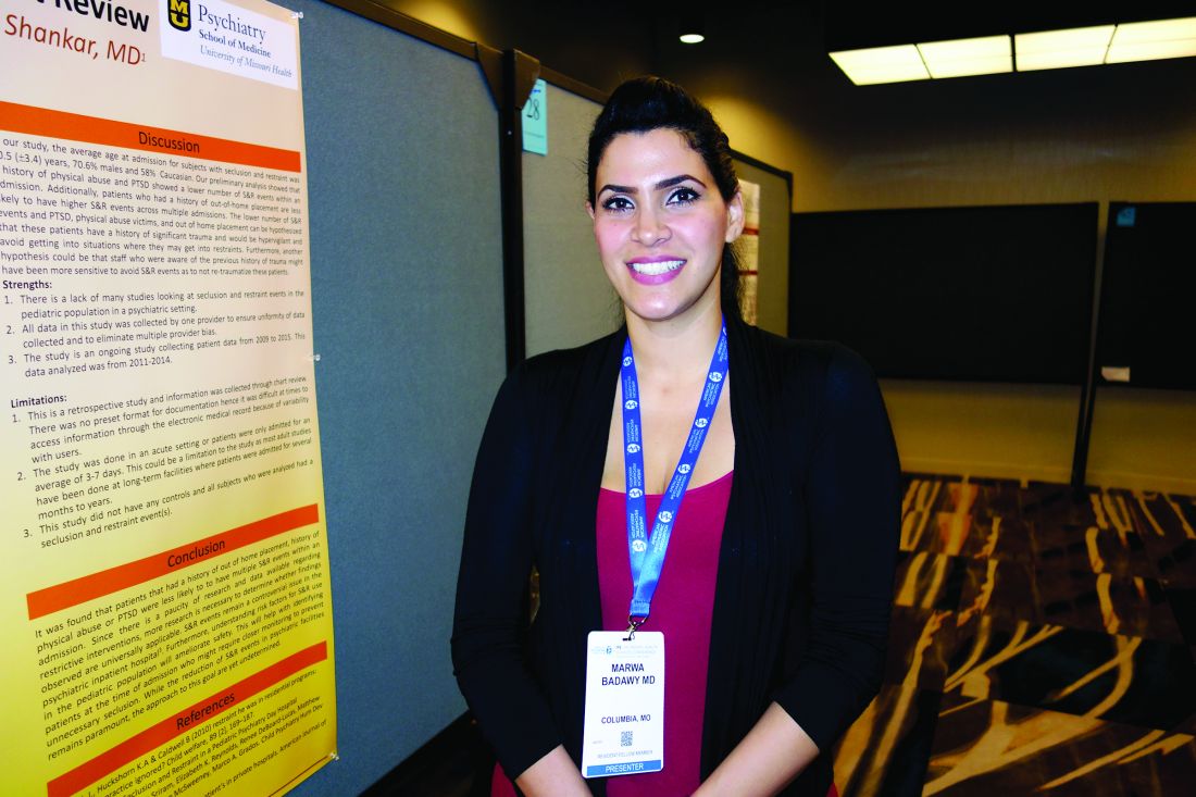
It’s well known that SR, a last-ditch effort to prevent physical harm, traumatizes patients, but research on how to avoid it has mostly focused on adults. The Missouri team wants to change that by identifying the children most at risk, so that something can be done beforehand to prevent it. Maybe extra one-on-one care would help, Dr. Badawy said.
The children in the review were an average of 10.5 years old, and most were admitted for 3-7 days. Fifty-five (60%) had multiple SR episodes, 34 in a single admission and 21 across multiple admissions; 71% of the episodes were in boys, 58% in white children, 27.7% in black children, and the rest in multiracial children. Twenty-one percent of the episodes were in children with intellectual problems. ADHD and oppositional defiant disorder were the most common diagnoses, each diagnosed in more than half of the subjects.
When asked how a 5-year-old child can end up in restraints, Dr. Badawy agreed that bad parenting is a factor. Parents who don’t know any better might escalate normal behavior, and others might simply not care that much about their kids, or have much empathy. “In the 5-10 year-olds, I do think a lot of it is parenting. Neglect is the number one form of abuse,” she said, “and it causes instability in children.”
But parents aren’t always the problem. Dr. Badawy mentioned an “extremely depressed” boy who tried to hang himself again and again, at 8 years old. “His parents were supportive and focused on getting him better,” she said. It’s unknown if anything happened to him when they weren’t around.
If SR is related to children’s own behavior, and if they are able to listen to staff afterward and control it, kids seem able to avoid another episode. Those who have PTSD, or who have been through physical abuse or out-of-home placement, seemed particularly adept in the review.
Avoidance seems less likely, however, when children are set off by other kids.
Sometimes there are clues of impending trouble, like the boy who walked around whistling before he lashed out. Staff at the university quickly learned to swoop in and calm him when they heard the whistling.
The investigators had no industry disclosures.
NEW ORLEANS – Attention-deficit/hyperactivity disorder and non-suicidal self-harm – cutting and head banging, for instance – strongly predicted longer seclusion and restraint episodes, sometimes past 2 hours, in a review at the University of Missouri, Columbia, pediatric inpatient psychiatric unit.
Meanwhile, children with histories of physical abuse, post-traumatic stress disorder (PTSD), or out-of-home placement were less likely to have multiple seclusion and restraint (SR) episodes during an admission, and had lower numbers of SR events overall. Perhaps hyper-vigilance due to past traumas helped them avoid situations that led to problems. Staff might also have used a lighter touch given the children’s histories.
The investigators reviewed 305 SR episodes from 2011-2014 among 92 children aged 5-18 years old. They plan to expand their study to 2009-2017 and add a prospective arm.
It’s well known that SR, a last-ditch effort to prevent physical harm, traumatizes patients, but research on how to avoid it has mostly focused on adults. The Missouri team wants to change that by identifying the children most at risk, so that something can be done beforehand to prevent it. Maybe extra one-on-one care would help, Dr. Badawy said.
The children in the review were an average of 10.5 years old, and most were admitted for 3-7 days. Fifty-five (60%) had multiple SR episodes, 34 in a single admission and 21 across multiple admissions; 71% of the episodes were in boys, 58% in white children, 27.7% in black children, and the rest in multiracial children. Twenty-one percent of the episodes were in children with intellectual problems. ADHD and oppositional defiant disorder were the most common diagnoses, each diagnosed in more than half of the subjects.
When asked how a 5-year-old child can end up in restraints, Dr. Badawy agreed that bad parenting is a factor. Parents who don’t know any better might escalate normal behavior, and others might simply not care that much about their kids, or have much empathy. “In the 5-10 year-olds, I do think a lot of it is parenting. Neglect is the number one form of abuse,” she said, “and it causes instability in children.”
But parents aren’t always the problem. Dr. Badawy mentioned an “extremely depressed” boy who tried to hang himself again and again, at 8 years old. “His parents were supportive and focused on getting him better,” she said. It’s unknown if anything happened to him when they weren’t around.
If SR is related to children’s own behavior, and if they are able to listen to staff afterward and control it, kids seem able to avoid another episode. Those who have PTSD, or who have been through physical abuse or out-of-home placement, seemed particularly adept in the review.
Avoidance seems less likely, however, when children are set off by other kids.
Sometimes there are clues of impending trouble, like the boy who walked around whistling before he lashed out. Staff at the university quickly learned to swoop in and calm him when they heard the whistling.
The investigators had no industry disclosures.
AT IPS 2017
Key clinical point:
Major finding: ADHD and non-suicidal self-harm – cutting and head banging – strongly predict longer seclusion and restraint episodes, sometimes past 2 hours.
Data source: A University of Missouri, Columbia, review of 305 seclusion and restraint episodes from 2011-2014 among 92 children aged 5-18 years old.
Disclosures: The investigators had no industry disclosures.
White House opioid panel calls for more addiction specialists
Increased addiction education, treatment certifications, and prescription drug monitoring program adherence were among the physician-focused recommendations in the final report of the President’s Commission on Combating Drug Addiction and the Opioid Crisis, released Nov. 1.
The report recommends more than 50 action points for President Trump and Congress to employ to address opioid and substance misuse, which the president declared a public health emergency.
Strategies focusing specifically on health providers targeted insufficient training and education regarding responsible prescribing, addiction prevention, and substance use disorder treatment.
“Estimates suggest there are currently about 4,400 actively practicing certified addiction specialist physicians (addiction medicine and addiction psychiatry) in the country, but data on the specialty workforce is limited,” according to the report. “About 8 years ago, an estimate was made of the need for 6,000 addiction specialists, but that number is now insufficient given the growth of the opioid epidemic.“
The report recommended increasing the number of medical schools accredited to offer addiction medicine fellowships from 46 to 125 over the next 5 years and recommends using funds allocated to the Health Resources and Services Administration by the 21st Century Cures Act to do so starting in 2018.
Increased counseling on addiction risk when patients receive an opioid prescription also is crucial, according to the report, which cited a survey in which only 36% of patients reported they were properly informed before or while taking their medication.
The low number of physicians holding waivers to dispense buprenorphine (medication-assisted treatment) was another source of concern, especially among rural communities.
“None of the large central metro counties, and 72% of the rural counties, did not have a waived physician,” according to the committee report, which cited registration data from the Drug Enforcement Administration. “Findings from focus groups of counselors in rural areas noted a dearth of good facilities, poor access due to clients living far away from treatment centers, reliance on friends or family for transportation, and a need for basic medical and dental services.”
The committee has advised increased outreach to physicians regarding buprenorphine prescribing waivers, although it acknowledged that with the cap on patients per waived physician, increasing the number of waived physicians “is not necessarily indicative of sufficient access.”
The report also stressed a need for physicians to adhere more strictly to opioid prescribing guidelines from the Centers for Disease Control & Prevention and regular registration of opioid prescriptions in state-level prescription drug monitoring programs (PDMPs).
“Providers often resist using PDMPs because these systems are not well integrated into the electronic health record systems they currently use in practice,” the report noted. “Simplifying the method of access to PDMPs for providers by integrating PDMP data into health information exchanges [may increase] the likelihood that prescription history information will be used in clinical decision-making.”
Despite the recommendation, the committee recommended $12 million be spent on restructuring the program in the fiscal year 2018 federal budget, $1 million less than in 2017.
The committee also encouraged CMS to revise payment policies, including some bundled payments, that currently disincentivize the use of nonopioid treatment options as well as remove pain management measurements from patient surveys.
CMS announced in 2016 it would work to revise patient surveys starting in January 2017.
The American Medical Association, in a statement, expressed support of the committee’s recommendations.
“The AMA commends The President’s Commission on Combating Drug Addiction and the Opioid Crisis for delivering a comprehensive report that provides an excellent road map for increasing access to medication-assisted treatment for patients with substance-use disorders and also demonstrates the need to eliminate barriers to accessing the full spectrum of multidisciplinary pain treatment options,” said Patrice A. Harris, MD, chair of the AMA opioid task force. “The AMA is pleased that the commission agreed with many of the AMA’s previous suggestions, and we look forward to working with the Administration and Congress on next steps, including the needed financial resources.”
The President’s Commission on Combating Drug Addition and the Opioid Crisis is comprised of Gov. Chris Christie (R-N.J.), chairman; Gov. Charlie Baker (R-Mass.); Gov. Roy Cooper (D-N.C.); former Rep. Patrick Kennedy (D-RI.); Bertha K. Madras, PhD, Harvard Medical School, Boston; and Attorney General Pam Bondi (R-Fla.).*
*This article was updated Nov. 8, 2017.