User login
Buprenorphine merits more attention for treatment of opioid use disorder
SAN DIEGO – Prescribing buprenorphine for the treatment of opioid use disorder requires strict discernment on the part of clinicians, Arwen Podesta, MD, said at the annual Psych Congress.

She encouraged clinicians to be prepared for a visit from the Drug Enforcement Administration, understand the unique properties of buprenorphine, and make sure that patients grasp the importance of sublingual administration.
Research shows that only 5% of physicians are allowed to prescribe buprenorphine – an opioid – by way of a DEA waiver, Dr. Podesta said. About half do not prescribe the drug. Barriers to prescribing buprenorphine include factors such as low reimbursement and untrained support staff, said Dr. Podesta, a board-certified psychiatrist who subspecializes in addiction medicine and practices in New Orleans.
But she noted that the Substance Abuse and Mental Health Services Administration has recommended that medication-assisted therapy (MAT) – methadone, buprenorphine, and naltrexone – be considered in all patients with opioid use disorder. The drugs are safe and effective when used correctly, the federal agency has said.
Remember, Dr. Podesta said, that “patients taking MAT are considered to be in recovery.” In the big picture, she added, “we have to improve access to care because we have so many people who don’t have access to treatment.”
Getting permission from the DEA to prescribe buprenorphine – a schedule III controlled substance – comes with a price, Dr. Podesta said. “We have special scrutiny from the DEA,” she said. They come in and want to see your records. It sounds very punitive, although it’s their jobs.”
The best approach is to document that you know what you’re doing, she said. “It’s your job to educate them about why you’re using buprenorphine and produce the records to show that.”
Being aware of buprenorphine’s unique properties is important, she said. The drug is safer on the overdose front than are other opioids, Dr. Podesta said, but it can be very dangerous in patients without opioid tolerance. According to the DEA, as an analgesic, buprenorphine is 20-30 times more potent than morphine. Also, like morphine, patients who take buprenorphine are likely to experience euphoria, papillary restriction, and respiratory depression and sedation.
The buprenorphine/naloxone formulation is preferred to treat opioid use disorder, she noted.
The reason that naloxone, which treats opioid overdoses, is part of the drug combo is because as an add-on, it reduces the risk that buprenorphine will be crushed and snorted for an opioid high, she said. Those who take the combo drug via that method could end up with sudden and nasty withdrawal symptoms.
When the drug combo is administered sublingually, the idea is that the “good stuff” (buprenorphine) is absorbed in the mouth, while the “bad stuff” (naloxone) is harmlessly absorbed in the gut, Dr. Podesta said. This happens because the drugs are absorbed differently.
But patients can mistakenly trigger symptoms of withdrawal if, for example, they put the combo drug on their tongue and then go to sleep. “That’s a peril,” she said, and it’s important to make sure patients know what to do – and what not to do.
Dr. Podesta emphasized the importance of choosing language related to patients with addictions carefully and respectfully.
“We have stigma,” she said. “We have been saying that patients are ‘dirty’ or ‘clean,’ and if they’re ‘clean,’ they’re the opposite of ‘dirty.’
She also suggested that clinicians drop the use of the word “contract” to describe treatment agreements between patients and clinicians. “Call it an ‘agreement,’ ” she said. “It seems more mutual and less punitive or risky for the patient to sign, especially when they’re in a precarious comfort zone.”
And consider that even the words “substance abuse” can be misleading, she said. “Many [patients] are taking the medications that the doctor prescribed and following instructions to the letter.”
Dr. Podesta disclosed consulting with Kaleo, Pear Therapeutics, and JayMac, and serving on the speakers bureau of Alkermes, Orexo, and US WorldMeds. She is the author of “Hooked: A Concise Guide to the Underlying Mechanics of Addiction and Treatment for Patients, Families, and Providers” (Dog Ear Publishing, 2016).
SAN DIEGO – Prescribing buprenorphine for the treatment of opioid use disorder requires strict discernment on the part of clinicians, Arwen Podesta, MD, said at the annual Psych Congress.
She encouraged clinicians to be prepared for a visit from the Drug Enforcement Administration, understand the unique properties of buprenorphine, and make sure that patients grasp the importance of sublingual administration.
Research shows that only 5% of physicians are allowed to prescribe buprenorphine – an opioid – by way of a DEA waiver, Dr. Podesta said. About half do not prescribe the drug. Barriers to prescribing buprenorphine include factors such as low reimbursement and untrained support staff, said Dr. Podesta, a board-certified psychiatrist who subspecializes in addiction medicine and practices in New Orleans.
But she noted that the Substance Abuse and Mental Health Services Administration has recommended that medication-assisted therapy (MAT) – methadone, buprenorphine, and naltrexone – be considered in all patients with opioid use disorder. The drugs are safe and effective when used correctly, the federal agency has said.
Remember, Dr. Podesta said, that “patients taking MAT are considered to be in recovery.” In the big picture, she added, “we have to improve access to care because we have so many people who don’t have access to treatment.”
Getting permission from the DEA to prescribe buprenorphine – a schedule III controlled substance – comes with a price, Dr. Podesta said. “We have special scrutiny from the DEA,” she said. They come in and want to see your records. It sounds very punitive, although it’s their jobs.”
The best approach is to document that you know what you’re doing, she said. “It’s your job to educate them about why you’re using buprenorphine and produce the records to show that.”
Being aware of buprenorphine’s unique properties is important, she said. The drug is safer on the overdose front than are other opioids, Dr. Podesta said, but it can be very dangerous in patients without opioid tolerance. According to the DEA, as an analgesic, buprenorphine is 20-30 times more potent than morphine. Also, like morphine, patients who take buprenorphine are likely to experience euphoria, papillary restriction, and respiratory depression and sedation.
The buprenorphine/naloxone formulation is preferred to treat opioid use disorder, she noted.
The reason that naloxone, which treats opioid overdoses, is part of the drug combo is because as an add-on, it reduces the risk that buprenorphine will be crushed and snorted for an opioid high, she said. Those who take the combo drug via that method could end up with sudden and nasty withdrawal symptoms.
When the drug combo is administered sublingually, the idea is that the “good stuff” (buprenorphine) is absorbed in the mouth, while the “bad stuff” (naloxone) is harmlessly absorbed in the gut, Dr. Podesta said. This happens because the drugs are absorbed differently.
But patients can mistakenly trigger symptoms of withdrawal if, for example, they put the combo drug on their tongue and then go to sleep. “That’s a peril,” she said, and it’s important to make sure patients know what to do – and what not to do.
Dr. Podesta emphasized the importance of choosing language related to patients with addictions carefully and respectfully.
“We have stigma,” she said. “We have been saying that patients are ‘dirty’ or ‘clean,’ and if they’re ‘clean,’ they’re the opposite of ‘dirty.’
She also suggested that clinicians drop the use of the word “contract” to describe treatment agreements between patients and clinicians. “Call it an ‘agreement,’ ” she said. “It seems more mutual and less punitive or risky for the patient to sign, especially when they’re in a precarious comfort zone.”
And consider that even the words “substance abuse” can be misleading, she said. “Many [patients] are taking the medications that the doctor prescribed and following instructions to the letter.”
Dr. Podesta disclosed consulting with Kaleo, Pear Therapeutics, and JayMac, and serving on the speakers bureau of Alkermes, Orexo, and US WorldMeds. She is the author of “Hooked: A Concise Guide to the Underlying Mechanics of Addiction and Treatment for Patients, Families, and Providers” (Dog Ear Publishing, 2016).
REPORTING FROM PSYCH CONGRESS 2019
Continuous NSAID use for ankylosing spondylitis may raise hypertension risk
Continuous use of NSAIDs could increase the risk of incident hypertension among patients with ankylosing spondylitis (AS), according to findings from a recent prospective study.

Against expectations, data also suggested that tumor necrosis factor inhibitor (TNFi) therapy could increase blood pressure, although this finding was not significant across all methods of analysis, reported lead author Jean W. Liew, MD, of the University of Washington, Seattle, and colleagues.
The investigators noted that patients with AS already have a greater risk of cardiovascular disease than that of the general population, making any added risks that much more concerning.
“[T]he evidence for increased cardiovascular disease burden and cardiovascular risk in patients with inflammatory rheumatic diseases is well recognized,” the investigators wrote in Arthritis Care & Research. “Multiple population-based studies have demonstrated increased cardiovascular events and cardiovascular-related mortality in AS. There is a high prevalence of cardiovascular risk factors among individuals with AS, particularly hypertension.”
Exacerbation of this risk by NSAIDs has been previously studied with mixed results, according to the investigators. Meta-analyses have suggested that NSAIDs increase blood pressure in normotensive and hypertensive individuals, but some data point to a cardioprotective effect among those with AS, possibly as a consequence of dampened inflammation, and/or improved physical activity, which could lead to a secondary CV benefit. Still, the relationship between NSAIDs and CV risk was unclear, prompting the current study.
The investigators enrolled 1,282 patients with AS at five centers in the United States and Australia. Using a combination of clinical evaluations and self-reporting, enrollees were monitored at regular intervals. Disease activity was tracked with the Bath Ankylosing Spondylitis Disease Activity Index (BASDAI), while the Bath Ankylosing Spondylitis Functional Index (BASFI) was used for functional impairments. Patients were also checked for a variety of comorbidities such as hypertension, coronary artery disease, mental health conditions, and renal disorders. Medication data included type, dosage, frequency, duration, and number of missed doses.
Including only baseline normotensive patients with at least 1 year of follow-up, 628 participants were eligible for analysis, of whom 200 used NSAIDs continuously. After a median of 7 years follow-up, 129 out of 628 patients developed hypertension. This translated to a hazard ratio (HR) for incident hypertension of 1.12 (95% confidence interval, 1.04-1.20), compared with nonusers or those who took NSAIDs intermittently. This relationship did not differ across subgroups defined by age, disease activity, body mass index, or TNFi use. Multiple sensitivity analyses added support to the association between continuous NSAID use and hypertension.
“The association of NSAIDs and incident hypertension remains particularly concerning, as the early development of hypertension may portend a higher risk of premature CV events due to cumulative exposure,” the investigators wrote.
In contrast with NSAIDs, TNFi therapy was not associated with hypertension across all models; however, against expectations, two models of analysis pointed to an 8% increased risk.
“Although TNFi use did not reach statistical significance in the main model, the direction of association was opposite that hypothesized based on prior data, specifically that TNFi use reduces CV risk by suppressing chronic inflammation,” the investigators wrote.
Considering the present findings, and previous studies, which have reported conflicting associations between TNFi use and hypertension, the investigators suggested that more research is needed, and offered specific methods to approach the topic.
“The association of TNFi use and incident hypertension requires further clarification in future studies,” they wrote, “which may be done by applying a marginal structural modeling (MSM) framework and inverse probability of treatment weighting (IPTW) statistical analyses to account for the relationships between TNFi use, disease activity, and NSAID use.”
In their concluding remarks, the investigators further emphasized the current knowledge gap in this area.
“There is an unmet need to clarify how treatment choices, particularly the use of NSAIDs and TNFi, impact CV risk factors and CV events in AS,” they wrote. “Further studies are needed to focus on precision medicine and predicting risk and benefit for patients in whom continuous NSAIDs are being considered. These further studies can inform the revision of guidelines to address the management of CV risk factors and CV disease in AS and axial spondyloarthritis more broadly.”
The investigators reported funding from the National Institutes of Health, the Assessment of Spondyloarthritis International Society, the Spondylitis Association of America, and the Russel Engleman Rheumatology Research Center at the University of California, San Francisco. Some authors reported ties with Eli Lilly, Novartis, and other pharmaceutical companies..
SOURCE: Liew et al. Arthritis Care Res. 2019 Sep 17. doi: 10.1002/acr.24070.
Continuous use of NSAIDs could increase the risk of incident hypertension among patients with ankylosing spondylitis (AS), according to findings from a recent prospective study.
Against expectations, data also suggested that tumor necrosis factor inhibitor (TNFi) therapy could increase blood pressure, although this finding was not significant across all methods of analysis, reported lead author Jean W. Liew, MD, of the University of Washington, Seattle, and colleagues.
The investigators noted that patients with AS already have a greater risk of cardiovascular disease than that of the general population, making any added risks that much more concerning.
“[T]he evidence for increased cardiovascular disease burden and cardiovascular risk in patients with inflammatory rheumatic diseases is well recognized,” the investigators wrote in Arthritis Care & Research. “Multiple population-based studies have demonstrated increased cardiovascular events and cardiovascular-related mortality in AS. There is a high prevalence of cardiovascular risk factors among individuals with AS, particularly hypertension.”
Exacerbation of this risk by NSAIDs has been previously studied with mixed results, according to the investigators. Meta-analyses have suggested that NSAIDs increase blood pressure in normotensive and hypertensive individuals, but some data point to a cardioprotective effect among those with AS, possibly as a consequence of dampened inflammation, and/or improved physical activity, which could lead to a secondary CV benefit. Still, the relationship between NSAIDs and CV risk was unclear, prompting the current study.
The investigators enrolled 1,282 patients with AS at five centers in the United States and Australia. Using a combination of clinical evaluations and self-reporting, enrollees were monitored at regular intervals. Disease activity was tracked with the Bath Ankylosing Spondylitis Disease Activity Index (BASDAI), while the Bath Ankylosing Spondylitis Functional Index (BASFI) was used for functional impairments. Patients were also checked for a variety of comorbidities such as hypertension, coronary artery disease, mental health conditions, and renal disorders. Medication data included type, dosage, frequency, duration, and number of missed doses.
Including only baseline normotensive patients with at least 1 year of follow-up, 628 participants were eligible for analysis, of whom 200 used NSAIDs continuously. After a median of 7 years follow-up, 129 out of 628 patients developed hypertension. This translated to a hazard ratio (HR) for incident hypertension of 1.12 (95% confidence interval, 1.04-1.20), compared with nonusers or those who took NSAIDs intermittently. This relationship did not differ across subgroups defined by age, disease activity, body mass index, or TNFi use. Multiple sensitivity analyses added support to the association between continuous NSAID use and hypertension.
“The association of NSAIDs and incident hypertension remains particularly concerning, as the early development of hypertension may portend a higher risk of premature CV events due to cumulative exposure,” the investigators wrote.
In contrast with NSAIDs, TNFi therapy was not associated with hypertension across all models; however, against expectations, two models of analysis pointed to an 8% increased risk.
“Although TNFi use did not reach statistical significance in the main model, the direction of association was opposite that hypothesized based on prior data, specifically that TNFi use reduces CV risk by suppressing chronic inflammation,” the investigators wrote.
Considering the present findings, and previous studies, which have reported conflicting associations between TNFi use and hypertension, the investigators suggested that more research is needed, and offered specific methods to approach the topic.
“The association of TNFi use and incident hypertension requires further clarification in future studies,” they wrote, “which may be done by applying a marginal structural modeling (MSM) framework and inverse probability of treatment weighting (IPTW) statistical analyses to account for the relationships between TNFi use, disease activity, and NSAID use.”
In their concluding remarks, the investigators further emphasized the current knowledge gap in this area.
“There is an unmet need to clarify how treatment choices, particularly the use of NSAIDs and TNFi, impact CV risk factors and CV events in AS,” they wrote. “Further studies are needed to focus on precision medicine and predicting risk and benefit for patients in whom continuous NSAIDs are being considered. These further studies can inform the revision of guidelines to address the management of CV risk factors and CV disease in AS and axial spondyloarthritis more broadly.”
The investigators reported funding from the National Institutes of Health, the Assessment of Spondyloarthritis International Society, the Spondylitis Association of America, and the Russel Engleman Rheumatology Research Center at the University of California, San Francisco. Some authors reported ties with Eli Lilly, Novartis, and other pharmaceutical companies..
SOURCE: Liew et al. Arthritis Care Res. 2019 Sep 17. doi: 10.1002/acr.24070.
FROM ARTHRITIS CARE & RESEARCH
Poll: Do you think that the electronic medical record has improved patient care?
[polldaddy:10424906]
[polldaddy:10424906]
[polldaddy:10424906]
Multigene testing for all patients with breast cancer may be cost-effective, life-saving
High-risk multigene testing for all patients with breast cancer may be a cost-effective way to reduce rates of breast cancer and ovarian cancer, according to investigators.
A microsimulation modeling study suggested that moving from standard BRCA testing based on clinical criteria or family history to widespread adoption of multigene testing could potentially save more than 2,400 lives per year in the United States, reported lead author Li Sun, MSc, of the London School of Hygiene & Tropical Medicine, and colleagues.
“Moving toward unselected BC testing may give an impetus for prevention in unaffected family members along with clinical implications for the patient with BC. Pathogenic variant carriers with newly diagnosed BC can opt for bilateral mastectomy rather than breast conservation at initial BC surgery,” the investigators wrote in JAMA Oncology. They noted that, presently, only 20%-30% of eligible patients are actually tested. “Testing all patients with breast cancer at diagnosis can increase testing access and uptake and identify many more pathogenic variant carriers for screening and prevention.”
To see if such a broad roll-out would be economically and medically feasible and beneficial, the investigators performed a modeling study involving 11,836 women with breast cancer, with data drawn from four large databases in the United Kingdom, the United States, and Australia.
The model compared annual costs, number of detected cases of breast cancer and ovarian cancer, and related rates of mortality between standard BRCA testing based on clinical criteria or family history versus high-risk multigene testing (BRCA1/BRCA2/PALB2) for all patients with breast cancer.
Resultant incremental cost-effectiveness ratios (ICERs) showed that in both the United Kingdom and the United States, multigene testing would cost, on average, significantly less than minimum willingness-to-pay thresholds of £30,000 per quality-adjusted life-year (QALY) and $100,000/QALY, respectively. In the United Kingdom, the estimated cost of multigene testing was £10,464/QALY from a payer’s perspective and £7,216/QALY from a societal perspective, the latter of which incorporates economic costs beyond the health care system. In the United States, these values rose to $65,661/QALY (payer perspective) and $61,618/QALY (societal perspective). Probabilistic sensitivity analysis suggested that multigene testing would be cost-effective for almost all health system simulations in the United Kingdom (98%-99%) and approximately two-thirds of those in the United States (64%-68%).
Epidemiologically, in the United Kingdom, multigene testing could potentially prevent 2,101 cases of breast cancer and ovarian cancer and 633 deaths per year, while in the United States, 9,733 cases might be prevented, with 2,406 lives saved.
“Our analysis suggests that an unselected testing strategy is extremely cost-effective for U.K. and U.S. health systems and provides a basis for change in current guidelines and policy to implement this strategy,” the investigators said.
The study was funded by the National Cancer Institute, the State of Washington, the Fred Hutchinson Cancer Research Center, and others. The investigators reported additional relationships with AstraZeneca, the Manchester National Institute for Health Research Biomedical Research Centre, Cancer Research UK, an others.
SOURCE: Sun et al. JAMA Oncol. 2019 Oct 3. doi: 10.1001/jamaoncol.2019.3323.
Although detection of more pathogenic variants among patients with breast cancer may initially appear beneficial, the hidden risks and costs associated with broader multigene testing would likely outweigh the benefits.
Present guidelines, such as those provided by the National Comprehensive Cancer Network, already include effective strategies for detecting BRCA1/2 variants, as only 0.6% of patients who do not meet screening guidelines are estimated to test positive
Beyond BRCA1/2, the clinical relevance of pathogenic variants becomes dubious, as many genetic aberrations are not actionable. Furthermore, misinterpretation of unfamiliar results may increase risks of treatment decisions that might actually harm patients. While some argue that broader testing could have benefits for family members, this is poorly supported with evidence.
Behind the allegedly falling costs of genetic sequencing, true costs are hidden with corporate policies that reduce patient financial liability. It is worth noting that two publicly held testing companies recently reported annual revenues of $144 million and $498 million, representing money drawn largely from third-party payers covering guideline-recommended testing. Instead of concrete science, the popularity of multigene testing is more likely driven by active marketing and technological convenience. More independent studies are needed.
Mark Robson, MD, is with the department of breast cancer medicine and clinical genetics at Memorial Sloan Kettering Cancer Center in New York. Susan Domchek, MD is with the Basser Center for BRCA at the University of Pennsylvania in Philadelphia. Dr. Robson disclosed financial relationships with AstraZeneca, McKesson, and Pfizer. Dr. Domchek reported personal fees from Clovis, AstraZeneca, and BMS. Their remarks are adapted from an editorial (JAMA Oncol. 2019 Oct 3. doi: 10.1001/jamaoncol.2019.4004).
High-risk multigene testing for all patients with breast cancer may be a cost-effective way to reduce rates of breast cancer and ovarian cancer, according to investigators.
A microsimulation modeling study suggested that moving from standard BRCA testing based on clinical criteria or family history to widespread adoption of multigene testing could potentially save more than 2,400 lives per year in the United States, reported lead author Li Sun, MSc, of the London School of Hygiene & Tropical Medicine, and colleagues.
“Moving toward unselected BC testing may give an impetus for prevention in unaffected family members along with clinical implications for the patient with BC. Pathogenic variant carriers with newly diagnosed BC can opt for bilateral mastectomy rather than breast conservation at initial BC surgery,” the investigators wrote in JAMA Oncology. They noted that, presently, only 20%-30% of eligible patients are actually tested. “Testing all patients with breast cancer at diagnosis can increase testing access and uptake and identify many more pathogenic variant carriers for screening and prevention.”
To see if such a broad roll-out would be economically and medically feasible and beneficial, the investigators performed a modeling study involving 11,836 women with breast cancer, with data drawn from four large databases in the United Kingdom, the United States, and Australia.
The model compared annual costs, number of detected cases of breast cancer and ovarian cancer, and related rates of mortality between standard BRCA testing based on clinical criteria or family history versus high-risk multigene testing (BRCA1/BRCA2/PALB2) for all patients with breast cancer.
Resultant incremental cost-effectiveness ratios (ICERs) showed that in both the United Kingdom and the United States, multigene testing would cost, on average, significantly less than minimum willingness-to-pay thresholds of £30,000 per quality-adjusted life-year (QALY) and $100,000/QALY, respectively. In the United Kingdom, the estimated cost of multigene testing was £10,464/QALY from a payer’s perspective and £7,216/QALY from a societal perspective, the latter of which incorporates economic costs beyond the health care system. In the United States, these values rose to $65,661/QALY (payer perspective) and $61,618/QALY (societal perspective). Probabilistic sensitivity analysis suggested that multigene testing would be cost-effective for almost all health system simulations in the United Kingdom (98%-99%) and approximately two-thirds of those in the United States (64%-68%).
Epidemiologically, in the United Kingdom, multigene testing could potentially prevent 2,101 cases of breast cancer and ovarian cancer and 633 deaths per year, while in the United States, 9,733 cases might be prevented, with 2,406 lives saved.
“Our analysis suggests that an unselected testing strategy is extremely cost-effective for U.K. and U.S. health systems and provides a basis for change in current guidelines and policy to implement this strategy,” the investigators said.
The study was funded by the National Cancer Institute, the State of Washington, the Fred Hutchinson Cancer Research Center, and others. The investigators reported additional relationships with AstraZeneca, the Manchester National Institute for Health Research Biomedical Research Centre, Cancer Research UK, an others.
SOURCE: Sun et al. JAMA Oncol. 2019 Oct 3. doi: 10.1001/jamaoncol.2019.3323.
FROM JAMA ONCOLOGY
Tennessee’s Medicaid block grant proposal could have deep impacts
In late September, Gov. Bill Lee (R) submitted a draft proposal to the Trump administration requesting the government convert the federal share of the state’s Medicaid funding into a $7.9 billion lump sum.
Under the proposal, the federal spending cap would apply to currently eligible children and adults, patients covered based on disability, and currently eligible seniors, but not to newly eligible patients, prescription drugs, dually eligible enrollees, and reimbursement to disproportionate-share hospitals and critical access hospitals. The proposed block grant would increase on a per capita basis for new patients and would adjust annually for inflation.
Gov. Lee contends the block grant would allow for more efficiency in the state’s Medicaid program – called TennCare – and generate significant savings, which the state would split 50/50 with the federal government, according to the proposal.
“The traditional model of Medicaid financing is an outdated model of fundamentally misaligned incentives,” Gov. Lee said in the waiver request. “New models of Medicaid financing are needed that reward states for promoting value and health, not merely spending more money. Tennessee’s Medicaid block grant proposal represents a natural progression of the state’s history of nationally recognized innovation and financial management. It also ensures that TennCare members continue to receive high-quality, cost-effective care well into the future.”

Tennessee’s proposal aims to provide care to low-income recipients more efficiently, while breaking Medicaid’s “perverse dynamic” in which states seek to extract more money from the federal government, said Doug Badger, a visiting fellow in domestic policy studies at the conservative-leaning Heritage Foundation.
“The waiver would incentivize Tennessee to reduce federal spending while maintaining access to care,” Mr. Badger said in an interview. “It is an idea worth testing. Tennessee will have to convince federal officials that its proposal would maintain quality of care without increasing federal spending. If they clear those hurdles, the federal government should approve the waiver.”
However, Dan Hawkins, senior vice president for public policy and research at the nonpartisan National Association of Community Health Centers said the proposal contains some concerning omissions and extreme demands that could affect access to care. For instance, the waiver calls for complete exemption from federal Medicaid requirements, including managed care rules that ensure access and network adequacy protections, pregnancy care rules, requirements pertaining to the treatment of disabled adults and children, and early and periodic screening, diagnostic, and treatment (EPSDT) requirements. Under the waiver, the state would no longer be subject to federal oversight for its enrollment, coverage, or management decisions.
“The most shocking thing is that [Tennessee has] provided no estimate of impact on the population served, either in terms of their health or their ability to access care or the amount of money it would cost to serve them,” Mr. Hawkins said in an interview. “They are essentially saying, ‘Send [the block grant] to us and go away.’ ”

Without the federal protections, Tennessee could take any number of measures to reduce costs, adds Andy Schneider a research professor at Georgetown University and an analyst for Georgetown University Health Policy Institute’s Center for Children and Families, both in Washington. Mr. Schneider served as a senior advisor at the Centers for Medicare & Medicaid Services under the Obama administration.
“What would the state do in a world where it no longer had to follow federal rules in contracting with risk-based managed care organizations [MCOs] and in a world where it wanted to save money?” Mr. Schneider said in an interview. “Well, without the federal protections, it could reduce its payments on behalf of enrolled beneficiaries to the MCOs, it could not require the MCOs to have adequate provider networks, not require the MCOs to describe what services they were or were not providing, it could eliminate appeals protections for beneficiaries. Just let your imagination run wild.”
It is uncertain whether the waiver will be approved. Mr. Hawkins noted that CMS leadership has expressed support for the notion of a block grant. In his 2020 fiscal year federal budget for example, President Trump proposed a number of changes to Medicaid, including the potential for state Medicaid block grants or per capita caps. The Office of Management and Budget, meanwhile, is currently preparing guidance for state governments on creating block grants for Medicaid.
“On the other hand, this proposal is so outrageous I have to believe that even the federal government, even CMS, would blanch and come back requiring some additional changes in return for approving the proposal,” Mr. Hawkins said.
Mr. Schneider believes if the block grant is approved, it will likely face immediate litigation.
“The Department of Health & Human Services does not have the authority to give the state a block grant, only Congress can do that,” said Mr. Schneider who recently blogged about the proposal. “The reason for that is that, as the state makes clear in its proposal, it wants to reimagine the traditional Medicaid financing arrangement. That traditional financing arrangement has been in place since 1965. The executive branch can’t [change] it. It simply does not have the authority.”
Mr. Badger emphasized there is no shortage of speculation about the impact of fixed Medicaid allotments. What is needed now, he said, is evidence.
“A well-designed demonstration project will yield that evidence,” he said. “The statute gives the [HHS] Secretary authority to authorize demonstrations precisely to replace speculation with data. The best way to assess the impact of a fixed allotment is to test it in the real world.”
Tennessee will hold public hearings about its proposal in early October and collect feedback through Oct. 18. A final proposal is expected in November.
In late September, Gov. Bill Lee (R) submitted a draft proposal to the Trump administration requesting the government convert the federal share of the state’s Medicaid funding into a $7.9 billion lump sum.
Under the proposal, the federal spending cap would apply to currently eligible children and adults, patients covered based on disability, and currently eligible seniors, but not to newly eligible patients, prescription drugs, dually eligible enrollees, and reimbursement to disproportionate-share hospitals and critical access hospitals. The proposed block grant would increase on a per capita basis for new patients and would adjust annually for inflation.
Gov. Lee contends the block grant would allow for more efficiency in the state’s Medicaid program – called TennCare – and generate significant savings, which the state would split 50/50 with the federal government, according to the proposal.
“The traditional model of Medicaid financing is an outdated model of fundamentally misaligned incentives,” Gov. Lee said in the waiver request. “New models of Medicaid financing are needed that reward states for promoting value and health, not merely spending more money. Tennessee’s Medicaid block grant proposal represents a natural progression of the state’s history of nationally recognized innovation and financial management. It also ensures that TennCare members continue to receive high-quality, cost-effective care well into the future.”
Tennessee’s proposal aims to provide care to low-income recipients more efficiently, while breaking Medicaid’s “perverse dynamic” in which states seek to extract more money from the federal government, said Doug Badger, a visiting fellow in domestic policy studies at the conservative-leaning Heritage Foundation.
“The waiver would incentivize Tennessee to reduce federal spending while maintaining access to care,” Mr. Badger said in an interview. “It is an idea worth testing. Tennessee will have to convince federal officials that its proposal would maintain quality of care without increasing federal spending. If they clear those hurdles, the federal government should approve the waiver.”
However, Dan Hawkins, senior vice president for public policy and research at the nonpartisan National Association of Community Health Centers said the proposal contains some concerning omissions and extreme demands that could affect access to care. For instance, the waiver calls for complete exemption from federal Medicaid requirements, including managed care rules that ensure access and network adequacy protections, pregnancy care rules, requirements pertaining to the treatment of disabled adults and children, and early and periodic screening, diagnostic, and treatment (EPSDT) requirements. Under the waiver, the state would no longer be subject to federal oversight for its enrollment, coverage, or management decisions.
“The most shocking thing is that [Tennessee has] provided no estimate of impact on the population served, either in terms of their health or their ability to access care or the amount of money it would cost to serve them,” Mr. Hawkins said in an interview. “They are essentially saying, ‘Send [the block grant] to us and go away.’ ”
Without the federal protections, Tennessee could take any number of measures to reduce costs, adds Andy Schneider a research professor at Georgetown University and an analyst for Georgetown University Health Policy Institute’s Center for Children and Families, both in Washington. Mr. Schneider served as a senior advisor at the Centers for Medicare & Medicaid Services under the Obama administration.
“What would the state do in a world where it no longer had to follow federal rules in contracting with risk-based managed care organizations [MCOs] and in a world where it wanted to save money?” Mr. Schneider said in an interview. “Well, without the federal protections, it could reduce its payments on behalf of enrolled beneficiaries to the MCOs, it could not require the MCOs to have adequate provider networks, not require the MCOs to describe what services they were or were not providing, it could eliminate appeals protections for beneficiaries. Just let your imagination run wild.”
It is uncertain whether the waiver will be approved. Mr. Hawkins noted that CMS leadership has expressed support for the notion of a block grant. In his 2020 fiscal year federal budget for example, President Trump proposed a number of changes to Medicaid, including the potential for state Medicaid block grants or per capita caps. The Office of Management and Budget, meanwhile, is currently preparing guidance for state governments on creating block grants for Medicaid.
“On the other hand, this proposal is so outrageous I have to believe that even the federal government, even CMS, would blanch and come back requiring some additional changes in return for approving the proposal,” Mr. Hawkins said.
Mr. Schneider believes if the block grant is approved, it will likely face immediate litigation.
“The Department of Health & Human Services does not have the authority to give the state a block grant, only Congress can do that,” said Mr. Schneider who recently blogged about the proposal. “The reason for that is that, as the state makes clear in its proposal, it wants to reimagine the traditional Medicaid financing arrangement. That traditional financing arrangement has been in place since 1965. The executive branch can’t [change] it. It simply does not have the authority.”
Mr. Badger emphasized there is no shortage of speculation about the impact of fixed Medicaid allotments. What is needed now, he said, is evidence.
“A well-designed demonstration project will yield that evidence,” he said. “The statute gives the [HHS] Secretary authority to authorize demonstrations precisely to replace speculation with data. The best way to assess the impact of a fixed allotment is to test it in the real world.”
Tennessee will hold public hearings about its proposal in early October and collect feedback through Oct. 18. A final proposal is expected in November.
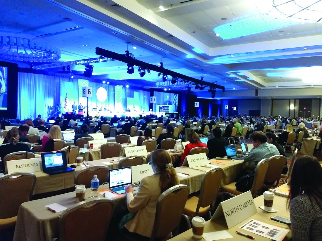
AAFP Congress adopts resolutions on physician privileges, medical education, employee benefits
PHILADELPHIA –
Practice enhancement
Hospital privileges were a hot topic for the reference committee on practice enhancement.
Adopted Resolution No. 304 calls on AAFP to oppose health insurance companies “privileging physicians based solely on their hospital privileges and hospital credentials.” The new rule also resolves that AAFP engage major national health insurance companies to develop methods to credential physicians that do not depend on hospital privileges.
The Congress also adopted Substitute Resolution No. 305, which calls on the AAFP to collaborate with the Joint Commission and other appropriate entities to create policy stating that hospitals remove undue barriers and restriction of privileges to hospitals and intensive care units for qualified family physicians who practice hospital medicine.
Delegates requested amendments to a resolution that called on AAFP to oppose nonphysician health care professionals making credentialing or privileging decisions regarding family physicians and that the AAFP oppose the use of nonphysician health care professionals in providing consultations requested of other physicians. The Congress could not agree on a final wording of the resolution.
Douglas J. Gruenbacher, MD, a Kansas delegate who works in a small hospital, said, “We actually credential in our hospital radiologists, orthopedists ... urologists. Do I know what they know? So we have to have many of our nonphysician providers, our nurse practitioners, help us. ... Should they be independent? No, of course not, but they do play an important part of our team.”
Douglas W. Curran, MD, a delegate from Texas, said, “I think we continue to give away stuff without taking care of ourselves. I have seen it for 4 years and, as result, we have seen this expansion of second-class care for people. ... Those are huge decisions, especially thinking about who’s going to do what in our hospitals. That includes small hospitals; I practice in a small hospital. I get all of that.”
After much debate, the Congress voted in favor of referring two proposed amendments to the board.
The Congress also adopted an amended version of Substitute Resolution No. 303, which calls on AAFP to support insurance coverage of acupuncture for pain control when ordered by a licensed physician or licensed collaborating advanced clinician on their practice team.
Education
Multipronged Substitute Resolution No. 606 – adopted by the Congress – aims to address racial inequities in medical education. Specifically, it calls on AAFP to do the following:
- Instruct the Liaison Committee for Medical Education to add race to its existing Cultural Competence and Health Care Disparities section 7.6 of Functions and Structure of a Medical School: Standards for Accreditation of Medical Education Programs Leading to the MD Degree.
- Ask the Accreditation Council for Graduate Medical Education to adopt an antiracism policy that includes corresponding curricular requirements,
- Develop and implement a policy on training in racism and implicit bias for officeholders and commission members.
- Take an active stance against racism when racist events occur within the medical community.
The Congress also adopted Resolution No. 611, which calls for the AAFP to encourage the expansion of clinical behavioral health fellowships for family medicine physicians.
The resolution received mixed testimony during the reference committee meeting, with those in favor of the resolution having cited the need for more education in behavioral health due to shortages in many communities. Opponents argued that completing the fellowship would not have added value in hospital privileging and insurance payment, because it would not lead physicians to earn a certificate of added qualification.
Delegates also passed Resolution 608, over the objections of the reference committee.
As adopted, the resolution calls for AAFP to express its concern that the American Board of Family Medicine (ABFM) Family Medicine Certification Longitudinal Assessment is the only alternative to 1-day-only certification exam, and for the AAFP to urge the ABFM to offer a longitudinal self-assessment process similar to the American Board of Obstetricians and Gynecologists self-assessment process to satisfy the cognitive component of ABFM’s continued certification requirement.
The Reference Committee on Education also referred several hotly debated resolutions back to the AAFP board of directors. No. 604 called on AAFP to support forgiving 1 year of federal medical student loans for every 2 years of full-time work in a primary care position, as well as tax credits for those working in rural or underserved areas.
Advocacy
The delegates also approved most of the recommendations of the reference committee on advocacy with little discussion.
Substitute Resolution No. 515, which was ultimately adopted with an amendment by the Congress, states that AAFP support policies that provide employees with reasonable benefits, including job security, wage replacement, and continued availability of health plan coverage in the event that leave by an employee becomes necessary for documented medical conditions, with protections for small businesses. Among the policies this resolution includes are the following:
- Medical leave for the employee, including pregnancy.
- Parental leave for the employee-parent, including leave for birth, adoption or foster care leading to adoption.
- Leave if medically appropriate to care for a member of the employee’s immediate family.
The Congress adopted several other resolutions recommended by the advocacy committee:
- Resolution No. 501, calling on AAFP to advocate for state-level adoption of the Interstate Medical Licensure Compact.
- Substitute Resolution No. 505, asking AAFP to request a National Coverage Determination for Cardiac Rehabilitation Programs to allow such programs to operate without physician supervision when an AED is immediately available, and the patient is attended by nursing staff currently trained in basic life support.
- Substitute Resolutions No. 506 and 507 to support and encourage the ability of parents to breastfeed in the workplace through its advocacy efforts, as well as promote the enforcements of current law.
- Resolution No. 508, to petition CMS, national health insurance companies, and pharmacy benefits managers to include all generic medications in a class within a health plan’s formulary and implement a system that informs the prescriber of all formulary alternatives to a medication when denying the same medication immediately upon denial, while also providing a mechanism to rapidly appeal the denial.
- Substitute Resolution No. 512, to petition the CMS to reevaluate its current policy on the time requirements for discharge summaries from hospitals and post-acute care facilities and specifically require such facilities to provide primary care physicians with discharge summaries within 7 days.
- Substitute Resolution No. 517, to unequivocally support the right of physicians to organize and bargain collectively.
- Substitute Resolution No. 519, to support legislation that decriminalizes people who are solicited for sex or sexual activities in exchange for money or goods, without supporting the legalization of the selling of sex, and advocate against legislation that decriminalizes sex-buying and third-parties who promote and/or profit from sex buying.
Some of the resolutions that incited many passionate responses during the reference committee on advocacy were not discussed during the Congress of Delegates meeting.
One of these asked the AAFP to oppose legislation of physician-patient decision making in child and adolescent gender-affirming care. Some in support of this resolution referred to this type of care as evidence-based medicine and said that legislators should be kept out of the exam room. Those opposed disagreed with classifying this type of care that way, noting that the long-term effects of some of the treatments are unknown.

During the reference committee, one opponent of the resolution, Lisa Gilbert, MD, claimed that gender-affirming care refers to blocking puberty, followed by cross-sex hormones,which would permanently sterilize the children. Dr. Gilbert, who identified herself as a member from Kansas speaking independently, added that if children have gone through puberty naturally, this would be a different discussion.
Kevin Wang, MD, an alternate delegate from Washington, who supported the resolution, noted that the rate of suicide in the transgender population is nine times that of the general population.
“I do want to emphasize that doing nothing does cause significant harm,” Dr. Wang said, The committee referred resolution No. 509 to the board for further clarification and study.
The merits of two other resolutions (No. 510 and No. 511), which called for the AAFP to no longer reject the use of “physician-assisted suicide” and “assisted suicide” and avoid the use of vague and euphemistic terms when referring to lethal medications prescribed with the intention of ending a patient’s life in statements and documents, also were heavily debated during the advocacy reference committee meeting. The committee recommended such resolutions be referred to the board for discussion.
The delegates approved the advocacy committee’s recommendations for Resolutions 509, 510, and 511.
PHILADELPHIA –
Practice enhancement
Hospital privileges were a hot topic for the reference committee on practice enhancement.
Adopted Resolution No. 304 calls on AAFP to oppose health insurance companies “privileging physicians based solely on their hospital privileges and hospital credentials.” The new rule also resolves that AAFP engage major national health insurance companies to develop methods to credential physicians that do not depend on hospital privileges.
The Congress also adopted Substitute Resolution No. 305, which calls on the AAFP to collaborate with the Joint Commission and other appropriate entities to create policy stating that hospitals remove undue barriers and restriction of privileges to hospitals and intensive care units for qualified family physicians who practice hospital medicine.
Delegates requested amendments to a resolution that called on AAFP to oppose nonphysician health care professionals making credentialing or privileging decisions regarding family physicians and that the AAFP oppose the use of nonphysician health care professionals in providing consultations requested of other physicians. The Congress could not agree on a final wording of the resolution.
Douglas J. Gruenbacher, MD, a Kansas delegate who works in a small hospital, said, “We actually credential in our hospital radiologists, orthopedists ... urologists. Do I know what they know? So we have to have many of our nonphysician providers, our nurse practitioners, help us. ... Should they be independent? No, of course not, but they do play an important part of our team.”
Douglas W. Curran, MD, a delegate from Texas, said, “I think we continue to give away stuff without taking care of ourselves. I have seen it for 4 years and, as result, we have seen this expansion of second-class care for people. ... Those are huge decisions, especially thinking about who’s going to do what in our hospitals. That includes small hospitals; I practice in a small hospital. I get all of that.”
After much debate, the Congress voted in favor of referring two proposed amendments to the board.
The Congress also adopted an amended version of Substitute Resolution No. 303, which calls on AAFP to support insurance coverage of acupuncture for pain control when ordered by a licensed physician or licensed collaborating advanced clinician on their practice team.
Education
Multipronged Substitute Resolution No. 606 – adopted by the Congress – aims to address racial inequities in medical education. Specifically, it calls on AAFP to do the following:
- Instruct the Liaison Committee for Medical Education to add race to its existing Cultural Competence and Health Care Disparities section 7.6 of Functions and Structure of a Medical School: Standards for Accreditation of Medical Education Programs Leading to the MD Degree.
- Ask the Accreditation Council for Graduate Medical Education to adopt an antiracism policy that includes corresponding curricular requirements,
- Develop and implement a policy on training in racism and implicit bias for officeholders and commission members.
- Take an active stance against racism when racist events occur within the medical community.
The Congress also adopted Resolution No. 611, which calls for the AAFP to encourage the expansion of clinical behavioral health fellowships for family medicine physicians.
The resolution received mixed testimony during the reference committee meeting, with those in favor of the resolution having cited the need for more education in behavioral health due to shortages in many communities. Opponents argued that completing the fellowship would not have added value in hospital privileging and insurance payment, because it would not lead physicians to earn a certificate of added qualification.
Delegates also passed Resolution 608, over the objections of the reference committee.
As adopted, the resolution calls for AAFP to express its concern that the American Board of Family Medicine (ABFM) Family Medicine Certification Longitudinal Assessment is the only alternative to 1-day-only certification exam, and for the AAFP to urge the ABFM to offer a longitudinal self-assessment process similar to the American Board of Obstetricians and Gynecologists self-assessment process to satisfy the cognitive component of ABFM’s continued certification requirement.
The Reference Committee on Education also referred several hotly debated resolutions back to the AAFP board of directors. No. 604 called on AAFP to support forgiving 1 year of federal medical student loans for every 2 years of full-time work in a primary care position, as well as tax credits for those working in rural or underserved areas.
Advocacy
The delegates also approved most of the recommendations of the reference committee on advocacy with little discussion.
Substitute Resolution No. 515, which was ultimately adopted with an amendment by the Congress, states that AAFP support policies that provide employees with reasonable benefits, including job security, wage replacement, and continued availability of health plan coverage in the event that leave by an employee becomes necessary for documented medical conditions, with protections for small businesses. Among the policies this resolution includes are the following:
- Medical leave for the employee, including pregnancy.
- Parental leave for the employee-parent, including leave for birth, adoption or foster care leading to adoption.
- Leave if medically appropriate to care for a member of the employee’s immediate family.
The Congress adopted several other resolutions recommended by the advocacy committee:
- Resolution No. 501, calling on AAFP to advocate for state-level adoption of the Interstate Medical Licensure Compact.
- Substitute Resolution No. 505, asking AAFP to request a National Coverage Determination for Cardiac Rehabilitation Programs to allow such programs to operate without physician supervision when an AED is immediately available, and the patient is attended by nursing staff currently trained in basic life support.
- Substitute Resolutions No. 506 and 507 to support and encourage the ability of parents to breastfeed in the workplace through its advocacy efforts, as well as promote the enforcements of current law.
- Resolution No. 508, to petition CMS, national health insurance companies, and pharmacy benefits managers to include all generic medications in a class within a health plan’s formulary and implement a system that informs the prescriber of all formulary alternatives to a medication when denying the same medication immediately upon denial, while also providing a mechanism to rapidly appeal the denial.
- Substitute Resolution No. 512, to petition the CMS to reevaluate its current policy on the time requirements for discharge summaries from hospitals and post-acute care facilities and specifically require such facilities to provide primary care physicians with discharge summaries within 7 days.
- Substitute Resolution No. 517, to unequivocally support the right of physicians to organize and bargain collectively.
- Substitute Resolution No. 519, to support legislation that decriminalizes people who are solicited for sex or sexual activities in exchange for money or goods, without supporting the legalization of the selling of sex, and advocate against legislation that decriminalizes sex-buying and third-parties who promote and/or profit from sex buying.
Some of the resolutions that incited many passionate responses during the reference committee on advocacy were not discussed during the Congress of Delegates meeting.
One of these asked the AAFP to oppose legislation of physician-patient decision making in child and adolescent gender-affirming care. Some in support of this resolution referred to this type of care as evidence-based medicine and said that legislators should be kept out of the exam room. Those opposed disagreed with classifying this type of care that way, noting that the long-term effects of some of the treatments are unknown.
During the reference committee, one opponent of the resolution, Lisa Gilbert, MD, claimed that gender-affirming care refers to blocking puberty, followed by cross-sex hormones,which would permanently sterilize the children. Dr. Gilbert, who identified herself as a member from Kansas speaking independently, added that if children have gone through puberty naturally, this would be a different discussion.
Kevin Wang, MD, an alternate delegate from Washington, who supported the resolution, noted that the rate of suicide in the transgender population is nine times that of the general population.
“I do want to emphasize that doing nothing does cause significant harm,” Dr. Wang said, The committee referred resolution No. 509 to the board for further clarification and study.
The merits of two other resolutions (No. 510 and No. 511), which called for the AAFP to no longer reject the use of “physician-assisted suicide” and “assisted suicide” and avoid the use of vague and euphemistic terms when referring to lethal medications prescribed with the intention of ending a patient’s life in statements and documents, also were heavily debated during the advocacy reference committee meeting. The committee recommended such resolutions be referred to the board for discussion.
The delegates approved the advocacy committee’s recommendations for Resolutions 509, 510, and 511.
REPORTING FROM AAFP Congress
Ribociclib/fulvestrant boosts survival in advanced breast cancer
BARCELONA – In postmenopausal women with hormone receptor–positive, HER2-negative advanced breast cancer, the combination of ribociclib (Kisqali) and fulvestrant (Faslodex) was associated with a significant survival benefit, compared with fulvestrant alone, investigators reported.

Among 726 postmenopausal patients in MONALEESA-3 randomly assigned to receive either ribociclib or placebo plus fulvestrant who were followed for a median of 39.4 months, the median overall survival (OS) was not reached for patients assigned to ribociclib, compared with 40 months for patients in the control (fulvestrant-only) arm, reported Dennis J. Slamon, MD, PhD from the University of California, Los Angeles
“The combined data set of MONALEESA-3 and MONALEESA-7 represent some 1,400 patients, and the largest body of evidence for overall survival for any [cyclin-dependent kinases 4/6] inhibitor, and these data demonstrate a consistent, meaningful prolongation of survival irrespective of menopausal status, pre or post, and irrespective of whether the patient is treated in the first line or the second line,” he said at the European Society for Medical Oncology Congress.
Results of the MONALEESA-7 trial, which showed an overall survival advantage for adding ribociclib to endocrine therapy in premenopausal women with hormone receptor–positive, HER2-negative breast cancer, were reported at the 2019 annual meeting of the American Society of Clinical Oncology.
MONALEESA-3 investigators enrolled 726 postmenopausal women with hormone receptor–positive/HER2-negative advanced breast cancer who had received not more than one prior line of endocrine therapy for advanced disease, stratified them by the presence of liver and/or lung metastases and prior endocrine therapy, and then randomly assigned them to receive intramuscular fulvestrant 500 mg every 28 days, with an additional dose on day 15 of cycle one, plus either ribociclib 600 mg per day, 3 weeks on and 1 week off for every cycle, or placebo.
Dr. Slamon presented results of the primary endpoint of progression-free survival at the 2018 ASCO annual meeting, which showed a median PFS with ribociclib plus fulvestrant of 20.5 months versus 12.8 months for fulvestrant alone (hazard ratio, 5.93; P less than .0001). The overall survival data presented by Dr. Slamon at ESMO 2019 were not mature at that time.
As noted, at a median follow-up of 39.4 months, median overall survival was not reached in the combination arm, compared with 40 months in the fulvestrant/placebo arm, translating into a HR for risk of death with ribociclib of 0.724 (P = .00455).
Estimated OS rates at 36 months were 67% in the ribociclib arm versus 58.2% in the placebo arm, and at 42 months were 57.8% and 45.9%, respectively.
The survival benefit appeared consistent both for patients treated in the first line (median OS not reached vs. 45.1 months; HR, 0.70) and for those treated after early relapse of therapy for early breast or in the second line (median OS, 40.2 vs. 32.5 months, respectively; HR, 0.73). In both comparisons, however, the 95% confidence interval crossed 1, indicating that the results were not statistically significant.
An analysis of OS by subgroup indicated significant benefit or trends for most categories except Asian race, and treatment in Asia or Latin America, although the numbers of patients in each of those subgroups was less than 20, making it difficult to draw significant inferences from the findings, Dr. Slamon cautioned.
A descriptive analysis of updated PFS results were very similar to those previously reported, with a median of 20.6 months with ribociclib versus 12.8 months with placebo (HR, 0.587). Median PFS by line of therapy was 33.6 versus 19.2 months, respectively, in the first line, and 14.5 versus 9.1 months in the second line (HR, 0.546 and 0.571; P values not shown).

“CDK4/6 inhibitors not only improve progression-free survival in first- and second-line metastatic breast cancer, it also translates into an overall survival improvement. What more do we really want?” said invited discussant Sybil Loibl, MD, from Goethe University in Frankfurt, Germany.
“Outcome improves irrespective of pretreatment, menopausal status, endocrine sensitivity and site of metastases, which is good knowledge for our patients,” she said.
To date there are no biomarkers identified that can select subgroups that could experience particular benefit from CDK4/6 inhibitors. It may require a meta-analysis of all available clinical trial data on these agents in both the first and second line therapy to reveal potential differences among subgroups, she said.
MONALEESA-3 is supported by Novartis. Dr. Slamon disclosed a consulting or advisory role, research funding, honoraria, and travel expenses from Novartis and others. Dr. Loibl disclosed honoraria and research grants from Novartis and others.
SOURCE: Slamon DJ et al. ESMO 2019, Abstract LBA7_PR.
BARCELONA – In postmenopausal women with hormone receptor–positive, HER2-negative advanced breast cancer, the combination of ribociclib (Kisqali) and fulvestrant (Faslodex) was associated with a significant survival benefit, compared with fulvestrant alone, investigators reported.
Among 726 postmenopausal patients in MONALEESA-3 randomly assigned to receive either ribociclib or placebo plus fulvestrant who were followed for a median of 39.4 months, the median overall survival (OS) was not reached for patients assigned to ribociclib, compared with 40 months for patients in the control (fulvestrant-only) arm, reported Dennis J. Slamon, MD, PhD from the University of California, Los Angeles
“The combined data set of MONALEESA-3 and MONALEESA-7 represent some 1,400 patients, and the largest body of evidence for overall survival for any [cyclin-dependent kinases 4/6] inhibitor, and these data demonstrate a consistent, meaningful prolongation of survival irrespective of menopausal status, pre or post, and irrespective of whether the patient is treated in the first line or the second line,” he said at the European Society for Medical Oncology Congress.
Results of the MONALEESA-7 trial, which showed an overall survival advantage for adding ribociclib to endocrine therapy in premenopausal women with hormone receptor–positive, HER2-negative breast cancer, were reported at the 2019 annual meeting of the American Society of Clinical Oncology.
MONALEESA-3 investigators enrolled 726 postmenopausal women with hormone receptor–positive/HER2-negative advanced breast cancer who had received not more than one prior line of endocrine therapy for advanced disease, stratified them by the presence of liver and/or lung metastases and prior endocrine therapy, and then randomly assigned them to receive intramuscular fulvestrant 500 mg every 28 days, with an additional dose on day 15 of cycle one, plus either ribociclib 600 mg per day, 3 weeks on and 1 week off for every cycle, or placebo.
Dr. Slamon presented results of the primary endpoint of progression-free survival at the 2018 ASCO annual meeting, which showed a median PFS with ribociclib plus fulvestrant of 20.5 months versus 12.8 months for fulvestrant alone (hazard ratio, 5.93; P less than .0001). The overall survival data presented by Dr. Slamon at ESMO 2019 were not mature at that time.
As noted, at a median follow-up of 39.4 months, median overall survival was not reached in the combination arm, compared with 40 months in the fulvestrant/placebo arm, translating into a HR for risk of death with ribociclib of 0.724 (P = .00455).
Estimated OS rates at 36 months were 67% in the ribociclib arm versus 58.2% in the placebo arm, and at 42 months were 57.8% and 45.9%, respectively.
The survival benefit appeared consistent both for patients treated in the first line (median OS not reached vs. 45.1 months; HR, 0.70) and for those treated after early relapse of therapy for early breast or in the second line (median OS, 40.2 vs. 32.5 months, respectively; HR, 0.73). In both comparisons, however, the 95% confidence interval crossed 1, indicating that the results were not statistically significant.
An analysis of OS by subgroup indicated significant benefit or trends for most categories except Asian race, and treatment in Asia or Latin America, although the numbers of patients in each of those subgroups was less than 20, making it difficult to draw significant inferences from the findings, Dr. Slamon cautioned.
A descriptive analysis of updated PFS results were very similar to those previously reported, with a median of 20.6 months with ribociclib versus 12.8 months with placebo (HR, 0.587). Median PFS by line of therapy was 33.6 versus 19.2 months, respectively, in the first line, and 14.5 versus 9.1 months in the second line (HR, 0.546 and 0.571; P values not shown).
“CDK4/6 inhibitors not only improve progression-free survival in first- and second-line metastatic breast cancer, it also translates into an overall survival improvement. What more do we really want?” said invited discussant Sybil Loibl, MD, from Goethe University in Frankfurt, Germany.
“Outcome improves irrespective of pretreatment, menopausal status, endocrine sensitivity and site of metastases, which is good knowledge for our patients,” she said.
To date there are no biomarkers identified that can select subgroups that could experience particular benefit from CDK4/6 inhibitors. It may require a meta-analysis of all available clinical trial data on these agents in both the first and second line therapy to reveal potential differences among subgroups, she said.
MONALEESA-3 is supported by Novartis. Dr. Slamon disclosed a consulting or advisory role, research funding, honoraria, and travel expenses from Novartis and others. Dr. Loibl disclosed honoraria and research grants from Novartis and others.
SOURCE: Slamon DJ et al. ESMO 2019, Abstract LBA7_PR.
REPORTING FROM ESMO 2019
‘Bridging leaders’ link quality, medical education
A new community emerges
In June 2019, a 5-hour preconference seminar at the annual Integrating Quality Conference of the Association of American Medical Colleges (AAMC) in Minneapolis highlighted the emergence of a new concept, and a new community, within the larger field of hospital medicine.

“Bridging leaders” are clinician-educators with a foot in two worlds: leading quality and safety initiatives within their teaching hospitals – with the hospitalist’s customary participation in a broad spectrum of quality improvement (QI) efforts in the hospital – while helping to train future and current physicians. “Bridging” also extends to the third piece of the quality puzzle, the hospital and/or health system’s senior administrators.
“About 8 years ago, another hospitalist and I found ourselves in this role, bridging graduate medical education with hospital quality and safety,” said Jennifer S. Myers, MD, FHM, director of quality and safety education in the department of medicine at the University of Pennsylvania, Philadelphia. “The role has since begun to proliferate, in teaching settings large and small, and about 30-50 of us with somewhat similar job responsibilities have been trying to create a community.”
Following the lead of the American College of Graduate Medical Education1 and its standards for clinical learning environments that include integration of patient safety and quality improvement, these have become graduate medical education (GME) priorities. Students need to learn the theory and practice of safety and quality improvement on the job as part of their professional development. Residency program directors and other trainers thus need to find opportunities for them to practice these techniques in the clinical practice environment.
At the same time, mobilizing those eager medical learners to plan and conduct quality improvement projects can enhance a hospital’s ability to advance its mission in the new health care environment of accountable care and population health.
New concept arises
Is bridging leaders a real thing? The short answer is yes, said Thomas Ciesielski, MD, GME medical director for patient safety, quality education, and clinical learning environment review program development at Washington University in St. Louis. “This is a new trend, but it’s still in the process of defining itself. Every bridging leader has their own identity based on their institution. Some play a bridging role for the entire institution; others play similar roles but only within a specific department or division. There’s a lot of learning going on in our community,” he said.
The first Bridging Leaders track was held last year at AAMC’s 2018 Integrating Quality Conference, an event which has been held annually for the past decade. The concept was also highlighted in a 2017 article in the Journal of Graduate Medical Education2 by bridging leaders, including many of the faculty at the subsequent AAMC sessions, highlighting their roles and programs at six academic medical centers.
One of those coauthors, hospitalist Vineet Arora, MD, MAPP, MHM, was recently appointed to a new position at University of Chicago Medicine: associate chief medical officer for the clinical learning environment – which pulls together many of the threads of the bridging leaders movement into a single job title. Dr. Arora said her job builds on her prior work in GME and improves the clinical learning environment for residents and fellows by integrating them into the health system’s institutional quality, safety, and value missions. It also expands on that work to include faculty and allied health professionals. “I just happen to come from the health system side,” she said.
Natural bridges: From clinical to educational
As with the early days of the hospitalist movement, bridging leaders are trying to build a community of peers with common interests.
“We’re just at the beginning,” Dr. Arora said. “Hospitalists have been the natural torch bearers for quality and safety in their hospitals, and also play roles in the education of residents and medical students, working alongside residency program directors. They are well-versed in quality and in education. So, they are the natural bridges between education and clinical care,” she said. “We also know this is a young group that comes to our meetings. One-third of them have been doing this for only the past 2 years or less – so they are early in their career paths.”
Front-line clinical providers, such as residents, often have good ideas, and bridging leaders can bring these ideas to the health system’s leaders, Dr. Arora said. “Bridging at the leadership level also involves thinking about the larger priorities of the system.” There are trust issues that these leaders can help to bridge, as well as internal communication barriers. “We also realize that health systems have to move quickly in response to a rapidly changing environment,” she noted.
“You don’t want a hundred quality improvement projects being done by students that are unaligned with the organization’s priorities. That leads to waste, and highlights the need for greater alignment,” Dr. Arora added. “Think about using front-line staff as agents of change, of engaging with learners as a win/win – as a way to actually solve the problems we are facing.”

A bridging leader occupies a role in which they can influence and affect these two parts of the mission of health care, somebody whose leadership responsibilities sit at the intersection of these two areas, said Darlene Tad-y, MD, director of GME quality and safety programs at the University of Colorado, Aurora. “Once, these people were mostly in academic medical centers, but that’s not so true anymore. A director of quality for a hospital medicine group is responsible for developing the group’s quality strategy, but at the same time responsible for teaching members of the group – not only doing QI but teaching others how to do it,” she said.
“Hospitalists make terrific bridging leaders. We really are in that sweet spot, and we can and should step into these leadership roles,” Dr. Tad-y said. “Because of our role in the hospital, we know the ins and outs of how processes work or don’t work. We have an insider’s view of the system’s dysfunction, which puts us in a great place to lead these efforts.”
The bridging leaders movement has been a grass-roots development, Dr. Tad-y explained. “It’s not like people started with the job title. But because all of this work was needed, a few people started doing it – and they began seeking each other out. Then they found that there were more than a few of us. We just hadn’t known what it was called.”
What is being bridged?
There has long been a relationship between individuals who lead in the clinical environment and those who lead in education, such as the program directors of residency programs, said Janis Orlowski, MD, chief health care officer for AAMC, which represents 154 MD-granting medical schools and their associated teaching hospitals.

“Our association’s three missions of research, education, and patient care really come together around the bridging leaders concept. So, this movement is well aligned. And as bridging leaders started to develop as a group, they found a home in AAMC and at our Integrating Quality Conference,” she said.
“Where we see this integration is in the teaching of residents and medical students in the clinical environment,” Dr. Orlowski said. “It’s not just their knowledge of disease or treatments or procedural skills that needs to be taught. They also need to understand the safe and effective clinical environment, and the role of learners in patient safety, quality improvement, and efficient and cost-effective hospital care. They need to understand value.” A new field of health systems science is emerging and quality improvement is evolving to incorporate population health. But traditional medical faculty may not be that comfortable teaching it.
Any physician who sees that they have a role in the clinical, administrative, and educational worlds can do the bridging, Dr. Orlowski said. “It could be any environment in which care is provided and learning takes place. I mentioned QI and patient safety, but among the other essential skills for the doctor of tomorrow are teamwork, inter-professional training in how to work with, for example, the pharmacist and dietitian, and understanding the value they bring.”
Whenever quality improvement projects are undertaken as part of post-graduate medical education, they should be aligned with the institution’s quality improvement plan and with the priorities of the health system, said Rob Dressler, MD, MBA, quality and safety officer at Christiana Health Care System in Newark, Del., and president of the Alliance of Independent Academic Medical Centers (AIAMC), which represents 80 hospital and health systems active in the emerging movement for bridging leaders.
“GME needs to keep the C-suite aware of its front-line efforts to improve quality and safety, so the institution’s return on investment can be recognized,” he said. “The AIAMC has consistently advocated for the building of bridges between GME leaders and their C-suites at our member hospitals. If you are doing process improvement, you need to be aligned with the organization and its priorities, or you’ll be less successful.”
AIAMC convenes the National Initiative – a multi-institutional collaborative in which residents lead multi-disciplinary teams in quality improvement projects. A total of 64 hospitals and health systems have participated since the program started in 2007. “We need to train our clinicians to solve the problems of tomorrow,” Dr. Dressler said.
Bridging leaders in action
The leaders contacted for this article offered some examples of bridging in action. Dr. Arora has used “crowd sourcing” – a technique employed extensively in her work with Costs of Care, a global nonprofit trying to drive better health care at lower cost – to implement a local program for front-line clinicians to generate ideas on how to improve value and reduce unnecessary treatment.
“We created our local ‘Choosing Wisely’ challenge for residents and staff at the University of Chicago – with the understanding that the winner would get analytic and time support to pursue their project,” she said. A resident winner was a finalist in the RIV (Research, Innovations and Clinical Vignettes) competition at a recent SHM Annual Conference.
At the University of Colorado, there is an associate program director who is responsible for the quality improvement curriculum for residents, Dr. Tad-y said. Because teaching QI means doing QI, the associate program director had to start implementing QI in the hospital, learning how to choose appropriate QI projects for the residents. That meant looking at quality priorities for the hospital – including VTE prophylaxis, fall prevention, and rates of central line–associated bloodstream infections and catheter-associated urinary tract infections. “A critical priority was to align the learners’ QI projects with what the hospital is already working on,” she explained.
“In our practice, all fellows need education and training in patient safety, how to recognize medical errors and close calls, and how to use our errors reporting system,” Dr. Myers said. “They also need to participate in errors analysis discussions. But we have struggled to get residents to attend those meetings. There’s not enough time in their schedules, and here at Penn, we have 1,500 residents and fellows, and maybe only 20 of these formal medical errors conferences per year,” she said.
Dr. Myers worked with the hospital’s patient safety officer and head of GME to design a simulated approach to fill the gap, a simulation of the root cause analysis process – how it works, the various roles played by different individuals, and what happens after it is done. “In my role, I trained one faculty member in each large residency program in how to identify a case and how to use the simulation,” she said. “They can now teach their own learners and make it more relevant to their specialty.”
Penn also has a blueprint for quality – a road map for how the organization socializes health care quality, safety, and value, Dr. Myers said. “Every 3 or 4 years our CEO looks at the road map and tries to get feedback on its direction from payers and insurers, quality leaders, academic department heads – and residents. I was in a good position to organize a session for a representative group of residents to get together and talk about where they see the quality and safety gaps in their everyday work.”
The role of the bridging leader is a viable career path or target for many hospitalists, Dr. Arora said. “But even if it’s not a career path for you, knowing that hospitalists are at the forefront of the bridging leaders movement could help you energize your health system. If you are seeing gaps in quality and safety, this is an issue you can bring before the system.”
These days doctors are wearing a lot of hats and filling roles that weren’t seen as much before, said Dr. Orlowski. “Bridging leaders are not an exclusive group but open to anyone who finds their passion in teaching quality and safety. Maybe you’re doing quality and safety, but not education, but you recognize its importance, or vice versa. First of all, look to see what this bridging leaders thing really is, and how it might apply to you. You might say: ‘That accurately describes what I’m doing now. I have the interest; I want to learn more.’”
References
1. Accreditation Council for Graduate Medical Education. CLER pathways to excellence.
2. Myers JS et al. Bridging leadership roles in quality and patient safety: Experience of 6 U.S. Academic Medical Centers. J Grad Med Educ. 2017 Feb;9(1): 9-13.
A new community emerges
A new community emerges
In June 2019, a 5-hour preconference seminar at the annual Integrating Quality Conference of the Association of American Medical Colleges (AAMC) in Minneapolis highlighted the emergence of a new concept, and a new community, within the larger field of hospital medicine.
“Bridging leaders” are clinician-educators with a foot in two worlds: leading quality and safety initiatives within their teaching hospitals – with the hospitalist’s customary participation in a broad spectrum of quality improvement (QI) efforts in the hospital – while helping to train future and current physicians. “Bridging” also extends to the third piece of the quality puzzle, the hospital and/or health system’s senior administrators.
“About 8 years ago, another hospitalist and I found ourselves in this role, bridging graduate medical education with hospital quality and safety,” said Jennifer S. Myers, MD, FHM, director of quality and safety education in the department of medicine at the University of Pennsylvania, Philadelphia. “The role has since begun to proliferate, in teaching settings large and small, and about 30-50 of us with somewhat similar job responsibilities have been trying to create a community.”
Following the lead of the American College of Graduate Medical Education1 and its standards for clinical learning environments that include integration of patient safety and quality improvement, these have become graduate medical education (GME) priorities. Students need to learn the theory and practice of safety and quality improvement on the job as part of their professional development. Residency program directors and other trainers thus need to find opportunities for them to practice these techniques in the clinical practice environment.
At the same time, mobilizing those eager medical learners to plan and conduct quality improvement projects can enhance a hospital’s ability to advance its mission in the new health care environment of accountable care and population health.
New concept arises
Is bridging leaders a real thing? The short answer is yes, said Thomas Ciesielski, MD, GME medical director for patient safety, quality education, and clinical learning environment review program development at Washington University in St. Louis. “This is a new trend, but it’s still in the process of defining itself. Every bridging leader has their own identity based on their institution. Some play a bridging role for the entire institution; others play similar roles but only within a specific department or division. There’s a lot of learning going on in our community,” he said.
The first Bridging Leaders track was held last year at AAMC’s 2018 Integrating Quality Conference, an event which has been held annually for the past decade. The concept was also highlighted in a 2017 article in the Journal of Graduate Medical Education2 by bridging leaders, including many of the faculty at the subsequent AAMC sessions, highlighting their roles and programs at six academic medical centers.
One of those coauthors, hospitalist Vineet Arora, MD, MAPP, MHM, was recently appointed to a new position at University of Chicago Medicine: associate chief medical officer for the clinical learning environment – which pulls together many of the threads of the bridging leaders movement into a single job title. Dr. Arora said her job builds on her prior work in GME and improves the clinical learning environment for residents and fellows by integrating them into the health system’s institutional quality, safety, and value missions. It also expands on that work to include faculty and allied health professionals. “I just happen to come from the health system side,” she said.
Natural bridges: From clinical to educational
As with the early days of the hospitalist movement, bridging leaders are trying to build a community of peers with common interests.
“We’re just at the beginning,” Dr. Arora said. “Hospitalists have been the natural torch bearers for quality and safety in their hospitals, and also play roles in the education of residents and medical students, working alongside residency program directors. They are well-versed in quality and in education. So, they are the natural bridges between education and clinical care,” she said. “We also know this is a young group that comes to our meetings. One-third of them have been doing this for only the past 2 years or less – so they are early in their career paths.”
Front-line clinical providers, such as residents, often have good ideas, and bridging leaders can bring these ideas to the health system’s leaders, Dr. Arora said. “Bridging at the leadership level also involves thinking about the larger priorities of the system.” There are trust issues that these leaders can help to bridge, as well as internal communication barriers. “We also realize that health systems have to move quickly in response to a rapidly changing environment,” she noted.
“You don’t want a hundred quality improvement projects being done by students that are unaligned with the organization’s priorities. That leads to waste, and highlights the need for greater alignment,” Dr. Arora added. “Think about using front-line staff as agents of change, of engaging with learners as a win/win – as a way to actually solve the problems we are facing.”
A bridging leader occupies a role in which they can influence and affect these two parts of the mission of health care, somebody whose leadership responsibilities sit at the intersection of these two areas, said Darlene Tad-y, MD, director of GME quality and safety programs at the University of Colorado, Aurora. “Once, these people were mostly in academic medical centers, but that’s not so true anymore. A director of quality for a hospital medicine group is responsible for developing the group’s quality strategy, but at the same time responsible for teaching members of the group – not only doing QI but teaching others how to do it,” she said.
“Hospitalists make terrific bridging leaders. We really are in that sweet spot, and we can and should step into these leadership roles,” Dr. Tad-y said. “Because of our role in the hospital, we know the ins and outs of how processes work or don’t work. We have an insider’s view of the system’s dysfunction, which puts us in a great place to lead these efforts.”
The bridging leaders movement has been a grass-roots development, Dr. Tad-y explained. “It’s not like people started with the job title. But because all of this work was needed, a few people started doing it – and they began seeking each other out. Then they found that there were more than a few of us. We just hadn’t known what it was called.”
What is being bridged?
There has long been a relationship between individuals who lead in the clinical environment and those who lead in education, such as the program directors of residency programs, said Janis Orlowski, MD, chief health care officer for AAMC, which represents 154 MD-granting medical schools and their associated teaching hospitals.
“Our association’s three missions of research, education, and patient care really come together around the bridging leaders concept. So, this movement is well aligned. And as bridging leaders started to develop as a group, they found a home in AAMC and at our Integrating Quality Conference,” she said.
“Where we see this integration is in the teaching of residents and medical students in the clinical environment,” Dr. Orlowski said. “It’s not just their knowledge of disease or treatments or procedural skills that needs to be taught. They also need to understand the safe and effective clinical environment, and the role of learners in patient safety, quality improvement, and efficient and cost-effective hospital care. They need to understand value.” A new field of health systems science is emerging and quality improvement is evolving to incorporate population health. But traditional medical faculty may not be that comfortable teaching it.
Any physician who sees that they have a role in the clinical, administrative, and educational worlds can do the bridging, Dr. Orlowski said. “It could be any environment in which care is provided and learning takes place. I mentioned QI and patient safety, but among the other essential skills for the doctor of tomorrow are teamwork, inter-professional training in how to work with, for example, the pharmacist and dietitian, and understanding the value they bring.”
Whenever quality improvement projects are undertaken as part of post-graduate medical education, they should be aligned with the institution’s quality improvement plan and with the priorities of the health system, said Rob Dressler, MD, MBA, quality and safety officer at Christiana Health Care System in Newark, Del., and president of the Alliance of Independent Academic Medical Centers (AIAMC), which represents 80 hospital and health systems active in the emerging movement for bridging leaders.
“GME needs to keep the C-suite aware of its front-line efforts to improve quality and safety, so the institution’s return on investment can be recognized,” he said. “The AIAMC has consistently advocated for the building of bridges between GME leaders and their C-suites at our member hospitals. If you are doing process improvement, you need to be aligned with the organization and its priorities, or you’ll be less successful.”
AIAMC convenes the National Initiative – a multi-institutional collaborative in which residents lead multi-disciplinary teams in quality improvement projects. A total of 64 hospitals and health systems have participated since the program started in 2007. “We need to train our clinicians to solve the problems of tomorrow,” Dr. Dressler said.
Bridging leaders in action
The leaders contacted for this article offered some examples of bridging in action. Dr. Arora has used “crowd sourcing” – a technique employed extensively in her work with Costs of Care, a global nonprofit trying to drive better health care at lower cost – to implement a local program for front-line clinicians to generate ideas on how to improve value and reduce unnecessary treatment.
“We created our local ‘Choosing Wisely’ challenge for residents and staff at the University of Chicago – with the understanding that the winner would get analytic and time support to pursue their project,” she said. A resident winner was a finalist in the RIV (Research, Innovations and Clinical Vignettes) competition at a recent SHM Annual Conference.
At the University of Colorado, there is an associate program director who is responsible for the quality improvement curriculum for residents, Dr. Tad-y said. Because teaching QI means doing QI, the associate program director had to start implementing QI in the hospital, learning how to choose appropriate QI projects for the residents. That meant looking at quality priorities for the hospital – including VTE prophylaxis, fall prevention, and rates of central line–associated bloodstream infections and catheter-associated urinary tract infections. “A critical priority was to align the learners’ QI projects with what the hospital is already working on,” she explained.
“In our practice, all fellows need education and training in patient safety, how to recognize medical errors and close calls, and how to use our errors reporting system,” Dr. Myers said. “They also need to participate in errors analysis discussions. But we have struggled to get residents to attend those meetings. There’s not enough time in their schedules, and here at Penn, we have 1,500 residents and fellows, and maybe only 20 of these formal medical errors conferences per year,” she said.
Dr. Myers worked with the hospital’s patient safety officer and head of GME to design a simulated approach to fill the gap, a simulation of the root cause analysis process – how it works, the various roles played by different individuals, and what happens after it is done. “In my role, I trained one faculty member in each large residency program in how to identify a case and how to use the simulation,” she said. “They can now teach their own learners and make it more relevant to their specialty.”
Penn also has a blueprint for quality – a road map for how the organization socializes health care quality, safety, and value, Dr. Myers said. “Every 3 or 4 years our CEO looks at the road map and tries to get feedback on its direction from payers and insurers, quality leaders, academic department heads – and residents. I was in a good position to organize a session for a representative group of residents to get together and talk about where they see the quality and safety gaps in their everyday work.”
The role of the bridging leader is a viable career path or target for many hospitalists, Dr. Arora said. “But even if it’s not a career path for you, knowing that hospitalists are at the forefront of the bridging leaders movement could help you energize your health system. If you are seeing gaps in quality and safety, this is an issue you can bring before the system.”
These days doctors are wearing a lot of hats and filling roles that weren’t seen as much before, said Dr. Orlowski. “Bridging leaders are not an exclusive group but open to anyone who finds their passion in teaching quality and safety. Maybe you’re doing quality and safety, but not education, but you recognize its importance, or vice versa. First of all, look to see what this bridging leaders thing really is, and how it might apply to you. You might say: ‘That accurately describes what I’m doing now. I have the interest; I want to learn more.’”
References
1. Accreditation Council for Graduate Medical Education. CLER pathways to excellence.
2. Myers JS et al. Bridging leadership roles in quality and patient safety: Experience of 6 U.S. Academic Medical Centers. J Grad Med Educ. 2017 Feb;9(1): 9-13.
Cancer burden: Multiple metrics needed to clarify the big picture
A new analysis of 40 years of U.S. cancer data underscores the importance of looking at multiple metrics to discern the complex interplay of factors influencing cancer burden in the population. Findings showed that the epidemiologic signature – a composite of two or three key metrics – differed across cancer types and was favorable in some cases and unfavorable in others.
“Epidemiologic signatures that illustrate trends in population-based data on cancer burden provide insight into true cancer occurrence, overdiagnosis, and treatment advances,” explain the analysts, led by H. Gilbert Welch, MD, MPH, Center for Surgery and Public Health, Brigham and Women’s Hospital, Boston. “They are important indicators of the potential contribution of environmental exposures, primary preventive interventions, new treatments, and changing diagnostic and screening practices.”
Dr. Welch and colleagues analyzed data for the years 1975 through 2015, assessing juxtaposed trends in incidence, mortality, and, when available, metastatic incidence (cancer already metastatic at diagnosis) for 11 cancers individually and for all cancers combined. Incidence data combining invasive and in situ cancers were obtained from the original nine Surveillance, Epidemiology, and End Results (SEER) registries, and mortality data were obtained from the National Vital Statistics System.
The analysts then explored implications of the epidemiologic signatures as they pertain to true cancer occurrence (the underlying incidence of clinically meaningful cancer), overdiagnosis (detection of cancers that will not cause symptoms or death), and treatment advances.
Individual cancers
Findings of the analysis, published in a special report in the New England Journal of Medicine, revealed three broad categories of epidemiologic signatures having different implications for the public health and oncology fields.
Desirable signatures showed, for example, declining mortality against a backdrop of stable incidence over the 40-year period, signaling improved treatment, as seen for chronic myeloid leukemia following introduction of imatinib (Gleevec), according to the analysts. Lung cancer incidence and mortality rose and fell in tandem, reflecting an increase in smoking followed by a decrease in response to prevention efforts. Stomach, cervical, and colorectal cancers had both falling incidence – likely reflecting a true decline in occurrence related to prevention and/or screening detection and subsequent treatment of precancerous lesions – and falling mortality.
Undesirable signatures showed a rising incidence juxtaposed with stable mortality and stable or rising metastatic incidence, signaling likely overdiagnosis, Dr. Welch and colleagues proposed. Three cancers—thyroid cancer, kidney cancer, and melanoma—fell into this category; for thyroid cancer and melanoma, fairly recent upticks in metastatic incidence may reflect upstaging.
Finally, some signatures showed mixed signals, with rising incidence and falling mortality. Breast cancer incidence rose and stabilized, coinciding with introduction of screening mammography, and possibly reflecting an increase in true cancer occurrence or overdiagnosis (with stable metastatic incidence favoring the latter), the analysts speculate. Declining mortality since the 1990s may be due to improved treatment or screening, or both. Prostate cancer incidence rose sharply with introduction of prostate-specific antigen screening but then fell to initial levels, suggesting sensitivity of this cancer to diagnostic scrutiny. Falling metastatic incidence indicates screening leads to earlier diagnosis in some cases, while declining mortality starting in the 1990s may again reflect improved treatment or screening, or both.
All cancers
The epidemiologic signature for all cancers combined differed somewhat by sex. Women had a rising incidence during the 1980s that was mainly driven by lung and breast cancers, according to Dr. Welch and colleagues; a continued rise since the mid-1990s was largely driven by melanoma, kidney cancer, and thyroid cancer. Declining mortality since 1990 has been primarily due to reductions in deaths from breast and colorectal cancers, and, more recently, lung cancer.
Men had a “volatile pattern” in the incidence of all cancers combined that was attributable to prostate cancer trends; drops in lung and colorectal cancer incidences were offset by rises in melanoma and kidney cancer incidences, the analysts proposed. Declining mortality since 1990 was more marked than that among women and reflects a longer period of decline in lung cancer mortality, plus reductions in deaths from prostate cancer and colorectal cancer.
“Falling mortality means that there has been real progress against cancer in the past 40 years – largely reflecting improved treatment and the decline of a uniquely powerful causal factor: cigarette smoking,” Dr. Welch and colleagues noted. “The lack of an accompanying fall in incidence is an unfortunate side effect of early cancer-detection efforts.”
Dr. Welch reported that he had no relevant disclosures. The analysis did not receive any specific funding.
SOURCE: Welch HG et al. N Engl J Med. 2019;381:1378-86. doi: 10.1056/NEJMsr1905447.
FROM NEW ENGLAND JOURNAL OF MEDICINE
Massachusetts tops state vaccination rankings
according to a new analysis from personal finance website WalletHub.
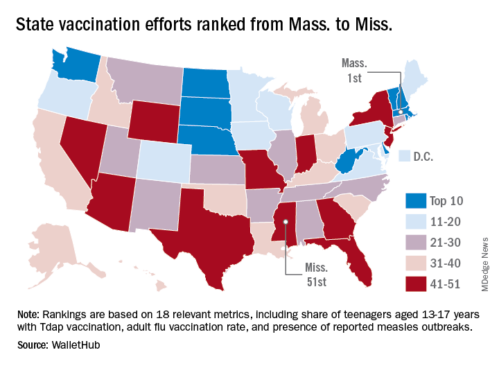
The Bay State’s top finish in the “children and teenagers immunization rates” category moved it ahead of Vermont in the overall rankings, which had the highest score in each of the other two broad categories – “adult and elderly vaccination rates” and “immunization uptake disparities and influencing factors” – but only finished 15th in child/teen immunization, Wallethub reported.
The state that ranked 51st in child/teen immunization – Mississippi – also finished 51st overall, behind every other state and Washington, D.C. The rest of the bottom five consisted of Texas (50th); Florida (49th), which ranked last in the adult/elderly category; Georgia (48th); and Indiana (47th). New Mexico, however, managed to show that last is not always least by earning a mid-pack overall rank of 30 despite its last-place showing in the disparities/influencing factors category, the WalletHub analysis showed.
Scores for the three broad categories were determined using 18 relevant metrics, including influenza vaccination rate in children aged 6 months to 17 years (1st, Rhode Island; 51st, Wyoming), share of adults aged 60 years and older with zoster vaccination (1st, Vermont; 51st, Mississippi), and share of population without health insurance coverage (1st, Massachusetts; 51st, Texas), WalletHub said.
“Each state should tailor its vaccines policy to its need, with an understanding that those needs may change,” Dorit Rubinstein Reiss of the University of California Hastings College of the Law, San Francisco, told WalletHub. When parents refuse to have their children vaccinated, it’s important to remember that “the state is not denying these children schooling. It is requiring that they be protected from disease first.”
according to a new analysis from personal finance website WalletHub.
The Bay State’s top finish in the “children and teenagers immunization rates” category moved it ahead of Vermont in the overall rankings, which had the highest score in each of the other two broad categories – “adult and elderly vaccination rates” and “immunization uptake disparities and influencing factors” – but only finished 15th in child/teen immunization, Wallethub reported.
The state that ranked 51st in child/teen immunization – Mississippi – also finished 51st overall, behind every other state and Washington, D.C. The rest of the bottom five consisted of Texas (50th); Florida (49th), which ranked last in the adult/elderly category; Georgia (48th); and Indiana (47th). New Mexico, however, managed to show that last is not always least by earning a mid-pack overall rank of 30 despite its last-place showing in the disparities/influencing factors category, the WalletHub analysis showed.
Scores for the three broad categories were determined using 18 relevant metrics, including influenza vaccination rate in children aged 6 months to 17 years (1st, Rhode Island; 51st, Wyoming), share of adults aged 60 years and older with zoster vaccination (1st, Vermont; 51st, Mississippi), and share of population without health insurance coverage (1st, Massachusetts; 51st, Texas), WalletHub said.
“Each state should tailor its vaccines policy to its need, with an understanding that those needs may change,” Dorit Rubinstein Reiss of the University of California Hastings College of the Law, San Francisco, told WalletHub. When parents refuse to have their children vaccinated, it’s important to remember that “the state is not denying these children schooling. It is requiring that they be protected from disease first.”