User login
Neurology Reviews covers innovative and emerging news in neurology and neuroscience every month, with a focus on practical approaches to treating Parkinson's disease, epilepsy, headache, stroke, multiple sclerosis, Alzheimer's disease, and other neurologic disorders.
PML
Progressive multifocal leukoencephalopathy
Rituxan
The leading independent newspaper covering neurology news and commentary.
NfL levels might presage MS disability
Neurofilament light chain (NfL) is a well-known and useful biomarker for multiple sclerosis (MS) disease activity, but its association with disease progression is not well understood. A new analysis of MS patients in California’s EPIC cohort suggests that
“We see evidence for accelerated neuroaxonal damage in the year preceding the first diagnosis of the progression events, [but] only if they were associated with evidence of focal inflammatory activity – that can be either clinical or imaging evidence,” said Ahmed Abdelhak, MD, during a presentation of the study at the annual meeting of the European Committee for Treatment and Research in Multiple Sclerosis (ECTRIMS).
“By the time we diagnose the EDSS progression, it’s already too late. Every damage or any accelerated neuroaxonal damage that has happened in association with this event already took place around a year ago. I think [this has] huge implications for the designing of clinical trials,” said Dr. Abdelhak, who is a postdoctoral researcher at the University of California, San Francisco.
In the study, researchers analyzed data from 609 MS cases, with a total of 3,906 office visits. The median age was 42 years, and 69.6% were female. Median disease duration was 6 years.
They examined the association between NfL scores and confirmed disease worsening, as recorded by an increase in EDSS score. There was an increase in NfL age-adjusted z score about 12 months in advance among patients with a progression association with a relapse in the past year, compared with individuals who did not experience disease progression. There was also a more modest increase among individuals who had disease progression without a recent relapse, but this was not statistically significant.
“Our findings suggest that the association between NfL levels and EDSS worsening is most prominent in the setting of relapse-associated events,” said Dr. Abdelhak.
Clinical implications and audience skepticism
During the Q&A following the talk, session moderator Charlotte Teunissen, PhD, professor of neurochemistry at Amsterdam University Medical Center, asked about the clinical implication of the finding. “It seems that you concluded that axonal damage has been done before the progression starts. Is that your conclusion? So it means that there is no option to interfere anymore, consequently.”
Dr. Abdelhak responded: “I think that’s a very important interpretation of the data, which I’m sure is a relatively new way of thinking about it. That means, indeed, that when we see these patients, measuring NfL wouldn’t deliver any additional value because they don’t differ between the groups at the time of EDSS worsening. And there is probably nothing more we can do about this event. But it’s still very important to know that any therapeutic intervention has also the need to prevent future disability progression, future neuroaxonal damage, but regarding what has happened already, I’m a little bit skeptical if we will be able to change anything.”
Dr. Teunissen expressed skepticism that there was no further neurodegeneration following the spike in NfL, and pointed out an important caveat, which was the study’s reliance on NfL. “You base your conclusions on what you observe for NfL, and it’s a far-fetched conclusion that there is no further axonal damage ongoing. Maybe NfL is just one marker, and it’s not the best biomarker to measure progression,” she said.
Dr. Abdelhak conceded that it will be necessary to confirm the findings with other biomarkers of neurological injury. Even different subunits of the NfL protein have been shown to have different dynamics in other neurological conditions. “So the data we have give definitely an incomplete picture because we [know] nothing about the other biomarkers of neuroaxonal injury, including the other subunits of NfL,” he said.
Later in the Q&A, Alasdair Coles, MD, professor of neurology at University of Cambridge (England), spoke from the audience. He suggested that the findings could be seen as dispiriting for clinicians. “Would the panel agree that actually for a clinician this is all rather disappointing, because none of these markers are telling us anything that we don’t otherwise know by examining the patient and doing scans?”
“I can attempt to tackle that provocative question,” replied Elias Sotirchos, MD, who also presented on an association between NfL and brain atrophy research during the session. He pointed out that all clinical tests are imperfect, and suggested that NfL isn’t something to be used in isolation. It could be useful when patients are experiencing new symptoms, or worsening symptoms, and in combination with MRI results. “My interpretation of NfL is that it does have incremental value, telling us which patients have lesions that are more destructive, potentially, given all of these consistent associations with brain atrophy and disability progression over time,” said Dr. Sotirchos, who is an assistant professor of neurology at Johns Hopkins Medicine, Baltimore.
Dr. Abdelhak and Dr. Teunissen have no relevant financial disclosures. Dr. Sotirchos has financial relationships with Alexion, Viela Bio, Horizon Therapeutics, Genentech, and Ad Scientiam.
FROM ECTRIMS 2022
Younger doctors call for more attention to patients with disabilities
As an undergraduate student at Northeastern University in Boston, Meghan Chin spent her summers working for a day program in Rhode Island. Her charges were adults with various forms of intellectual and developmental disabilities (IDD).
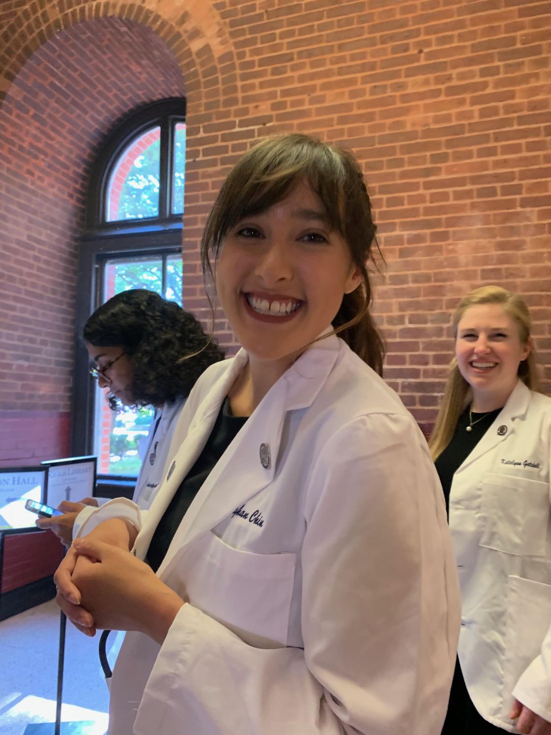
“I was very much a caretaker,” Ms. Chin, now 29, said. “It was everything from helping them get dressed in the morning to getting them to medical appointments.”
During one such visit Ms. Chin got a lesson about how health care looks from the viewpoint of someone with an IDD.
The patient was a woman in her 60s and she was having gastrointestinal issues; symptoms she could have articulated, if asked. “She was perfectly capable of telling a clinician where it hurt, how long she had experienced the problem, and what she had done or not done to alleviate it,” Ms. Chin said.
And of comprehending a response. But she was not given the opportunity.
“She would explain what was going on to the clinician,” Ms. Chin recalled. “And the clinician would turn to me and answer. It was this weird three-way conversation – as if she wasn’t even there in the room with us.”
Ms. Chin was incensed at the rude and disrespectful way the patient had been treated. But her charge didn’t seem upset or surprised. Just resigned. “Sadly, she had become used to this,” Ms. Chin said.
For the young aide, however, the experience was searing. “It didn’t seem right to me,” Ms. Chin said. “That’s why, when I went to medical school, I knew I wanted to do better for this population.”

Serendipity led her to Georgetown University, Washington, where she met Kim Bullock, MD, one of the country’s leading advocates for improved health care delivery to those with IDDs.
Dr. Bullock, an associate professor of family medicine, seeks to create better training and educational opportunities for medical students who will likely encounter patients with these disabilities in their practices.
When Dr. Bullock heard Ms. Chin’s story about the patient being ignored, she was not surprised.
“This is not an unusual or unique situation,” said Dr. Bullock, who is also director of Georgetown’s community health division and a faculty member of the university’s Center for Excellence for Developmental Disabilities. “In fact, it’s quite common and is part of what spurred my own interest in educating pre-med and medical students about effective communication techniques, particularly when addressing neurodiverse patients.”
More than 13% of Americans, or roughly 44 million people, have some form of disability, according to the National Institute on Disability at the University of New Hampshire, a figure that does not include those who are institutionalized. The Centers for Disease Control and Prevention estimates that 17% of children aged 3-17 years have a developmental disability.
Even so, many physicians feel ill-prepared to care for disabled patients. A survey of physicians, published in the journal Health Affairs, found that some lacked the resources and training to properly care for patients with disabilities, or that they struggled to coordinate care for such individuals. Some said they did not know which types of accessible equipment, like adjustable tables and chair scales, were needed or how to use them. And some said they actively try to avoid treating patients with disabilities.
Don’t assume
The first step at correcting the problem, Dr. Bullock said, is to not assume that all IDD patients are incapable of communicating. By talking not to the patient but to their caregiver or spouse or child, as the clinician did with Ms. Chin years ago, “we are taking away their agency, their autonomy to speak for and about themselves.”
Change involves altering physicians’ attitudes and assumptions toward this population, through education. But how?
“The medical school curriculum is tight as it is,” Dr. Bullock acknowledged. “There’s a lot of things students have to learn. People wonder: where we will add this?”
Her suggestion: Incorporate IDD all along the way, through programs or experiences that will enable medical students to see such patients “not as something separate, but as people that have special needs just as other populations have.”
Case in point: Operation House Call, a program in Massachusetts designed to support young health care professionals, by building “confidence, interest, and sensitivity” toward individuals with IDD.
Eight medical and allied health schools, including those at Harvard Medical School and Yale School of Nursing, participate in the program, the centerpiece of which is time spent by teams of medical students in the homes of families with neurodiverse members. “It’s transformational,” said Susan Feeney, DNP, NP-C, director of adult gerontology and family nurse practitioner programs at the graduate school of nursing at the University of Massachusetts, Worcester. “They spend a few hours at the homes of these families, have this interaction with them, and journal about their experiences.”
Dr. Feeney described as “transformational” the experience of the students after getting to know these families. “They all come back profoundly changed,” she told this news organization. “As a medical or health care professional, you meet people in an artificial environment of the clinic and hospital. Here, they become human, like you. It takes the stigma away.”
One area of medicine in which this is an exception is pediatrics, where interaction with children with IDD and their families is common – and close. “They’re going to be much more attuned to this,” Dr. Feeney said. “The problem is primary care or internal medicine. Once these children get into their mid and later 20s, and they need a practitioner to talk to about adult concerns.”
And with adulthood come other medical needs, as the physical demands of age fall no less heavily on individuals with IDDs than those without. For example: “Neurodiverse people get pregnant,” Dr. Bullock said. They also can get heart disease as they age; or require the care of a rheumatologist, a neurologist, an orthopedic surgeon, or any other medical specialty.
Generation gap
Fortunately, the next generation of physicians may be more open to this more inclusionary approach toward a widely misunderstood population.
Like Ms. Chin, Sarah Bdeir had experience with this population prior to beginning her training in medicine. She had volunteered at a school for people with IDD.
“It was one of the best experiences I’ve ever had,” Ms. Bdeir, now 23 and a first-year medical student at Wayne State University, Detroit, said. She found that the neurodiverse individuals she worked with had as many abilities as disabilities. “They are capable of learning, but they do it differently,” she said. “You have to adjust to the way they learn. And you have to step out of your own box.”
Ms. Bdeir also heard about Dr. Bullock’s work and is assisting her in a research project on how to better improve nutritional education for people with IDDs. And although she said it may take time for curriculum boards at medical schools to integrate this kind of training into their programs, she believes they will, in part because the rising cohort of medical students today have an eagerness to engage with and learn more about IDD patients.
As does Ms. Chin.
“When I talk to my peers about this, they’re very receptive,” Ms. Chin said. “They want to learn how to better support the IDD population. And they will learn. I believe in my generation of future doctors.”
A version of this article first appeared on Medscape.com.
As an undergraduate student at Northeastern University in Boston, Meghan Chin spent her summers working for a day program in Rhode Island. Her charges were adults with various forms of intellectual and developmental disabilities (IDD).
“I was very much a caretaker,” Ms. Chin, now 29, said. “It was everything from helping them get dressed in the morning to getting them to medical appointments.”
During one such visit Ms. Chin got a lesson about how health care looks from the viewpoint of someone with an IDD.
The patient was a woman in her 60s and she was having gastrointestinal issues; symptoms she could have articulated, if asked. “She was perfectly capable of telling a clinician where it hurt, how long she had experienced the problem, and what she had done or not done to alleviate it,” Ms. Chin said.
And of comprehending a response. But she was not given the opportunity.
“She would explain what was going on to the clinician,” Ms. Chin recalled. “And the clinician would turn to me and answer. It was this weird three-way conversation – as if she wasn’t even there in the room with us.”
Ms. Chin was incensed at the rude and disrespectful way the patient had been treated. But her charge didn’t seem upset or surprised. Just resigned. “Sadly, she had become used to this,” Ms. Chin said.
For the young aide, however, the experience was searing. “It didn’t seem right to me,” Ms. Chin said. “That’s why, when I went to medical school, I knew I wanted to do better for this population.”
Serendipity led her to Georgetown University, Washington, where she met Kim Bullock, MD, one of the country’s leading advocates for improved health care delivery to those with IDDs.
Dr. Bullock, an associate professor of family medicine, seeks to create better training and educational opportunities for medical students who will likely encounter patients with these disabilities in their practices.
When Dr. Bullock heard Ms. Chin’s story about the patient being ignored, she was not surprised.
“This is not an unusual or unique situation,” said Dr. Bullock, who is also director of Georgetown’s community health division and a faculty member of the university’s Center for Excellence for Developmental Disabilities. “In fact, it’s quite common and is part of what spurred my own interest in educating pre-med and medical students about effective communication techniques, particularly when addressing neurodiverse patients.”
More than 13% of Americans, or roughly 44 million people, have some form of disability, according to the National Institute on Disability at the University of New Hampshire, a figure that does not include those who are institutionalized. The Centers for Disease Control and Prevention estimates that 17% of children aged 3-17 years have a developmental disability.
Even so, many physicians feel ill-prepared to care for disabled patients. A survey of physicians, published in the journal Health Affairs, found that some lacked the resources and training to properly care for patients with disabilities, or that they struggled to coordinate care for such individuals. Some said they did not know which types of accessible equipment, like adjustable tables and chair scales, were needed or how to use them. And some said they actively try to avoid treating patients with disabilities.
Don’t assume
The first step at correcting the problem, Dr. Bullock said, is to not assume that all IDD patients are incapable of communicating. By talking not to the patient but to their caregiver or spouse or child, as the clinician did with Ms. Chin years ago, “we are taking away their agency, their autonomy to speak for and about themselves.”
Change involves altering physicians’ attitudes and assumptions toward this population, through education. But how?
“The medical school curriculum is tight as it is,” Dr. Bullock acknowledged. “There’s a lot of things students have to learn. People wonder: where we will add this?”
Her suggestion: Incorporate IDD all along the way, through programs or experiences that will enable medical students to see such patients “not as something separate, but as people that have special needs just as other populations have.”
Case in point: Operation House Call, a program in Massachusetts designed to support young health care professionals, by building “confidence, interest, and sensitivity” toward individuals with IDD.
Eight medical and allied health schools, including those at Harvard Medical School and Yale School of Nursing, participate in the program, the centerpiece of which is time spent by teams of medical students in the homes of families with neurodiverse members. “It’s transformational,” said Susan Feeney, DNP, NP-C, director of adult gerontology and family nurse practitioner programs at the graduate school of nursing at the University of Massachusetts, Worcester. “They spend a few hours at the homes of these families, have this interaction with them, and journal about their experiences.”
Dr. Feeney described as “transformational” the experience of the students after getting to know these families. “They all come back profoundly changed,” she told this news organization. “As a medical or health care professional, you meet people in an artificial environment of the clinic and hospital. Here, they become human, like you. It takes the stigma away.”
One area of medicine in which this is an exception is pediatrics, where interaction with children with IDD and their families is common – and close. “They’re going to be much more attuned to this,” Dr. Feeney said. “The problem is primary care or internal medicine. Once these children get into their mid and later 20s, and they need a practitioner to talk to about adult concerns.”
And with adulthood come other medical needs, as the physical demands of age fall no less heavily on individuals with IDDs than those without. For example: “Neurodiverse people get pregnant,” Dr. Bullock said. They also can get heart disease as they age; or require the care of a rheumatologist, a neurologist, an orthopedic surgeon, or any other medical specialty.
Generation gap
Fortunately, the next generation of physicians may be more open to this more inclusionary approach toward a widely misunderstood population.
Like Ms. Chin, Sarah Bdeir had experience with this population prior to beginning her training in medicine. She had volunteered at a school for people with IDD.
“It was one of the best experiences I’ve ever had,” Ms. Bdeir, now 23 and a first-year medical student at Wayne State University, Detroit, said. She found that the neurodiverse individuals she worked with had as many abilities as disabilities. “They are capable of learning, but they do it differently,” she said. “You have to adjust to the way they learn. And you have to step out of your own box.”
Ms. Bdeir also heard about Dr. Bullock’s work and is assisting her in a research project on how to better improve nutritional education for people with IDDs. And although she said it may take time for curriculum boards at medical schools to integrate this kind of training into their programs, she believes they will, in part because the rising cohort of medical students today have an eagerness to engage with and learn more about IDD patients.
As does Ms. Chin.
“When I talk to my peers about this, they’re very receptive,” Ms. Chin said. “They want to learn how to better support the IDD population. And they will learn. I believe in my generation of future doctors.”
A version of this article first appeared on Medscape.com.
Concerning trend of growing subarachnoid hemorrhage rates in Black people
Results of a new study based on hospital discharge data show Black people have disproportionately high rates of SAH versus other racial groups. Compared with White and Hispanic people, who had an average of 10 cases per 100,000, or Asian people, with 8 per 100,000 people, Black people had an average of 15 cases per 100,000 population.
Whereas case rates held steady for other racial groups in the study over a 10-year period, Black people were the only racial group for whom SAH incidence increased over time, at a rate of 1.8% per year.
“Root causes of the higher SAH incidence in Black [people] are complex and likely extend beyond simple differences in risk factor characteristics to other socioeconomic factors including level of education, poverty level, lack of insurance, access to quality care, and structural racism,” study investigator Fadar Oliver Otite, MD, assistant professor of neurology at SUNY Upstate Medical University, Syracuse, said in an interview.
“Addressing this racial disparity will require multidisciplinary factors targeted not just at subarachnoid hemorrhage risk factors but also at socioeconomic equity,” he added.
The study was published online in Neurology.
Uncontrolled hypertension
The average incidence of SAH for all participants was 11 cases per 100,000 people. Men had an average rate of 10 cases and women an average rate of 13 cases per 100,000.
As expected, incidence increased with age: For middle-aged men, the average was four cases per 100,000 people whereas for men 65 and older, the average was 22 cases.
Dr. Otite and his team combined U.S. Census data with two state hospitalization databases in New York and Florida and found that there were nearly 40,000 people hospitalized for SAH between 2007 and 2017. To find annual incidences of SAH per 100,000 population, they calculated the number of SAH cases and the total adult population for the year.
“Smoking and hypertension are two of the strongest risk factors for subarachnoid hemorrhage,” Dr. Otite said. “Hypertension is more prevalent in Black people in the United States, and Black patients with hypertension are more likely to have it uncontrolled.”
Racism, toxic stress
Anjail Sharieff, MD, associate professor of neurology at UT Health, Houston, said aside from a high rate of common SAH risk factors such as hypertension, Black Americans also face a barrage of inequities to health education and quality health care that contributes to higher SAH rates.
“The impact of toxic stress related to racism and discrimination experiences, and chronic stress related to poverty, can contribute to hypertension in Black people,” Dr. Sharieff said, adding that these factors contribute to stroke risk and are not usually accounted for in studies.
Dr. Sharieff said many of her first-time patients end up in her office due to a heart attack or stroke because they were previously uninsured and did not have access to primary care. “We need to begin leveraging trust with people in communities – meeting people where they are,” to educate them about hypertension and other health issues, she said.
A shining example of community engagement to reduce hypertension in Black communities was the Cedars-Sinai Barbershop Study, where 52 barbershops in Los Angeles implemented blood pressure checks and interventions among customers. A year later, the project was still working.
“Once we can identify the health problems in Black communities,” said Dr. Sharieff, “we can treat them.”
Dr. Otite and Dr. Sharieff report no relevant financial relationships.
A version of this article first appeared on Medscape.com.
FROM NEUROLOGY
A better way to predict fall risk in patients with MS?
Compared with patients with MS who didn’t fall, those who did fall had worse neuromuscular function as evidenced by a reduced rate of force development.
“Our study suggests that instead of looking at reduced maximum muscle strength, perhaps we should start looking at reduced rate of force development when trying to identify potential fallers,” said Laurits Taul-Madsen, PhD student, Aarhus University, Denmark.
The study was presented at the annual meeting of the European Committee for Treatment and Research in Multiple Sclerosis (ECTRIMS).
Explosive strength
In contrast to maximal muscle strength, the rate of force development is a measure of explosive strength, or simply the amount of force that an individual can produce over a given time period. When a patient is about to fall, what’s most important is not how strong the person is, but how quickly they can produce enough force to counteract the balance perturbation, thus avoid falling, said Dr. Taul-Madsen.
“If a person is very slow to produce this force, [that person] will have fallen before he or she has produced enough force to counteract the balance perturbation that the person is experiencing,” he added.
Research has shown a reduced rate of force development (RFD) in patients with MS, compared with healthy controls. However, little is known about the impact of RFD on falls in those with MS.
To investigate, researchers studied 53 adults with MS: Twenty-four had no fall history in the prior year, 16 had one to two prior falls, and 13 had three or more falls. The two groups of fallers were both slightly older and had a slightly higher Expanded Disability Status Scale (EDSS) scores, “which may not be so surprising,” Dr. Taul-Madsen said.
Knee extensor neuromuscular function, including maximum muscle strength and RFD at 50 and 200 milliseconds, was assessed via isokinetic dynamometry.
A high RFD is “good and the non-fallers had the highest RFD at 50 ms.” On this measure, “we saw quite a big difference between the non-fallers and the two groups of fallers,” Dr. Taul-Madsen reported.
At 200 ms, the RFD was again highest in the group of non-fallers but the difference was somewhat smaller. Non-fallers also had greater maximum muscle strength than that of the fallers.
There was “good” correlation between these neuromuscular measurements and falls, Dr. Taul-Madsen said.
He noted that RFD, which can be improved with resistance training, “seems like a specialized and difficult measurement, but it doesn’t have to be. It can be measured with just a linear encoder and a chair to perform the sit-to-stand test, so in clinical practice, it’s quite easily measured.”
‘Highly promising’ approach
“There are some data on predictors of falls in persons with MS, but not yet on neuromuscular function, as has been done in other populations,” said Brian Sandroff, PhD, senior research scientist, Kessler Foundation, West Orange, N.J.
This study is “interesting in that recurrent fallers were distinguished based on having worse neuromuscular function,” said Dr. Sandroff, who was not part of the research team.
“Although this relationship is somewhat intuitive,” RFD provides a “potentially sensitive measure that can be addressed via specific resistance exercise programs as a highly promising approach for reducing fall risk and falls themselves in persons with MS,” Dr. Sandroff said.
More generally, he said this study provides “more evidence on the multisystemic benefits of exercise training and having better physical fitness in persons with MS.
“The evidence seems to be converging more and more on this, as research groups across countries and continents are reporting on similar themes,” said Dr. Sandroff.
The study had no specific funding. Dr. Taul-Madsen and Dr. Sandroff report no relevant financial relationships.
A version of this article first appeared on Medscape.com.
FROM ECTRIMS 2022
‘Reassuring data’ for two MS meds in pregnancy
new research shows. Results of two studies show that preterm birth, spontaneous abortions, and major congenital anomalies were rare with anti-CD20 drugs ocrelizumab or natalizumab, even when continued well into the third trimester. However, hematologic abnormalities were common in newborns who were exposed to MS therapies during pregnancy.
The research comes at a time when the incidence of MS is on the rise worldwide and pregnancy in MS patients is becoming more common (Neurology. 2018 Oct 23;91[17]:e1559-69). “The results of our study should lead to a distinct risk-benefit discussion between neurologists and pregnant natalizumab-treated women to maintain treatment up to the 30th or even the 34th week of gestation, in combination with an early restart during the first 4 weeks after delivery,” Sandra Thiel, PhD, an investigator in one of the studies, told delegates attending the annual meeting of the European Committee for Treatment and Research in Multiple Sclerosis (ECTRIMS). Dr. Thiel is in the department of neurology at St. Josef Hospital and Ruhr University Bochum (Germany).
Rising number of pregnancies
In a second study, researchers from Spain presented results from the largest dataset of pregnancy outcomes for an anti-CD20 therapy in MS. The trial included 2,020 pregnancies, most of which were followed prospectively. The study included data on pregnancies since 2008. The largest single-year increase among participants was in the past year, in which there was a 65% rise in the number of pregnancies.
“The number of women with MS exposed to ocrelizumab before, during, and after the pregnancy is increasing and increasing,” Celia Oreja-Guevara, MD, PhD, vice-chair of neurology and head of the Multiple Sclerosis Center of the University Hospital San Carlos, Madrid, told conference attendees. “We have more evidence, we have more knowledge, patients and physicians trust ocrelizumab more. They know that it’s a safe treatment, and more patients are becoming pregnant.”
Dr. Oreja-Guevara highlighted a decrease in the number of elective abortions, which dropped from 50% in 2021 to 11% in the past year. The number was higher for patients with known exposure to ocrelizumab (11.5% vs. 3.7%).
Among the prospective cases, ectopic pregnancies occurred in 1.8% of those who were not exposed to ocrelizumab versus 1.4% of those exposed to the medication. The overall rate of spontaneous abortion was 11.8%, which, Dr. Oreja-Guevara said, is lower than the rate among the general population in Spain.
In the total cohort, 79.0% of pregnancies resulted in live births. Rates were similar regardless of ocrelizumab exposure. About 57% of births were full term, and 10.0% were preterm. Gestational age was unknown in 32.9% of the cases.
Overall, 0.9% of infants had a major congenital anomaly, which Dr. Oreja-Guevara said is lower than the rate of 2%-3% in all children born in Europe.
A look at natalizumab
To examine outcomes following use of natalizumab during pregnancy, researchers examined data from the German Multiple Sclerosis and Pregnancy Registry, which was created in 2006 and follows people during pregnancy and up to 6 years post partum. Of the 350 pregnant people included in the study, 171 continued natalizumab therapy beyond the first trimester. Discontinuation occurred at a median of 30.9 gestational weeks.
Most patients did not experience MS relapse during pregnancy, but the number was higher for patients who continued taking natalizumab later into pregnancy compared with those who stopped taking the drug in the first trimester (94.8% vs. 67.6%).
In the group analysis, women who continued treatment after the first trimester had significantly fewer relapses during pregnancy (5.9% vs. 32.4%; P < .001) and in the postpartum period (22.8% vs. 49.7%, P < .001). Resuming treatment with natalizumab within 4 weeks after birth significantly reduced relapse risk post partum (odds ratio, 0.32; P < .001).
The researchers also examined relapse risk with respect to treatment duration after the first trimester. Significantly more women who discontinued natalizumab before the 30th gestational week experienced postpartum relapses, compared with those whose treatment extended beyond that point (38.5% vs. 16.0%; P = .008), especially during the first postpartum trimester (26.9% vs. 8.00%; P = .009).
There were no significant differences in preterm births or major congenital abnormalities between groups. However, about half of the infants exposed to natalizumab beyond the first trimester were born with anemia or other hematologic abnormalities.
Another unexpected finding was the number of infants who were small for their gestational age (SGA). About 19% of infants born to women who continued treatment later in pregnancy were SGA. Among those in the group that discontinued therapy in the first trimester, the figure was just under 17%.
“Pregnancy outcomes were within the expected range, but a potential increased risk for small for gestational age newborns justified further investigation,” Dr. Thiel said.
Reassuring data
Angie Child Jelin, MD, director of the first trimester screening program and associate professor of gynecology and obstetrics at Johns Hopkins University, Baltimore, said that the increase in the number of pregnant patients with MS seen in their clinic could be caused in part by the growing body of work on MS treatment during pregnancy.
“It’s very reassuring that these data have come out and showed how safe they are generally in pregnancy,” Dr. Jalin said.
Although hematologic abnormalities in newborns are thought to be acute, Dr. Jalin said long-term study is needed to better understand any potential lasting effects from those abnormalities, as well as any effects in SGA newborns.
“Long-term outcomes are very hard to acquire, but ideally you’d want long-term outcomes on all of these measures,” Dr. Jalin said.
Funding for the natalizumab study was not disclosed. The ocrelizumab study was funded by F. Hoffmann–La Roche. Dr. Oreja-Guevara received honoraria for consulting and serving on advisory boards from Biogen Idec, F. Hoffmann–La Roche, Genzyme, Merck, Novartis, and Teva. Dr. Thiel has received speaker honoraria from Bayer Healthcare. Dr. Jalin reported no relevant financial relationships.
A version of this article first appeared on Medscape.com.
FROM ECTRIMS 2022
Viagra, Cialis, and Alzheimer’s risk: New data
The findings contradict results from a previous study that suggested that individuals who take sildenafil (Viagra) were significantly less likely to develop Alzheimer’s.
The new research, part of a larger effort to identify existing medications that could be repurposed to treat ADRD, employed a study design that reduced the risk for potential bias that may have influenced the earlier findings, the investigators note.
“That study came out last fall and was widely covered in the media, and we thought there were some methodological shortcomings that might have explained the results,” lead investigator Rishi Desai, PhD, assistant professor of medicine at Harvard Medical School and an associate epidemiologist at Brigham and Women’s Hospital, both in Boston, said in an interview.
The new study was published online in Brain Communications.
Not the final word?
Animal studies suggest that phosphodiesterase-5 (PDE5) inhibitors, a drug class that includes the ED drugs sildenafil and tadalafil (Cialis), improve memory and cognitive function and reduce amyloid burden. But studies in humans have yielded conflicting results.*
Although the new research and the work published last year both drew on Medicare data, they examined different patient populations.
The first study compared those who took sildenafil for any reason to those who did not take it. That design likely resulted in an analysis of a comparison of individuals with ED – the most common indication for sildenafil – to generally older individuals with diabetes or hypertension, Dr. Desai said.
In contrast, the current study included only those with pulmonary arterial hypertension (PAH), which is also an indication for PDE5 inhibitors. The researchers compared ADRD incidence in those who took PDE5 inhibitors with the incidence among those who took a different medication to treat their PAH. They used propensity matching to create two groups with similar characteristics and examined the data using four analytic strategies.
The investigators found no significant difference between groups in the incidence of ADRD, regardless of the strategy they used. Cell culture studies also revealed no protective effect from PDE5 inhibitors.
“No study of this kind should claim the final word,” Dr. Desai said. “It is extremely difficult to nail down causality from these types of data sources.”
Impressive study design
Commenting on the findings, David Knopman, MD, professor of neurology at Mayo Clinic, Rochester, Minn., described the study design as “impressive” for its efforts to minimize bias, a key limitation in the previous study.
“It was always the case that the claims about sildenafil needed further developmental work prior to testing the drug in randomized controlled trials,” Dr. Knopman said. “The evidence for the use of the drug was never sufficient for clinicians to use it in their patients.”
The study was funded by National Institute on Aging. Dr. Desai is an investigator who receives research grants from Bayer, Vertex, and Novartis that were given to the Brigham and Women’s Hospital for unrelated projects. Dr. Knopman has disclosed no relevant financial relationships.
A version of this article first appeared on Medscape.com.
Correction, 11/3/22: An earlier version of this article misstated the abbreviation for phosphodiesterase-5. It is PDE-5.
FROM BRAIN COMMUNICATIONS
I’m a physician battling long COVID. I can assure you it’s real
One in 5. It almost seems unimaginable that this is the real number of people who are struggling with long COVID, especially considering how many people in the United States have had COVID-19 at this point (more than 96 million).
Even more unimaginable at this time is that it’s happening to me. I’ve experienced not only the disabling effects of long COVID, but I’ve also seen, firsthand, the frustration of navigating diagnosis and treatment. It’s given me a taste of what millions of other patients are going through.
Vaxxed, masked, and (too) relaxed
I caught COVID-19 (probably Omicron BA.5) that presented as sniffles, making me think it was probably just allergies. However, my resting heart rate was up on my Garmin watch, so of course I got tested and was positive.
With my symptoms virtually nonexistent, it seemed, at the time, merely an inconvenience, because I was forced to isolate away from family and friends, who all stayed negative.
But 2 weeks later, I began to have urticaria – hives – after physical exertion. Did that mean my mast cells were angry? There’s some evidence these immune cells become overactivated in some patients with COVID. Next, I began to experience lightheadedness and the rapid heartbeat of tachycardia. The tachycardia was especially bad any time I physically exerted myself, including on a walk. Imagine me – a lover of all bargain shopping – cutting short a trip to the outlet mall on a particularly bad day when my heart rate was 140 after taking just a few steps. This was orthostatic intolerance.
Then came the severe worsening of my migraines – which are often vestibular, making me nauseated and dizzy on top of the throbbing.
I was of course familiar with these symptoms, as professor and chair of the department of rehabilitation medicine at the Joe R. and Teresa Lozano Long School of Medicine at University of Texas Health Science Center, San Antonio. I developed a post-COVID recovery clinic to help patients.
So I knew about postexertional malaise (PEM) and postexertional symptom exacerbation (PESE), but I was now experiencing these distressing symptoms firsthand.
Clinicians really need to look for this cardinal sign of long COVID as well as evidence of myalgic encephalomyelitis or chronic fatigue syndrome (ME/CFS). ME/CFS is marked by exacerbation of fatigue or symptoms after an activity that could previously be done without these aftereffects. In my case, as an All-American Masters miler with several marathons under my belt, running 5 miles is a walk in the park. But now, I pay for those 5 miles for the rest of the day on the couch or with palpitations, dizziness, and fatigue the following day. Busy clinic day full of procedures? I would have to be sitting by the end of it. Bed by 9 PM was not always early enough.
Becoming a statistic
Here I am, one of the leading experts in the country on caring for people with long COVID, featured in the national news and having testified in front of Congress, and now I am part of that lived experience. Me – a healthy athlete, with no comorbidities, a normal BMI, vaccinated and boosted, and after an almost asymptomatic bout of COVID-19, a victim to long COVID.
You just never know how your body is going to react. Neuroinflammation occurred in studies with mice with mild respiratory COVID and could be happening to me. I did not want a chronic immune-mediated vasculopathy.
So, I did what any other hyperaware physician-researcher would do. I enrolled in the RECOVER trial – a study my own institution is taking part in and one that I recommend to my own patients.
I also decided that I need to access care and not just ignore my symptoms or try to treat them myself.
That’s when things got difficult. There was a wait of at least a month to see my primary care provider – but I was able to use my privileged position as a physician to get in sooner.
My provider said that she had limited knowledge of long COVID, and she hesitated to order some of the tests and treatments that I recommended because they were not yet considered standard of care. I can understand the hesitation. It is engrained in medical education to follow evidence based on the highest-quality research studies. We are slowly learning more about long COVID, but acknowledging the learning curve offers little to patients who need help now.
This has made me realize that we cannot wait on an evidence-based approach – which can take decades to develop – while people are suffering. And it’s important that everyone on the front line learn about some of the manifestations and disease management of long COVID.
I left this first physician visit feeling more defeated than anything and decided to try to push through. That, I quickly realized, was not the right thing to do.
So again, after a couple of significant crashes and days of severe migraines, I phoned a friend: Ratna Bhavaraju-Sanka, MD, the amazing neurologist who treats patients with long COVID alongside me. She squeezed me in on a non-clinic day. Again, I had the privilege to see a specialist most people wait half a year to see. I was diagnosed with both autonomic dysfunction and intractable migraine.
She ordered some intravenous fluids and IV magnesium that would probably help both. But then another obstacle arose. My institution’s infusion center is focused on patients with cancer, and I was unable to schedule treatments there.
Luckily, I knew about the concierge mobile IV hydration therapy companies that come to your house – mostly offering a hangover treatment service. And I am thankful that I had the health literacy and financial ability to pay for some fluids at home.
On another particularly bad day, I phoned other friends – higher-ups at the hospital – who expedited a slot at the hospital infusion center and approval for the IV magnesium.
Thanks to my access, knowledge, and other privileges, I got fairly quick if imperfect care, enrolled in a research trial, and received medications. I knew to pace myself. The vast majority of others with long COVID lack these advantages.
The patient with long COVID
Things I have learned that others can learn, too:
- Acknowledge and recognize that long COVID is a disease that is affecting 1 in 5 Americans who catch COVID. Many look completely “normal on the outside.” Please listen to your patients.
- Autonomic dysfunction is a common manifestation of long COVID. A 10-minute stand test goes a long way in diagnosing this condition, from the American Academy of Physical Medicine and Rehabilitation. It is not just anxiety.
- “That’s only in research” is dismissive and harmful. Think outside the box. Follow guidelines. Consider encouraging patients to sign up for trials.
- Screen for PEM/PESE and teach your patients to pace themselves, because pushing through it or doing graded exercises will be harmful.
- We need to train more physicians to treat postacute sequelae of SARS-CoV-2 infection () and other postinfectious conditions, such as ME/CFS.
If long COVID is hard for physicians to understand and deal with, imagine how difficult it is for patients with no expertise in this area.
It is exponentially harder for those with fewer resources, time, and health literacy. My lived experience with long COVID has shown me that being a patient is never easy. You put your body and fate into the hands of trusted professionals and expect validation and assistance, not gaslighting or gatekeeping.
Along with millions of others, I am tired of waiting.
Dr. Gutierrez is Professor and Distinguished Chair, department of rehabilitation medicine, University of Texas Health Science Center at San Antonio. She reported receiving honoraria for lecturing on long COVID and receiving a research grant from Co-PI for the NIH RECOVER trial.
A version of this article first appeared on Medscape.com.
Should McDonald criteria include optical nerve lesions?
The McDonald criteria for diagnosing multiple sclerosis (MS) has been around since 2001, with revisions in 2005, 2010, and 2017. They focus on lesions disseminated in space (DIS) and disseminated in time (DIT). As with any diagnostic, new science and methods inform changes.
At the annual meeting of the European Committee for Treatment and Research in Multiple Sclerosis (ECTRIMS), several speakers made the case for the addition of ONL to the next McDonald revision. Arguments ranged from the inherent injustice that patients with a diagnosis of optic lesions should have to meet three symptoms of MS to get a diagnosis, while other patients require only two, to the possibility that early presentation with ocular symptoms could be an indicator of a more severe prognosis.
Still, many conditions can mimic the symptoms of ONL, so it is critical to be sure of the diagnosis before considering it a symptom of MS. For example, central serious maculopathy in young to middle-aged patients is often painless and could lead to an inadvertent MS diagnosis if it were added to the criteria. However, treatment with steroids can make it worse, according to Laura Balcer, MD, who spoke at the session. Dr. Balcer is a neuro-ophthalmologist at NYU Langone Health.
Adding ONL to McDonald
In the first talk of the session, Frederik Barkhof, MD, PhD, discussed some of the history of the McDonald criteria and laid a groundwork for why it should be updated. He began by pointing out that the latest criteria require either symptomatic or asymptomatic MRI lesions to determine DIS or DIT.
Dr. Barkhof has led the Magnetic Imaging in Multiple Sclerosis (MAGNIMS) group, which had some doubts about the 2017 revision to the McDonald criteria. “We discussed this and said, ‘Well, that is interesting. So you can have a brainstem lesion, which can be the symptomatic lesion and then you need one further region, so you have two regions, and then you have MS. But now if you have an optic nerve presentation, and you can find the optic nerve, it doesn’t count, and then you need two more regions. So there’s a bit of an imbalance. Why would you need three regions if you have an optic nerve, and only two if you have a spinal cord or a brainstem presentation?” recalled Dr. Barkhof, professor of radiology and neuroscience at Vrije Universiteit Amsterdam.
Identifying optic nerve lesions requires a good MRI that should use fat-saturation techniques.
Dr. Barkhof pointed out that the ONLs are a common presentation among MS patients. “We’re now preparing for the next revisions of the McDonnell criteria, and I’m confident and hopeful that we’ll make it this time,” said Dr. Barkhof.
Confirm the MS diagnosis
In the second talk, Angela Vidal-Jordana, MD, PhD, discussed ways to implement ONLs in the clinic. She emphasized the importance of ruling out other causes. “Even when a patient comes to us with a diagnosis of optic neuritis, we should question that. Different studies have shown that the optic neuritis overdiagnosis rate at referral might be as high as 60% of the cases,” said Dr. Vidal-Jordana, a neurologist at Centre d’Esclerosi Multiple de Catalunya in Barcelona. Studies show that about half of those are due to misinterpretation of the clinical history and examination, she said.
“So when presenting new visual symptoms, we should ask whether this is suggestive of an inflammatory etiology or not, and whether they are typical for MS or not. If both answers to these questions are yes, then we should apply the diagnostic criteria,” said Dr. Vidal-Jordana.
She presented results from a longitudinal, prospective study conducted within the MAGNIMS network supporting the inclusion of ONL into DIS criteria for MS. It included use of optic nerve MRI, OCT, and VEP [visual evoked potential]. “All of the modified DIS criteria, that is including one of the tests at each time, or a combination of them, led to a higher sensitivity of the diagnostic criteria (when optic nerve involvement was included), albeit with a small decrease in specificity which mainly was due to the fact that we only had 3 years of follow-up. The same held true when analyzing the secondary outcome of new T2 lesions or second relapse during the follow up,” said Dr. Vidal-Jordana.
“As a summary, I would say for sure we will need first of all an MRI, and we need to know if we can evaluate the optic nerve by MRI or not. If not, and we still do not have a diagnosis of a MS, maybe we can order VEP or OCT and then the test selection should be based on the time elapsed since first CIS [clinically isolated syndrome]. If it’s less than 3 months, I would go for a VEP. If it’s more than 3 months, then I probably want to go for OCT,” said Dr. Vidal-Jordana.
An aid to earlier diagnosis
In the last talk, Dr. Balcer discussed non-MRI methods for assessing ONLs. She noted that in about 25% of MS patients, ONLs are the first clinical demyelinating event. “Adding the optic nerve to the MS diagnostic criteria will allow us to diagnose our patients even earlier, (it) may help their vision, and could also help us to reduce the overall burden of MS disability over a lifetime. Importantly, entry into MS diagnosis can be delayed among patients for whom optic neuritis or even asymptomatic optic nerve lesions are noted at presentation, and there has been an enormous amount of data that have emerged over the past 5 years demonstrating the importance of the optic nerve in the MS diagnosis with implications for early therapy,” said Dr. Balcer.
She discussed optical coherence tomography (OCT), which is a key technique for diagnosing optic neuropathy. It measures the thickness of the retinal nerve fiber and ganglion cell layers, which have been associated with vision impairment and can reveal asymptomatic involvement of optic nerves in MS.
Beware of misdiagnosis
In the Q&A period following the talks, much of the discussion turned to reliability of ONL diagnoses.
“Misdiagnosis is a huge problem. That’s my experience: People who are referred to me with optic neuritis often don’t have optic neuritis,” said comoderator Wallace Brownlee, MBChB, PhD, a consultant neurologist at Cleveland Clinic London. Misha Pless, MD, spoke up from the audience to second that. “I’m delighted that the optic nerve will finally get a place at the table. I’ve been practicing neuro-ophthalmology and I have also [been] an MS doctor for about 25 years. What I see here in this fantastic discussion is that a lot of neurologists are going to be – I hate to use the word ‘misled’ – into relying on technology like OCT, VEP, and MRI to make the diagnosis. I will submit to this panel that that will lead to a number of MS misdiagnoses because I have been doing this for 25 years and the number of patients that I’ve received with rule-out optic neuritis that had macular disease, optical changes, or corneal abrasions are too numerous to count. If you’re going to add optic neuritis in the list of criteria [for MS], you definitely have to have a little asterisk in my opinion and say, ‘with prior blessing from an ophthalmologist to rule out ocular disease,’ because I don’t know any neurologist that knows how to look in the back of the eye, and I don’t know any neurologist that has an OCT in their office,” said Dr. Pless, professor of ophthalmology and a neurologist at Mayo Clinic in Jacksonville, Fla.
The panelists generally agreed with his point, although the hot mic picked up when one panelist whispered to another, ‘I have one,’ referring to an office OCT. “Using OCT in that regard, but also collaborating with your neuro-ophthalmologist and ophthalmologist is critical,” said Dr. Balcer.
Dr. Barkhof has financial relationships with Biogen, Merck, Roche, EISAI, Prothena, IXICO, Jansen, Combinostics, Novartis, GE, Queen Square, and Analytics. Dr. Vidal-Jordana has financial relationships with Roche, Novartis, Merck, and Sanofi. Dr. Balcer has no relevant financial disclosures. Dr. Brownlee has financial relationships with Biogen, Celgene, Merck, Mylan, Novartis, Roche, and Sanofi.
FROM ECTRIMS 2022
Stem cell transplantation ‘substantially’ superior to fingolimod in cutting MS relapse risk
, a new study suggests.
The multicenter observational study showed that AHSCT was associated with a better overall annual relapse rate and significantly greater disability improvement versus fingolimod. Stem cell transplantation also offered a slight advantage compared with natalizumab but showed no benefit over ocrelizumab.
“Based on these results, we can conclude that in this observational study AHSCT is substantially superior to fingolimod and marginally superior to natalizumab in reducing the risk of posttreatment relapses,” study investigator Tomas Kalincik, MD, PhD, said at the annual meeting of the European Committee for Treatment and Research in Multiple Sclerosis (ECTRIMS).
Research gap
Prior studies have suggested that AHSCT is associated with better disability outcomes than other immunotherapies in patients with active secondary progressive MS. Findings from another study on long-term efficacy in relapse prevention are also promising.
“The information that we are still lacking is the head-to-head comparison of the effectiveness of AHSCT in relapsing-remitting disease to concrete, highly effective therapies, which is the aim of this current study,” said Dr. Kalincik, professor of neurology at the University of Melbourne and head of the Multiple Sclerosis and Neuroimmunology Service at the Royal Melbourne Hospital, Australia.
The study included 167 patients with relapsing-remitting MS from six centers in Ottawa, Uppsala, Sheffield, Bergen, Sydney, and Melbourne, combined with 1,675 patients from MSBase. Patients were included if they were treated with AHSCT or fingolimod (n = 769), natalizumab (n = 730) or ocrelizumab (n = 343).
Researchers used propensity matching to ensure the AHSCT and disease-modifying therapy (DMT) groups had similar relapse rates and disability scores at baseline. At 1 year, patients who underwent AHSCT were 74% more likely than were those on fingolimod to be relapse free (hazard ratio [HR], 0.26; P < .0001).
The AHSCT group had a lower overall annualized relapse rate (0.09 vs. 0.19, respectively; P < .0001) and a higher cumulative probability of remaining relapse free at 2 years (90% vs. 68%, respectively) and 5 years (85% vs. 54%, respectively). AHSCT was associated with significantly better confirmed disability improvement at 6 months compared with fingolimod (HR, 2.70; P < .0001).
The researchers found better outcomes with AHSCT compared with natalizumab, but the results weren’t as promising. The annual relapse rate with AHSCT was statistically significant (P = .03) but the clinical benefit was marginal, Dr. Kalincik said. However, the confirmed disability improvement was significantly better with AHSCT (HR, 2.68; P < .0001).
There was no significant difference in outcomes with AHSCT compared to ocrelizumab.
Adverse events were common with stem cell transplantation, a sticking point for at least one conference attendee who raised safety concerns. Among those treated with AHSCT, 23% had febrile neutropenia, 11% had serum sickness, and 36% experienced complications after discharge, mostly infection. There was one AHSCT treatment-related death.
“We’re showing that AHSCT is a highly potent therapy that definitely is competitive vis a vis the most potent standard conventional disease modifying therapies,” said Dr. Kalincik, who noted the high rate of adverse events. Still, treatment-related mortality from AHSCT has improved since the therapy was first used in MS patients, he added.
“I’m hoping the data provides some evidence to convince you that this treatment still has a place in our armamentarium,” Dr. Kalincik said.
Questions remain
Eva Kubala Havrdová, MD, PhD, professor of neurology in the Multiple Sclerosis Center at Charles University in Prague, Czech Republic, said the results add to a growing body of data on the efficacy of AHSCT in patients with highly active relapsing-remitting MS, but questions remain.
“It is important to understand that AHSCT provides advantage in terms of effectiveness compared with most of the DMTs,” Dr. Kubala Havrdová said. “However, it is a therapy associated with high risk of adverse events and this needs to be considered when one decides whether other, less invasive options have been exploited.”
Study funding was not reported. Dr. Kalincik disclosed ties with BioCSL, Biogen, BMS, Celgene, Eisai, Janssen, Merck, Novartis, Roche, Sanofi Genzyme, Teva, and WebMD Global. Dr. Kubala Havrdová reported no relevant financial relationships.
A version of this article first appeared on Medscape.com.
FROM ECTRIMS 2022
Has the pandemic affected babies’ brain development?
There’s some good overall news in a large analysis that looked at whether a mother’s COVID-19 infection or birth during the pandemic could affect a baby’s brain development.
Researchers studied 21,419 infants who had neurodevelopmental screening during the pandemic (from January 2020 to January 2021) and compared them with babies born before the pandemic (2015-2019).
They found in an analysis of eight studies that, generally, brain development in infants ages 6-12 months old was not changed by COVID-19.
Communication skill scores lower than prepandemic
However, one area did see a significant difference when they looked at answers to the Ages and Stages Questionnaire, 3rd edition (ASQ-3): Scores were lower in communication skills.
Compared with the prepandemic babies, the pandemic group of babies was more likely to have communication impairment (odds were 1.7 times higher).
Additionally, mothers’ SARS-CoV-2 infection was not associated with significant differences in any neurodevelopment sector in offspring, with one exception: Odds were 3.5 times higher for fine motor impairment in the pandemic baby group.
The babies in this study were either exposed in the womb to the SARS-CoV-2 infection or screened during the pandemic regardless of whether they were exposed to the virus.
The study, led by Kamran Hessami, MD, with the Maternal Fetal Care Center at Boston Children’s Hospital and Harvard Medical School in Boston, was published in JAMA Network Open.
Potential reasons for lower communication skills
The study points to some factors of the pandemic that may be tied to impaired communication skills.
“Higher levels of COVID-19–related stress were reported for both mothers and fathers of infants aged 0-6 months and were associated with insensitive parenting practices, including decreased emotional responsiveness in only mothers, which could lessen the reciprocal exchanges that support language development in early childhood,” they write. “Additionally, opportunities to promote language and social development through new experiences outside the home, including visits with extended family and friends or attendance at a child care center, were lessened for many during the pandemic.”
Viviana M. Fajardo Martinez, MD, with neonatal/perinatal medicine at University of California, Los Angeles, Health, told this publication her team is also studying child development before and after the pandemic over a 3-year period, and delayed communication skills is something she is seeing in clinic there.
She says some parents have been concerned, saying their babies aren’t talking enough or are behind in vocabulary.
Babies can catch up after 12 months
One thing she tells parents is that babies who are a bit delayed at 12 months can catch up.
Up to 18 months, they can catch up, she said, adding that they can be reevaluated then for improvement. If, at that point, the baby is not catching up, “that’s when we refer for early intervention,” she said.
Dr. Martinez also tells parents concerned about their infant’s communication skills that it’s important to talk, read, and sing to their child. She said amid pandemic stress, corners may have been cut in asking children to use language skills.
For instance, if a child points to an apple, a stressed parent may just give the child the apple instead of asking the child to request it by name and repeat the word several times.
She also said a limitation of this study is the use of the ASQ-3 questionnaire, which is filled out by parents. Answers are subjective, she notes, and sometimes differ between one child’s two parents. The questionnaire was commonly used during the pandemic because a more objective, professional evaluation has been more difficult.
However, a measure like the Bayley Scales of Infant and Toddler Development Screening Test adds objectivity and will likely give a better picture as research progresses, Dr. Martinez said.
Some information missing
Andréane Lavallée, PhD, and Dani Dumitriu, MD, PhD, both with the department of pediatrics at Columbia University, New York, write in an invited commentary that the overall positive message of the study “should not make researchers complacent” and results should be viewed with caution.
They point out that the precise effects of this novel virus are still unclear and the age group and variables studied may not tell the whole story.
“It should be noted that this systematic review did not consider timing of exposure during pregnancy, maternal infection severity, or exposure to various SARS-CoV-2 variants – all factors that could eventually be proven to contribute to subtle adverse neurodevelopmental outcomes,” they write.
Additionally, past pandemics “such as the 1918 Spanish flu, 1964 rubella, and 2009 H1N1” have taught researchers to watch for increases in diagnoses such as autism spectrum disorder (ASD) and schizophrenia in subsequent years.
“ASD is generally diagnosed at age 3-5 years (and often not until early teens), while schizophrenia is generally diagnosed in mid-to-late 20s,” the editorialists point out. The authors agree and emphasize the need for long-term studies.
Authors report no relevant financial relationships. Editorialist Dr. Dumitriu reports grants from National Institute of Mental Health, the U.S. Centers for Disease Control and Prevention, and the W. K. Kellogg Foundation; and has received gift funds from Einhorn Collaborative during the conduct of the study to the Nurture Science Program, for which Dr Dumitriu serves as director. Dr. Dumitriu received personal fees from Medela outside the submitted work; and is the corresponding author for one of the studies (Shuffrey et al., 2022) included in the systematic review conducted by Dr. Hessami et al. Dr. Lavallée reports grants from the Canadian Institutes of Health Research. Dr. Martinez reports no relevant financial relationships.
FROM JAMA NETWORK OPEN