User login
Clinicians, Parents Still Confused About Vitamin D
SAN FRANCISCO – Dermatologists don’t need to check the serum vitamin D levels of every patient, according to Dr. Shelia Fallon Friedlander.
The test costs $100. Unless someone is in a high-risk group, which includes the elderly, dark-skinned, or obese patients; has fat malabsorption; or has limited sun exposure, there is no need to check vitamin D levels, she said at the Women’s and Pediatric Dermatology Seminar, sponsored by Skin Disease Education Foundation (SDEF).

"I don’t think every person of color who walks into your office needs to get blood levels" checked, but if they have other risk factors, order the test, she said. Many clinicians now routinely check blood levels in the elderly, she added.
Besides dermatologists, patients are seeking clarity about vitamin D supplementation. Among the questions they most frequently ask their physicians:
• Does everyone need supplementation? It is not an absolute, she said. Alaskans who are eating salmon day and night may not need to be supplemented, but the Institute of Medicine (IOM) suggests that supplementation is reasonable for everyone unless it is clear the person does not need it, said Dr. Friedlander of the University of California, San Diego.
• What kind of supplement is best? Some foods can be great sources for vitamin D, especially salmon, shiitake mushrooms, and milk. Vitamin D3 (cholecalciferol) supplements are thought to be better than vitamin D2 at raising blood levels, and have a longer shelf life.
• How much is best? For now, stick with the IOM recommendations of 400 IU/day for ages younger than 1 year, 600 IU/day for ages 1-70 years, and 800 IU/day for older people, except for people in high-risk groups, Dr. Friedlander advised at the seminar.
All breast-fed infants also are considered to have a higher risk for insufficient vitamin D because often the vitamin D status of the mother is unknown. Supplementation with 400 IU/day of vitamin D is recommended and is "not a bad idea for all children," she said.
For people aged 9-70 years who are at high risk, "1,000 IU/day is the number that has been batted around" for supplementation, though the IOM recommendations allow up to 4,000 IU/day, she said.
• Is more vitamin D better? No one knows, and there are suggestions in some studies of a detrimental effect from too much vitamin D. It’s clear, however, that too little vitamin D is a problem and a moderate amount of vitamin D is good for the body. But the data in 30-40 studies on vitamin D’s effects on risks for cancer and other problems aren’t clear enough yet to say whether a lot of vitamin D is helpful or harmful, Dr. Friedlander said.
Studies looking at colorectal cancer are supportive but not confirming of the beneficial effects of high levels of vitamin D, while studies on prostate and breast cancer are "really scary," she said. Many studies suggest a protective effect of high levels of vitamin D, but some show increased risks, including a small study showing a higher risk for kidney stones. "There’s a lot of controversy in the literature," she said.
The IOM based its recommendations on the principles of doing no harm and not relying on imprecise, suboptimal data. Dr. Friedlander reminded listeners of what happened with beta carotene supplementation: It was thought to decrease skin cancer risk until data showed it increased the risk for other cancers.
"Vitamin D has a beneficial effect until you hit a certain number. Nobody knows what the number is. In certain situations, more vitamin D may have a negative effect on your health," she said.
• Does vitamin D support bone health? Good studies make it absolutely clear that it does. Supplement levels above the recommended 600 IU/day for most people would be even better for bone health, "but there’s nothing to show that you need to get a gazillion units a day," she said.
• Does vitamin D reduce the risk for other diseases? The evidence that vitamin D may protect against multiple sclerosis, cardiovascular disease, and cancer is imprecise, inconclusive, inconsistent, and insufficient. This will change as more randomized, controlled trials are conducted, she said. "We may find that everyone needs to be on 1,000 IU/day, but right now the data aren’t there," she said.
• Should we get vitamin D from the sun? "No, no, no," Dr. Friedlander stressed. Dermatologists have advised patients to stay out of the sun for good reason. "This is a pseudocontroversy. Sunlight is a known carcinogen. You can’t make vitamin D in your skin without inducing DNA damage." People can get sufficient vitamin D from incidental sun exposure, a reasonable diet, and supplements.
Dr. Friedlander said she has no relevant financial disclosures.
SDEF and this news organization are owned by Elsevier.
SAN FRANCISCO – Dermatologists don’t need to check the serum vitamin D levels of every patient, according to Dr. Shelia Fallon Friedlander.
The test costs $100. Unless someone is in a high-risk group, which includes the elderly, dark-skinned, or obese patients; has fat malabsorption; or has limited sun exposure, there is no need to check vitamin D levels, she said at the Women’s and Pediatric Dermatology Seminar, sponsored by Skin Disease Education Foundation (SDEF).
"I don’t think every person of color who walks into your office needs to get blood levels" checked, but if they have other risk factors, order the test, she said. Many clinicians now routinely check blood levels in the elderly, she added.
Besides dermatologists, patients are seeking clarity about vitamin D supplementation. Among the questions they most frequently ask their physicians:
• Does everyone need supplementation? It is not an absolute, she said. Alaskans who are eating salmon day and night may not need to be supplemented, but the Institute of Medicine (IOM) suggests that supplementation is reasonable for everyone unless it is clear the person does not need it, said Dr. Friedlander of the University of California, San Diego.
• What kind of supplement is best? Some foods can be great sources for vitamin D, especially salmon, shiitake mushrooms, and milk. Vitamin D3 (cholecalciferol) supplements are thought to be better than vitamin D2 at raising blood levels, and have a longer shelf life.
• How much is best? For now, stick with the IOM recommendations of 400 IU/day for ages younger than 1 year, 600 IU/day for ages 1-70 years, and 800 IU/day for older people, except for people in high-risk groups, Dr. Friedlander advised at the seminar.
All breast-fed infants also are considered to have a higher risk for insufficient vitamin D because often the vitamin D status of the mother is unknown. Supplementation with 400 IU/day of vitamin D is recommended and is "not a bad idea for all children," she said.
For people aged 9-70 years who are at high risk, "1,000 IU/day is the number that has been batted around" for supplementation, though the IOM recommendations allow up to 4,000 IU/day, she said.
• Is more vitamin D better? No one knows, and there are suggestions in some studies of a detrimental effect from too much vitamin D. It’s clear, however, that too little vitamin D is a problem and a moderate amount of vitamin D is good for the body. But the data in 30-40 studies on vitamin D’s effects on risks for cancer and other problems aren’t clear enough yet to say whether a lot of vitamin D is helpful or harmful, Dr. Friedlander said.
Studies looking at colorectal cancer are supportive but not confirming of the beneficial effects of high levels of vitamin D, while studies on prostate and breast cancer are "really scary," she said. Many studies suggest a protective effect of high levels of vitamin D, but some show increased risks, including a small study showing a higher risk for kidney stones. "There’s a lot of controversy in the literature," she said.
The IOM based its recommendations on the principles of doing no harm and not relying on imprecise, suboptimal data. Dr. Friedlander reminded listeners of what happened with beta carotene supplementation: It was thought to decrease skin cancer risk until data showed it increased the risk for other cancers.
"Vitamin D has a beneficial effect until you hit a certain number. Nobody knows what the number is. In certain situations, more vitamin D may have a negative effect on your health," she said.
• Does vitamin D support bone health? Good studies make it absolutely clear that it does. Supplement levels above the recommended 600 IU/day for most people would be even better for bone health, "but there’s nothing to show that you need to get a gazillion units a day," she said.
• Does vitamin D reduce the risk for other diseases? The evidence that vitamin D may protect against multiple sclerosis, cardiovascular disease, and cancer is imprecise, inconclusive, inconsistent, and insufficient. This will change as more randomized, controlled trials are conducted, she said. "We may find that everyone needs to be on 1,000 IU/day, but right now the data aren’t there," she said.
• Should we get vitamin D from the sun? "No, no, no," Dr. Friedlander stressed. Dermatologists have advised patients to stay out of the sun for good reason. "This is a pseudocontroversy. Sunlight is a known carcinogen. You can’t make vitamin D in your skin without inducing DNA damage." People can get sufficient vitamin D from incidental sun exposure, a reasonable diet, and supplements.
Dr. Friedlander said she has no relevant financial disclosures.
SDEF and this news organization are owned by Elsevier.
EXPERT ANALYSIS FROM SDEF WOMEN’S & PEDIATRIC DERMATOLOGY SEMINAR
Five or More Infantile Hemangiomas? Order Liver Ultrasound
SAN FRANCISCO – Infants with five or more hemangiomas should undergo a liver ultrasound according to new research, said Dr. Ilona J. Frieden.
Clinicians know that when they see infants with multiple cutaneous hemangiomas, they need to worry about internal hemangiomas, said Dr. Frieden at the seminar, sponsored by Skin Disease Education Foundation (SDEF). And now, for the first time, a prospective, multicenter study identified how many infantile hemangiomas should trigger alarm.

The study screened infants younger than 6 months in the United States, Canada, and Spain with abdominal ultrasound and found that 16% of 151 infants with five or more skin hemangiomas had liver hemangiomas, compared with no liver hemangiomas in a control group of infants with one to four skin hemangiomas, she said.
"We certainly recommend doing liver ultrasound in children with five or more hemangiomas," said Dr. Frieden, professor of dermatology and pediatrics at the University of California, San Francisco. "Five does seem to be the right threshold for doing this screening test."
Interestingly, only 2 of the 24 infants with hepatic hemangiomas received treatment specifically for the hepatic hemangiomas (Pediatr. Dermatol. 2011;28:245-53). "Liver hemangiomas are probably a lot like skin hemangiomas – they don’t all need treatment. That’s an important fact," she said.
In general, segmental hemangiomas are more likely to develop complications, and facial segmental hemangiomas are more likely to need systemic treatment. The main indication for treating cutaneous infantile hemangiomas is a risk of scarring and disfigurement, especially with facial segmental hemangiomas, ulcerated hemangiomas, and hemangiomas located on the ear or on any prominent facial site but especially the nasal tip or perioral areas.
The study found that 16% of 151 infants with five or more skin hemangiomas had liver hemangiomas.
Treatment should also be considered in patients with periocular or liver hemangiomas, hemangiomas of the airway, spinal dysraphism or genitourinary complications, or in PHACE syndrome, she said.
A previous study by Dr. Frieden and her associates showed that 30% of infants with large facial hemangiomas were shown to have PHACE syndrome (Pediatrics 2010;126:e418-26).
Dr. Frieden, who played a leading role in defining PHACE syndrome, estimated it to be a more common neurocutaneous syndrome than Sturge-Weber: "Not super-rare, but not common," she said.
Any infant with a facial hemangioma measuring 5 cm or more in diameter probably deserves cardiac echocardiography to look for transverse coarctation of the aorta, MRI and magnetic resonance angiography to look for arterial anomalies, and an eye exam to look for structural eye abnormalities even if there is no periocular hemangioma, she advised.
Infants with lumbosacral hemangiomas are at greater risk for having a tethered spinal cord or other problems such as genitourinary abnormalities. Approximately half of infants with typical lumbosacral hemangiomas will have a tethered spinal cord, prospective studies suggest.
Lumbosacral hemangiomas tend to stay flatter and be less bulky than hemangiomas on other body areas and so may not raise the suspicion of problems that would be warranted. Dr. Frieden has diagnosed tethered spinal cord in several infants with lumbosacral hemangiomas who were referred only after they developed ulcerations.
Dr. Frieden has been an advisor or consultant to Pierre Fabre and Topaz Pharmaceuticals.
SDEF and this news organization are owned by Elsevier.
SAN FRANCISCO – Infants with five or more hemangiomas should undergo a liver ultrasound according to new research, said Dr. Ilona J. Frieden.
Clinicians know that when they see infants with multiple cutaneous hemangiomas, they need to worry about internal hemangiomas, said Dr. Frieden at the seminar, sponsored by Skin Disease Education Foundation (SDEF). And now, for the first time, a prospective, multicenter study identified how many infantile hemangiomas should trigger alarm.
The study screened infants younger than 6 months in the United States, Canada, and Spain with abdominal ultrasound and found that 16% of 151 infants with five or more skin hemangiomas had liver hemangiomas, compared with no liver hemangiomas in a control group of infants with one to four skin hemangiomas, she said.
"We certainly recommend doing liver ultrasound in children with five or more hemangiomas," said Dr. Frieden, professor of dermatology and pediatrics at the University of California, San Francisco. "Five does seem to be the right threshold for doing this screening test."
Interestingly, only 2 of the 24 infants with hepatic hemangiomas received treatment specifically for the hepatic hemangiomas (Pediatr. Dermatol. 2011;28:245-53). "Liver hemangiomas are probably a lot like skin hemangiomas – they don’t all need treatment. That’s an important fact," she said.
In general, segmental hemangiomas are more likely to develop complications, and facial segmental hemangiomas are more likely to need systemic treatment. The main indication for treating cutaneous infantile hemangiomas is a risk of scarring and disfigurement, especially with facial segmental hemangiomas, ulcerated hemangiomas, and hemangiomas located on the ear or on any prominent facial site but especially the nasal tip or perioral areas.
The study found that 16% of 151 infants with five or more skin hemangiomas had liver hemangiomas.
Treatment should also be considered in patients with periocular or liver hemangiomas, hemangiomas of the airway, spinal dysraphism or genitourinary complications, or in PHACE syndrome, she said.
A previous study by Dr. Frieden and her associates showed that 30% of infants with large facial hemangiomas were shown to have PHACE syndrome (Pediatrics 2010;126:e418-26).
Dr. Frieden, who played a leading role in defining PHACE syndrome, estimated it to be a more common neurocutaneous syndrome than Sturge-Weber: "Not super-rare, but not common," she said.
Any infant with a facial hemangioma measuring 5 cm or more in diameter probably deserves cardiac echocardiography to look for transverse coarctation of the aorta, MRI and magnetic resonance angiography to look for arterial anomalies, and an eye exam to look for structural eye abnormalities even if there is no periocular hemangioma, she advised.
Infants with lumbosacral hemangiomas are at greater risk for having a tethered spinal cord or other problems such as genitourinary abnormalities. Approximately half of infants with typical lumbosacral hemangiomas will have a tethered spinal cord, prospective studies suggest.
Lumbosacral hemangiomas tend to stay flatter and be less bulky than hemangiomas on other body areas and so may not raise the suspicion of problems that would be warranted. Dr. Frieden has diagnosed tethered spinal cord in several infants with lumbosacral hemangiomas who were referred only after they developed ulcerations.
Dr. Frieden has been an advisor or consultant to Pierre Fabre and Topaz Pharmaceuticals.
SDEF and this news organization are owned by Elsevier.
EXPERT ANALYSIS FROM THE SDEF WOMEN’S & PEDIATRIC DERMATOLOGY SEMINAR
Major Finding: Sixteen percent of infants with five or more hemangiomas had hepatic hemangiomas.
Data Source: Prospective, multicenter study of 151 infants younger than 6 months of age with at least 5 infantile hemangiomas and a control group of infants with 1-4 hemangiomas.
Disclosures: Dr. Frieden has been an advisor or consultant to Pierre Fabre and Topaz Pharmaceuticals. SDEF and this news organization are owned by Elsevier.
Age, Location of Bruises Flag Child Abuse
SAN FRANCISCO – The location of bruising and the age of a child can help hone clinical suspicion of child abuse, thanks to a study identifying these predictors.
Because of this study, clinicians now have a better way of raising the topic with parents, which is always a difficult scenario, Dr. Robert Sidbury said.

Instead of physicians having to say that they need to discuss the possibility the bruises might be from nonaccidental trauma, they can now can say, "I’ve got this paper that says bruises in this certain location in a child of this age make me have to check this out," he said at the Women’s and Pediatric Dermatology Seminar, sponsored by Skin Disease Education Foundation (SDEF). "To me, that sounds different."
The study compared the characteristics of bruises on 95 infants aged 0-48 months seen in a pediatric ICU, 53 of whom had accidental trauma and 42 of whom were victims of abuse. Bruising on the torso, ear, or neck ("Think TEN," he suggested) in a child younger than 4 years of age increased the possibility of abuse (Pediatrics 2010;125:67-74).
"Does that mean a child can’t fall and bruise an ear? Of course not," said Dr. Sidbury, chief of dermatology at Seattle Children’s Hospital. "It is one thing to add to the list when we’re doing an assessment of the interaction with the parent, interaction with the child, [and] any other signs of trauma – all the things we go through" when considering the possibility of abuse.
"Remember, if they can’t cruise, they can’t bruise." Also, bruising anywhere on an infant younger than 4 months of age was suggestive of abuse. "Remember, if they can’t cruise, they can’t bruise," he said. "Is that evidence of abuse? It is not. Is it something we should pay attention to? I think it is."
Bruising on multiple sites was not in the study’s model, but also is suggestive of child abuse, Dr. Sidbury added.
He described seeing a 2-month-old patient with multiple linear, angulated bruises, some of them in the TEN locations. "The index of suspicion was high, and sadly, this was absolutely a case of abuse," he said.
The more data like this that can be gathered, the easier it will make the physician’s job when assessing a child that might be a victim of abuse.
"It is a wrenching issue," he said. "It is wrenching if it is abuse, and it is equally wrenching if you falsely raise the specter of abuse."
Dr. Sidbury said he had no relevant conflicts of interest.
SDEF and this news organization are owned by Elsevier.
SAN FRANCISCO – The location of bruising and the age of a child can help hone clinical suspicion of child abuse, thanks to a study identifying these predictors.
Because of this study, clinicians now have a better way of raising the topic with parents, which is always a difficult scenario, Dr. Robert Sidbury said.
Instead of physicians having to say that they need to discuss the possibility the bruises might be from nonaccidental trauma, they can now can say, "I’ve got this paper that says bruises in this certain location in a child of this age make me have to check this out," he said at the Women’s and Pediatric Dermatology Seminar, sponsored by Skin Disease Education Foundation (SDEF). "To me, that sounds different."
The study compared the characteristics of bruises on 95 infants aged 0-48 months seen in a pediatric ICU, 53 of whom had accidental trauma and 42 of whom were victims of abuse. Bruising on the torso, ear, or neck ("Think TEN," he suggested) in a child younger than 4 years of age increased the possibility of abuse (Pediatrics 2010;125:67-74).
"Does that mean a child can’t fall and bruise an ear? Of course not," said Dr. Sidbury, chief of dermatology at Seattle Children’s Hospital. "It is one thing to add to the list when we’re doing an assessment of the interaction with the parent, interaction with the child, [and] any other signs of trauma – all the things we go through" when considering the possibility of abuse.
"Remember, if they can’t cruise, they can’t bruise." Also, bruising anywhere on an infant younger than 4 months of age was suggestive of abuse. "Remember, if they can’t cruise, they can’t bruise," he said. "Is that evidence of abuse? It is not. Is it something we should pay attention to? I think it is."
Bruising on multiple sites was not in the study’s model, but also is suggestive of child abuse, Dr. Sidbury added.
He described seeing a 2-month-old patient with multiple linear, angulated bruises, some of them in the TEN locations. "The index of suspicion was high, and sadly, this was absolutely a case of abuse," he said.
The more data like this that can be gathered, the easier it will make the physician’s job when assessing a child that might be a victim of abuse.
"It is a wrenching issue," he said. "It is wrenching if it is abuse, and it is equally wrenching if you falsely raise the specter of abuse."
Dr. Sidbury said he had no relevant conflicts of interest.
SDEF and this news organization are owned by Elsevier.
EXPERT ANALYSIS FROM THE SDEF WOMEN’S & PEDIATRIC DERMATOLOGY SEMINAR
Case Reports Link ADHD Meds to Skin Eruptions
SAN FRANCISCO – Cutaneous reactions are starting to be seen in children on stimulant medications for attention deficit hyperactivity disorder.
Methylphenidate hydrochloride (Ritalin) was associated with acute generalized exanthematous pustulosis (AGEP) in a recent report of a 9-year-old boy being treated for ADHD (Arch. Dermatol. 2011;147:872-3). Dextroamphetamine plus amphetamine (Adderall) was associated with pernio in a separate report of a 9-year-old girl also being treated for ADHD (J. Am. Acad. Dermatol. 2011;64:1218-9).
Before the report of AGEP and Ritalin use, "I had not heard of it related to stimulant therapy," Dr. Robert Sidbury said at the SDEF Women’s and Pediatric Dermatology Seminar, sponsored by Skin Disease Education Foundation (SDEF).
One could be skeptical about a single report of AGEP associated with Ritalin, he acknowledged, but in the case of pernio and stimulants, "I’ve now had three cases of kids who developed this reaction to stimulant therapy," he said. "I believe this to be true."
Both have dramatic presentations that can alarm parents and physicians. "It’s nice to know you can just link it to a certain medication," said Dr. Sidbury, chief of dermatology at Seattle Children’s Hospital. "That can cause a lot of relief."
AGEP is a shower of very small pustules that can be related to a variety of medications. The child in the first report had been taking Ritalin for 6 weeks. Pathology showed sterile intracorneal pustules with mixed eosinophilic infiltrate. A lymphocyte transformation test was positive for AGEP. The patient stopped Ritalin, was treated with topical and systemic steroids for the AGEP, and "did just fine," he said.
Previously, Ritalin has been associated with other cutaneous reactions including morbilliform eruption, urticaria, and even alopecia.
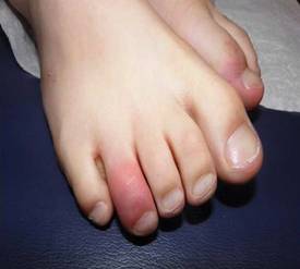
Pernio, also known as chilblain, is a distinctive vasoactive reaction with asymptomatic erythema progressing to blue discoloration of a toe. Its dramatic presentation – "blue toes kind of coming out of nowhere" – can make physicians worry about potential vasculitis, connective disease, or other problems, Dr. Sidbury said.
The second published report described a child who had been taking extended-release Adderall for ADHD for 6 months, was otherwise well, and had no known triggers for pernio such as exposure to cold. The pernio resolved 4 weeks after discontinuing the medication.
Pernio traditionally has been described in Wisconsin hunters with wet socks and cold (but not extremely cold) exposure of the extremities. "In a susceptible population, you get almost a Raynaud’s-type phenomenon," he said. In the case of pernio from stimulants, patients don’t get the classic red, white, and blue cutaneous changes described in the hunters, "you just get a very classic blue-looking toe."
Mechanistically, it makes sense that stimulants could have this sort of reaction in susceptible individuals, he added.
Dr. Sidbury said he had no relevant conflicts of interest.
SDEF and this news organization are owned by Elsevier.
SAN FRANCISCO – Cutaneous reactions are starting to be seen in children on stimulant medications for attention deficit hyperactivity disorder.
Methylphenidate hydrochloride (Ritalin) was associated with acute generalized exanthematous pustulosis (AGEP) in a recent report of a 9-year-old boy being treated for ADHD (Arch. Dermatol. 2011;147:872-3). Dextroamphetamine plus amphetamine (Adderall) was associated with pernio in a separate report of a 9-year-old girl also being treated for ADHD (J. Am. Acad. Dermatol. 2011;64:1218-9).
Before the report of AGEP and Ritalin use, "I had not heard of it related to stimulant therapy," Dr. Robert Sidbury said at the SDEF Women’s and Pediatric Dermatology Seminar, sponsored by Skin Disease Education Foundation (SDEF).
One could be skeptical about a single report of AGEP associated with Ritalin, he acknowledged, but in the case of pernio and stimulants, "I’ve now had three cases of kids who developed this reaction to stimulant therapy," he said. "I believe this to be true."
Both have dramatic presentations that can alarm parents and physicians. "It’s nice to know you can just link it to a certain medication," said Dr. Sidbury, chief of dermatology at Seattle Children’s Hospital. "That can cause a lot of relief."
AGEP is a shower of very small pustules that can be related to a variety of medications. The child in the first report had been taking Ritalin for 6 weeks. Pathology showed sterile intracorneal pustules with mixed eosinophilic infiltrate. A lymphocyte transformation test was positive for AGEP. The patient stopped Ritalin, was treated with topical and systemic steroids for the AGEP, and "did just fine," he said.
Previously, Ritalin has been associated with other cutaneous reactions including morbilliform eruption, urticaria, and even alopecia.
Pernio, also known as chilblain, is a distinctive vasoactive reaction with asymptomatic erythema progressing to blue discoloration of a toe. Its dramatic presentation – "blue toes kind of coming out of nowhere" – can make physicians worry about potential vasculitis, connective disease, or other problems, Dr. Sidbury said.
The second published report described a child who had been taking extended-release Adderall for ADHD for 6 months, was otherwise well, and had no known triggers for pernio such as exposure to cold. The pernio resolved 4 weeks after discontinuing the medication.
Pernio traditionally has been described in Wisconsin hunters with wet socks and cold (but not extremely cold) exposure of the extremities. "In a susceptible population, you get almost a Raynaud’s-type phenomenon," he said. In the case of pernio from stimulants, patients don’t get the classic red, white, and blue cutaneous changes described in the hunters, "you just get a very classic blue-looking toe."
Mechanistically, it makes sense that stimulants could have this sort of reaction in susceptible individuals, he added.
Dr. Sidbury said he had no relevant conflicts of interest.
SDEF and this news organization are owned by Elsevier.
EXPERT ANALYSIS FROM THE SDEF WOMEN'S & PEDIATRIC DERMATOLOGY SEMINAR
Candida Antigen Injections Clear Pediatric Warts
SAN FRANCISCO – The current savior of dermatologists who treat pediatric warts is immunotherapy with Candida antigen, according to Dr. Sheila Fallon Friedlander.
"Warts are either the bane of your existence or the bread and butter," she said at the Women’s & Pediatric Dermatology Seminar sponsored by Skin Disease Education Foundation (SDEF). Candida antigen injections are not her first-line treatment choice, and injections aren’t desirable for some warts (such as those on the face), but she said Candida antigen is effective.
"There’s a lot of experience out there now. Candida truly does work, but you’ve got to be aware of side effects," said Dr. Friedlander, professor of clinical pediatrics and medicine at the University of California, San Diego.
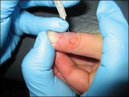
Articles in the medical literature suggest injecting up to 0.3 cc of Candida antigen directly into the substance of the largest wart. "Who are they kidding?" she scoffed. The wart would have to be a giant one to hold that much. "Get what you can directly into the wart," she suggested.
There’s controversy about whether it’s most important to get Candida antigen into the wart, the dermis, or both. Dr. Friedlander said she tries to do both. There’s no need to treat all warts; usually injecting one to three will suffice. Repeat the treatment every 3 weeks for a total of three to five treatments, she said.
One study found a relapse rate of 5% after 2 years, compared with relapse rates of 39% with cryotherapy and 10% with laser therapy (Arch. Dermatol. 2001;137:451-5).
Be sure to warn the family that the treatment may cause discomfort, redness, and swelling. Dr. Friedlander said she is cautious about injecting distal fingers because of a report of complications from Candida antigen injections of a patient’s distal left thumb and the distal subungual area of the left index finger. Within 24 hours the patient had a painful purple index finger with tremendous edema requiring incision (Dermatitis 2005;16:38-40).
To choose the best treatment for pediatric warts, consider some key questions, she advised. Are the lesions truly warts or something else (molluscum or Spitz nevi)? Is the patient well or immunocompromised? Are the warts large or small? Do they create a cosmetic problem? Most importantly, does the patient care that the warts are there?
Unlike acne, which raises concerns about scarring, pediatric warts usually disappear over time without leaving a scar. "Remember, you are the patient’s advocate," which sometimes means clashing with parents over whether to treat a wart, she said.
If you pursue treatment, design a plan the family can live with. Some want only gentle, organic products while others want to take a blowtorch to warts.

Her favorite approach is to start with cryotherapy "for the brave," and in between cryotherapy treatments, have the family use "the triple whammy" – topical salicylic acid, Mediplast, and duct tape, she said. If this isn’t working, and the family wants more, she quickly switches to intralesional Candida for multiple warts. For facial warts, however, she said she prefers tretinoin cream, and for periungual warts she prefers cantharidin (blister beetle extract).
Dr. Friedlander recommended a recent review in the May/June 2011 issue of Pediatric Dermatology of these and other treatments for warts that include a complex algorithm for choosing therapies (Ped. Derm. 2011;28:217-29).
A separate prospective randomized trial reported that a triple combination of cantharidin, podophyllotoxin, and salicylic acid worked better than cryotherapy to remove plantar warts (J. Eur. Acad. Dermatol. Venereol. 2011 July 26 [doi:10.1111/j.1468-3083.2011.04186.x]).
Some physicians are using photodynamic therapy to treat warts, but "I think that’s a sledgehammer approach," she said. A Cochrane review of 60 randomized controlled trials reported that topical salicylic acid has the best evidence for treating warts, but that review did not include Candida antigen, she added (Cochrane Database Syst. Rev. 2006 July 19;3:CD001781).
Dr. Friedlander said she has been a speaker, consultant, or researcher for Pierre-Favre, Onset Therapeutics, Ortho/Johnson & Johnson, and Galderma.
SDEF and this news organization are owned by Elsevier.
SAN FRANCISCO – The current savior of dermatologists who treat pediatric warts is immunotherapy with Candida antigen, according to Dr. Sheila Fallon Friedlander.
"Warts are either the bane of your existence or the bread and butter," she said at the Women’s & Pediatric Dermatology Seminar sponsored by Skin Disease Education Foundation (SDEF). Candida antigen injections are not her first-line treatment choice, and injections aren’t desirable for some warts (such as those on the face), but she said Candida antigen is effective.
"There’s a lot of experience out there now. Candida truly does work, but you’ve got to be aware of side effects," said Dr. Friedlander, professor of clinical pediatrics and medicine at the University of California, San Diego.
Articles in the medical literature suggest injecting up to 0.3 cc of Candida antigen directly into the substance of the largest wart. "Who are they kidding?" she scoffed. The wart would have to be a giant one to hold that much. "Get what you can directly into the wart," she suggested.
There’s controversy about whether it’s most important to get Candida antigen into the wart, the dermis, or both. Dr. Friedlander said she tries to do both. There’s no need to treat all warts; usually injecting one to three will suffice. Repeat the treatment every 3 weeks for a total of three to five treatments, she said.
One study found a relapse rate of 5% after 2 years, compared with relapse rates of 39% with cryotherapy and 10% with laser therapy (Arch. Dermatol. 2001;137:451-5).
Be sure to warn the family that the treatment may cause discomfort, redness, and swelling. Dr. Friedlander said she is cautious about injecting distal fingers because of a report of complications from Candida antigen injections of a patient’s distal left thumb and the distal subungual area of the left index finger. Within 24 hours the patient had a painful purple index finger with tremendous edema requiring incision (Dermatitis 2005;16:38-40).
To choose the best treatment for pediatric warts, consider some key questions, she advised. Are the lesions truly warts or something else (molluscum or Spitz nevi)? Is the patient well or immunocompromised? Are the warts large or small? Do they create a cosmetic problem? Most importantly, does the patient care that the warts are there?
Unlike acne, which raises concerns about scarring, pediatric warts usually disappear over time without leaving a scar. "Remember, you are the patient’s advocate," which sometimes means clashing with parents over whether to treat a wart, she said.
If you pursue treatment, design a plan the family can live with. Some want only gentle, organic products while others want to take a blowtorch to warts.
Her favorite approach is to start with cryotherapy "for the brave," and in between cryotherapy treatments, have the family use "the triple whammy" – topical salicylic acid, Mediplast, and duct tape, she said. If this isn’t working, and the family wants more, she quickly switches to intralesional Candida for multiple warts. For facial warts, however, she said she prefers tretinoin cream, and for periungual warts she prefers cantharidin (blister beetle extract).
Dr. Friedlander recommended a recent review in the May/June 2011 issue of Pediatric Dermatology of these and other treatments for warts that include a complex algorithm for choosing therapies (Ped. Derm. 2011;28:217-29).
A separate prospective randomized trial reported that a triple combination of cantharidin, podophyllotoxin, and salicylic acid worked better than cryotherapy to remove plantar warts (J. Eur. Acad. Dermatol. Venereol. 2011 July 26 [doi:10.1111/j.1468-3083.2011.04186.x]).
Some physicians are using photodynamic therapy to treat warts, but "I think that’s a sledgehammer approach," she said. A Cochrane review of 60 randomized controlled trials reported that topical salicylic acid has the best evidence for treating warts, but that review did not include Candida antigen, she added (Cochrane Database Syst. Rev. 2006 July 19;3:CD001781).
Dr. Friedlander said she has been a speaker, consultant, or researcher for Pierre-Favre, Onset Therapeutics, Ortho/Johnson & Johnson, and Galderma.
SDEF and this news organization are owned by Elsevier.
EXPERT ANALYSIS FROM THE SDEF WOMEN'S & PEDIATRIC DERMATOLOGY SEMINAR
Itchy and Pregnant? Consider Range of Skin Conditions
SAN FRANCISCO – When a pregnant woman presents with a complaint of itching, consider a range of causes, not just those triggered by pregnancy, Dr. Bethanee J. Schlosser advised.
Although some dermatoses of pregnancy are common, a pregnant woman’s itching may have nothing to do with her pregnancy and could be the result of contact dermatitis, drug eruption, scabies, folliculitis, or another cause, Dr. Schlosser said at SDEF Women’s & Pediatric Dermatology Seminar sponsored by Skin Disease Education Foundation (SDEF).

"Just because they’re pregnant doesn’t mean they only have to fit in the pregnancy dermatoses box," Dr. Schlosser of the department of dermatology, and director of the women’s skin health program, at Northwestern University in Chicago said in an interview.
With that said, the two most prominent dermatoses of pregnancy are pruritic urticarial papules and plaques of pregnancy, a condition now known under the umbrella term polymorphic eruption of pregnancy, and pemphigoid gestationis, previously called herpes gestationis.
Polymorphic eruption of pregnancy occurs in about 1 in 300 pregnancies and is generally associated with multiple gestations and increased maternal weight gain. It is also more common in women having their first child. The mean onset is at about 35 weeks, but in about 15% of cases, the onset can be post partum, according to Dr. Schlosser.
Pemphigoid gestationis is a rare acquired autoimmune blistering disease unique to pregnancy. It occurs in 1 in 50,000 pregnancies and is probably the least common dermatosis of pregnancy. The onset is usually in the second or third trimester, but in about 14% of cases, the onset can occur post partum. With pemphigoid gestationis, there is no change in maternal outcome, but there are risks to the fetus including being small for gestational age, preterm delivery, and neonatal pemphigoid disease.
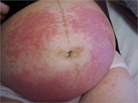
Typically, patients with the polymorphic eruption present with "hivelike" or urticarial papules and plaques, but no blisters, while women with pemphigoid gestationis often have more blistering. However, the clinical presentations and the routine histopathology can be identical, Dr. Schlosser explained.
"I’ve seen patients with both entities, with both kinds of clinical features," she noted. "If it’s in your differential diagnosis and you can’t distinguish 100% clinically, then that’s where the utility of biopsy comes in."
Cutaneous biopsy is a common procedure and is low risk, she reported, even in the context of pregnancy. Routine histopathology and direct immunofluorescence are essential in terms of differentiating between pemphigoid gestationis and polymorphic eruption.
The first-line treatment for both conditions is topical corticosteroids and oral antihistamines when the condition is mild or localized and systemic corticosteroids in severe cases. Although the treatments are generally the same, the difference between the two conditions is not academic, Dr. Schlosser said, because the potential sequelae and considerations for mother and child are different.

Dr. Schlosser also recommended that dermatologists make it a priority to communicate with the referring physician, specifically to review the risks to both the mother and child that may be associated with a particular skin condition or its treatments.
For example, polymorphic eruption of pregnancy is generally nonthreatening to the mother and child. But Dr. Schlosser said she has seen patients with widespread, severe polymorphic eruptions who have needed treatment with systemic corticosteroids. That’s essential information for the ob.gyn.; if the patient has a cesarean delivery, the patient will likely require stress-dose corticosteroids. Similarly, the newborn would need to be monitored for hypoglycemia during the immediate after-birth period.
"That doesn’t mean that dermatologists shouldn’t treat pregnant women aggressively, when appropriate," Dr. Schlosser said. "But the entire multidisciplinary care team needs to be kept informed so that the risks can be managed."
Dr. Schlosser said she had no relevant financial disclosures. SDEF and this news organization are owned by Elsevier.
SAN FRANCISCO – When a pregnant woman presents with a complaint of itching, consider a range of causes, not just those triggered by pregnancy, Dr. Bethanee J. Schlosser advised.
Although some dermatoses of pregnancy are common, a pregnant woman’s itching may have nothing to do with her pregnancy and could be the result of contact dermatitis, drug eruption, scabies, folliculitis, or another cause, Dr. Schlosser said at SDEF Women’s & Pediatric Dermatology Seminar sponsored by Skin Disease Education Foundation (SDEF).
"Just because they’re pregnant doesn’t mean they only have to fit in the pregnancy dermatoses box," Dr. Schlosser of the department of dermatology, and director of the women’s skin health program, at Northwestern University in Chicago said in an interview.
With that said, the two most prominent dermatoses of pregnancy are pruritic urticarial papules and plaques of pregnancy, a condition now known under the umbrella term polymorphic eruption of pregnancy, and pemphigoid gestationis, previously called herpes gestationis.
Polymorphic eruption of pregnancy occurs in about 1 in 300 pregnancies and is generally associated with multiple gestations and increased maternal weight gain. It is also more common in women having their first child. The mean onset is at about 35 weeks, but in about 15% of cases, the onset can be post partum, according to Dr. Schlosser.
Pemphigoid gestationis is a rare acquired autoimmune blistering disease unique to pregnancy. It occurs in 1 in 50,000 pregnancies and is probably the least common dermatosis of pregnancy. The onset is usually in the second or third trimester, but in about 14% of cases, the onset can occur post partum. With pemphigoid gestationis, there is no change in maternal outcome, but there are risks to the fetus including being small for gestational age, preterm delivery, and neonatal pemphigoid disease.
Typically, patients with the polymorphic eruption present with "hivelike" or urticarial papules and plaques, but no blisters, while women with pemphigoid gestationis often have more blistering. However, the clinical presentations and the routine histopathology can be identical, Dr. Schlosser explained.
"I’ve seen patients with both entities, with both kinds of clinical features," she noted. "If it’s in your differential diagnosis and you can’t distinguish 100% clinically, then that’s where the utility of biopsy comes in."
Cutaneous biopsy is a common procedure and is low risk, she reported, even in the context of pregnancy. Routine histopathology and direct immunofluorescence are essential in terms of differentiating between pemphigoid gestationis and polymorphic eruption.
The first-line treatment for both conditions is topical corticosteroids and oral antihistamines when the condition is mild or localized and systemic corticosteroids in severe cases. Although the treatments are generally the same, the difference between the two conditions is not academic, Dr. Schlosser said, because the potential sequelae and considerations for mother and child are different.
Dr. Schlosser also recommended that dermatologists make it a priority to communicate with the referring physician, specifically to review the risks to both the mother and child that may be associated with a particular skin condition or its treatments.
For example, polymorphic eruption of pregnancy is generally nonthreatening to the mother and child. But Dr. Schlosser said she has seen patients with widespread, severe polymorphic eruptions who have needed treatment with systemic corticosteroids. That’s essential information for the ob.gyn.; if the patient has a cesarean delivery, the patient will likely require stress-dose corticosteroids. Similarly, the newborn would need to be monitored for hypoglycemia during the immediate after-birth period.
"That doesn’t mean that dermatologists shouldn’t treat pregnant women aggressively, when appropriate," Dr. Schlosser said. "But the entire multidisciplinary care team needs to be kept informed so that the risks can be managed."
Dr. Schlosser said she had no relevant financial disclosures. SDEF and this news organization are owned by Elsevier.
EXPERT ANALYSIS FROM THE SDEF WOMEN'S & PEDIATRIC DERMATOLOGY SEMINAR