User login
American College of Emergency Physicians (ACEP): Annual Scientific Assembly
Passive leg raise may predict fluid response in sepsis
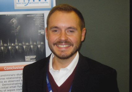
SEATTLE – Septic patients are more likely to respond to fluid therapy if their velocity time integral – a Doppler ultrasound measurement of blood flow across the left ventricular outflow tract – increases by 15% or more with a passive single-leg raise, according to a preliminary, observational study of 32 patients at New York Methodist Hospital.
A passive leg raise to 45 degrees simulates a 250- to 500-cc fluid bolus. "We have found that people who don’t respond with a VTI greater than 15% have higher repeat lactate levels. Instead of giving them 2 L [of fluid] and then reassessing, maybe they’re patients you want to start on pressors right away," Dr. Andrew Balk said at the annual meeting of the American College of Emergency Physicians.

Echocardiogram machines can automatically calculate VTI. The measurement, which Dr. Balk and his associates obtained from the apical five-chamber view, is a surrogate for, and can be used to calculate, cardiac output. Poor response to fluid challenge indicates that fluids are less likely to increase cardiac output and more likely to cause fluid overload, said Dr. Balk, associate director of the clinical ultrasound division at the hospital.
The patients’ mean age was 68 years, and those with valvular pathology and atrial fibrillation were excluded from the study.
The group’s mean baseline VTI was 22 cm (range, 15-29 cm), which leg raise elevated to a mean of 26 cm (18-34 cm), an increase of about 18% (4%-36%). A subsequent 2-L normal saline challenge increased VTI to a mean of 33 cm.
The mean baseline lactate level was 3.2 mmol/L (1.2-5.2 mmol/L), and 2 mmol/L (1-3 mmol/L) after the 2-L challenge. The percent change in VTI correlated significantly with the percent change in serum lactate levels. "Below-average responsiveness to the initial small fluid bolus was associated with a higher repeat lactate value ... which suggests an inverse relationship between a patient’s fluid responsiveness as observed by the change in VTI and the severity of sepsis," the researchers concluded.
The VTI/leg-raise approach looks promising as a possible quick bedside marker that identifies patients who need aggressive treatment, without the need for central line measurements, Dr. Balk said."The quickest initial fluid bolus you can get is a passive leg raise. You can watch for changes" in real time, and don’t have to move the probe from the point of maximum impact.
He reported having no disclosures.
Dr. Steven Q. Simpson, FCCP, comments: The search for noninvasive measures of or predictors for volume responsiveness in patients with sepsis continues. VTI is the integral of velocity and time, that is, the distance a small blood bolus travels. When multiplied by the cross-sectional area of the aortic outflow tract, this would result in stroke volume.
Since one would not expect the cross-sectional area to change significantly after a fluid bolus, alterations in VTI should reflect alterations in stroke volume.
While promising, this technique is not as easy as the authors make it sound and is operator dependent, even though the machine does the calculating. The incident angle of the probe must remain constant during the leg raise (for a period of at least 90 seconds). The user must know whether valve pathology or left ventricular impairment is present and, if so, the degree.
In addition, massively volume-depleted patients may fail to respond adequately to a passive leg raise.
One would be remiss to rely on this small study, which does not report sensitivity or specificity, to establish a reliable percent increase for predicting lactate response or to guide fluid therapy.
However, this research by Dr. Balk and colleagues is certainly aimed in the right direction.
SEATTLE – Septic patients are more likely to respond to fluid therapy if their velocity time integral – a Doppler ultrasound measurement of blood flow across the left ventricular outflow tract – increases by 15% or more with a passive single-leg raise, according to a preliminary, observational study of 32 patients at New York Methodist Hospital.
A passive leg raise to 45 degrees simulates a 250- to 500-cc fluid bolus. "We have found that people who don’t respond with a VTI greater than 15% have higher repeat lactate levels. Instead of giving them 2 L [of fluid] and then reassessing, maybe they’re patients you want to start on pressors right away," Dr. Andrew Balk said at the annual meeting of the American College of Emergency Physicians.
Echocardiogram machines can automatically calculate VTI. The measurement, which Dr. Balk and his associates obtained from the apical five-chamber view, is a surrogate for, and can be used to calculate, cardiac output. Poor response to fluid challenge indicates that fluids are less likely to increase cardiac output and more likely to cause fluid overload, said Dr. Balk, associate director of the clinical ultrasound division at the hospital.
The patients’ mean age was 68 years, and those with valvular pathology and atrial fibrillation were excluded from the study.
The group’s mean baseline VTI was 22 cm (range, 15-29 cm), which leg raise elevated to a mean of 26 cm (18-34 cm), an increase of about 18% (4%-36%). A subsequent 2-L normal saline challenge increased VTI to a mean of 33 cm.
The mean baseline lactate level was 3.2 mmol/L (1.2-5.2 mmol/L), and 2 mmol/L (1-3 mmol/L) after the 2-L challenge. The percent change in VTI correlated significantly with the percent change in serum lactate levels. "Below-average responsiveness to the initial small fluid bolus was associated with a higher repeat lactate value ... which suggests an inverse relationship between a patient’s fluid responsiveness as observed by the change in VTI and the severity of sepsis," the researchers concluded.
The VTI/leg-raise approach looks promising as a possible quick bedside marker that identifies patients who need aggressive treatment, without the need for central line measurements, Dr. Balk said."The quickest initial fluid bolus you can get is a passive leg raise. You can watch for changes" in real time, and don’t have to move the probe from the point of maximum impact.
He reported having no disclosures.
Major finding: The percent change in the velocity time integral on passive leg raise correlates significantly with the percent change in serial serum lactate levels (P = .05).
Data source: Prospective, observational study in 32 septic adults.
Disclosures: Dr. Balk reported having no disclosures.
Work cardiac resuscitations in the field, not the ambulance
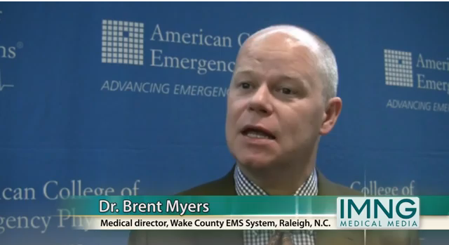
SEATTLE – In almost all cases, it’s best to stay on the scene to work a cardiac arrest resuscitation until the return of spontaneous circulation or efforts are stopped because of futility, according to Dr. Brent Myers, director of emergency medical services in Raleigh/Wake County, N.C.
The probability of neurologically intact survival is at least 10-fold higher, and by some estimates up to 35-fold higher, when resuscitation is achieved in the field, instead of en route to the emergency department, Dr. Myers said at the annual meeting of the American College of Emergency Physicians. Neurologically intact survival quadrupled to 11.5% for patients in ventricular fibrillation and to 40.8% for those with ventricular tachycardia after the approach was adopted in Wake County, he reported. (Ann. Emerg. Med. 2010;56:348-57).
"Load-and-go is not the appropriate approach; resuscitation is a prehospital exercise," Dr. Myers said. "In the vast majority of cases, if we interrupt compressions to move patients" – which is unavoidable with even the most careful transport – "we have sealed their fate."
An ever-increasing number of emergency medicine experts agree, but some EMS systems still have forced moves at, for example, 20 minutes. That standard probably accounts for much of the considerable regional variations in survival of cardiac arrest patients. "The variability, in large portion, can be explained simply by who’s on the chest and who’s not. As you stay on the chest above 80% of the time, your odds of survival triple," he said (for example, Circulation 2010;122:S685-705; JAMA 2008;300:1423-31).
"How long we should stay on the scene still needs further research, [but] the old rule that says you shouldn’t go beyond 30 minutes is clearly not true. We just don’t know what the new time is; it’s somewhere north of 30 minutes," he said.
In general, Wake County crews keep at resuscitation until asystole or disorganized pulseless electrical activity (PEA). "We don’t ever terminate [a patient with ventricular fibrillation] or good-looking PEA, which has resulted in prolonged resuscitation times" of sometimes 50 minutes or more. "They are very unlikely to survive after 40 minutes; but if they do, they are just as likely to be neurologically intact. We are not creating persistently vegetative states," Dr. Myers said.
In-home termination – which happens in almost half of Wake County cases – goes hand in hand with the approach. "The systems with the highest resuscitation rates also have the highest field termination rates. I don’t think that’s an accident; it’s a marker that [they] are focusing resuscitations on the scene," he said.
"You have to have the capacity to terminate, [and] be ready to [help] families" with grief. "Every published paper indicates that family members want to be present for the resuscitation." Among the most important things for them are knowing their loved one is not in pain and being able to touch the body; the cleanliness of the body is also important (Ann. Emerg. Med. 2002;40:521-3).
There may be a small number of patients who benefit from intra-arrest transport, but "we do not have sufficient data" to identify them. One group might be young, otherwise-healthy people in VF who can be whisked straight into a cath lab. "It sounds good, but we haven’t proven it yet," Dr. Myers said.
Rearrest is most likely within 10 minutes of a successful resuscitation, so, just in case, Dr. Myers’ crews stay put with patients for 10 minutes after they come around, before transport. If they re-arrest en route to the ED, in most cases crews "pull over and work the arrest right there in the ambulance," he said.
Dr. Myers reported having no disclosures.
EXPERT ANALYSIS FROM THE 2013 ACEP SCIENTIFIC ASSEMBLY
Keep atropine in reserve for pediatric rapid-sequence intubation
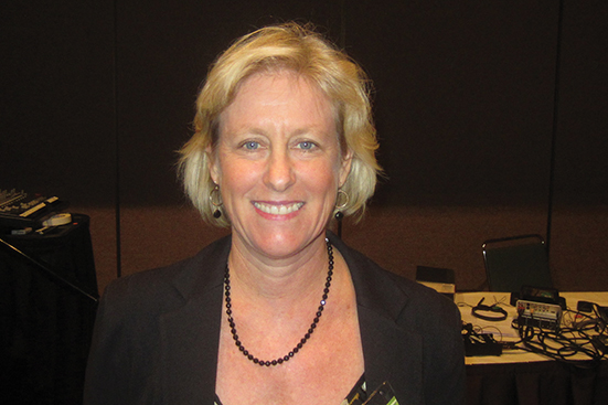
SEATTLE – Routine atropine is "optional, not standard" for preventing reflex bradycardia in children during rapid-sequence intubation, according to Dr. Marianne Gausche-Hill, associate professor of emergency medicine at the University of California, Los Angeles.
A common-sense guideline is to have atropine available in case you need it, she said. If bradycardia occurs, "the first thing I do ... is stop the intubation and begin bag-mask ventilation, and then, if they don’t respond, atropine is a reasonable choice," she said at the annual meeting of the American College of Emergency Physicians (ACEP).

Dr. Gausche-Hill uses a high-dose nasal cannula during the apneic period to prevent hypoxia – 5 L/min in neonates and young children, and 15 L/min in older children – and correct hypovolemia, when possible, before intubation. She also uses suction to deal with secretions.
When atropine is needed during rapid-sequence intubation (RSI), Dr. Gausche-Hill opts for a dose of 0.01-0.02 mg/kg for infants under 5 kg, based on research that found that dose to be both safe and effective (Pediatrics 2011;127:783-4).
"Atropine can prevent bradycardia associated with the administration of succinylcholine, but we don’t know how effective that is in the face of hypoxia. It appears that hypoxia is really the issue here, and not forgoing pretreatment with atropine," she said, echoing a meta-analysis of the issue (Emerg. Med. J. 2007;24:361-2).
Increasingly, emergency departments are not routinely using atropine in pediatric RSI, but the approach hasn’t been adopted everywhere, according to an informal poll taken during Dr. Gausche-Hill’s presentation at the ACEP Scientific Assembly.
In addition, Advanced Pediatric Life Support (APLS) guidelines recommend routine use of atropine in infants under 1 year old, in children 1-5 years old who receive succinylcholine, and in older children who receive a second dose of succinylcholine during RSI, she said.
The notion that children need atropine to prevent reflex bradycardia is based on pediatric surgery studies using inhaled anesthetics and succinylcholine. Researchers "felt that with those inhalation anesthetics, atropine would decrease bradycardia associated with use of high-dose succinylcholine, [but] we are not using those inhalation agents, which may in themselves have resulted in mild hypoxia and bradycardia," she said.
In one study, atropine made no difference in pediatric RSI. Among 68 young children who got atropine, 3 became bradycardic, as did 3 of 75 other children who didn’t get atropine (Pediatr. Emerg. Care 2004;20:651-5).
It appears hypoxia, not forgoing pretreatment with atropine, is a strong predictor of patients who will develop reflex bradycardia, she said.
Further, atropine masks the ability to monitor heart rate changes; increases the risk of ventricular arrhythmias; relaxes the lower esophageal sphincter, possibly increasing the risk of aspiration; and increases temperature and the risk of malignant hyperthermia.
Dr. Gausche-Hill had no relevant disclosures.
SEATTLE – Routine atropine is "optional, not standard" for preventing reflex bradycardia in children during rapid-sequence intubation, according to Dr. Marianne Gausche-Hill, associate professor of emergency medicine at the University of California, Los Angeles.
A common-sense guideline is to have atropine available in case you need it, she said. If bradycardia occurs, "the first thing I do ... is stop the intubation and begin bag-mask ventilation, and then, if they don’t respond, atropine is a reasonable choice," she said at the annual meeting of the American College of Emergency Physicians (ACEP).
Dr. Gausche-Hill uses a high-dose nasal cannula during the apneic period to prevent hypoxia – 5 L/min in neonates and young children, and 15 L/min in older children – and correct hypovolemia, when possible, before intubation. She also uses suction to deal with secretions.
When atropine is needed during rapid-sequence intubation (RSI), Dr. Gausche-Hill opts for a dose of 0.01-0.02 mg/kg for infants under 5 kg, based on research that found that dose to be both safe and effective (Pediatrics 2011;127:783-4).
"Atropine can prevent bradycardia associated with the administration of succinylcholine, but we don’t know how effective that is in the face of hypoxia. It appears that hypoxia is really the issue here, and not forgoing pretreatment with atropine," she said, echoing a meta-analysis of the issue (Emerg. Med. J. 2007;24:361-2).
Increasingly, emergency departments are not routinely using atropine in pediatric RSI, but the approach hasn’t been adopted everywhere, according to an informal poll taken during Dr. Gausche-Hill’s presentation at the ACEP Scientific Assembly.
In addition, Advanced Pediatric Life Support (APLS) guidelines recommend routine use of atropine in infants under 1 year old, in children 1-5 years old who receive succinylcholine, and in older children who receive a second dose of succinylcholine during RSI, she said.
The notion that children need atropine to prevent reflex bradycardia is based on pediatric surgery studies using inhaled anesthetics and succinylcholine. Researchers "felt that with those inhalation anesthetics, atropine would decrease bradycardia associated with use of high-dose succinylcholine, [but] we are not using those inhalation agents, which may in themselves have resulted in mild hypoxia and bradycardia," she said.
In one study, atropine made no difference in pediatric RSI. Among 68 young children who got atropine, 3 became bradycardic, as did 3 of 75 other children who didn’t get atropine (Pediatr. Emerg. Care 2004;20:651-5).
It appears hypoxia, not forgoing pretreatment with atropine, is a strong predictor of patients who will develop reflex bradycardia, she said.
Further, atropine masks the ability to monitor heart rate changes; increases the risk of ventricular arrhythmias; relaxes the lower esophageal sphincter, possibly increasing the risk of aspiration; and increases temperature and the risk of malignant hyperthermia.
Dr. Gausche-Hill had no relevant disclosures.
EXPERT ANALYSIS AT THE ACEP SCIENTIFIC ASSEMBLY 2013
Passive leg raise may predict fluid responsiveness in sepsis
SEATTLE – Septic patients are more likely to respond to fluid therapy if their velocity time integral – a Doppler ultrasound measurement of blood flow across the left ventricular outflow tract – increases by 15% or more with a passive single-leg raise, according to a preliminary, observational study of 32 patients at New York Methodist Hospital in Brooklyn.
A passive leg raise to 45 degrees simulates a 250- to 500-cc fluid bolus. "We have found that people who don’t respond with a VTI greater than 15% have higher repeat lactate levels. Instead of giving them 2 L [of fluid] and then reassessing, maybe they’re patients you want to start on pressors right away," Dr. Andrew Balk said at the annual meeting of the American College of Emergency Physicians.
Echocardiogram machines can automatically calculate VTI. The measurement, which Dr. Balk and his associates obtained from the apical five-chamber view, is a surrogate for, and can be used to calculate, cardiac output. Poor response to fluid challenge indicates that fluids are less likely to increase cardiac output and more likely to cause fluid overload, said Dr. Balk, associate director of the clinical ultrasound division at the hospital.
The patients’ mean age was 68 years, and those with valvular pathology and atrial fibrillation were excluded from the study.
The group’s mean baseline VTI was 22 cm (range, 15-29 cm), which leg raises raised to a mean of 26 cm (18-34 cm), an increase of about 18% (4%-36%). A subsequent 2-L normal saline challenge increased VTI to a mean of 33 cm.
The mean baseline lactate level was 3.2 mmol/L (1.2-5.2 mmol/L), and 2 mmol/L (1-3 mmol/L) after the 2-L challenge. The percent change in VTI correlated significantly with the percent change in serum lactate levels. "Below-average responsiveness to the initial small fluid bolus was associated with a higher repeat lactate value, ... which suggests an inverse relationship between a patient’s fluid responsiveness as observed by the change in VTI and the severity of sepsis," the researchers concluded.
The VTI/leg-raise approach looks promising as a possible quick bedside marker that identifies patients who need aggressive treatment, without the need for central line measurements, Dr. Balk said. "The quickest initial fluid bolus you can get is a passive leg raise. You can watch for changes" in real time, and don’t have to move the probe from the point of maximum impact.
Dr. Balk reported having no disclosures.

Dr. Steven Q. Simpson commented: The search for noninvasive measures of or predictors for volume responsiveness in septic patients continues. VTI is the integral of velocity and time, i.e., the distance a small blood bolus travels. When multiplied by cross-sectional area of the aortic outflow tract, this would result in stroke volume. Since one would not expect the cross-sectional area to change significantly after a fluid bolus, alterations in VTI should reflect alterations in stroke volume. While promising, this technique is not as easy as the authors make it sound
and is operator dependent, even though the machine does the calculating. The incident angle of the probe must remain constant during the leg raise (at least 90 seconds). The user must know whether valve pathology or LV impairment are present and, if so, the degree. Massively volume depleted patients may fail to respond adequately to a passive leg raise.
One would be remiss to rely on this small study, which does not report sensitivity or specificity, to establish a reliable percent increase for predicting lactate response or to guide fluid therapy. However, this research is certainly aimed in the right direction.
Dr. Steven Q. Simpson is professor of medicine and director of fellowship training in the pulmonary disease and critical care medicine division at the University of Kansas Medical Center, Kansas City.
SEATTLE – Septic patients are more likely to respond to fluid therapy if their velocity time integral – a Doppler ultrasound measurement of blood flow across the left ventricular outflow tract – increases by 15% or more with a passive single-leg raise, according to a preliminary, observational study of 32 patients at New York Methodist Hospital in Brooklyn.
A passive leg raise to 45 degrees simulates a 250- to 500-cc fluid bolus. "We have found that people who don’t respond with a VTI greater than 15% have higher repeat lactate levels. Instead of giving them 2 L [of fluid] and then reassessing, maybe they’re patients you want to start on pressors right away," Dr. Andrew Balk said at the annual meeting of the American College of Emergency Physicians.
Echocardiogram machines can automatically calculate VTI. The measurement, which Dr. Balk and his associates obtained from the apical five-chamber view, is a surrogate for, and can be used to calculate, cardiac output. Poor response to fluid challenge indicates that fluids are less likely to increase cardiac output and more likely to cause fluid overload, said Dr. Balk, associate director of the clinical ultrasound division at the hospital.
The patients’ mean age was 68 years, and those with valvular pathology and atrial fibrillation were excluded from the study.
The group’s mean baseline VTI was 22 cm (range, 15-29 cm), which leg raises raised to a mean of 26 cm (18-34 cm), an increase of about 18% (4%-36%). A subsequent 2-L normal saline challenge increased VTI to a mean of 33 cm.
The mean baseline lactate level was 3.2 mmol/L (1.2-5.2 mmol/L), and 2 mmol/L (1-3 mmol/L) after the 2-L challenge. The percent change in VTI correlated significantly with the percent change in serum lactate levels. "Below-average responsiveness to the initial small fluid bolus was associated with a higher repeat lactate value, ... which suggests an inverse relationship between a patient’s fluid responsiveness as observed by the change in VTI and the severity of sepsis," the researchers concluded.
The VTI/leg-raise approach looks promising as a possible quick bedside marker that identifies patients who need aggressive treatment, without the need for central line measurements, Dr. Balk said. "The quickest initial fluid bolus you can get is a passive leg raise. You can watch for changes" in real time, and don’t have to move the probe from the point of maximum impact.
Dr. Balk reported having no disclosures.
Dr. Steven Q. Simpson commented: The search for noninvasive measures of or predictors for volume responsiveness in septic patients continues. VTI is the integral of velocity and time, i.e., the distance a small blood bolus travels. When multiplied by cross-sectional area of the aortic outflow tract, this would result in stroke volume. Since one would not expect the cross-sectional area to change significantly after a fluid bolus, alterations in VTI should reflect alterations in stroke volume. While promising, this technique is not as easy as the authors make it sound
and is operator dependent, even though the machine does the calculating. The incident angle of the probe must remain constant during the leg raise (at least 90 seconds). The user must know whether valve pathology or LV impairment are present and, if so, the degree. Massively volume depleted patients may fail to respond adequately to a passive leg raise.
One would be remiss to rely on this small study, which does not report sensitivity or specificity, to establish a reliable percent increase for predicting lactate response or to guide fluid therapy. However, this research is certainly aimed in the right direction.
Dr. Steven Q. Simpson is professor of medicine and director of fellowship training in the pulmonary disease and critical care medicine division at the University of Kansas Medical Center, Kansas City.
AT THE ACEP SCIENTIFIC ASSEMBLY 2013

IV antihypertensives are overused, single-center ED chart audit finds
SEATTLE – About a third of the patients who received intravenous bolus antihypertensives in the emergency department at the Detroit Receiving Hospital did not need them, according to the results of a retrospective review at the 340-bed level 1 trauma center.
The findings were presented at the annual meeting of the American College of Emergency Physicians.

Of the 295 patients in the study, all had blood pressure of at least 180/110 mm Hg, but 95 did not have signs and symptoms of end-organ damage and had failed on oral therapy. These patients received no further workup or diagnosis related to acute hypertension after being given at least one IV push of, most commonly, labetalol. Atropine was needed to correct subsequent bradycardia in 1 of those 95 patients, and vasopressors in 1 of 2 patients who became hypotensive.
"A lot of physicians jump the gun and treat the number, not the patient," but in the absence of hypertensive emergencies, patients "can get away with oral therapy. You really don’t want to drop their blood pressure too quickly; you just want to bring them down slowly to their normal," which may only be a bit below their presenting pressure, said Suprat Saely, Pharm. D., an emergency department pharmacist at the hospital.
For the study, pharmacy orders were matched to patient medical records. There were no statistical differences in survival to discharge and 30-day revisits to the ED between the 200 patients treated appropriately and the 95 who were unnecessarily given IV antihypertensives.
These days, "as soon as I get these orders, I assess whether or not they are appropriate. Usually they are, but if not, I’ll go ask the physician if they really meant to order it," said Dr. Saely, who had no disclosures.
SEATTLE – About a third of the patients who received intravenous bolus antihypertensives in the emergency department at the Detroit Receiving Hospital did not need them, according to the results of a retrospective review at the 340-bed level 1 trauma center.
The findings were presented at the annual meeting of the American College of Emergency Physicians.
Of the 295 patients in the study, all had blood pressure of at least 180/110 mm Hg, but 95 did not have signs and symptoms of end-organ damage and had failed on oral therapy. These patients received no further workup or diagnosis related to acute hypertension after being given at least one IV push of, most commonly, labetalol. Atropine was needed to correct subsequent bradycardia in 1 of those 95 patients, and vasopressors in 1 of 2 patients who became hypotensive.
"A lot of physicians jump the gun and treat the number, not the patient," but in the absence of hypertensive emergencies, patients "can get away with oral therapy. You really don’t want to drop their blood pressure too quickly; you just want to bring them down slowly to their normal," which may only be a bit below their presenting pressure, said Suprat Saely, Pharm. D., an emergency department pharmacist at the hospital.
For the study, pharmacy orders were matched to patient medical records. There were no statistical differences in survival to discharge and 30-day revisits to the ED between the 200 patients treated appropriately and the 95 who were unnecessarily given IV antihypertensives.
These days, "as soon as I get these orders, I assess whether or not they are appropriate. Usually they are, but if not, I’ll go ask the physician if they really meant to order it," said Dr. Saely, who had no disclosures.
AT THE ACEP SCIENTIFIC ASSEMBLY 2013

Emergency department electronic record systems fell short in Boston bombings
The massive number of injured patients from the April 2013 Boston Marathon bombings overwhelmed electronic medical records and information systems in some Boston emergency departments.
Their experiences offer an important lesson at a time when most hospitals have switched to electronic systems and providers are soon to face financial penalties for not doing so.
During mass-casualty events, paper and pen might be best, said Dr. Andrew Ulrich, executive vice chair of emergency medicine at the Boston Medical Center (BMC).

"The ability to get names into the system fast enough to order tests and establish a medical record limited us" on April 15, he noted. "There were so many patients coming through so quickly [23 at BMC right after the bombs went off] that it was difficult to know exactly who had what done and where they were going. You couldn’t leave the patient to" go to a computer, log on, get an update, and place orders, Dr. Ulrich said.
Other hospitals "had similar experiences. Communication was one of the more difficult components of this event," he said.
At Brigham and Women’s Hospital, "the first issue was our naming convention. We found that when every patient is ‘unidentified’ with a string of numbers, there’s a lot of confusion and many near misses. [Also,] if your system is inundated with multiple patients at once, it takes more time to register them so you can" go to work, said Dr. Eric Goralnick, the hospital’s medical director of emergency preparedness, who helped care for the 19 blast victims his ED received in the first half hour, and more later.

Now, at Brigham and Women’s, unknown patients are identified with a color, state, or other quickly recognized word.
Since the bombings, the EDs at Boston Medical Center and Brigham and Women’s have stocked up on old-school paper-and-pen trauma packets – an envelope with a wrist band, presigned orders, and other care documents all prestamped with a unique identifier – so that they are ready for the next mass-casualty event.
"We are much more flexible, much more adaptable if we go back to basics on this one, and that’s paper and pen. We are preparing to do that" for the next disaster, so "we can pull a bracelet out of the packet, throw it on the wrist," and start to work. It’s "much faster than getting [patients] onto the [electronic] medical record," Dr. Ulrich said.
Also from the old school department: "We [may also] have a clipboard attached to the patient’s bed, so when the patient rolls somewhere else, whoever gets them just has to look at the paperwork" to know what’s going on "instead of separating information from the patient" with the computer system, he said. "In a situation where there’s so much going on, it’s a clearer way of exchanging information."
Electronic record problems aren’t uncommon in EDs.
"There are many places where the computer system is a barrier to care. If you are trying to handle [an event] like this with a system that’s optimized for internal medicine clinics, it’s not going to work," said Dr. Larry Nathanson, who helped tend to the 22 bombing victims Beth Israel Deaconess Medical Center received in the first hour, almost half of whom were in critical condition.
Things went more smoothly at Beth Israel, where the electronic information system worked well. Part of the reason is that the hospital uses a home-grown system designed by Dr. Nathanson, a computer programmer as well as an emergency physician. The system is quick and provides patient information with few clicks. To identify unknown patients, the system uses a convention similar to the one used to name hurricanes (Annie, Bruce, Candace, etc.); the record can be easily updated once a patient’s identity is known.

The system had been tested prior to the bombings via simulation including 500 patients. "We made sure beforehand that [it] could handle the influx and quickly register large volumes of critical patients," said Dr. Nathanson, who serves as the ED’s director of emergency medical informatics.
"The reason it worked well in a disaster is because it works well every day," he said.
Based on feedback from his colleagues, he’s since added an interface that pops up when two or more mass-casualty victims are in the ED. Pressing it displays them all with a summary of their situations, so providers can home in on them.
Dr. Ulrich and Dr. Goralnick have no relevant disclosures. Dr. Nathanson owns stock in Forerun Inc., the commercialized version of the system he developed for Beth Israel.
The massive number of injured patients from the April 2013 Boston Marathon bombings overwhelmed electronic medical records and information systems in some Boston emergency departments.
Their experiences offer an important lesson at a time when most hospitals have switched to electronic systems and providers are soon to face financial penalties for not doing so.
During mass-casualty events, paper and pen might be best, said Dr. Andrew Ulrich, executive vice chair of emergency medicine at the Boston Medical Center (BMC).
"The ability to get names into the system fast enough to order tests and establish a medical record limited us" on April 15, he noted. "There were so many patients coming through so quickly [23 at BMC right after the bombs went off] that it was difficult to know exactly who had what done and where they were going. You couldn’t leave the patient to" go to a computer, log on, get an update, and place orders, Dr. Ulrich said.
Other hospitals "had similar experiences. Communication was one of the more difficult components of this event," he said.
At Brigham and Women’s Hospital, "the first issue was our naming convention. We found that when every patient is ‘unidentified’ with a string of numbers, there’s a lot of confusion and many near misses. [Also,] if your system is inundated with multiple patients at once, it takes more time to register them so you can" go to work, said Dr. Eric Goralnick, the hospital’s medical director of emergency preparedness, who helped care for the 19 blast victims his ED received in the first half hour, and more later.
Now, at Brigham and Women’s, unknown patients are identified with a color, state, or other quickly recognized word.
Since the bombings, the EDs at Boston Medical Center and Brigham and Women’s have stocked up on old-school paper-and-pen trauma packets – an envelope with a wrist band, presigned orders, and other care documents all prestamped with a unique identifier – so that they are ready for the next mass-casualty event.
"We are much more flexible, much more adaptable if we go back to basics on this one, and that’s paper and pen. We are preparing to do that" for the next disaster, so "we can pull a bracelet out of the packet, throw it on the wrist," and start to work. It’s "much faster than getting [patients] onto the [electronic] medical record," Dr. Ulrich said.
Also from the old school department: "We [may also] have a clipboard attached to the patient’s bed, so when the patient rolls somewhere else, whoever gets them just has to look at the paperwork" to know what’s going on "instead of separating information from the patient" with the computer system, he said. "In a situation where there’s so much going on, it’s a clearer way of exchanging information."
Electronic record problems aren’t uncommon in EDs.
"There are many places where the computer system is a barrier to care. If you are trying to handle [an event] like this with a system that’s optimized for internal medicine clinics, it’s not going to work," said Dr. Larry Nathanson, who helped tend to the 22 bombing victims Beth Israel Deaconess Medical Center received in the first hour, almost half of whom were in critical condition.
Things went more smoothly at Beth Israel, where the electronic information system worked well. Part of the reason is that the hospital uses a home-grown system designed by Dr. Nathanson, a computer programmer as well as an emergency physician. The system is quick and provides patient information with few clicks. To identify unknown patients, the system uses a convention similar to the one used to name hurricanes (Annie, Bruce, Candace, etc.); the record can be easily updated once a patient’s identity is known.
The system had been tested prior to the bombings via simulation including 500 patients. "We made sure beforehand that [it] could handle the influx and quickly register large volumes of critical patients," said Dr. Nathanson, who serves as the ED’s director of emergency medical informatics.
"The reason it worked well in a disaster is because it works well every day," he said.
Based on feedback from his colleagues, he’s since added an interface that pops up when two or more mass-casualty victims are in the ED. Pressing it displays them all with a summary of their situations, so providers can home in on them.
Dr. Ulrich and Dr. Goralnick have no relevant disclosures. Dr. Nathanson owns stock in Forerun Inc., the commercialized version of the system he developed for Beth Israel.

Oral contrast rarely needed before CT scans of adults with acute abdomen
SEATTLE – Oral contrast is almost always unnecessary when performing a CT scan to work up adults with acute abdomens. Further, intravenous contrast is needed only when vascular causes of pain are suspected, according to Dr. Phillips Perera, a clinical associate professor of emergency medicine at Stanford (Calif.) University Medical Center.
As with everything in medicine, there are rare exceptions to this advice, he said. Oral contrast can be helpful to confirm the presence of a fistula in a postcolectomy patient, for example.
With the growth of endovascular repair of abdominal aortic aneurysms, patients with procedural complications including leaks and postop pain are increasingly presenting in emergency departments. Intravenous contrast is needed in those cases, and "more and more of these patients will be coming into the emergency department in the next few years," he said.

Forgoing oral contrast "allows us to get our patients through the emergency department much faster, and we don’t lose [diagnostic] accuracy." It also reduces radiation exposure, because noncontrast CT studies take less time, he said. The sensitivity of noncontrast CT is 93% for detecting acute appendicitis, with a specificity of 96% (Ann. Emerg. Med. 2010;55:51-59).
Recent studies indicate noncontrast CTs work well to diagnose most causes of acute abdominal pain in adults, including appendicitis, diverticulitis, kidney stones, and large ovarian cysts at risk for ovarian torsion (J. Endourol. 2008;22:2441-5).
"You only [lose] about two percentage points" on diagnostic accuracy by forgoing contrast, and the difference in one large study (World J. Surg. 2010;34:699-703) "was not statistically significant, which I think is the most important thing," Dr. Perera said in a literature review discussion at the annual meeting of the American College of Emergency Physicians.
Radiologists still require emergency physicians in some places "to make patients drink those big bottles" of contrast. "It takes about 6 hours to drink that contrast and let it pass through; that bed is pretty much done for your shift," he observed.
"We would like not to have to do IV contrast [too], but we need to move with radiologists" on that decision, and the literature has not reached that conclusion, he said. Meanwhile, "if you’re thinking about mesenteric ischemia, thrombosis, abdominal aortic aneurysm" or some other vascular cause of abdominal pain, "you want to consider giving IV contrast."
Pancreatic and intestinal fluid alone adequately opacifies the lumen of the bowel, enabling visualization of bowel loops and abrupt, diagnostic changes in lumen caliber, he said.
Alternatively, IV contrast is needed to detect bowel ischemia. The wall of ischemic intestines will not take up contrast, and the twisting of mesenteric vessels will often be apparent.
Dr. Perera reported having no relevant conflicts of interest.
SEATTLE – Oral contrast is almost always unnecessary when performing a CT scan to work up adults with acute abdomens. Further, intravenous contrast is needed only when vascular causes of pain are suspected, according to Dr. Phillips Perera, a clinical associate professor of emergency medicine at Stanford (Calif.) University Medical Center.
As with everything in medicine, there are rare exceptions to this advice, he said. Oral contrast can be helpful to confirm the presence of a fistula in a postcolectomy patient, for example.
With the growth of endovascular repair of abdominal aortic aneurysms, patients with procedural complications including leaks and postop pain are increasingly presenting in emergency departments. Intravenous contrast is needed in those cases, and "more and more of these patients will be coming into the emergency department in the next few years," he said.
Forgoing oral contrast "allows us to get our patients through the emergency department much faster, and we don’t lose [diagnostic] accuracy." It also reduces radiation exposure, because noncontrast CT studies take less time, he said. The sensitivity of noncontrast CT is 93% for detecting acute appendicitis, with a specificity of 96% (Ann. Emerg. Med. 2010;55:51-59).
Recent studies indicate noncontrast CTs work well to diagnose most causes of acute abdominal pain in adults, including appendicitis, diverticulitis, kidney stones, and large ovarian cysts at risk for ovarian torsion (J. Endourol. 2008;22:2441-5).
"You only [lose] about two percentage points" on diagnostic accuracy by forgoing contrast, and the difference in one large study (World J. Surg. 2010;34:699-703) "was not statistically significant, which I think is the most important thing," Dr. Perera said in a literature review discussion at the annual meeting of the American College of Emergency Physicians.
Radiologists still require emergency physicians in some places "to make patients drink those big bottles" of contrast. "It takes about 6 hours to drink that contrast and let it pass through; that bed is pretty much done for your shift," he observed.
"We would like not to have to do IV contrast [too], but we need to move with radiologists" on that decision, and the literature has not reached that conclusion, he said. Meanwhile, "if you’re thinking about mesenteric ischemia, thrombosis, abdominal aortic aneurysm" or some other vascular cause of abdominal pain, "you want to consider giving IV contrast."
Pancreatic and intestinal fluid alone adequately opacifies the lumen of the bowel, enabling visualization of bowel loops and abrupt, diagnostic changes in lumen caliber, he said.
Alternatively, IV contrast is needed to detect bowel ischemia. The wall of ischemic intestines will not take up contrast, and the twisting of mesenteric vessels will often be apparent.
Dr. Perera reported having no relevant conflicts of interest.
EXPERT ANALYSIS FROM THE ACEP SCIENTIFIC ASSEMBLY 2013