User login
American Burn Association (ABA): Annual Meeting
ABA: Engineered skin substitutes trim pediatric burn mortality
CHICAGO – Autologous engineered skin substitutes (EES) reduce mortality and donor-site harvesting in children with extensive, deep burns, a randomized, paired-site comparison showed.
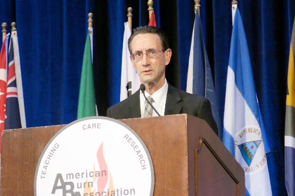
“Autologous ESS offer an alternative therapy for closure of extensive, excised, full-thickness burns,” Steven Boyce, Ph.D., said at the annual meeting of the American Burn Association.
The skin substitutes were engineered using split-thickness skin biopsies from 16 children with full-thickness burns covering an average total body surface area (TBSA) of 78% and a high of 95%.
The ESS were composed of autologous cultured keratinocytes and dermal fibroblasts attached to a collagen-based, biopolymer scaffold.
Clinicians are increasingly turning to tissue engineering as a way to address the shortfalls associated with the prevailing standard of care, autologous skin grafting, including lack of donor sites in larger burns, increased morbidity from autograft harvesting and skin grafts, and scarring.
At the 2014 ABA meeting, Canadian researchers reported on an autologous skin substitute that had both dermal and epidermal components. It took 2 months to prepare, whereas the Cincinnati model requires about 1 month to prepare and offers greater availability and stability of the closed wounds, Dr. Boyce said in an interview.
For the current study, Dr. Boyce and his associates compared mortality after treatment with ESS in 16 children with data from 1,008 patients from the National Burn Repository who were of similar ages, had 77% full-thickness TBSA burns, and were treated with meshed and expanded split-thickness skin autografts (AG). One patient died before ESS were prepared, leaving 15 patients in whom 2,056 ESS grafts were applied in 59 operations. Their average age was 6.3 years, 13 were male, and the average time to first ESS was 32 days.
Mortality was 6.25% (1/16) with the skin substitutes and 30.3% (305/1,008) with the autografts (P value < .05), said Dr. Boyce, a professor with the University of Cincinnati and director of the Skin Substitute Laboratories, Shriners Hospitals for Children, both in Cincinnati.
Rates of engraftment at postoperative day 14, however, significantly favored the AG group at 96.5 vs. 83.5% for ESS (P < .05).
One major reason for reduced rates of engraftment is lack of blood vessels in current models of engineered skin, he explained.
At day 28, the percentage of TBSA closed was approximately 30% for ESS and 47% for AG, while the ratio of closed-to-donor areas was significantly greater for ESS (108.7 vs. 4.0; P < .001).
The correlation between the percentage TBSA closed with ESS and TBSA burned generated an R-squared value of 0.65 (P < .001), Dr. Boyce said.
Regraftment rates were similar between graft types. Antibody formation to the biopolymer scaffold at postoperative day 28 was similar between patients receiving 1 or multiple ESS.
“Stable wound closure [with ESS] is interpreted to result from preservation of epidermal progenitor cells, formation in vitro of basement membrane between fibroblasts and keratinocytes, and generation of fibrovascular tissue by the biopolymer scaffold,” he said.
Tissue engineering offers patients the opportunity to survive with otherwise statistically lethal burns and “the data are very clear that happens with this product,” session comoderator and past ABA president Dr. David Ahrenholz said in an interview.
That said, “the product is very expensive, so I would not use it for patients in whom I had adequate donor sites, but it’s fairly easy to identify those patients on admission,” he added.
The study was supported by grants from the U.S. Department of Defense, National Institute of General Medical Sciences, and Shriners Hospitals for Children. The authors reported no current relevant financial disclosures.
On Twitter @pwendl
AT THE ABA ANNUAL MEETING
Key clinical point: Availability of autologous engineered skin substitutes for treatment of extensive, deep burns may reduce time to wound closure, long-term morbidity, and mortality.
Major finding: Mortality was 6.25% with ESS and 30.3% with autografts (P < .05).
Data source: Randomized, paired-site comparison in 16 pediatric burn patients.
Disclosures: The study was supported by grants from the U.S. Department of Defense, National Institute of General Medical Sciences, and Shriners Hospitals for Children. The authors reported no current relevant financial disclosures.
ABA: Childhood burn survivors risk more physical, mental disorders
CHICAGO – Adult survivors of childhood burns have significantly higher rates of Axis I mental and physical disorders years after their injury, a population-based study shows.
“We think it is really important to screen for, identify, and treat these illnesses not only in that acute period and shortly after the burn injury, but well into adulthood,” study author James Stone said at the annual meeting of the American Burn Association.
He reported on 745 adult burn survivors identified using administrative data from a regional pediatric burn center registry in Manitoba, Canada, who were matched 1:5 with 3,725 controls from the general Manitoba population based on age, sex, and geographic location. The burn survivors had an average age of 5.9 years at the time of burn injury, burns involved an average 12% of total body surface area, and 65% of burn survivors were male. The average follow-up was nearly 15 years (range 2.8-24.7 years).
In unadjusted univariate analysis, adult survivors had significantly higher rates than matched controls for any lifetime physical disorder (rate ratio, 1.17), arthritis (RR, 1.23), cancer (RR, 1.94), diabetes (RR, 1.69), fractures (RR, 1.45), and total respiratory morbidity (RR, 1.15).
After adjustment for gender, geography, and income, any physical disorder (RR, 1.15; P value < .01), arthritis (RR, 1.24; P < .01), fractures (RR, 1.37; P .001), and total respiratory morbidity (RR, 1.13; P < .05) remained significant, Mr. Stone, from the University of Manitoba, Winnipeg, Canada, reported.
Further, 81% of burn survivors had a lifetime physical illness compared with 69% of controls.
“The fact that 81% of our burn cohort was diagnosed with a physical illness is definitely concerning,” he said. “We hypothesize that the prolonged hyperinflammatory and hypermetabolic state that has been previously reported makes these individuals more susceptible to these illnesses down the road.”
The burn cohort also had significantly higher unadjusted rate ratios for any Axis 1 mental disorder (RR, 1.62), major depressive disorder (RR, 1.64), anxiety (RR, 1.57), substance abuse (RR, 2.86), and suicide attempts (RR, 5.00).
All disorders remained statistically significant after adjustment with rate ratios of 1.54 (P < .001), 1.54 (P < .001), 1.50 (P < .001), 2.35 (P < .001), and 4.33 (P < .01), respectively.
The high rates of substance abuse and suicide attempts are consistent with previous clinical interview studies, but are still cause for great concern, Mr. Stone said.
The risk for any mental or physical disorder was not significantly impacted by burn location or by burns that affected more than 30% of total body surface area. Age older than 5 years at the time of the burn significantly increased the risk of any mental disorder (relative risk, 1.92; P < .001).
Limitations of the study include the potential for bias because the data relied on individuals presenting to physicians, discrepancies between ICD codes for physician billings and hospital claims, and some survivors may have moved out of the province, Mr. Stone said. The study, however, had a sample size three times greater than the next largest study of its kind, and importantly, matched burn patients to the general population.
AT THE ABA ANNUAL MEETING
Key clinical point: Adult survivors of childhood burn injuries have increased rates of Axis I mental and physical disorders.
Major finding: 81% of burn survivors had a physical disorder vs. 69% of matched controls.
Data source: Population-based study in 745 adult survivors of childhood burns.
Disclosures: The study was funded by grants from the University of Manitoba and the Manitoba Firefighters Burn Fund. The authors declared no conflicts of interest.
ABA: Rehab time linked to outcomes in all burn patients
CHICAGO – Rehabilitation time is directly associated with a reduced rate of contracture and a better range of motion, irrespective of the extent of burn injury, results from the prospective ACT study showed.
The Acuity, Contractures, Time (ACT) study examined outcomes and rehabilitation time in patients with burns involving 10% or less of their total body surface area (n = 177) and in those with burns involving more than 10% of total body surface area (n = 130). Joint range of motion was recorded based on cutaneous functional units at the time of discharge for all 307 patients enrolled from September 2010 through December 2013 at five verified burn centers across the United States. Most patients were men (71%).
Overall, 79% of the patients had burn scar contracture. Based on range of motion for 8,068 joints analyzed, 66% had burn scar contracture.
For patients with burns affecting 10% or less of their bodies, only rehabilitation time per cutaneous functional unit significantly differed between those with and without burn scar contracture (4.6 minutes vs. 2.4 minutes; P<i/>= .002). The same was true for patients with burns affecting more than 10% of their bodies (3.3 minutes vs. 1.4 minutes; P<i/> < .0001).
“One of the impetuses to do this study in this manner was the realization of what most therapists say all the time: They don’t have enough time to treat their patients in the hospital,” Reg Richard, P.T., said at the annual meeting of the American Burn Association.
During a discussion of the study at the meeting, Mr. Richard said he hoped the results would be used to improve rehabilitation services for burn patients and called on the ABA to lead the charge.
ABA President David Ahrenholz of Regions Hospital Burn Center, St. Paul, Minn., said in an interview that coverage of rehabilitation services varies from insurer to insurer, and physicians can’t point to an absolute number to say this is the minimum number of rehabilitation days needed to obtain a good outcome in a specific patient.
“This is the first pass of an unbelievably large data set,” Dr. Ahrenholz said “I think it will be transformative ... to help the insurers to understand that it isn’t just a number of sessions, but that we’re looking for an outcome and that the therapy should continue until we get the desired outcome.”
In the ACT study, a univariate analysis found patients with no contracture were significantly more likely than those with contracture to have shorter hospital stays (12 days vs. 14 days; P value = .02), burns with a lower percentage of body surface area involvement (5% vs. nearly 10%; P<i/> < .0001), fewer skin grafts (2.3% vs. 4%; P<i/> = .001), more rehabilitation time per total body surface area involvement (6.1 minutes vs. 4.5 minutes; P<i/> = .003 ), and more rehabilitation time per cutaneous functional unit (4.4 minutes vs. 2 minutes; P<i/> < .0001), Mr. Richard from the U.S. Army Institute of Surgical Research, JBSA Fort Sam Houston, Texas, reported.
In multivariate regression analysis, rehabilitation time per cutaneous functional unit was the only significant predictor of preventing burn scar contracture and lost range of motion for patients with small burns (Odds ratio, 1.07; 95% confidence interval, 1.02-1.12) and for those with larger burns (OR, 1.36; 95% CI, 1.18-1.74).
Further analyses are warranted, as the results were based on the variables used and present analysis methods, he said.
On Twitter @pwendl
AT THE ABA ANNUAL MEETING
Key clinical point: Rehabilitation time was associated with reduced risk of scar contracture and lost range of motion in burn patients.
Major finding: For patients with burns affecting 10% or less of their bodies, only rehabilitation time per cutaneous functional unit significantly differed between those with and without burn scar contracture (4.6 minutes vs. 2.4 minutes; P = .002). The same was true for patients with burns affecting more than 10% of their bodies (3.3 minutes vs. 1.4 minutes; P < .0001).
Data source: Prospective, observational study in 307 burn patients.
Disclosures: The study was funded by a U.S. Department of Defense grant.
