User login
Bringing you the latest news, research and reviews, exclusive interviews, podcasts, quizzes, and more.
div[contains(@class, 'header__large-screen')]
div[contains(@class, 'read-next-article')]
div[contains(@class, 'nav-primary')]
nav[contains(@class, 'nav-primary')]
section[contains(@class, 'footer-nav-section-wrapper')]
footer[@id='footer']
div[contains(@class, 'main-prefix')]
section[contains(@class, 'nav-hidden')]
div[contains(@class, 'ce-card-content')]
nav[contains(@class, 'nav-ce-stack')]
Reduced-Dose Vaccines Protect Patients With HIV Against Mpox
The smallpox vaccine effectively induces immunity against mpox virus infection (formerly simian smallpox) in patients with human immunodeficiency virus (HIV) infection, although patients with lymphocyte counts below 500 cells/mm3 require booster doses, according to data from a study published in the Journal of Medical Virology.
The data come from the prospective observational study conducted by researchers at the Infection Biology Laboratory of the Department of Medicine and Life Sciences at Pompeu Fabra University and the HIV Unit of the Hospital del Mar Medical Research Institute in Barcelona, Spain. The investigators analyzed T-cell responses induced by vaccination with JYNNEOS.
Despite the substantial decrease in the reporting frequency of mpox cases from the global peak in August 2022 (30,894 cases) to 804 monthly cases in the last six months of 2023, mpox continues to circulate, and there is no specific vaccine. The JYNNEOS vaccine, with protective cross-reactivity against orthopoxviruses, is approved by the US Food and Drug Administration and the European Medicines Agency for the prevention of smallpox and mpox in adults at high risk for infection.
During the 2022 outbreak in the United States and Europe, vaccine shortages led to the emergency use authorization of a lower intradermal dose. This strategy was aimed at increasing vaccine supply up to fivefold.
Further clinical trials are needed to evaluate responses to JYNNEOS vaccination and compare different administration routes in patients with HIV infection. Protecting this population against mpox is a priority because people with high viral loads or loCD4+ T-lymphocyte counts are especially susceptible to severe disease.
Vaccination Responses
The study assessed the immune response to the JYNNEOS vaccine in patients with HIV who were receiving antiretroviral therapy as outpatients at the Infectious Diseases Unit of Hospital del Mar in Barcelona, Spain. Participants had viral loads controlled by antiretroviral therapy and CD4+ T-lymphocyte counts ≤ 500/mm3 (loCD4 group) or ≥ 500/mm3 (hiCD4 group) in blood. Vaccine responses were compared with those of vaccinated controls without the disease. The study included cases that received the standard subcutaneous vaccine (before August 2022) or the emergency dose-saving intradermal vaccine after its approval in August 2022.
The results demonstrated that the intradermal dose-saving vaccination route is preferable to the subcutaneous route and that patients in the loCD4 group may require at least one booster to generate an efficient response of specific T cells for mpox, wrote the authors.
“This study has two relevant points,” study author Robert Güerri-Fernandez, MD, PhD, head of infectious diseases at the Hospital del Mar Medical Research Institute, told this news organization. “In the subgroup of patients with HIV with effective treatment but without an immune response (ie, loCD4), the vaccine response is worse than in people who have recovered immunity or do not have HIV. Therefore, they need a booster dose.
“The second point is that the intradermal route with one-fifth of the standard subcutaneous dose has a better immune response than the standard subcutaneous route.” He added that it was a good strategy to save doses and be able to vaccinate many more people when vaccine shortages occurred.
“A general conclusion cannot be drawn,” he said. “It needs to be validated with many more subjects, of course, but in some way, it reinforced our confidence in the strategy of health authorities to promote intradermal vaccination. There we had evidence that the patients we were vaccinating intradermally were responding well.”
In Spain, although there is no shortage of vaccines today, they continue to be administered intradermally with a fractionated dose equivalent to one fifth of a standard dose, said Dr. Güerri-Fernandez.
However, in his opinion, observations regarding the two administration routes signal a need for further research. The main message should be that for patients with HIV infection who do not have an immune response, the vaccine response is incomplete, and they need booster doses as well as monitoring of the vaccine immune response, said Dr. Güerri-Fernandez.
More Studies Required
The research, which prospectively collected data and blood samples from patients with HIV who received the JYNNEOS vaccine, is small and included only 24 patients with HIV infection, with seven hospital workers who also received the vaccine and seven unvaccinated individuals as controls. “I am one of the control subjects of the study, and intradermal vaccination is not especially pleasant,” said Dr. Güerri-Fernandez. “It is a very innervated area, and the moment of introducing the liquid is uncomfortable. But it is perfectly bearable.”
Outpatient HIV-infected patients from the Infectious Diseases Unit of Hospital del Mar on antiretroviral therapy and with undetectable viral loads were grouped according to their CD4+ T-lymphocyte counts. Those with CD4+ T-lymphocyte counts ≤ 500/mm3 required at least one booster vaccine to exhibit efficient virus-specific T-lymphocyte responses. The magnitude of the T-cell response after this booster correlated directly with the CD4+ T-lymphocyte count of those vaccinated.
For Argentine infectious disease specialist Julián García, MD, clinical researcher at the Huésped Foundation in Buenos Aires, Argentina, who did not participate in the study, it is always productive to know that T-cell responses develop in patients with HIV infection, with CD4+ T-lymphocyte counts > and < 500/mm3, through an intradermal administration route.
Dr. García emphasized that the most novel aspect is that the JYNNEOS vaccine induces a specific T-cell response in patients with HIV infection that increases with higher CD4+ T-lymphocyte levels. However, he noted that the number of patients was less than 10 in most study groups, and the control group had only intradermal administration, which limits the interpretation of the results. “It will be necessary to verify this in studies with larger groups with control groups from all routes and with a correlate of protection.”
Dr. García referred to this latter point as a significant source of uncertainty. “The study is fundamentally based on the cellular response, but nowadays, there is no immune correlate of real-life protection.” He concluded that the study builds knowledge, which is essential for a vaccine that began to be used for mpox and the effectiveness of which is based on estimates.
Dr. Güerri-Fernandez and Dr. Garcia declared no relevant financial conflicts of interest.
This story was translated from the Medscape Spanish edition using several editorial tools, including AI, as part of the process. Human editors reviewed this content before publication. A version of this article appeared on Medscape.com.
Communicating Bad News to Patients
Communicating bad news to patients is one of the most stressful and challenging clinical tasks for any physician, regardless of his or her specialty. This task is more frequent for physicians caring for oncology patients and can also affect the physician’s emotional state.
The manner in which bad news is communicated plays a significant role in the psychological burden on the patient, and various communication techniques and guidelines have been developed to enable physicians to perform this difficult task effectively.
Revealing bad news in person whenever possible, to address the emotional responses of patients or relatives, is part of the prevailing expert recommendations. However, it has been acknowledged that in certain situations, communicating bad news over the phone is more feasible.
Since the beginning of the COVID-19 pandemic, the disclosure of bad news over the phone has become a necessary substitute for in-person visits and an integral part of clinical practice worldwide. It remains to be clarified what the real psychological impact on patients and their closest relatives is when delivering bad news over the phone compared with delivering it in person.
Right and Wrong Ways
The most popular guideline for communicating bad news is SPIKES, a six-phase protocol with a special application for cancer patients. It is used in various countries (eg, the United States, France, and Germany) as a guide for this sensitive practice and for training in communication skills in this context. The SPIKES acronym refers to the following six recommended steps for delivering bad news:
- Setting: Set up the conversation.
- Perception: Assess the patient’s perception.
- Invitation: Ask the patient what he or she would like to know.
- Knowledge: Provide the patient with knowledge and information, breaking it down into small parts.
- Emotions: Acknowledge and empathetically address the patient’s emotions.
- Strategy and Summary: Summarize and define a medical action plan.
The lesson from SPIKES is that when a person experiences strong emotions, it is difficult to continue discussing anything, and they will struggle to hear anything. Allowing for silence is fundamental. In addition, empathy allows the patient to express his or her feelings and concerns, as well as provide support. The aim is not to argue but to allow the expression of emotions without criticism. However, these recommendations are primarily based on expert opinion and less on empirical evidence, due to the difficulty of studies in assessing patient outcomes in various phases of these protocols.
A recent study analyzed the differences in psychological distress between patients who received bad news over the phone vs those who received it in person. The study was a systematic review and meta-analysis.
The investigators examined 5944 studies, including 11 qualitative analysis studies, nine meta-analyses, and four randomized controlled trials.
In a set of studies ranging from moderate to good quality, no difference in psychological distress was found when bad news was disclosed over the phone compared with in person, regarding anxiety, depression, and posttraumatic stress disorder.
There was no average difference in patient satisfaction levels when bad news was delivered over the phone compared with in person. The risk for dissatisfaction was similar between groups.
Clinical Practice Guidelines
The demand for telemedicine, including the disclosure of bad news, is growing despite the limited knowledge of potential adverse effects. The results of existing studies suggest that the mode of disclosure may play a secondary role, and the manner in which bad news is communicated may be more important.
Therefore, it is paramount to prepare patients or their families for the possibility of receiving bad news well in advance and, during the conversation, to ensure first and foremost that they are in an appropriate environment. The structure and content of the conversation may be relevant, and adhering to dedicated communication strategies can be a wise choice for the physician and the interlocutor.
This story was translated from Univadis Italy, which is part of the Medscape professional network, using several editorial tools, including AI, as part of the process. Human editors reviewed this content before publication. A version of this article appeared on Medscape.com.
The Ghost Research Haunting Nordic Medical Trials
Campaigners for greater transparency in medical science have reiterated calls for more to be done to avoid “medical research waste” after an investigation found that results from more than a fifth of clinical trials across five Nordic countries have never been made public.
Nonpublication of clinical trial results wastes public money, harms patients, and undermines public health, the researchers said.
There is already a well-defined ethical responsibility to publish trial results. Article 36 of the Declaration of Helsinki on Ethical Principles for Medical Research Involving Human Subjects states that “researchers have a duty to make publicly available the results of their research on human subjects,” and World Health Organization best practice protocols call for results to be uploaded onto trial registries within 12 months of trial completion.
Research Waste Is a ‘Pervasive Problem’
So, how and why do so many trials end up gathering dust in a drawer? The latest study, published February 5 as a preprint, evaluated the reporting outcomes of 2113 clinical trials at medical universities and university hospitals in Nordic countries between 2016 and 2019. It found that across the five countries, 22% of all clinical trial results had not been shared. Furthermore, only 27% of all trial results were made public, either on registries or in journals, within 12 months. Even 2 years after trials ended, only around half of results (51.7%) had been put into the public domain.
The authors concluded that missing and delayed results from academically-led clinical trials was a “pervasive problem” in Nordic countries and that institutions, funding bodies, and policymakers needed to ensure that regulations around reporting results were adhered to so that important findings are not lost.
Study first author, Gustav Nilsonne, MD, PHD, from the Department of Clinical Neuroscience at the Karolinska Institutet, Sweden, told this news organization: “Most people I talk to — most colleagues who are clinical scientists — tend to think that the main reason is that negative results are not as interesting to publish and therefore they get lower priority, and they get published later and sometimes not at all.”
Experts stressed that the problem is not confined to Nordic countries and that wasted medical research persists elsewhere in Europe and remains a global problem. For instance, a report published in the Journal of Clinical Epidemiology found that 30% of German trials completed between 2014 and 2017 remained unpublished 5 years after completion.
The Case for Laws, Monitoring, and Fines
Till Bruckner, PHD, from TranspariMED, which campaigns to end evidence distortion in medicine, told this news organization: “What is needed to comprehensively fix the problem is a national legal requirement to make all trial results public, coupled with effective monitoring, and followed by sanctions in the rare cases where institutions refuse to comply.”
Dr. Nilsonne added: “We have argued that the sponsors need to take greater responsibility, but also that there needs to be somebody whose job it is to monitor clinical trials reporting. It shouldn’t have to be that we do this as researchers on a shoestring with no dedicated resources. It should be somebody’s job.”
Since January 31, 2023, all initial clinical trial applications in the European Union must be submitted through the EU Clinical Trials Information System. Dr. Bruckner said that “the picture is not yet clear” in Europe, as the first trial results under the system are not expected until later this year. Even then, enforcement lies with regulators in individual countries. And while Denmark has already indicated it will enforce the regulations, he warned that other countries “might turn a blind eye.”
He pointed out that existing laws don’t apply to all types of trials. “That means that for many trials, nobody is legally responsible for ensuring that results are made public, and no government agency has any oversight or mandate,” he said.
Outside the EU, the United Kingdom has helped lead the way through the NHS Health Research Authority (HRA), which registers trials run in the country. One year after a trial has been completed, the HRA checks to see if the results have been uploaded to the registry and issues reminders if they haven’t.
In an update of its work in January, the authority said that compliance had hovered at just below 90% between 2018 and 2021 but that it was working to increase this to 100% by working with stakeholders across the research sector.
Dr. Nilsonne considers the UK system of central registration and follow-up an attractive option. “I would love to see something along those lines in other countries too,” he said.
‘Rampant Noncompliance’ in the United States
In the United States, a requirement to make trial results public is backed by law. Despite this, there’s evidence of “rampant noncompliance” and minimal government action, according to Megan Curtin from Universities Allied for Essential Medicines (UAEM), which has been tracking the issue in the United States and working to push universities and others to make their findings available.
The US Food and Drug Administration (FDA) shares responsibility with the National Institutes of Health for enforcement of clinical trial results reporting, but the UAEM says nearly 4000 trials are currently out of compliance with reporting requirements. In January last year, the UAEM copublished a report with the National Center for Health Research and TranspariMED, which found that 3627 American children participated in clinical trials whose results remain unreported.
The FDA can levy a fine of up to $10,000 USD for a violation of the law, but UAEM said that, as of January 2023, the FDA had sent only 92 preliminary notices of noncompliance and four notices of noncompliance. “A clear difference between the EU field of clinical trial operation and US clinical trials is that there are clear laws for reporting within 12 months, which can be enforced, but they’re not being enforced by the FDA,” Ms. Curtin told this news organization.
The UAEM is pushing the FDA to issue a minimum of 250 preliminary notices of noncompliance each year to noncompliant trial sponsors.
Dr. Nilsonne said: “I do believe we have a great responsibility to the patients that do contribute. We need to make sure that the harms and risks that a clinical trial entails are really balanced by knowledge gain, and if the results are never reported, then we can’t have a knowledge gain.”
A version of this article appeared on Medscape.com.
Use of Biologics for Psoriasis Found to Confer a Survival Benefit
Among patients with psoriasis, the risk of mortality was strongly associated with hepatic injury, cardiovascular disease, and psychiatric affective disorders, but was reduced among those who received systemic therapy with biologics, researchers from Canada report.
Those are key findings from a large retrospective registry study of patients with psoriasis, published in The Journal of the American Academy of Dermatology.
“Psoriasis, a chronic inflammatory condition affecting approximately 3% of the western populations, bears a higher risk of mortality compared to healthy individuals, possibly by inducing systemic inflammation associated with numerous comorbidities, especially cardiovascular diseases, metabolic syndrome, and others,” wrote corresponding author Robert Gniadecki, MD, PhD, of the division of dermatology at the University of Alberta, Canada, and colleagues. “It has been argued that the use of systemic immunomodulatory agents quenches systemic inflammation and potentially improves patient survival. However, the evidence to support this hypothesis is limited.”
To investigate the impact of comorbidities and systemic therapies on all-cause mortality in psoriasis, the researchers used the Alberta Health Services Data Repository of Reporting database from January 1, 2012, to June 1, 2019, which represents a population base of 4.47 million individuals. They extracted data on 18,618 psoriasis cases and 55,854 controls, stratified cases according to the Charlson Comorbidity Index (CCI), a surrogate measure for comorbidity burden, and by the type of therapy received, and conducted statistical analyses including Cox proportional hazards regression to determine absolute hazard ratios representing relative effects of specific demographic and comorbidity factors on mortality within groups.
The median age in both cohorts was 48 years, and 51% were male. The researchers observed that mortality in the psoriasis cohort was significantly higher than in the controls (5.7% vs. 3.8%, respectively; P < .05), with a median age at the time of death of 72 vs. 74.4 years.
The CCI and comorbidities strongly predicted mortality, especially drug-induced liver injury (hazard ratio [HR], 1.78), bipolar disorder and suicidal ideation (HR, 1.24-1.58), and major cardiovascular diseases, which included myocardial infarction (MI), congestive heart failure (CHF), and cerebrovascular disease (CVA) (HR, 1.2-1.4).
Among patients in the psoriasis cohort, survival of those treated with biologic agents was higher than in controls, even after matching for CCI (3.2% vs. 4.4%, respectively, P < .05). “These patients also exhibit reduced overall mortality compared to those treated with methotrexate or topical agents,” Dr. Gniadecki and colleagues wrote. “There was no difference in mortality between methotrexate patients and the topical therapy patients, but any of those treatment groups had superior survival compared to the no-treatment cohort.”
They added that despite better survival among patients treated with biologic agents, no significant improvements were detected in their comorbidity profiles. “Notably, the frequency of major cardiovascular disease (MI, CHF, CVA) was the same as in the controls, and overall, the frequency of diseases coded as cardiovascular was slightly increased,” they wrote.
The fact that some factors could not be measured, including the type and severity of psoriasis, response to treatment, smoking history, and alcohol intake, was a study limitation, they noted.
Joel M. Gelfand, MD, director of the psoriasis and phototherapy treatment center at the University of Pennsylvania, Philadelphia, who was asked to comment on the analysis, said the study confirms prior work indicating that having psoriasis is a predictor of mortality. In addition, “there is a strong healthy user affect among patients who take and stay on biologics for psoriasis,” he told this news organization.

“The results are encouraging but are not able to establish a causal relationship between treating psoriasis with biologics and lowering mortality risk. Ultimately, randomized comparative trials will be needed to determine which approach or approaches to treating psoriasis, if any, lower the risk of psoriatic arthritis, cardiovascular disease, and mortality,” said Dr. Gelfand, who was not involved with the study.
Asked to comment on the results, Adam Friedman, MD, professor and chair of dermatology at George Washington University, Washington, who was not involved with the study, said that “data such as these enable us to rationalize the cost of our fleet of biologics, as managing the outpatient/inpatient burden of many of these comorbidities will actually drain the healthcare system, more so than managing psoriasis in the first place. Certainly other interventions to address the well known comorbidities, such as cardiovascular and hepatic, are warranted, but what if you could prevent the problem in the first place? To be continued for that answer.”

The study was funded by Canadian Dermatology Foundation, Alberta Innovates, and by a Health Sciences TD Bank Studentship Award. Dr. Gniadecki reported conducting clinical trials for Bausch Health, AbbVie and Janssen, and he has received honoraria as consultant and/or speaker from AbbVie, Bausch Health, Eli Lilly, Janssen, Mallinckrodt, Novartis, Kyowa Kirin, Sun Pharma and Sanofi. The other authors had no disclosures. Dr. Gelfand reported serving as a consultant for AbbVie, Artax, Bristol-Myers Squibb, GlaxoSmithKline, and other companies. He is on the board of directors for the International Psoriasis Council and the Medical Dermatology Society. Dr. Friedman disclosed that he is a speaker for Janssen and Bristol Myers Squibb. He has received grants from Janssen, Pfizer, Bristol Myers Squibb, and Lilly, and has served as an advisor for Arcutis, Dermavant, and Janssen.
Among patients with psoriasis, the risk of mortality was strongly associated with hepatic injury, cardiovascular disease, and psychiatric affective disorders, but was reduced among those who received systemic therapy with biologics, researchers from Canada report.
Those are key findings from a large retrospective registry study of patients with psoriasis, published in The Journal of the American Academy of Dermatology.
“Psoriasis, a chronic inflammatory condition affecting approximately 3% of the western populations, bears a higher risk of mortality compared to healthy individuals, possibly by inducing systemic inflammation associated with numerous comorbidities, especially cardiovascular diseases, metabolic syndrome, and others,” wrote corresponding author Robert Gniadecki, MD, PhD, of the division of dermatology at the University of Alberta, Canada, and colleagues. “It has been argued that the use of systemic immunomodulatory agents quenches systemic inflammation and potentially improves patient survival. However, the evidence to support this hypothesis is limited.”
To investigate the impact of comorbidities and systemic therapies on all-cause mortality in psoriasis, the researchers used the Alberta Health Services Data Repository of Reporting database from January 1, 2012, to June 1, 2019, which represents a population base of 4.47 million individuals. They extracted data on 18,618 psoriasis cases and 55,854 controls, stratified cases according to the Charlson Comorbidity Index (CCI), a surrogate measure for comorbidity burden, and by the type of therapy received, and conducted statistical analyses including Cox proportional hazards regression to determine absolute hazard ratios representing relative effects of specific demographic and comorbidity factors on mortality within groups.
The median age in both cohorts was 48 years, and 51% were male. The researchers observed that mortality in the psoriasis cohort was significantly higher than in the controls (5.7% vs. 3.8%, respectively; P < .05), with a median age at the time of death of 72 vs. 74.4 years.
The CCI and comorbidities strongly predicted mortality, especially drug-induced liver injury (hazard ratio [HR], 1.78), bipolar disorder and suicidal ideation (HR, 1.24-1.58), and major cardiovascular diseases, which included myocardial infarction (MI), congestive heart failure (CHF), and cerebrovascular disease (CVA) (HR, 1.2-1.4).
Among patients in the psoriasis cohort, survival of those treated with biologic agents was higher than in controls, even after matching for CCI (3.2% vs. 4.4%, respectively, P < .05). “These patients also exhibit reduced overall mortality compared to those treated with methotrexate or topical agents,” Dr. Gniadecki and colleagues wrote. “There was no difference in mortality between methotrexate patients and the topical therapy patients, but any of those treatment groups had superior survival compared to the no-treatment cohort.”
They added that despite better survival among patients treated with biologic agents, no significant improvements were detected in their comorbidity profiles. “Notably, the frequency of major cardiovascular disease (MI, CHF, CVA) was the same as in the controls, and overall, the frequency of diseases coded as cardiovascular was slightly increased,” they wrote.
The fact that some factors could not be measured, including the type and severity of psoriasis, response to treatment, smoking history, and alcohol intake, was a study limitation, they noted.
Joel M. Gelfand, MD, director of the psoriasis and phototherapy treatment center at the University of Pennsylvania, Philadelphia, who was asked to comment on the analysis, said the study confirms prior work indicating that having psoriasis is a predictor of mortality. In addition, “there is a strong healthy user affect among patients who take and stay on biologics for psoriasis,” he told this news organization.
“The results are encouraging but are not able to establish a causal relationship between treating psoriasis with biologics and lowering mortality risk. Ultimately, randomized comparative trials will be needed to determine which approach or approaches to treating psoriasis, if any, lower the risk of psoriatic arthritis, cardiovascular disease, and mortality,” said Dr. Gelfand, who was not involved with the study.
Asked to comment on the results, Adam Friedman, MD, professor and chair of dermatology at George Washington University, Washington, who was not involved with the study, said that “data such as these enable us to rationalize the cost of our fleet of biologics, as managing the outpatient/inpatient burden of many of these comorbidities will actually drain the healthcare system, more so than managing psoriasis in the first place. Certainly other interventions to address the well known comorbidities, such as cardiovascular and hepatic, are warranted, but what if you could prevent the problem in the first place? To be continued for that answer.”
The study was funded by Canadian Dermatology Foundation, Alberta Innovates, and by a Health Sciences TD Bank Studentship Award. Dr. Gniadecki reported conducting clinical trials for Bausch Health, AbbVie and Janssen, and he has received honoraria as consultant and/or speaker from AbbVie, Bausch Health, Eli Lilly, Janssen, Mallinckrodt, Novartis, Kyowa Kirin, Sun Pharma and Sanofi. The other authors had no disclosures. Dr. Gelfand reported serving as a consultant for AbbVie, Artax, Bristol-Myers Squibb, GlaxoSmithKline, and other companies. He is on the board of directors for the International Psoriasis Council and the Medical Dermatology Society. Dr. Friedman disclosed that he is a speaker for Janssen and Bristol Myers Squibb. He has received grants from Janssen, Pfizer, Bristol Myers Squibb, and Lilly, and has served as an advisor for Arcutis, Dermavant, and Janssen.
FROM THE JOURNAL OF THE AMERICAN ACADEMY OF DERMATOLOGY
Democratic Lawmakers Press Pfizer on Chemotherapy Drug Shortages
In a statement about their February 21 action, the legislators, led by Rep. Jamie Raskin (D-Md.), the committee’s ranking minority member, described their work as a follow up to an earlier investigation into price hikes of generic drugs. While the committee members queried Pfizer over the three oncology medications only, they also sent letters to drugmakers Teva and Sandoz with respect to shortages in other drug classes.
A representative for Pfizer confirmed to MDedge Oncology that the company had received the representatives’ letter but said “we have no further details to provide at this time.”
What is the basis for concern?
All three generic chemotherapy drugs are mainstay treatments used across a broad array of cancers. Though shortages have been reported for several years, they became especially acute after December 2022, when an inspection by the US Food and Drug Administration (FDA) led to regulatory action against an Indian manufacturer, Intas, that produced up to half of the platinum-based therapies supplied globally. The National Comprehensive Cancer Care Network reported in October 2023 that more than 90% of its member centers were struggling to maintain adequate supplies of carboplatin, and 70% had trouble obtaining cisplatin, while the American Society of Clinical Oncology published clinical guidance on alternative treatment strategies.
What has the government done in response to the recent shortages?
The White House and the FDA announced in September that they were working with several manufacturers to help increase supplies of the platinum-based chemotherapies and of methotrexate, and taking measures that included relaxing rules on imports. Recent guidance under a pandemic-era federal law, the 2020 CARES Act, strengthened manufacturer reporting requirements related to drug shortages, and other measures have been proposed. While federal regulators have many tools with which to address drug shortages, they cannot legally oblige a manufacturer to increase production of a drug.
What can the lawmakers expect to achieve with their letter?
By pressuring Pfizer publicly, the lawmakers may be able to nudge the company to take measures to assure more consistent supplies of the three drugs. The lawmakers also said they hoped to glean from Pfizer more insight into the root causes of the shortages and potential remedies. They noted that, in a May 2023 letter by Pfizer to customers, the company had warned of depleted and limited supplies of the three drugs and said it was “working diligently” to increase output. However, the lawmakers wrote, “the root cause is not yet resolved and carboplatin, cisplatin, and methotrexate continue to experience residual delays.”
Why did the committee target Pfizer specifically?
Pfizer and its subsidiaries are among the major manufacturers of the three generic chemotherapy agents mentioned in the letter. The legislators noted that “pharmaceutical companies may not be motivated to produce generic drugs like carboplatin, cisplatin, and methotrexate, because they are not as lucrative as producing patented brand name drugs,” and that “as a principal supplier of carboplatin, cisplatin, and methotrexate, it is critical that Pfizer continues to increase production of these life-sustaining cancer medications, even amidst potential lower profitability.”
The committee members also made reference to news reports of price-gouging with these medications, as smaller hospitals or oncology centers are forced to turn to unscrupulous third-party suppliers.
What is being demanded of Pfizer?
Pfizer was given until March 6 to respond, in writing and in a briefing with committee staff, to a six questions. These queries concern what specific steps the company has taken to increase supplies of the three generic oncology drugs, what Pfizer is doing to help avert price-gouging, whether further oncology drug shortages are anticipated, and how the company is working with the FDA on the matter.
Autoimmune Disease Risk May Rise Following Cushing Disease Remission After Surgery
Patients with Cushing disease have an increased risk for new-onset autoimmune disease in the 3 years after surgical remission, according to a new retrospective study published on February 20 in Annals of Internal Medicine.
Outcomes for patients with Cushing disease were compared against those with nonfunctioning pituitary adenomas (NFPAs). New-onset autoimmune disease occurred in 10.4% with Cushing disease and 1.6% among patients with NFPA (hazard ratio, 7.80; 95% CI, 2.88-21.10).
“Understanding and recognizing new and recurrent autoimmune disease in this setting is important to avoid misclassifying such patients with glucocorticoid withdrawal syndrome, which could result in failure to treat underlying autoimmune disease, as well as erroneous diagnosis of steroid withdrawal cases,” wrote Dennis Delasi Nyanyo of Massachusetts General Hospital and Harvard Medical School, Boston, and colleagues.
Given the general population’s annual incidence of major autoimmune diseases, estimated at about 100 cases per 100,000 people, and the 3-year incidence of 10.4% found in this study’s cohort, “our findings suggest that Cushing disease remission may trigger development of autoimmune disease,” the authors wrote.
Monitor Patients With Family History of Autoimmune Disease?
The study results were not necessarily surprising to Anthony P. Heaney, MD, PhD, an endocrinologist and professor of medicine at the University of California, Los Angeles, because past research has raised similar questions. The authors’ suggestion that the rapid postsurgical drop in cortisol that occurs as a result of treating Cushing disease becomes some sort of autoimmune trigger is interesting but remains speculative, Dr. Heaney pointed out.
If future evidence supports that possibility, “it would suggest, in terms of managing those patients in the postoperative setting, that there may be some merit to giving them higher concentrations of glucocorticoids for a short period of time,” Dr. Heaney said, thereby bringing their levels down more gradually rather than taking them off a cliff, in a sense. Or, if more evidence bears out the authors’ hypothesis, another approach might be treating patients with medicine to bring down the cortisol before surgery, though there are challenges to that approach, Dr. Heaney said.
At the same time, those who developed new autoimmune disease remain a small subset of patients with Cushing disease, so such approaches may become only potentially appropriate to consider in patients with risk factors, such as a family history of autoimmune disease.
The researchers conducted a retrospective chart review of adult patients who underwent transsphenoidal surgery for either Cushing disease or NFPA at Massachusetts General Hospital between 2005 and 2019.
The study involved 194 patients with Cushing disease who had postsurgical remission and at least one follow-up visit with a pituitary expert and 92 patients with NFPA who were matched to patients with Cushing disease based on age and sex. The authors regarded autoimmune disease diagnosed within 36 months of the surgery to be temporally associated with Cushing disease remission. Among the autoimmune diseases considered were “rheumatoid arthritis, Sjögren syndrome, systemic lupus erythematosus, autoimmune thyroiditis, celiac disease, psoriasis, vitiligo, autoimmune neuropathy, multiple sclerosis, myasthenia gravis, and ulcerative colitis.”
Patients differed in average body mass index and tumor size, but family history of autoimmune disease was similar in both groups. Average BMI was 34.5 in the Cushing group and 29.5 in the NFPA group. Average tumor size was 5.7 mm in the Cushing group and 21.3 mm in the NFPA group.
Before surgery, 2.9% of patients with Cushing disease and 15.4% of patients with NFPA had central hypothyroidism, and 8% in the Cushing group and 56.8% in the NFPA group had hyperprolactinemia. Central adrenal insufficiency occurred in 11% with NFPA and in all with Cushing disease, by definition.
After surgery, 93.8% in the Cushing group and 16.5% in the NFPA group had adrenal insufficiency. In addition, patients with Cushing disease had lower postsurgical nadir serum cortisol levels (63.8 nmol/L) than those with NFPA (282.3 nmol/L).
Of the 17 patients with Cushing disease — all women — who developed autoimmune disease within 3 years, 6 had a personal history of autoimmune disease and 7 had a family history of it. In addition, 41.2% of them had adrenal insufficiency when they developed the new autoimmune disease. Among the diseases were six autoimmune thyroiditis cases, three Sjögren syndrome cases, and two autoimmune seronegative spondyloarthropathy.
Dr. Heaney said he found it interesting that more than half of the new autoimmune diseases in patients with Cushing disease were related to the thyroid. “In this kind of setting, where you have a patient who has been producing too much steroid over a period of time and then you take that away, it’s almost like you release a brake on the TSH [thyroid-stimulating hormone],” Dr. Heaney said. “So, there’s probably some rebound in TSH that occurs, and that could be driving the thyroiditis, to some extent, that we see in these patients.”
Only one patient with NFPA developed new-onset autoimmune disease, a woman who developed Graves disease 22 months after surgery. When the researchers excluded patients in both groups with central hypothyroidism, new-onset autoimmune disease was still significantly higher (11.4%) in the Cushing group than in the NFPA group (1.9%; HR, 7.02; 95% CI, 2.54-19.39).
Could Postoperative Adrenal Insufficiency Contribute to Risk?
Within the Cushing cohort, those who developed autoimmune disease had a lower BMI (31.8 vs 34.8) and larger tumor size (7.2 vs 5.6 mm) than those who didn’t develop new autoimmune disease. Patients who developed autoimmune disease also had a lower baseline urine free cortisol ratio (2.7 vs 6.3) before surgery and more family history of autoimmune disease (41.2% vs 20.9%) than those who didn’t develop one.
“The higher prevalence of adrenal insufficiency and the lower nadir serum cortisol levels in the Cushing disease group suggest that the postoperative adrenal insufficiency in the Cushing disease group might have contributed to autoimmune disease pathogenesis,” the authors wrote. “This finding is clinically significant because cortisol plays a pivotal role in modulating the immune system.”
Most postoperative management among patients with Cushing disease was similar, with all but one patient receiving 0.5 or 1 mg daily dexamethasone within the first week after surgery. (The one outlier received 5 mg daily prednisone.) However, fewer patients who developed autoimmune disease (17.6%) received supraphysiologic doses of glucocorticoid — equivalent to at least 25 mg hydrocortisone — compared with patients who didn’t develop autoimmune disease (41.8%).
“Although the daily average hydrocortisone equivalent replacement doses within the first month and during long-term follow-up were within the physiologic range in both subgroups, patients with Cushing disease who had autoimmune disease received slightly lower doses of glucocorticoid replacement within the first month after surgery,” the authors reported. “The immediate postoperative period might be a critical window where supraphysiologic glucocorticoids seem to be protective with regard to development of autoimmune disease,” they wrote, though they acknowledged the study’s retrospective design as a limitation in drawing that conclusion.
At the least, they suggested that new symptoms in patients with Cushing disease, particularly those with a family history of autoimmune disease, should prompt investigation of potential autoimmune disease.
Recordati Rare Diseases funded the study. The research was also conducted with support from Harvard Catalyst (the Harvard Clinical and Translational Science Center) as well as financial contributions from Harvard University and its affiliated academic healthcare centers. One author reported holding stocks in Pfizer and Amgen, and another reported receiving consulting fees from Corcept. Dr. Heaney reported receiving institutional grants for trials from Corcept, Ascendis, Crinetics, and Sparrow Pharm; serving on the advisory board for Xeris, Recordati, Corcept, Novo Nordisk, Lundbeck, and Crinetics; and serving as a speaker for Chiesi, Novo Nordisk, and Corcept.
A version of this article appeared on Medscape.com.
FROM ANNALS OF INTERNAL MEDICINE
Prognosis of Polyarteritis Nodosa: What Are the Predictors?
TOPLINE:
Age older than 65 years and serum creatinine greater than 140 micromol/L at the time of systemic polyarteritis nodosa (PAN) diagnosis were significant predictors of mortality.
METHODOLOGY:
- A total of 358 patients diagnosed with PAN between 1990 and 2020 were identified from retrospective chart reviews and prospective cohorts from nine countries as a part of GLOBAL-PAN, a collaboration of the European Vasculitis Society, the Vasculitis Clinical Research Consortium, and other networks.
- The goal of the retrospective chart review was to characterize the nature, presentation, and survival rates of patients with PAN.
- The study population included 174 female and 184 male patients; 282 had systemic PAN (sPAN) and 76 had cutaneous PAN (cPAN); the mean age at diagnosis was 44.3 years.
TAKEAWAY:
- Overall survival rates at 1, 5, and 10 years for patients with sPAN were 97.1%, 94.0%, and 89.0%, respectively.
- Significant independent predictors of mortality were age ≥ 65 years at the time of sPAN diagnosis (hazard ratio [HR], 3.85), serum creatinine > 140 micromol/L at the time of diagnosis (HR, 4.93), gastrointestinal involvement (HR, 3.51), and central nervous system involvement (HR, 3.56).
- Constitutional symptoms were significantly more common in patients with sPAN vs cPAN (78.8% vs 44.7%), while patients with cPAN were significantly more likely to be female and have more skin nodules than patients with sPAN.
- Relapse over a median disease duration of 59.6 months was slightly higher for cPAN vs sPAN (38.8% vs 32.1%).
IN PRACTICE:
“This study helps better define the demographic and clinical characteristics of patients with PAN and differentiates sPAN from cPAN,” the researchers wrote.
SOURCE:
The lead author of the study was Omer Karadag, MD, of Hacettepe University, Ankara, Turkey. The study was published online on February 12 in Arthritis & Rheumatology.
LIMITATIONS:
Study limitations included the combination of prospective and retrospective data, varying approaches to patient assessment, and lack of data on treatment effects.
DISCLOSURES:
The study was supported by the Vasculitis Clinical Research Consortium, which received funding from the National Center for Advancing Translational Science, the National Institute of Arthritis and Musculoskeletal and Skin Diseases, and the National Center for Research Resources. Dr. Karadag disclosed research grants from AbbVie, Novartis, Viela-Bio, and TR-Pharma, and consulting fees from AbbVie, Abdi Ibrahim, Celltrion, Novartis, Pfizer, Sandoz, and UCB.
A version of this article appeared on Medscape.com.
Study Eyes Longer IV Ertapenem for Recalcitrant Hidradenitis Suppurativa
at the completion of treatment, a retrospective study showed.
“These findings suggest a course of 12 to 16 weeks of ertapenem may be appropriate as a new standard length of therapy in HS patients, which is at least twice the current recommendation of the North American treatment guidelines,” wrote corresponding author Steven R. Cohen, MD, MPH, of the departments of dermatology at Weill Cornell Medicine and Albert Einstein College of Medicine, New York, and his coauthors. The results were published online February 14, 2024, in JAMA Dermatology.
In an earlier study , some of the same researchers evaluated the efficacy of daily IV ertapenem for 6 weeks in seven patients with HS. The patients experienced “notable remediation of disease that was rapidly lost within 1 month of withdrawal.”

Treatment guidelines published in 2019 recommend ertapenem as a highly effective third-line therapy limited to one 6-week course “as rescue therapy or during surgical planning, given the practical barriers to home infusions and concerns about antibiotic resistance” .
For the current analysis, Dr. Cohen and colleagues explored the effects of a longer duration of treatment with ertapenem in this patient population. They retrospectively reviewed the medical records of 98 patients with HS who received care at Albert Einstein College of Medicine’s Montefiore HS Center between 2018 and 2022. Each patient used an elastomeric pump to self-administer 1 g IV ertapenem daily for 12-16 weeks.
Key outcome measures of interest were the HS Physician Global Assessment (PGA) score (a 6-point scale ranging from clear to very severe) and a numerical rating scale (NRS) for pain (an 11-point scale in which a score of 0 indicates no pain and a score of 10 indicates the worst possible pain) and markers of inflammation such as leukocytes, erythrocyte sedimentation rate, C-reactive protein (CRP), and interleukin (IL)-6. The researchers measured these outcomes at baseline, the midcourse of IV ertapenem treatment, at the end of the course, and post therapy.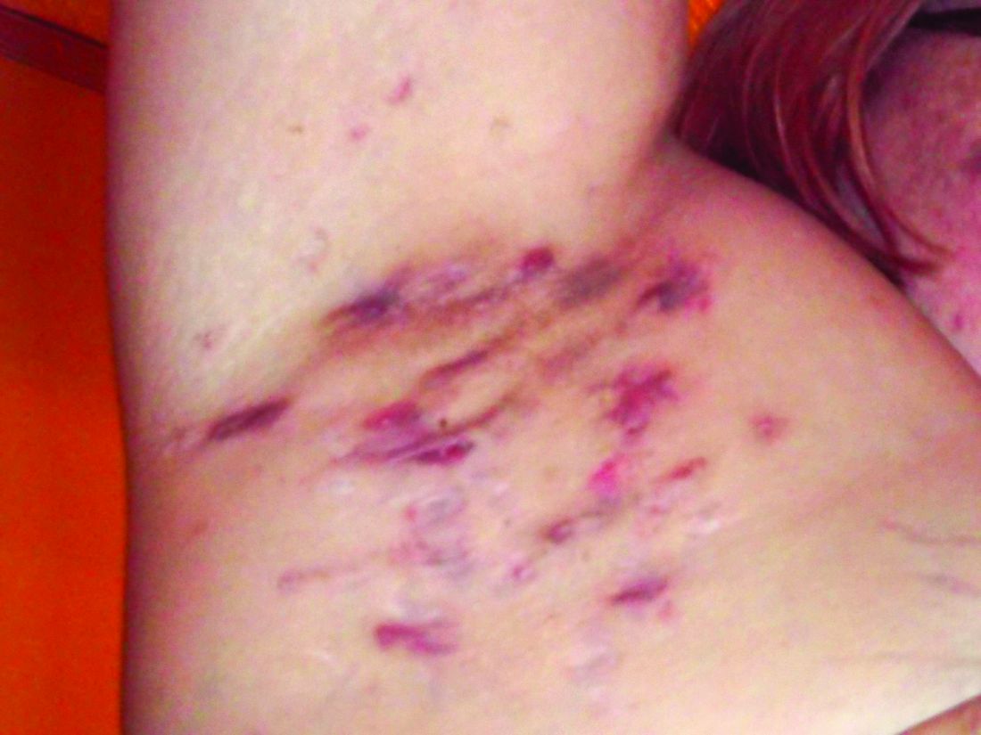
The mean age of the patients was 35.8 years, 62.2% were female, and 60.2% were Black. The mean treatment duration was 13.1 weeks and the mean posttherapy follow-up occurred after a mean of 7.8 weeks.
Between baseline and posttherapy follow-up, the HS PGA scores dropped from a mean of 3.9 to 2.7 and the NRS for pain dropped from 4.2 to 1.8 (P < .001 for both associations). Markers of inflammation also dropped between baseline and post therapy.
Specifically, values for CRP dropped from 5.4 to 2.4 mg/dL; IL-6 dropped from 25.2 to 13.7, and leukocytes dropped from 11.3 to 10.0 (P < .001 for all associations). Among the 76 patients who participated in a follow-up telephone survey, 63 (80.3%) reported medium to high satisfaction with their course of ertapenem, and 69 (90.8%) said they would recommend the treatment to other patients with HS.
The authors noted certain limitations of their study, including its retrospective, single-center design, the lack of a control group, and the fact that the HS-PGA scores at each visit did not meet the threshold of a 2-point decrease that is considered a clinically meaningful in the medical literature.
The definitive mechanism of ertapenem efficacy remains elusive, the authors pointed out. “Although oral antibiotics are generally accepted as a core therapeutic approach to HS, much less is known about the efficacy of IV antibiotics, especially ertapenem, a parenteral carbapenem possessing activity against many gram-positive bacteria, gram-negative bacteria, and anaerobic organisms,” they wrote.
In an accompanying editorial, Haley B. Naik, MD, MHSc, a dermatologist at the University of California, San Francisco, said that adopting prolonged courses of ertapenem treatment “comes with substantial individual and public health considerations”.

“Even though HS is a noninfectious disease, microbes might play a role in inciting HS immune dysregulation, prompting the inclusion of antimicrobial therapy in treatment regimens. However, broad-spectrum antibiotics for HS are associated with high levels of antibiotic resistance,” she wrote. Prolonged use of ertapenem and other carbapenems in HS treatment “will likely increase antimicrobial resistance, thereby limiting management of both HS and comorbid infections.”
Jennifer L. Hsiao, MD, a dermatologist who directs the HS clinic at the University of Southern California, Los Angeles, who was asked to comment on the study, said that, despite significant advances in the management of HS over the past decade, there are still patients who do not respond adequately to standard treatments.

For these patients, IV ertapenem can serve as a valuable bridge to a longer-term therapeutic option, “be it surgery or escalated immunomodulation,” such as dual biologic therapy, she said. “In my personal experience, IV ertapenem, which like the authors I also typically use for a 12-week course, delivers impressive and fast results even in the worst disease cases.
“It can be difficult to maintain the therapeutic benefit of ertapenem after it is discontinued, which is why patients should be on concomitant medications as they were in this study and have a post-ertapenem treatment plan in place,” said Dr. Hsiao, who was not involved with the study. “Hopefully, we will be able to one day understand why ertapenem is so effective for HS and be able to harness that benefit for patients without concern for antimicrobial resistance.”
Dr. Cohen reported receiving personal fees from Verrica Pharmaceuticals and belonging to the Board of Trustees of the American Skin Association outside the submitted work. No other disclosures were reported. Dr. Naik reported having received grants from AbbVie and the National Institutes of Health; personal fees from Novartis, UCB, Boehringer Ingelheim, 23andMe, Aristea Therapeutics, Medscape, Sonoma Biotherapeutics, DAVA Oncology, and Pfizer; and shares from Radera during the conduct of the study. She is a board member of the Hidradenitis Suppurativa Foundation. Dr. Hsiao disclosed that she is a member of the board of directors for the Hidradenitis Suppurativa Foundation. She has served as a consultant for AbbVie, Aclaris, Boehringer Ingelheim, Incyte, Novartis, UCB, as a speaker for AbbVie, Novartis, and UCB, and as an investigator for Amgen, Boehringer Ingelheim, and Incyte.
at the completion of treatment, a retrospective study showed.
“These findings suggest a course of 12 to 16 weeks of ertapenem may be appropriate as a new standard length of therapy in HS patients, which is at least twice the current recommendation of the North American treatment guidelines,” wrote corresponding author Steven R. Cohen, MD, MPH, of the departments of dermatology at Weill Cornell Medicine and Albert Einstein College of Medicine, New York, and his coauthors. The results were published online February 14, 2024, in JAMA Dermatology.
In an earlier study , some of the same researchers evaluated the efficacy of daily IV ertapenem for 6 weeks in seven patients with HS. The patients experienced “notable remediation of disease that was rapidly lost within 1 month of withdrawal.”
Treatment guidelines published in 2019 recommend ertapenem as a highly effective third-line therapy limited to one 6-week course “as rescue therapy or during surgical planning, given the practical barriers to home infusions and concerns about antibiotic resistance” .
For the current analysis, Dr. Cohen and colleagues explored the effects of a longer duration of treatment with ertapenem in this patient population. They retrospectively reviewed the medical records of 98 patients with HS who received care at Albert Einstein College of Medicine’s Montefiore HS Center between 2018 and 2022. Each patient used an elastomeric pump to self-administer 1 g IV ertapenem daily for 12-16 weeks.
Key outcome measures of interest were the HS Physician Global Assessment (PGA) score (a 6-point scale ranging from clear to very severe) and a numerical rating scale (NRS) for pain (an 11-point scale in which a score of 0 indicates no pain and a score of 10 indicates the worst possible pain) and markers of inflammation such as leukocytes, erythrocyte sedimentation rate, C-reactive protein (CRP), and interleukin (IL)-6. The researchers measured these outcomes at baseline, the midcourse of IV ertapenem treatment, at the end of the course, and post therapy.
The mean age of the patients was 35.8 years, 62.2% were female, and 60.2% were Black. The mean treatment duration was 13.1 weeks and the mean posttherapy follow-up occurred after a mean of 7.8 weeks.
Between baseline and posttherapy follow-up, the HS PGA scores dropped from a mean of 3.9 to 2.7 and the NRS for pain dropped from 4.2 to 1.8 (P < .001 for both associations). Markers of inflammation also dropped between baseline and post therapy.
Specifically, values for CRP dropped from 5.4 to 2.4 mg/dL; IL-6 dropped from 25.2 to 13.7, and leukocytes dropped from 11.3 to 10.0 (P < .001 for all associations). Among the 76 patients who participated in a follow-up telephone survey, 63 (80.3%) reported medium to high satisfaction with their course of ertapenem, and 69 (90.8%) said they would recommend the treatment to other patients with HS.
The authors noted certain limitations of their study, including its retrospective, single-center design, the lack of a control group, and the fact that the HS-PGA scores at each visit did not meet the threshold of a 2-point decrease that is considered a clinically meaningful in the medical literature.
The definitive mechanism of ertapenem efficacy remains elusive, the authors pointed out. “Although oral antibiotics are generally accepted as a core therapeutic approach to HS, much less is known about the efficacy of IV antibiotics, especially ertapenem, a parenteral carbapenem possessing activity against many gram-positive bacteria, gram-negative bacteria, and anaerobic organisms,” they wrote.
In an accompanying editorial, Haley B. Naik, MD, MHSc, a dermatologist at the University of California, San Francisco, said that adopting prolonged courses of ertapenem treatment “comes with substantial individual and public health considerations”.
“Even though HS is a noninfectious disease, microbes might play a role in inciting HS immune dysregulation, prompting the inclusion of antimicrobial therapy in treatment regimens. However, broad-spectrum antibiotics for HS are associated with high levels of antibiotic resistance,” she wrote. Prolonged use of ertapenem and other carbapenems in HS treatment “will likely increase antimicrobial resistance, thereby limiting management of both HS and comorbid infections.”
Jennifer L. Hsiao, MD, a dermatologist who directs the HS clinic at the University of Southern California, Los Angeles, who was asked to comment on the study, said that, despite significant advances in the management of HS over the past decade, there are still patients who do not respond adequately to standard treatments.
For these patients, IV ertapenem can serve as a valuable bridge to a longer-term therapeutic option, “be it surgery or escalated immunomodulation,” such as dual biologic therapy, she said. “In my personal experience, IV ertapenem, which like the authors I also typically use for a 12-week course, delivers impressive and fast results even in the worst disease cases.
“It can be difficult to maintain the therapeutic benefit of ertapenem after it is discontinued, which is why patients should be on concomitant medications as they were in this study and have a post-ertapenem treatment plan in place,” said Dr. Hsiao, who was not involved with the study. “Hopefully, we will be able to one day understand why ertapenem is so effective for HS and be able to harness that benefit for patients without concern for antimicrobial resistance.”
Dr. Cohen reported receiving personal fees from Verrica Pharmaceuticals and belonging to the Board of Trustees of the American Skin Association outside the submitted work. No other disclosures were reported. Dr. Naik reported having received grants from AbbVie and the National Institutes of Health; personal fees from Novartis, UCB, Boehringer Ingelheim, 23andMe, Aristea Therapeutics, Medscape, Sonoma Biotherapeutics, DAVA Oncology, and Pfizer; and shares from Radera during the conduct of the study. She is a board member of the Hidradenitis Suppurativa Foundation. Dr. Hsiao disclosed that she is a member of the board of directors for the Hidradenitis Suppurativa Foundation. She has served as a consultant for AbbVie, Aclaris, Boehringer Ingelheim, Incyte, Novartis, UCB, as a speaker for AbbVie, Novartis, and UCB, and as an investigator for Amgen, Boehringer Ingelheim, and Incyte.
FROM JAMA DERMATOLOGY
Herpes Zoster and Varicella Encephalitis Following the Recombinant Zoster Vaccine
To the Editor:
Reported adverse effects following the recombinant zoster vaccine (RZV) include pyrexia, myalgia, and fatigue.1 We report the case of a patient who developed herpes zoster and subsequent varicella encephalitis within 8 days of receiving the second dose of the RZV.
A 75-year-old man presented to the emergency department with burning pain and pruritus involving the left hip and calf 2 days after receiving the second dose of the RZV. He had a history of chronic lymphocytic leukemia (CLL) and was being clinically monitored. He received the first dose of the RZV without complication 3 months prior. In the emergency department, he was diagnosed with “nerve pain,” given acetaminophen, and discharged home; however, he continued to have worsening pain 8 days later followed by a vesicular eruption that wrapped around the left leg and was concentrated on the inner thigh/groin area in a dermatomal distribution. His primary care physician diagnosed him with herpes zoster and prescribed valacyclovir 1000 mg every 8 hours for 7 days. Two days later, the patient developed weakness and confusion and returned to the emergency department. Upon admission, computed tomography and magnetic resonance imaging/magnetic resonance angiography of the brain was normal. A lumbar puncture confirmed varicella encephalitis via a polymerase chain reaction assay. He was treated with intravenous acyclovir and discharged to a rehabilitation facility. His course was further complicated by a subarachnoid hemorrhage and normal pressure hydrocephalus. He did not require a shunt but continues to have memory impairment, weakness, and cognitive impairment. He is steadily improving with rehabilitative services.
The RZV is an inactivated vaccine composed of the varicella-zoster virus (VZV) glycoprotein E antigen and an adjuvant, AS01B, that boosts both innate and adaptive immunity.2 It was approved by the US Food and Drug Administration in 2017 for prevention of herpes zoster in adults aged 50 years or older. It requires 2 separate injections administered 2 to 6 months apart. Its efficacy for the prevention of cutaneous herpes zoster and postherpetic neuralgia is 97% and 80% to 91%, respectively. It was developed to improve on the existing zoster vaccine live, which contains a live attenuated virus, with efficacy ranging from 38% to 70%.3
The Centers for Disease Control and Prevention initially recommended the RZV for immunocompetent individuals or those taking low-dose immunosuppressant medications as well those who have recovered from an immunocompromising illness. In immunocompetent patients, reported adverse effects include injection site pain and redness, headache, myalgia, fatigue, shivering, fever, and gastrointestinal tract symptoms; however, when the vaccine first came out, many of the studies excluded patients with CLL.4 Our patient’s herpes zoster and varicella encephalitis occurred following administration of the second dose of the RZV. Herpes zoster occurs from declining VZV-specific cell-mediated immunity. Given that the vaccine contains inactive virus, it is unlikely that our patient’s infection was the direct result of dissemination of the virus contained within the vaccine. The RZV specifically generates T-cell responses to the glycoprotein E subunit of VZV, which is thought to be responsible for the high levels of VZV-specific memory T cells with the RZV compared to the zoster vaccine live.5 However, this response does not occur until after the second dose of RZV. Although our patient already had 1 dose of RZV, it was unlikely that he had a substantial number of glycoprotein E and VZV-specific memory T cells to combat virus reactivation. Additionally, his CLL, though mild, may have resulted in an aberrant T-cell response in the presence of already low VZV-specific lymphocytes, allowing for reactivation and dissemination of the virus. Since then, there has been more of an emphasis on looking at the immunogenicity elicited by the vaccine in patients with CLL—both those who are treatment naive and those treated with Bruton tyrosine kinase inhibitors. Both groups of patients have demonstrated reduced immunogenicity in response to RZV, leaving the opportunity for viral reactivation in this patient population.6,7
The safety of the RZV has now been demonstrated in patients with CLL.7 However, even after RZV vaccination, patients with CLL are still at risk for herpes zoster reactivation and may have an aberrant response due to immune cell dysregulation. Our case demonstrates the need to increase monitoring of CLL patients for signs of viral reactivation and shift our focus to providing antiviral therapy quickly after symptom occurrence.
- Centers for Disease Control and Prevention. Shingles: about the vaccine. Updated January 24, 2022. Accessed February 7, 2024. https://www.cdc.gov/vaccines/vpd/shingles/hcp/shingrix/about-vaccine.html
- Dooling KL, Guo A, Patel M, et al. Recommendations of the advisory committee on immunization practices for use of herpes zoster vaccines. MMWR Morb Mortal Wkly Rep. 2018;67:103-108. doi:10.15585/mmwr.mm6703a5external icon
- Hunter P, Fryhofer SA, Szilagyi PG. Vaccination of adults in general medical practice. Mayo Clin Proc. 2020;95:169-183. doi:10.1016/j.mayocp.2019.02.024
- Dagnew AF, Ilhan O, Lee WS, et al. Immunogenicity and safety of the adjuvanted recombinant zoster vaccine in adults with haematological malignancies: a phase 3, randomised, clinical trial and post-hoc efficacy analysis [published correction appears in Lancet Infect Dis. 2020;20:E1]. Lancet Infect Dis. 2019;19:988-1000. doi:10.1016/S1473-3099(19)30163-X
- Levin MJ, Kroehl ME, Johnson MJ, et al. Th1 memory differentiates recombinant from live herpes zoster vaccines. J Clin Invest. 2018;128:4429-4440.
- Pleyer C, Laing KJ, Ali MA, et al. BTK inhibitors impair humoral and cellular responses to recombinant zoster vaccine in CLL. Blood Adv. 2022;6:1732-1740. doi:10.1182/bloodadvances.2021006574
- Pleyer C, Cohen J, Soto S, et al. Response to the Shingrix varicella zoster virus (VZV) vaccine in patients with chronic lymphocytic leukemia (CLL) that are treatment naive or treated with a Bruton’s tyrosine kinase inhibitor (BTK-I). Blood. 2019;134(suppl 1):3053. doi:10.1182/blood-2019-121675
To the Editor:
Reported adverse effects following the recombinant zoster vaccine (RZV) include pyrexia, myalgia, and fatigue.1 We report the case of a patient who developed herpes zoster and subsequent varicella encephalitis within 8 days of receiving the second dose of the RZV.
A 75-year-old man presented to the emergency department with burning pain and pruritus involving the left hip and calf 2 days after receiving the second dose of the RZV. He had a history of chronic lymphocytic leukemia (CLL) and was being clinically monitored. He received the first dose of the RZV without complication 3 months prior. In the emergency department, he was diagnosed with “nerve pain,” given acetaminophen, and discharged home; however, he continued to have worsening pain 8 days later followed by a vesicular eruption that wrapped around the left leg and was concentrated on the inner thigh/groin area in a dermatomal distribution. His primary care physician diagnosed him with herpes zoster and prescribed valacyclovir 1000 mg every 8 hours for 7 days. Two days later, the patient developed weakness and confusion and returned to the emergency department. Upon admission, computed tomography and magnetic resonance imaging/magnetic resonance angiography of the brain was normal. A lumbar puncture confirmed varicella encephalitis via a polymerase chain reaction assay. He was treated with intravenous acyclovir and discharged to a rehabilitation facility. His course was further complicated by a subarachnoid hemorrhage and normal pressure hydrocephalus. He did not require a shunt but continues to have memory impairment, weakness, and cognitive impairment. He is steadily improving with rehabilitative services.
The RZV is an inactivated vaccine composed of the varicella-zoster virus (VZV) glycoprotein E antigen and an adjuvant, AS01B, that boosts both innate and adaptive immunity.2 It was approved by the US Food and Drug Administration in 2017 for prevention of herpes zoster in adults aged 50 years or older. It requires 2 separate injections administered 2 to 6 months apart. Its efficacy for the prevention of cutaneous herpes zoster and postherpetic neuralgia is 97% and 80% to 91%, respectively. It was developed to improve on the existing zoster vaccine live, which contains a live attenuated virus, with efficacy ranging from 38% to 70%.3
The Centers for Disease Control and Prevention initially recommended the RZV for immunocompetent individuals or those taking low-dose immunosuppressant medications as well those who have recovered from an immunocompromising illness. In immunocompetent patients, reported adverse effects include injection site pain and redness, headache, myalgia, fatigue, shivering, fever, and gastrointestinal tract symptoms; however, when the vaccine first came out, many of the studies excluded patients with CLL.4 Our patient’s herpes zoster and varicella encephalitis occurred following administration of the second dose of the RZV. Herpes zoster occurs from declining VZV-specific cell-mediated immunity. Given that the vaccine contains inactive virus, it is unlikely that our patient’s infection was the direct result of dissemination of the virus contained within the vaccine. The RZV specifically generates T-cell responses to the glycoprotein E subunit of VZV, which is thought to be responsible for the high levels of VZV-specific memory T cells with the RZV compared to the zoster vaccine live.5 However, this response does not occur until after the second dose of RZV. Although our patient already had 1 dose of RZV, it was unlikely that he had a substantial number of glycoprotein E and VZV-specific memory T cells to combat virus reactivation. Additionally, his CLL, though mild, may have resulted in an aberrant T-cell response in the presence of already low VZV-specific lymphocytes, allowing for reactivation and dissemination of the virus. Since then, there has been more of an emphasis on looking at the immunogenicity elicited by the vaccine in patients with CLL—both those who are treatment naive and those treated with Bruton tyrosine kinase inhibitors. Both groups of patients have demonstrated reduced immunogenicity in response to RZV, leaving the opportunity for viral reactivation in this patient population.6,7
The safety of the RZV has now been demonstrated in patients with CLL.7 However, even after RZV vaccination, patients with CLL are still at risk for herpes zoster reactivation and may have an aberrant response due to immune cell dysregulation. Our case demonstrates the need to increase monitoring of CLL patients for signs of viral reactivation and shift our focus to providing antiviral therapy quickly after symptom occurrence.
- Centers for Disease Control and Prevention. Shingles: about the vaccine. Updated January 24, 2022. Accessed February 7, 2024. https://www.cdc.gov/vaccines/vpd/shingles/hcp/shingrix/about-vaccine.html
- Dooling KL, Guo A, Patel M, et al. Recommendations of the advisory committee on immunization practices for use of herpes zoster vaccines. MMWR Morb Mortal Wkly Rep. 2018;67:103-108. doi:10.15585/mmwr.mm6703a5external icon
- Hunter P, Fryhofer SA, Szilagyi PG. Vaccination of adults in general medical practice. Mayo Clin Proc. 2020;95:169-183. doi:10.1016/j.mayocp.2019.02.024
- Dagnew AF, Ilhan O, Lee WS, et al. Immunogenicity and safety of the adjuvanted recombinant zoster vaccine in adults with haematological malignancies: a phase 3, randomised, clinical trial and post-hoc efficacy analysis [published correction appears in Lancet Infect Dis. 2020;20:E1]. Lancet Infect Dis. 2019;19:988-1000. doi:10.1016/S1473-3099(19)30163-X
- Levin MJ, Kroehl ME, Johnson MJ, et al. Th1 memory differentiates recombinant from live herpes zoster vaccines. J Clin Invest. 2018;128:4429-4440.
- Pleyer C, Laing KJ, Ali MA, et al. BTK inhibitors impair humoral and cellular responses to recombinant zoster vaccine in CLL. Blood Adv. 2022;6:1732-1740. doi:10.1182/bloodadvances.2021006574
- Pleyer C, Cohen J, Soto S, et al. Response to the Shingrix varicella zoster virus (VZV) vaccine in patients with chronic lymphocytic leukemia (CLL) that are treatment naive or treated with a Bruton’s tyrosine kinase inhibitor (BTK-I). Blood. 2019;134(suppl 1):3053. doi:10.1182/blood-2019-121675
Practice Points
- Patients with chronic lymphocytic leukemia (CLL) are at risk for herpes zoster reactivation even with vaccination due to a decreased immune response. These patients may have an aberrant response due to immune cell dysregulation.
- It is important to increase monitoring of CLL patients for signs of viral reactivation and shift the focus to providing antiviral therapy quickly if herpes zoster symptoms occur.
Rapidly Progressive Necrotizing Myositis Mimicking Pyoderma Gangrenosum
To the Editor:
Necrotizing myositis (NM) is an exceedingly rare necrotizing soft-tissue infection (NSTI) that is characterized by skeletal muscle involvement. β -Hemolytic streptococci, such as Streptococcus pyogenes , are the most common causative organisms. The overall prevalence and incidence of NM is unknown. A review of the literature by Adams et al 2 identified only 21 cases between 1900 and 1985.
Timely treatment of this infection leads to improved outcomes, but diagnosis can be challenging due to the ambiguous presentation of NM and lack of specific cutaneous changes.3 Clinical manifestations including bullae, blisters, vesicles, and petechiae become more prominent as infection progresses.4 If NM is suspected due to cutaneous manifestations, it is imperative that the underlying cause be identified; for example, NM must be distinguished from the overlapping presentation of pyoderma gangrenosum (PG). Because NM has nearly 100% mortality without prompt surgical intervention, early identification is critical.5 Herein, we report a case of NM that illustrates the correlation of clinical, histological, and imaging findings required to diagnose this potentially fatal infection.
An 80-year-old man presented to the emergency department with worsening pain, edema, and spreading redness of the right wrist over the last 5 weeks. He had a history of atopic dermatitis that was refractory to topical steroids and methotrexate; he was dependent on an oral steroid (prednisone 30 mg/d) for symptom control. The patient reported minor trauma to the area after performing home renovations. He received numerous rounds of oral antibiotics as an outpatient for presumed cellulitis and reported he was “getting better” but that the signs and symptoms of the condition grew worse after outpatient arthrocentesis. Dermatology was consulted to evaluate for a necrotizing neutrophilic dermatosis such as PG.
At the current presentation, the patient was tachycardic and afebrile (temperature, 98.2 °F [36.8 °C]). Physical examination revealed large, exquisitely tender, ill-defined necrotic ulceration of the right wrist with purulent debris and diffuse edema (Figure 1). Sequential evaluation at 6-hour intervals revealed notably increasing purulence, edema, and tenderness. Interconnected sinus tracts that extended to the fascial plane were observed.

Laboratory workup was notable for a markedly elevated C-reactive protein level of 18.9 mg/dL (reference range, 0–0.8 mg/dL) and an elevated white blood cell count of 19.92×10⁹/L (reference range, 4.5–11.0×10⁹/L). Blood and tissue cultures were positive for methicillin-sensitive Staphylococcus aureus. Computed tomography and magnetic resonance imaging (MRI) prior to biopsy demonstrated findings consistent with extensive subcutaneous and intramuscular areas of loculation and foci of gas (Figure 2). These findings were consistent with intramuscular involvement. A punch biopsy revealed a necrotic epidermis filled with neutrophilic pustules and a dense dermal infiltrate of neutrophilic inflammation consistent with infection (Figure 3).
, contrast-enhancing erosions of the carpus (arrows), and rim-enhancing")
Emergency surgery was performed with debridement of necrotic tissue and muscle. Postoperatively, he became more clinically stable after being placed on cefazolin through a peripherally inserted central catheter. He underwent 4 additional washouts over the ensuing month, as well as tendon reconstructions, a radial forearm flap, and reverse radial forearm flap reconstruction of the forearm. At the time of publication, there has been no recurrence. The patient’s atopic dermatitis is well controlled on dupilumab and topical fluocinonide alone, with a recent IgA level of 1 g/L and a body surface area measurement of 2%. Dupilumab was started 3 months after surgery.
.")
Necrotizing myositis is a rare, rapidly progressive infection involving muscle that can manifest as superficial cutaneous involvement. The clinical manifestation of NM is harder to recognize than other NSTIs such as necrotizing fasciitis, likely due to the initial prodromal phase of NM, which consists of nonspecific constitutional symptoms.3 Systemic findings such as tachycardia, fever, hypotension, and shock occur in only 10% to 40% of NM patients.4,5
In our patient, clues of NM included fulfillment of criteria for systemic inflammatory response syndrome at admission and a presumed source of infection; taken together, these findings should lead to a diagnosis of sepsis until otherwise proven. The patient also reported pain that was not proportional to the skin findings, which suggested an NSTI. His lack of constitutional symptoms may have been due to the effects of prednisone, which was changed to dupilumab during hospitalization.
The clinical and histological findings of NM are nonspecific. Clinical findings include skin discoloration with bullae, blisters, vesicles, or petechiae.4 Our case adds to the descriptive morphology by including marked edema with ulceration, progressive purulence, and interconnected sinuses tracking to the fascial plane. Histologic findings can include confluent necrosis extending from the epidermis to the underlying muscle with dense neutrophilic inflammation. Notably, these findings can mirror necrotizing neutrophilic dermatoses in the absence of an infectious cause. Failure to recognize simple systemic inflammatory response syndrome criteria in NM patients due to slow treatment response or incorrect treatment can can lead to loss of a limb or death.
Workup reveals overlap with necrotizing neutrophilic dermatoses including PG, which is the prototypical neutrophilic dermatosis. Morphologically, PG presents as an ulcer with a purple and undermined border, often having developed from an initial papule, vesicle, or pustule. A neutrophilic infiltrate of the ulcer edge is the major criterion required to diagnose PG6; minor criteria include a positive pathergy test, history of inflammatory arthritis or inflammatory bowel disease, and exclusion of infection.6 When compared directly to an NSTI such as NM, the most important variable that sets PG apart is the absence of bacterial growth on blood and tissue cultures.7
Imaging studies can aid in the clinical diagnosis of NM and help distinguish the disease from PG. Computed tomography and MRI may demonstrate hallmarks of extensive necrotizing infection, such as gas formation and consequent fascial swelling, thickening and edema of involved muscle, and subfascial fluid collection.3,4 Distinct from NM, imaging findings in PG are more subtle, suggesting cellulitic inflammation with edema.8 A defining radiographic feature of NM can be foci of gas within muscle or fascia, though absence of this finding does not exclude NM.1,4
In conclusion, NM is a rare intramuscular infection that can be difficult to diagnose due to its nonspecific presentation and lack of constitutional symptoms. Dermatologists should maintain a high level of suspicion for NM in the setting of rapidly progressive clinical findings; accurate diagnosis requires a multimodal approach with complete correlation of clinical, histological, and imaging findings. Computed tomography and MRI can heighten the approach, even when necrotizing neutrophilic dermatoses and NM have similar clinical and histological appearances. Once a diagnosis of NM is established, prompt surgical and medical intervention improves the prognosis.
- Stevens DL, Baddour LM. Necrotizing soft tissue infections. UpToDate. Updated October 7, 2022. Accessed February 13, 2024. https://www.uptodate.com/contents/necrotizing-soft-tissue-infections?search=Necrotizing%20soft%20tissue%20infections&source=search_result&selectedTitle=1~150&usage_type=default&display_rank=1
- Adams EM, Gudmundsson S, Yocum DE, et al. Streptococcal myositis. Arch Intern Med . 1985;145:1020-1023.
- Khanna A, Gurusinghe D, Taylor D. Necrotizing myositis: highlighting the hidden depths—case series and review of the literature. ANZ J Surg . 2020;90:130-134. doi:10.1111/ans.15429
- Boinpally H, Howell RS, Ram B, et al. Necrotizing myositis: a rare necrotizing soft tissue infection involving muscle. Wounds . 2018;30:E116-E120.
- Anaya DA, Dellinger EP. Necrotizing soft-tissue infection: diagnosis and management. Clin Infect Dis . 2007;44:705-710. doi:10.1086/511638
- Maverakis E, Ma C, Shinkai K, et al. Diagnostic criteria of ulcerative pyoderma gangrenosum: a Delphi consensus of international experts. JAMA Dermatol . 2018;154:461-466. doi:10.1001/jamadermatol.2017.5980
- Sanchez IM, Lowenstein S, Johnson KA, et al. Clinical features of neutrophilic dermatosis variants resembling necrotizing fasciitis. JAMA Dermatol . 2019;155:79-84. doi:10.1001/jamadermatol.2018.3890
- Demirdover C, Geyik A, Vayvada H. Necrotising fasciitis or pyoderma gangrenosum: a fatal dilemma. Int Wound J . 2019;16:1347-1353. doi:10.1111/iwj.13196
To the Editor:
Necrotizing myositis (NM) is an exceedingly rare necrotizing soft-tissue infection (NSTI) that is characterized by skeletal muscle involvement. β -Hemolytic streptococci, such as Streptococcus pyogenes , are the most common causative organisms. The overall prevalence and incidence of NM is unknown. A review of the literature by Adams et al 2 identified only 21 cases between 1900 and 1985.
Timely treatment of this infection leads to improved outcomes, but diagnosis can be challenging due to the ambiguous presentation of NM and lack of specific cutaneous changes.3 Clinical manifestations including bullae, blisters, vesicles, and petechiae become more prominent as infection progresses.4 If NM is suspected due to cutaneous manifestations, it is imperative that the underlying cause be identified; for example, NM must be distinguished from the overlapping presentation of pyoderma gangrenosum (PG). Because NM has nearly 100% mortality without prompt surgical intervention, early identification is critical.5 Herein, we report a case of NM that illustrates the correlation of clinical, histological, and imaging findings required to diagnose this potentially fatal infection.
An 80-year-old man presented to the emergency department with worsening pain, edema, and spreading redness of the right wrist over the last 5 weeks. He had a history of atopic dermatitis that was refractory to topical steroids and methotrexate; he was dependent on an oral steroid (prednisone 30 mg/d) for symptom control. The patient reported minor trauma to the area after performing home renovations. He received numerous rounds of oral antibiotics as an outpatient for presumed cellulitis and reported he was “getting better” but that the signs and symptoms of the condition grew worse after outpatient arthrocentesis. Dermatology was consulted to evaluate for a necrotizing neutrophilic dermatosis such as PG.
At the current presentation, the patient was tachycardic and afebrile (temperature, 98.2 °F [36.8 °C]). Physical examination revealed large, exquisitely tender, ill-defined necrotic ulceration of the right wrist with purulent debris and diffuse edema (Figure 1). Sequential evaluation at 6-hour intervals revealed notably increasing purulence, edema, and tenderness. Interconnected sinus tracts that extended to the fascial plane were observed.
Laboratory workup was notable for a markedly elevated C-reactive protein level of 18.9 mg/dL (reference range, 0–0.8 mg/dL) and an elevated white blood cell count of 19.92×10⁹/L (reference range, 4.5–11.0×10⁹/L). Blood and tissue cultures were positive for methicillin-sensitive Staphylococcus aureus. Computed tomography and magnetic resonance imaging (MRI) prior to biopsy demonstrated findings consistent with extensive subcutaneous and intramuscular areas of loculation and foci of gas (Figure 2). These findings were consistent with intramuscular involvement. A punch biopsy revealed a necrotic epidermis filled with neutrophilic pustules and a dense dermal infiltrate of neutrophilic inflammation consistent with infection (Figure 3).
Emergency surgery was performed with debridement of necrotic tissue and muscle. Postoperatively, he became more clinically stable after being placed on cefazolin through a peripherally inserted central catheter. He underwent 4 additional washouts over the ensuing month, as well as tendon reconstructions, a radial forearm flap, and reverse radial forearm flap reconstruction of the forearm. At the time of publication, there has been no recurrence. The patient’s atopic dermatitis is well controlled on dupilumab and topical fluocinonide alone, with a recent IgA level of 1 g/L and a body surface area measurement of 2%. Dupilumab was started 3 months after surgery.
Necrotizing myositis is a rare, rapidly progressive infection involving muscle that can manifest as superficial cutaneous involvement. The clinical manifestation of NM is harder to recognize than other NSTIs such as necrotizing fasciitis, likely due to the initial prodromal phase of NM, which consists of nonspecific constitutional symptoms.3 Systemic findings such as tachycardia, fever, hypotension, and shock occur in only 10% to 40% of NM patients.4,5
In our patient, clues of NM included fulfillment of criteria for systemic inflammatory response syndrome at admission and a presumed source of infection; taken together, these findings should lead to a diagnosis of sepsis until otherwise proven. The patient also reported pain that was not proportional to the skin findings, which suggested an NSTI. His lack of constitutional symptoms may have been due to the effects of prednisone, which was changed to dupilumab during hospitalization.
The clinical and histological findings of NM are nonspecific. Clinical findings include skin discoloration with bullae, blisters, vesicles, or petechiae.4 Our case adds to the descriptive morphology by including marked edema with ulceration, progressive purulence, and interconnected sinuses tracking to the fascial plane. Histologic findings can include confluent necrosis extending from the epidermis to the underlying muscle with dense neutrophilic inflammation. Notably, these findings can mirror necrotizing neutrophilic dermatoses in the absence of an infectious cause. Failure to recognize simple systemic inflammatory response syndrome criteria in NM patients due to slow treatment response or incorrect treatment can can lead to loss of a limb or death.
Workup reveals overlap with necrotizing neutrophilic dermatoses including PG, which is the prototypical neutrophilic dermatosis. Morphologically, PG presents as an ulcer with a purple and undermined border, often having developed from an initial papule, vesicle, or pustule. A neutrophilic infiltrate of the ulcer edge is the major criterion required to diagnose PG6; minor criteria include a positive pathergy test, history of inflammatory arthritis or inflammatory bowel disease, and exclusion of infection.6 When compared directly to an NSTI such as NM, the most important variable that sets PG apart is the absence of bacterial growth on blood and tissue cultures.7
Imaging studies can aid in the clinical diagnosis of NM and help distinguish the disease from PG. Computed tomography and MRI may demonstrate hallmarks of extensive necrotizing infection, such as gas formation and consequent fascial swelling, thickening and edema of involved muscle, and subfascial fluid collection.3,4 Distinct from NM, imaging findings in PG are more subtle, suggesting cellulitic inflammation with edema.8 A defining radiographic feature of NM can be foci of gas within muscle or fascia, though absence of this finding does not exclude NM.1,4
In conclusion, NM is a rare intramuscular infection that can be difficult to diagnose due to its nonspecific presentation and lack of constitutional symptoms. Dermatologists should maintain a high level of suspicion for NM in the setting of rapidly progressive clinical findings; accurate diagnosis requires a multimodal approach with complete correlation of clinical, histological, and imaging findings. Computed tomography and MRI can heighten the approach, even when necrotizing neutrophilic dermatoses and NM have similar clinical and histological appearances. Once a diagnosis of NM is established, prompt surgical and medical intervention improves the prognosis.
- Stevens DL, Baddour LM. Necrotizing soft tissue infections. UpToDate. Updated October 7, 2022. Accessed February 13, 2024. https://www.uptodate.com/contents/necrotizing-soft-tissue-infections?search=Necrotizing%20soft%20tissue%20infections&source=search_result&selectedTitle=1~150&usage_type=default&display_rank=1
- Adams EM, Gudmundsson S, Yocum DE, et al. Streptococcal myositis. Arch Intern Med . 1985;145:1020-1023.
- Khanna A, Gurusinghe D, Taylor D. Necrotizing myositis: highlighting the hidden depths—case series and review of the literature. ANZ J Surg . 2020;90:130-134. doi:10.1111/ans.15429
- Boinpally H, Howell RS, Ram B, et al. Necrotizing myositis: a rare necrotizing soft tissue infection involving muscle. Wounds . 2018;30:E116-E120.
- Anaya DA, Dellinger EP. Necrotizing soft-tissue infection: diagnosis and management. Clin Infect Dis . 2007;44:705-710. doi:10.1086/511638
- Maverakis E, Ma C, Shinkai K, et al. Diagnostic criteria of ulcerative pyoderma gangrenosum: a Delphi consensus of international experts. JAMA Dermatol . 2018;154:461-466. doi:10.1001/jamadermatol.2017.5980
- Sanchez IM, Lowenstein S, Johnson KA, et al. Clinical features of neutrophilic dermatosis variants resembling necrotizing fasciitis. JAMA Dermatol . 2019;155:79-84. doi:10.1001/jamadermatol.2018.3890
- Demirdover C, Geyik A, Vayvada H. Necrotising fasciitis or pyoderma gangrenosum: a fatal dilemma. Int Wound J . 2019;16:1347-1353. doi:10.1111/iwj.13196
Practice Points
- The accurate diagnosis of necrotizing myositis (NM) requires a multimodal approach with complete clinical, histological, and radiographic correlation.
- Necrotizing myositis can manifest as violaceous erythematous plaques, bullae, blisters, or vesicles with petechiae, marked edema with ulceration, progressive purulence, and interconnected sinuses tracking to the fascial plane.
- The differential diagnosis of NM includes pyoderma gangrenosum.
