User login
Cardiology News is an independent news source that provides cardiologists with timely and relevant news and commentary about clinical developments and the impact of health care policy on cardiology and the cardiologist's practice. Cardiology News Digital Network is the online destination and multimedia properties of Cardiology News, the independent news publication for cardiologists. Cardiology news is the leading source of news and commentary about clinical developments in cardiology as well as health care policy and regulations that affect the cardiologist's practice. Cardiology News Digital Network is owned by Frontline Medical Communications.
The fate of the ACA now rests with the U.S. Supreme Court
The U.S. Supreme Court has agreed to hear Texas v. California, a closely watched case that could upend the Affordable Care Act.

The justices will hear oral arguments in the case in fall 2020, with a ruling likely in 2021.
The Texas case, consolidated with a similar challenge, stems from a lawsuit by 20 Republican state attorneys general and governors that was filed after Congress zeroed out the ACA’s individual mandate penalty in 2017. The plaintiffs contend the now-valueless mandate is no longer constitutional and thus, the entire ACA should be struck down. Since the Trump administration declined to defend the ACA, a coalition of Democratic attorneys general and governors intervened in the case as defendants.
In 2018, a Texas district court ruled in favor of the plaintiffs and declared the entire health care law invalid. The 5th U.S. Circuit Court of Appeals partially affirmed the district court’s decision, ruling that the mandate was unconstitutional, but sending the case back to the lower court for more analysis on severability. On March 2, the U.S. Supreme Court granted two petitions by the defendants requesting that the high court review the appeals court decision.
The review follows a previous look at the ACA’s mandate by the Supreme Court in 2012. In National Federation of Independent Business v. Sebelius, justices upheld the ACA’s insurance mandate as constitutional, ruling the requirement was authorized by Congress’ power to levy taxes. The vote was 5-4, with Chief Justice John G. Roberts Jr. in agreement with the court’s four more liberal members.
The U.S. Supreme Court has agreed to hear Texas v. California, a closely watched case that could upend the Affordable Care Act.
The justices will hear oral arguments in the case in fall 2020, with a ruling likely in 2021.
The Texas case, consolidated with a similar challenge, stems from a lawsuit by 20 Republican state attorneys general and governors that was filed after Congress zeroed out the ACA’s individual mandate penalty in 2017. The plaintiffs contend the now-valueless mandate is no longer constitutional and thus, the entire ACA should be struck down. Since the Trump administration declined to defend the ACA, a coalition of Democratic attorneys general and governors intervened in the case as defendants.
In 2018, a Texas district court ruled in favor of the plaintiffs and declared the entire health care law invalid. The 5th U.S. Circuit Court of Appeals partially affirmed the district court’s decision, ruling that the mandate was unconstitutional, but sending the case back to the lower court for more analysis on severability. On March 2, the U.S. Supreme Court granted two petitions by the defendants requesting that the high court review the appeals court decision.
The review follows a previous look at the ACA’s mandate by the Supreme Court in 2012. In National Federation of Independent Business v. Sebelius, justices upheld the ACA’s insurance mandate as constitutional, ruling the requirement was authorized by Congress’ power to levy taxes. The vote was 5-4, with Chief Justice John G. Roberts Jr. in agreement with the court’s four more liberal members.
Mortality sevenfold higher post TAVR with severe kidney injury
NATIONAL HARBOR, MD. – Acute kidney injury (AKI), a potentially modifiable risk factor in some cases, predicts increased mortality within the first year after transcatheter aortic valve transplantation (TAVR), according to an analysis of a U.S. registry presented at CRT 2020 sponsored by MedStar Heart & Vascular Institute.
“After adjustment, there are higher rates of all-cause mortality regardless of the severity of AKI,” reported Howard M. Julien, MD, of the University of Pennsylvania, Philadelphia.
Relative to the absence of AKI (stage 0), the hazard ratio for death at 1 year was more than threefold greater (HR, 3.26), even for those with stage 1 AKI. When unadjusted for covariates, it remained more than twice as high (HR, 2.67; P less than .001), Dr. Julien reported.
For stage 3 AKI, the unadjusted risk was more than nine times higher and remained roughly seven times greater after adjustment (HR, 7.04; P less than .001). Stage 2 AKI was linked with an adjusted risk of about the same magnitude.
Drawn from the National Cardiovascular TAVR Registry, which is maintained jointly by the Society of Thoracic Surgeons and the American College of Cardiology, data were analyzed on more than 100,000 TAVRs performed during 2012-2018. A subset of TAVRs performed between January 2016 and June 2018 served as a source of trends in what Dr. Julien described as the “modern era” of this procedure.
The incidence of AKI overall was about 10%, but rates were higher at the earliest time point in the analysis and fell modestly over the study period for all three stages. In a logistic regression analysis, the factors associated with the greatest odds ratio of developing AKI in patients following TAVR were conversion to open heart surgery (OR, 10.84, P less than .001), nonfemoral access (OR, 2.33; P less than .001), anemia (OR, 1.90; P less than .001), general versus moderate sedation (OR, 1.62; P less than .001), diabetes (OR, 1.61; P less than .001), and cardiogenic shock within 24 hours (OR, 1.60; P less than .023).
Other factors with a significant but lower relative risk association with AKI included a high contrast volume (OR, 1.004; P less than .001), use of a self-expanding valve (HR, 1.22; P = .009), severe lung disease (OR, 1.21; P = .043) and prior peripheral artery disease (HR, 1.20; P = .043).
“The message from these data is that there appears to be a cluster of patients who are unstable at the time of their procedure and are more likely to develop the most severe forms of AKI,” Dr. Julien reported.
The higher rate of AKI in patients who have diabetes is “not surprising,” but several of the factors associated with AKI are potentially modifiable. This includes choices in regard to sedation and arterial access. The value of modifying the amount of contrast is less clear, because the volume of contrast was no longer significant after an adjustment with multivariate analysis.
In fact, all of these factors require validation. Dr. Julien warned that neither the cause of AKI nor its temporal relationship to TAVR could be consistently determined from the registry data. In addition, retrospective analyses always include the potential for unrecognized residual confounders.
Still, these data are useful for drawing attention to the fact that AKI is a common complication of TAVR and one that is associated with adverse outcomes, including reduced survival at 1 year.
“The factors taken from these data might be useful to help identify patients who are at risk of the most severe forms of AKI and, hopefully, lead to prevention strategies that take these characteristics into consideration,” Dr. Julien said.
Dr. Julien reported no potential financial conflicts of interest.
REPORTING FROM CRT 2020
Incidence of cardiovascular events is doubled in patients with MS
WEST PALM BEACH, FLA. – The incidence rate of many cardiovascular events is more than doubled in patients with multiple sclerosis (MS), compared with matched controls without MS, according to a study presented at ACTRIMS Forum 2020. The risk of a major adverse cardiac event (MACE) – that is, a first myocardial infarction, stroke, or cardiac arrest – is approximately twofold higher. Venous thromboembolism and peripheral vascular disease also occur at notably increased rates, reported Rebecca Persson, MPH, and colleagues at the meeting held by the Americas Committee for Treatment and Research in Multiple Sclerosis. Ms. Persson is an epidemiologist at the Boston Collaborative Drug Surveillance Program in Lexington, Mass.
Vascular comorbidities are more prevalent in patients with MS than in the general population, but few studies have reported on the incidence of cardiovascular disease after MS diagnosis. To describe rates of incident cardiovascular disease after MS diagnosis and compare them with rates in a matched population without MS, the researchers analyzed data from a U.S. Department of Defense database.
The study included a cohort of 6,406 patients with MS diagnosed and treated during Jan. 2004–Aug. 2017 who had at least one prescription for an MS disease-modifying treatment.
A cohort of 66,281 patients without MS were matched to the patients with MS 10:1 based on age, sex, geographic region, and cohort entry date. The researchers excluded patients with a history of cardiovascular disease or select comorbidities such as dyslipidemia, atrial fibrillation, or a disorder related to peripheral vascular disease. They also excluded patients with a history of treated hypertension or treated type 2 diabetes, defined as diagnosis and treatment within 90 days of each other.
Researchers considered a patient to have a cardiovascular disease outcome – including MI, stroke, cardiac arrest, heart failure, angina or unspecified ischemic heart disease, transient ischemic attack or unspecified cerebrovascular disease, venous thromboembolism, peripheral vascular disease, pericardial disease, bradycardia or heart block, or arrhythmia other than atrial fibrillation or atrial flutter – if the disease was recorded five or more times.
The researchers followed patients from cohort entry until study outcome (separate for each outcome), loss of eligibility, death, or end of data collection. Ms. Persson and colleagues calculated incidence rates (IRs) using the Byar method and incidence rate ratios (IRRs) using Poisson regression for each outcome.
The median age at MS diagnosis or at the matched date was 38 years, and 71% were female. The median duration of record after patients entered the cohort was 7.2 years for patients with MS and 5.3 years for patients without MS.
The IRs of all cardiovascular disease types, with the exception of bradycardia or heart block, were higher for patients with MS, compared with non-MS patients, the researchers reported. Many cardiovascular disease outcomes had IRRs greater than 2. “The incidence of MI was higher among MS patients than among non-MS patients,” the researchers said (IR, 12.4 vs. 5.9 per 10,000 person-years; IRR, 2.11).
“Risk of MACE and risk of stroke were higher among MS patients than among non-MS patients,” the researchers said. Relative risks also were higher among women than among men (2.47 vs. 1.55 for MACE, and 2.19 vs. 1.71 for stroke). When the investigators performed a sensitivity analysis to address the possibility that physicians might misdiagnosis MS symptoms as stroke, the rate of stroke was attenuated among patients with MS, but remained elevated relative to the rate among patients without MS (IRR, 1.63).
The IR of venous thromboembolism was more than 2 times higher among patients with MS than among non-MS patients (38.4 vs. 15.1 per 10,000 person-years; IRR, 2.54), as was the risk of peripheral vascular disease (14.9 vs. 6.0 per 10,000 person-years; IRR, 2.49). The relative risk of peripheral vascular disease was higher in women than men, and the risk in patients with MS increased after age 40 years.
The study was funded by a grant from Celgene, a subsidiary of Bristol-Myers Squibb. Four of Ms. Persson’s coauthors are employees of BMS, and one works for a company that has a business relationship with Celgene.
SOURCE: Persson R et al. ACTRIMS Forum 2020. Abstract P082.
REPORTING FROM ACTRIMS FORUM 2020
After PCI, stopping antiplatelet therapy for surgery appears safe
NATIONAL HARBOR, MD. – Following a percutaneous intervention with a second-generation drug-eluting stent, a judicious interruption of antiplatelet therapy for noncardiac surgery does not increase risk of net adverse clinical events, according to a large dataset presented at CRT 2020 sponsored by MedStar Heart & Vascular Institute.

Drawn from a multicenter registry in South Korea, it is likely that those in whom antiplatelet therapy was stopped during the perioperative period were at a lower relative risk, but the data remain reassuring, according to Jung-Sun Kim, MD, PhD, professor of medicine at Yonsei University, Seoul, South Korea.
In the registry of patients with a second-generation drug-eluting stent (DES) undergoing noncardiac surgery, “antiplatelet therapy was discontinued in almost half of the patients,” Dr. Kim reported. When these patients were compared with those who did not discontinue antiplatelet therapy, the data, called an “exploratory analysis,” suggested “no increased risk” of a composite of major adverse cardiac events (MACE) or major bleeding.
The retrospective analysis involved 3,582 percutaneous intervention (PCI) patients who had received a second-generation DES and subsequently underwent noncardiac surgery. In 1,750 of these patients, antiplatelet therapy was temporarily discontinued. The remaining 1,832 remained on some form of antiplatelet treatment, whether aspirin, a P2Y12 inhibitor, or dual-antiplatelet therapy.
There were no significant differences in crude rates between groups in rates at 30 days of a composite endpoint of MACE, major bleeding as defined by the International Society on Thrombosis and Haemostasis, or net adverse clinical events (NACE), a composite of adverse events that included MACE and major bleeding.
Relative risks for antiplatelet discontinuation remained generally low even after multiple stratifications performed to explore different variables, including the types of antiplatelet therapy being taken at the time of discontinuation, the types of noncardiac surgery performed, and the duration of discontinuation.
Of these variables, the interval of discontinuation appeared to be most relevant. Antiplatelet discontinuation of 3 days or less appeared to be associated with a higher risk of bleeding, although the difference did not reach significance. Discontinuations of 9 days or more were associated with increased risk of MACE, and this difference did reach statistical significance (hazard ratio, 3.38; 95% confidence interval, 1.36-8.38).
“Discontinuation of antiplatelet therapy for a period of 4-8 days appears to be optimal,” Dr. Kim said.
In general, risk of MACE, major bleeding, or NACE could not be linked to type of surgery, with the exception of intra-abdominal surgery. For this procedure, there appeared to be a lower risk of MACE in those who discontinued relative to those who remained on antiplatelet therapy, Dr. Kim reported.
Importantly, because of the fact that the decision to stop antiplatelet treatment was made by treating physicians, the characteristics of those who discontinued or remained on antiplatelet therapy differed meaningfully. Specifically, those in the discontinuation group were younger and were less likely to have additional risks for thrombotic events such as diabetes or chronic kidney disease. In those who discontinued antiplatelets, the average time since PCI was 23 months versus 16 months in the continuation group.
In addition, “more of the patients underwent higher-risk surgeries in the discontinuation group,” Dr. Kim added.
Relative rates of MACE and NACE remained similar even after risk adjustment, but Dr. Kim advised that the data should be “interpreted cautiously” because of the retrospective nature of the analysis.
A panel of experts invited to comment on the presentation agreed. These data were considered reassuring for clinicians considering an interruption of antiplatelet therapy following PCI with a second-generation DES, but there was uncertainty about their value for defining which patients are the best candidates.
The decision to discontinue antiplatelet drugs for noncardiac surgery is an important and common dilemma, but these data might be best characterized as “a testament to Korean cardiologists making good decisions,” said David J. Moliterno, MD, chairman of the department of medicine at University of Kentucky Health Care, Lexington.
Dr. Kim reported no potential financial conflicts of interest.
NATIONAL HARBOR, MD. – Following a percutaneous intervention with a second-generation drug-eluting stent, a judicious interruption of antiplatelet therapy for noncardiac surgery does not increase risk of net adverse clinical events, according to a large dataset presented at CRT 2020 sponsored by MedStar Heart & Vascular Institute.
Drawn from a multicenter registry in South Korea, it is likely that those in whom antiplatelet therapy was stopped during the perioperative period were at a lower relative risk, but the data remain reassuring, according to Jung-Sun Kim, MD, PhD, professor of medicine at Yonsei University, Seoul, South Korea.
In the registry of patients with a second-generation drug-eluting stent (DES) undergoing noncardiac surgery, “antiplatelet therapy was discontinued in almost half of the patients,” Dr. Kim reported. When these patients were compared with those who did not discontinue antiplatelet therapy, the data, called an “exploratory analysis,” suggested “no increased risk” of a composite of major adverse cardiac events (MACE) or major bleeding.
The retrospective analysis involved 3,582 percutaneous intervention (PCI) patients who had received a second-generation DES and subsequently underwent noncardiac surgery. In 1,750 of these patients, antiplatelet therapy was temporarily discontinued. The remaining 1,832 remained on some form of antiplatelet treatment, whether aspirin, a P2Y12 inhibitor, or dual-antiplatelet therapy.
There were no significant differences in crude rates between groups in rates at 30 days of a composite endpoint of MACE, major bleeding as defined by the International Society on Thrombosis and Haemostasis, or net adverse clinical events (NACE), a composite of adverse events that included MACE and major bleeding.
Relative risks for antiplatelet discontinuation remained generally low even after multiple stratifications performed to explore different variables, including the types of antiplatelet therapy being taken at the time of discontinuation, the types of noncardiac surgery performed, and the duration of discontinuation.
Of these variables, the interval of discontinuation appeared to be most relevant. Antiplatelet discontinuation of 3 days or less appeared to be associated with a higher risk of bleeding, although the difference did not reach significance. Discontinuations of 9 days or more were associated with increased risk of MACE, and this difference did reach statistical significance (hazard ratio, 3.38; 95% confidence interval, 1.36-8.38).
“Discontinuation of antiplatelet therapy for a period of 4-8 days appears to be optimal,” Dr. Kim said.
In general, risk of MACE, major bleeding, or NACE could not be linked to type of surgery, with the exception of intra-abdominal surgery. For this procedure, there appeared to be a lower risk of MACE in those who discontinued relative to those who remained on antiplatelet therapy, Dr. Kim reported.
Importantly, because of the fact that the decision to stop antiplatelet treatment was made by treating physicians, the characteristics of those who discontinued or remained on antiplatelet therapy differed meaningfully. Specifically, those in the discontinuation group were younger and were less likely to have additional risks for thrombotic events such as diabetes or chronic kidney disease. In those who discontinued antiplatelets, the average time since PCI was 23 months versus 16 months in the continuation group.
In addition, “more of the patients underwent higher-risk surgeries in the discontinuation group,” Dr. Kim added.
Relative rates of MACE and NACE remained similar even after risk adjustment, but Dr. Kim advised that the data should be “interpreted cautiously” because of the retrospective nature of the analysis.
A panel of experts invited to comment on the presentation agreed. These data were considered reassuring for clinicians considering an interruption of antiplatelet therapy following PCI with a second-generation DES, but there was uncertainty about their value for defining which patients are the best candidates.
The decision to discontinue antiplatelet drugs for noncardiac surgery is an important and common dilemma, but these data might be best characterized as “a testament to Korean cardiologists making good decisions,” said David J. Moliterno, MD, chairman of the department of medicine at University of Kentucky Health Care, Lexington.
Dr. Kim reported no potential financial conflicts of interest.
REPORTING FROM CRT 2020
HDL hypothesis: New trial expected to show why prior ones failed
NATIONAL HARBOR, MD. – If positive, a major ongoing phase 3 trial of CSL112, an agent designed to promote efflux of cholesterol from macrophages, is positioned to prove the HDL hypothesis, according to an outline of the rationale of the trial at CRT 2020 sponsored by MedStar Heart & Vascular Institute.

“Twenty papers now show better efflux means better outcomes independent of standard risk factors” and “we know this drug improves efflux,” explained C. Michael Gibson, MD, an interventional cardiologist at Beth Israel Deaconess Hospital, Boston.
The HDL hypothesis was derived from the Framingham Heart Study, which correlated high levels of HDL cholesterol with a reduced risk of adverse cardiovascular (CV) outcomes, according to Dr. Gibson. Just as elevated LDL proved to be a treatable risk factor for CV events, reduced HDL was the target of numerous trials to achieve the same types of benefits.
All have failed.
The problem has been in seeing HDL as a number without addressing its function, Dr. Gibson said. In essence, he believes “the HDL hypothesis not been really tested to date.”
CSL112 is a novel formulation of apolipoprotein A-1 (apoA-1) that has been purified from human plasma and reconstituted to form HDL. In the experimental and clinical setting, including the AEGIS I pilot study, weekly infusions of CSL112 have been associated with a degree of cholesterol efflux that predicts major CV risk reductions.
At the same time that the multinational event-driven AEGIS II trial will determine whether cholesterol efflux with CSL112 does translate into protection from CV events, it will also examine the HDL side of the lipid equation. Dr. Gibson said that it is specifically designed to circumvent the weaknesses of previous efforts to target HDL for reducing CV risk.
“The previous studies were conducted in the wrong patients with the wrong drugs given in the wrong doses at the wrong times,” said Dr. Gibson, who is also professor of medicine at Harvard Medical School, Boston.
One major difference from previous trials is that AEGIS II is enrolling patients with an acute coronary syndrome rather than stable atherosclerosis. Many of those being enrolled have had a recent event. Also, rather than raising HDL, the goal of CSL112 is to increase cholesterol efflux, which is now considered to be the key function of HDL. Furthermore, the time frame for the primary outcome, which is a composite of major adverse cardiac outcomes (MACE), is 90 days rather than several years.
In patients with ACS, “it is the early period of vulnerability where efflux of cholesterol really appears to have the greatest influence on outcomes,” Dr. Gibson explained.
The failure of previous efforts to treat HDL now appears to be based on an incomplete understanding of the goals, according to Dr. Gibson. The doomed cholesteryl ester transfer protein (CETP) drugs, for example, effectively increased HDL levels, but generated a form of HDL that “was not all that functional.”
He noted that niacin raises HDL but has off-target effects. Apo-A1 Milano, a mutant variation of apo-A1, is now understood to reduce the endogenous form, which Dr. Gibson said might explain its counterproductive effect on CV protection.
Using a garbage truck analogy to explain the growing appreciation of factors involved in cholesterol accumulation in the macrophage, Dr. Gibson characterized ABCA1, a transporter protein sitting on the surface of the macrophage, as the loader. He described LCAT (lecithin-cholesterol acyltransferase), an enzyme that converts cholesterol into cholesteryl ester, as the compactor. He sees CRL112 as an empty garbage truck sent into the macrophage to reverse the process.
“We are moving beyond thinking of HDL as a number to try to better appreciate its function,” Dr. Gibson said.
The AEGIS II trial was opened in March of 2018. It has a planned enrollment of 17,400 patients, with an estimated completion date of October 2021.
Dr. Gibson reports financial relationships with Bayer, Janssen, Johnson & Johnson, and CSL Behring, the sponsor of the AEGIS II trial.
NATIONAL HARBOR, MD. – If positive, a major ongoing phase 3 trial of CSL112, an agent designed to promote efflux of cholesterol from macrophages, is positioned to prove the HDL hypothesis, according to an outline of the rationale of the trial at CRT 2020 sponsored by MedStar Heart & Vascular Institute.
“Twenty papers now show better efflux means better outcomes independent of standard risk factors” and “we know this drug improves efflux,” explained C. Michael Gibson, MD, an interventional cardiologist at Beth Israel Deaconess Hospital, Boston.
The HDL hypothesis was derived from the Framingham Heart Study, which correlated high levels of HDL cholesterol with a reduced risk of adverse cardiovascular (CV) outcomes, according to Dr. Gibson. Just as elevated LDL proved to be a treatable risk factor for CV events, reduced HDL was the target of numerous trials to achieve the same types of benefits.
All have failed.
The problem has been in seeing HDL as a number without addressing its function, Dr. Gibson said. In essence, he believes “the HDL hypothesis not been really tested to date.”
CSL112 is a novel formulation of apolipoprotein A-1 (apoA-1) that has been purified from human plasma and reconstituted to form HDL. In the experimental and clinical setting, including the AEGIS I pilot study, weekly infusions of CSL112 have been associated with a degree of cholesterol efflux that predicts major CV risk reductions.
At the same time that the multinational event-driven AEGIS II trial will determine whether cholesterol efflux with CSL112 does translate into protection from CV events, it will also examine the HDL side of the lipid equation. Dr. Gibson said that it is specifically designed to circumvent the weaknesses of previous efforts to target HDL for reducing CV risk.
“The previous studies were conducted in the wrong patients with the wrong drugs given in the wrong doses at the wrong times,” said Dr. Gibson, who is also professor of medicine at Harvard Medical School, Boston.
One major difference from previous trials is that AEGIS II is enrolling patients with an acute coronary syndrome rather than stable atherosclerosis. Many of those being enrolled have had a recent event. Also, rather than raising HDL, the goal of CSL112 is to increase cholesterol efflux, which is now considered to be the key function of HDL. Furthermore, the time frame for the primary outcome, which is a composite of major adverse cardiac outcomes (MACE), is 90 days rather than several years.
In patients with ACS, “it is the early period of vulnerability where efflux of cholesterol really appears to have the greatest influence on outcomes,” Dr. Gibson explained.
The failure of previous efforts to treat HDL now appears to be based on an incomplete understanding of the goals, according to Dr. Gibson. The doomed cholesteryl ester transfer protein (CETP) drugs, for example, effectively increased HDL levels, but generated a form of HDL that “was not all that functional.”
He noted that niacin raises HDL but has off-target effects. Apo-A1 Milano, a mutant variation of apo-A1, is now understood to reduce the endogenous form, which Dr. Gibson said might explain its counterproductive effect on CV protection.
Using a garbage truck analogy to explain the growing appreciation of factors involved in cholesterol accumulation in the macrophage, Dr. Gibson characterized ABCA1, a transporter protein sitting on the surface of the macrophage, as the loader. He described LCAT (lecithin-cholesterol acyltransferase), an enzyme that converts cholesterol into cholesteryl ester, as the compactor. He sees CRL112 as an empty garbage truck sent into the macrophage to reverse the process.
“We are moving beyond thinking of HDL as a number to try to better appreciate its function,” Dr. Gibson said.
The AEGIS II trial was opened in March of 2018. It has a planned enrollment of 17,400 patients, with an estimated completion date of October 2021.
Dr. Gibson reports financial relationships with Bayer, Janssen, Johnson & Johnson, and CSL Behring, the sponsor of the AEGIS II trial.
EXPERT ANALYSIS FROM CRT 2020
More evidence backs LDL below 70 to reduce recurrent stroke
LOS ANGELES – In a subanalysis of the TST (Treat Stroke to Target) trial, restricting analysis to only French participants followed for an average of 5 years demonstrated an even more robust potential to reduce recurrent stroke and other major cardiovascular events by treating patients to an LDL target of below 70 mg/dL. Treating LDL to a mean of 66 mg/dL versus 96 mg/dL was associated with a 26% relative risk reduction for the composite endpoint of ischemic stroke, MI, new symptoms requiring urgent coronary or carotid revascularization, and vascular death in an adjusted analysis.
“The results are similar to the main paper but even more spectacular, with no increase in hemorrhagic stroke whatsoever, and positive results on any stroke,” study investigator Pierre Amarenco, MD, professor and chair of the department of neurology and Stroke Centre, Bichat University Hospital, Paris, said.
Dr. Amarenco presented the findings as a late-breaking abstract at the International Stroke Conference sponsored by the American Heart Association. The trial was published simultaneously in the journal Stroke.
In the full TST trial population, risk was reduced by 22% with more-aggressive LDL-lowering treatment, compared with the more lax 90-110 mg/dL target.
The TST cohort included both French and Korean participants. Dr. Amarenco and colleagues focused on the French population in the current study because the group was larger (2,148 vs. 742 Korean participants) and had a longer follow-up, an average of 5.3 years compared to 2.0 years among Korean patients. The initial study had shown “very significant results in the French patients and no apparent effect in Korean patients,” he said. The longer duration of treatment in the French cohort could have contributed to the greater risk reduction, said Dr. Amarenco.
A 2017 European Atherosclerosis Society Consensus Panel statement noted that exposure time to lipid-lowering drugs correlates with outcomes. The European Stroke Organization and the American Heart Association/American Stroke Association guidelines each recommend intensive statin treatment to lower serum lipids following an ischemic stroke of atherosclerotic origin or after a transient ischemic attack (TIA). However, the current researchers noted that the recommendations do not specify specific target numbers.
“Therefore, there is uncertainty about the target levels of LDL cholesterol,” he said.
Aiming at different targets
To learn more, Dr. Amarenco and colleagues randomly assigned 1,073 of the French patients to a target LDL treatment group of 70 mg/dL and another 1,075 to a target range of 90-110 mg/dL. They enrolled participants at 61 sites in France. Mean age was 67 years. All participants had experienced an ischemic stroke within 3 months or a TIA within 15 days of baseline. They presented either with a modified Rankin Scale poststroke score of 0-3 or a TIA that included at least arm and leg motor deficit or speech disturbance that lasted more than 10 minutes.
Investigators could use any type and any dose of statin to reach the respective targets. Statins could be prescribed as monotherapy or in combination with ezetimibe (Zetia) or other agents. The baseline mean LDL cholesterol level was 137 mg/dL in the lower target group and 138 mg/dL in the higher target group, respectively (3.5 mmol/L in both groups). Dr. Amarenco and colleagues measured LDL cholesterol levels at 3 weeks postrandomization and then every 6 months.
A smaller proportion of the lower LDL cholesterol target group experienced the adverse composite outcome, 9.6%, compared with 12.9% of the higher LDL cholesterol target group. This translated to a hazard ratio of 0.73 (95% confidence interval, 0.57-0.94; P = .015). The absolute risk reduction was 3.3% with a number needed to treat of 30.
An analysis adjusted for covariates showed a hazard ratio of 0.74 (95% CI, 0.57-0.95; P = .019).
Cerebral infarction and acute cerebral artery revascularization were reduced by 27% (HR, 0.73; 95% CI, 0.54-0.99; P = .046). Cerebral infarction or intracranial hemorrhage (all strokes) were reduced by 28% (HR, 0.72; 95% CI, 0.54-0.98; P = .023). In this case, there was an absolute risk reduction of 2.9% and a number needed to treat of 34.
In contrast, MI or urgent coronary revascularization following new symptoms were not significantly reduced (HR, 0.66; 95% CI, 0.67-1.20; P = .18). The investigators also reported nonsignificant results regarding vascular death (HR, 0.76; 95% CI, 0.44-1.32; P = .32] and all deaths (HR, 1.0; 95% CI, 0.74-1.35; P = .99).
Dr. Amarenco and colleagues also tracked adverse events. They found intracranial hemorrhage occurred in 13 (1.2%) patients assigned an LDL cholesterol below 70 mg/dL and in 11 (1%) patients assigned an LDL cholesterol of 100 ± 10 mg/dL. In this analysis, the hazard ratio was 1.17 (95% CI, 0.53-2.62; P = .70), and the absolute difference was 0.2%.
The investigators also reported that 10.3% of the lower LDL target group vs 13.6% of the higher LDL target group experienced either the primary outcome or intracranial hemorrhage. This translated to a 25% relative risk reduction (HR, 0.75; 95% CI, 0.58-0.96; P = .021), an absolute risk reduction of 3.3% and a number needed to treat of 30.
Avoiding one in four events
Assessing the French participants in the TST trial showed that targeting LDL below 70 mg/dL for more than 5 years avoided more than one in four subsequent major cardiovascular events among adults who experienced a recent ischemic stroke or TIA.
Furthermore, more intense LDL lowering also avoided more than one in four recurrent cerebral infarctions or urgent carotid revascularizations following a TIA, as well as one in four recurrent cerebral infarctions or hemorrhages (all strokes), compared with the higher LDL target.
“This was obtained without increasing the risk of intracranial hemorrhage with a number needed to treat of 30,” the researchers noted. “In the context of all randomized clinical trials with statin and other lipid-lowering drugs, there is no reason to think that Asian patients do not benefit from statin treatment and from a lower target LDL cholesterol,” the researchers added.
Therefore, they plan to continue assessing the 742 Korean participants until they reach a median of 5 years of follow-up.
Clinically validating results
“My feeling is that these data are highly supportive of a practice that many of us have been using for years without this level of evidence,” Mitchell S.V. Elkind, MD, said when asked to comment on the study.
Prior secondary analyses of studies, including research into patients with intracranial atherosclerosis, demonstrated benefit from treating to this lower LDL cholesterol target. “These studies were suggestive enough that many of us were treating patients aggressively with statins,” added Dr. Elkind, professor of neurology and epidemiology and chief of the division of neurology clinical outcomes research and population sciences at Columbia University in New York.
“But this really confirms that [fact] with clinical trial evidence,” said Dr. Elkind, “and I think will be very useful to us as clinicians.”
The results could be used to counsel patients about the potential benefits of statin therapy or to motivate primary care providers to treat patients more aggressively, said Dr. Elkind, who will begin his term as president of the American Heart Association/American Stroke Association in July.
This study was supported by a grant from the French Ministry of Health and from SOS-Attaque Cérébrale Association, with unrestricted grants from Pfizer, AstraZeneca, and Merck for French sites and from Pfizer for South Korean sites.
Dr. Amarenco receives research grant support and consulting fees from Pfizer, Merck, and AstraZeneca. Elkind had has disclosed no relevant financial relationships.
A version of this article originally appeared on Medscape.com.
SOURCE: Amarenko P et al. ISC 2020. Late-breaking abstract 9.
REPORTING FROM ISC 2020
Are patient portals living up to the hype? Ask your mother-in-law!
While preparing to write this technology column, I received a great deal of insight from the unlikeliest of sources: my mother-in-law.

Now don’t get me wrong – she’s a truly lovely, intelligent, and capable woman. I have sought her advice often on many things and have always been impressed by her wisdom and pragmatism, but I’ve just never thought of asking her for her opinion on medicine or technology, as I considered her knowledge of both subjects to be limited.
This occasion changed my opinion. In fact, I believe that, as health care IT becomes more complex, people like my mother-in-law may be exactly who we should be looking to for answers.
A few weeks ago, my mother-in-law and I were discussing her recent trip to the doctor. When she mentioned some lab tests, I suggested that we log in to her patient portal to view the results. This elicited several questions and a declaration of frustration.
“Which portal?” she asked. “I have so many and can’t keep all of the websites and passwords straight! Why can’t all of my doctors use the same portal, and why do they all have different password requirements?”
As she spoke these words, I was immediately struck with an unfortunate reality of EHRs: We have done a brilliant job creating state-of-the-art digital castles and have filled them with the data needed to revolutionize care and improve population health – but we haven’t given our patients the keys to get inside.
We must ask ourselves if, in trying to construct fortresses of information around our patients, we have lost sight of the individuals in the center. I believe that we can answer this question and improve the benefits of patient portals, but we all must agree to a few simple steps to streamline the experience for everyone.
Make it easy
A study recently published in the Journal of General Internal Medicine surveyed several hospitals on their usage of patient portals. After determining whether or not the institutions had such portals, the authors then investigated to find out what, if any, guidance was provided to patients about how to use them.
Their findings are frustrating, though not surprising. While 89% of hospitals had some form of patient portal, only 65% of those “had links that were easily found, defined as links accessible within two clicks from the home page.”
Furthermore, even in cases where portals were easily found, good instructions on how to use them were missing. Those instructions that did exist centered on rules and restrictions and laying out “terms and conditions” and informing patients on “what not to do,” rather than explaining how to make the most of the experience.
According to the authors, “this focus on curtailing behavior, and the hurdles placed on finding and understanding guidance, suggest that some hospitals may be prioritizing reducing liability over improving the patient experience with portals.”
If we want our patients to use them, portals must be easy to access and intuitive to use. They also must provide value.
Make it meaningful
Patient portals have proliferated exponentially over the last 10 years, thanks to government incentive programs. One such program, known as “meaningful use,” is primarily responsible for this, as it made implementation of a patient portal one of its core requirements.
Sadly, in spite of its oft-reviled name, the meaningful use program never defined patient-friendly standards of usability for patient portals. As a result, current portals just aren’t very good. Patients like my mother-in-law find them to be too numerous, too unfriendly to use, and too limited, so they are not being used to their full potential.
In fact, many institutions may choose not to enable all of the available features in order to limit technical issues and reduce the burden on providers. In the study referenced above, only 63% of portals offered the ability for patients to communicate directly with their physicians, and only 43% offered the ability to refill prescriptions.
When enabled, these functions improve patient engagement and efficiency. Without them, patients are less likely to log on, and physicians are forced to rely on less-efficient telephone calls or traditional letters to communicate results to their patients.
Put the patient, not the portal, at the center
History has all but forgotten the attempts by tech giants such as Google and Microsoft to create personal health records. While these initially seemed like a wonderful concept, they sadly proved to be a total flop. Some patients embraced the idea, but security concerns and the lack of buy-in from EHR vendors significantly limited their uptake.
They may simply have been ahead of their time.
A decade later, wearable technology and telemedicine are ushering in a new era of patient-centric care. Individuals have been embracing a greater share of the responsibility for their own personal health information, yet most EHRs lack the ability to easily incorporate data acquired outside physicians’ offices.
It’s time for EHR vendors to go all in and change that. Instead of enslaving patients to the tyranny of fragmented health records, they should prioritize the creation of a robust, standardized, and portable health record that travels with the patient, not the other way around.
Have any other ideas on how to improve patient engagement? We’d love to hear about them and share them in a future column.
If you want to contribute but don’t have any ideas, we have a suggestion: Ask your mother-in-law. You may be surprised at what you learn!
Dr. Notte is a family physician and associate chief medical information officer for Abington (Pa.) Hospital–Jefferson Health. Follow him on twitter (@doctornotte). Dr. Skolnik is professor of family and community medicine at Sidney Kimmel Medical College, Philadelphia, and associate director of the family medicine residency program at Abington Hospital–Jefferson Health.
Reference
Lee JL et al. J Gen Intern Med. 2019 Nov 12. doi: 10.1007/s11606-019-05528-z.
While preparing to write this technology column, I received a great deal of insight from the unlikeliest of sources: my mother-in-law.
Now don’t get me wrong – she’s a truly lovely, intelligent, and capable woman. I have sought her advice often on many things and have always been impressed by her wisdom and pragmatism, but I’ve just never thought of asking her for her opinion on medicine or technology, as I considered her knowledge of both subjects to be limited.
This occasion changed my opinion. In fact, I believe that, as health care IT becomes more complex, people like my mother-in-law may be exactly who we should be looking to for answers.
A few weeks ago, my mother-in-law and I were discussing her recent trip to the doctor. When she mentioned some lab tests, I suggested that we log in to her patient portal to view the results. This elicited several questions and a declaration of frustration.
“Which portal?” she asked. “I have so many and can’t keep all of the websites and passwords straight! Why can’t all of my doctors use the same portal, and why do they all have different password requirements?”
As she spoke these words, I was immediately struck with an unfortunate reality of EHRs: We have done a brilliant job creating state-of-the-art digital castles and have filled them with the data needed to revolutionize care and improve population health – but we haven’t given our patients the keys to get inside.
We must ask ourselves if, in trying to construct fortresses of information around our patients, we have lost sight of the individuals in the center. I believe that we can answer this question and improve the benefits of patient portals, but we all must agree to a few simple steps to streamline the experience for everyone.
Make it easy
A study recently published in the Journal of General Internal Medicine surveyed several hospitals on their usage of patient portals. After determining whether or not the institutions had such portals, the authors then investigated to find out what, if any, guidance was provided to patients about how to use them.
Their findings are frustrating, though not surprising. While 89% of hospitals had some form of patient portal, only 65% of those “had links that were easily found, defined as links accessible within two clicks from the home page.”
Furthermore, even in cases where portals were easily found, good instructions on how to use them were missing. Those instructions that did exist centered on rules and restrictions and laying out “terms and conditions” and informing patients on “what not to do,” rather than explaining how to make the most of the experience.
According to the authors, “this focus on curtailing behavior, and the hurdles placed on finding and understanding guidance, suggest that some hospitals may be prioritizing reducing liability over improving the patient experience with portals.”
If we want our patients to use them, portals must be easy to access and intuitive to use. They also must provide value.
Make it meaningful
Patient portals have proliferated exponentially over the last 10 years, thanks to government incentive programs. One such program, known as “meaningful use,” is primarily responsible for this, as it made implementation of a patient portal one of its core requirements.
Sadly, in spite of its oft-reviled name, the meaningful use program never defined patient-friendly standards of usability for patient portals. As a result, current portals just aren’t very good. Patients like my mother-in-law find them to be too numerous, too unfriendly to use, and too limited, so they are not being used to their full potential.
In fact, many institutions may choose not to enable all of the available features in order to limit technical issues and reduce the burden on providers. In the study referenced above, only 63% of portals offered the ability for patients to communicate directly with their physicians, and only 43% offered the ability to refill prescriptions.
When enabled, these functions improve patient engagement and efficiency. Without them, patients are less likely to log on, and physicians are forced to rely on less-efficient telephone calls or traditional letters to communicate results to their patients.
Put the patient, not the portal, at the center
History has all but forgotten the attempts by tech giants such as Google and Microsoft to create personal health records. While these initially seemed like a wonderful concept, they sadly proved to be a total flop. Some patients embraced the idea, but security concerns and the lack of buy-in from EHR vendors significantly limited their uptake.
They may simply have been ahead of their time.
A decade later, wearable technology and telemedicine are ushering in a new era of patient-centric care. Individuals have been embracing a greater share of the responsibility for their own personal health information, yet most EHRs lack the ability to easily incorporate data acquired outside physicians’ offices.
It’s time for EHR vendors to go all in and change that. Instead of enslaving patients to the tyranny of fragmented health records, they should prioritize the creation of a robust, standardized, and portable health record that travels with the patient, not the other way around.
Have any other ideas on how to improve patient engagement? We’d love to hear about them and share them in a future column.
If you want to contribute but don’t have any ideas, we have a suggestion: Ask your mother-in-law. You may be surprised at what you learn!
Dr. Notte is a family physician and associate chief medical information officer for Abington (Pa.) Hospital–Jefferson Health. Follow him on twitter (@doctornotte). Dr. Skolnik is professor of family and community medicine at Sidney Kimmel Medical College, Philadelphia, and associate director of the family medicine residency program at Abington Hospital–Jefferson Health.
Reference
Lee JL et al. J Gen Intern Med. 2019 Nov 12. doi: 10.1007/s11606-019-05528-z.
AI algorithm finds diagnostic AFib signatures in normal ECGs
NATIONAL HARBOR, MD. – Researchers have created an artificial intelligence algorithm that can evaluate a 10-second ECG recording of a person in normal sinus rhythm and tell with a sensitivity and specificity of almost 80% whether or not that person ever had atrial fibrillation episodes some time in the past or will have a first arrhythmia episode in the near future.

Although this algorithm – derived from and then validated with a dataset of nearly 650,000 ECG recordings from more than 180,000 patients – still needs prospective validation, it offers the prospect for a potential revolution in screening for atrial fibrillation (AFib), Paul A. Friedman, MD, cautioned at the annual International AF Symposium. If initial clinical findings are confirmed, it would show that a 10-second, 12-lead ECG recording can provide the same screening scope as what otherwise takes weeks of ambulatory ECG recording with a Holter monitor or an implanted device, explained Dr. Friedman, professor of medicine and chair of the department of cardiovascular medicine at the Mayo Clinic in Rochester, Minn.
This finding “could have important implications for atrial fibrillation screening and for the management of patients with unexplained stroke,” Dr. Friedman and his associates noted in the published report of their study (Lancet. 2019 Sep 7;394[10201]:861-7). “We’re still working to define the window of ECG” recording time that provides the optimal assessment for a history of asymptomatic AFib, but the “possibilities this opens are huge,” Dr. Friedman said in his talk at the symposium. This work sprang from the premise that “subtle signatures” in a brief, apparently normal sinus rhythm ECG tracing can harbor reliable clues about AFib history or an imminent episode.
The 2019 report by Dr. Friedman and associates documented that in the validation phase of their study, the trained artificial intelligence (AI) program identified patients with a history of AFib or an impending arrhythmia event from a single, 10-second ECG that to the naked eye seemed to show normal sinus rhythm with a sensitivity of 79.0%, a specificity of 79.5%, and an accuracy of 79.4%. It also showed an area under a receiver operating characteristic curve of 0.87, meaning that screening for AFib by this method compared favorably with the area-under-the-curve (AUC) results tallied by several widely accepted screening tools, including Pap smears for cervical cancer (AUC of 0.70), mammograms for breast cancer (AUC of 0.85), and CHA2DS2-VASc scoring for estimating stroke risk in AFib patients (AUC of 0.57-0.72), Dr. Friedman said.
The researchers developed the AI algorithm with more than 450,000 10-second ECG tracings collected from roughly 126,000 patients who underwent at least one ECG recording as part of their routine care at the Mayo Clinic during 1993-2017. The goal was for the program to find and validate recurring characteristics in the ECG that consistently linked with a history of or an impending AFib episode and that did not appear in ECG recordings from people without any AFib history. The program this effort produced then underwent further adjustment with the use of more than 64,340 ECGs from an additional 18,116 patients, and then the final product underwent validation testing with a further 130,802 ECGs collected from an additional 36,280 people, the study phase that resulted in the reported sensitivity and specificity estimates.
It’s currently unclear to Dr. Friedman and associates what specific features the program uses to classify patients. It’s an important question, but if the results are reproducible and reliable, this uncertainty shouldn’t slow clinical adoption, he said in an interview.
While “this particular algorithm needs prospective vetting,” a similar algorithm developed by Dr. Friedman and the same research team that uses a 10-second ECG to identify patients with a left ventricular ejection fraction of 35% or less is further advanced in development, and a device that uses this algorithm will soon receive Food and Drug Administration review under a fast track designation that the agency approved in late 2019.
The researchers developed this algorithm for estimating left ventricular function using a strategy similar to their development of a tool for diagnosing AFib (Nat Med. 2019 Jan 7;25[1]:70-4), and results from 100 patients prospectively studied with this approach to ECG analysis and reported at the American Heart Association scientific sessions in November 2019 showed that the algorithm identified substantial left ventricular dysfunction with an AUC of 0.906 (Circulation. 2019 Nov 19;140[suppl 1]:A13447). The same team of investigators has developed an AI algorithm that can calculate a person’s physiologic age based on the ECG recording (Circ Arrhythm Electrophysiol. 2019 Sep;12[9]: 10.1161/CIRCEP.119.007284).
The study received no commercial funding, and Dr. Friedman and coauthors had no relevant disclosures. The Mayo Clinic has licensed a related artificial intelligence algorithm to EKO, and Dr. Friedman may benefit financially from this arrangement.
NATIONAL HARBOR, MD. – Researchers have created an artificial intelligence algorithm that can evaluate a 10-second ECG recording of a person in normal sinus rhythm and tell with a sensitivity and specificity of almost 80% whether or not that person ever had atrial fibrillation episodes some time in the past or will have a first arrhythmia episode in the near future.
Although this algorithm – derived from and then validated with a dataset of nearly 650,000 ECG recordings from more than 180,000 patients – still needs prospective validation, it offers the prospect for a potential revolution in screening for atrial fibrillation (AFib), Paul A. Friedman, MD, cautioned at the annual International AF Symposium. If initial clinical findings are confirmed, it would show that a 10-second, 12-lead ECG recording can provide the same screening scope as what otherwise takes weeks of ambulatory ECG recording with a Holter monitor or an implanted device, explained Dr. Friedman, professor of medicine and chair of the department of cardiovascular medicine at the Mayo Clinic in Rochester, Minn.
This finding “could have important implications for atrial fibrillation screening and for the management of patients with unexplained stroke,” Dr. Friedman and his associates noted in the published report of their study (Lancet. 2019 Sep 7;394[10201]:861-7). “We’re still working to define the window of ECG” recording time that provides the optimal assessment for a history of asymptomatic AFib, but the “possibilities this opens are huge,” Dr. Friedman said in his talk at the symposium. This work sprang from the premise that “subtle signatures” in a brief, apparently normal sinus rhythm ECG tracing can harbor reliable clues about AFib history or an imminent episode.
The 2019 report by Dr. Friedman and associates documented that in the validation phase of their study, the trained artificial intelligence (AI) program identified patients with a history of AFib or an impending arrhythmia event from a single, 10-second ECG that to the naked eye seemed to show normal sinus rhythm with a sensitivity of 79.0%, a specificity of 79.5%, and an accuracy of 79.4%. It also showed an area under a receiver operating characteristic curve of 0.87, meaning that screening for AFib by this method compared favorably with the area-under-the-curve (AUC) results tallied by several widely accepted screening tools, including Pap smears for cervical cancer (AUC of 0.70), mammograms for breast cancer (AUC of 0.85), and CHA2DS2-VASc scoring for estimating stroke risk in AFib patients (AUC of 0.57-0.72), Dr. Friedman said.
The researchers developed the AI algorithm with more than 450,000 10-second ECG tracings collected from roughly 126,000 patients who underwent at least one ECG recording as part of their routine care at the Mayo Clinic during 1993-2017. The goal was for the program to find and validate recurring characteristics in the ECG that consistently linked with a history of or an impending AFib episode and that did not appear in ECG recordings from people without any AFib history. The program this effort produced then underwent further adjustment with the use of more than 64,340 ECGs from an additional 18,116 patients, and then the final product underwent validation testing with a further 130,802 ECGs collected from an additional 36,280 people, the study phase that resulted in the reported sensitivity and specificity estimates.
It’s currently unclear to Dr. Friedman and associates what specific features the program uses to classify patients. It’s an important question, but if the results are reproducible and reliable, this uncertainty shouldn’t slow clinical adoption, he said in an interview.
While “this particular algorithm needs prospective vetting,” a similar algorithm developed by Dr. Friedman and the same research team that uses a 10-second ECG to identify patients with a left ventricular ejection fraction of 35% or less is further advanced in development, and a device that uses this algorithm will soon receive Food and Drug Administration review under a fast track designation that the agency approved in late 2019.
The researchers developed this algorithm for estimating left ventricular function using a strategy similar to their development of a tool for diagnosing AFib (Nat Med. 2019 Jan 7;25[1]:70-4), and results from 100 patients prospectively studied with this approach to ECG analysis and reported at the American Heart Association scientific sessions in November 2019 showed that the algorithm identified substantial left ventricular dysfunction with an AUC of 0.906 (Circulation. 2019 Nov 19;140[suppl 1]:A13447). The same team of investigators has developed an AI algorithm that can calculate a person’s physiologic age based on the ECG recording (Circ Arrhythm Electrophysiol. 2019 Sep;12[9]: 10.1161/CIRCEP.119.007284).
The study received no commercial funding, and Dr. Friedman and coauthors had no relevant disclosures. The Mayo Clinic has licensed a related artificial intelligence algorithm to EKO, and Dr. Friedman may benefit financially from this arrangement.
THE AF SYMPOSIUM 2020
FDA promises rigorous review of new renal denervation trials
NATIONAL HARBOR, MD. – Just a month before results from the first of several new pivotal trials with a renal denervation device are to be presented, a Food and Drug Administration medical officer speaking at CRT 2020 sponsored by MedStar Heart & Vascular Institute explained which data will most attract the scrutiny of regulators.
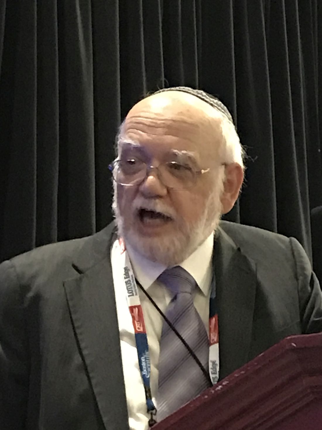
“The FDA is very interested in these devices. We recognize that there is a clinical need, but a reasonable benefit-to-risk relationship has to be established,” said Meir Shinnar, MD, PhD, who works in the division of cardiac devices in the FDA’s Office of Device Evaluation.
The field of renal denervation is expected to heat up again if the results of the SPYRAL HTN OFF MED pivotal trial, planned as a late-breaking presentation at the annual meeting of the American College of Cardiology in March 2020, are positive. However, long-term safety will remain a concern, and positive results will not diminish the rigor with which the relative safety and efficacy of other devices in late stages of clinical testing are evaluated.
“The safety profile is unique to the device design and the procedural technique,” Dr. Shinnar said. For example, vascular injury from the energy employed for denervation, whether radiofrequency or another modality, is an important theoretical risk. A minor initial injury might have no immediate consequences but pose major risks if it leads to altered kidney function over time.
“Most of the follow-up data we have now [with renal denervation devices] is about 1-3 years, but I think long-term safety requires a minimum of 5 years of safety data,” Dr. Shinnar said. “We do not expect all that data to be available at the time of approval, but postmarketing studies will be needed.”
Almost 6 years after the SYMPLICITY HTN-3 trial failed to show a significant reduction in blood pressure among patients with resistant hypertension treated with renal denervation rather than a sham procedure (N Engl J Med 2014;370:1393-401), this treatment is again considered promising. The surprising SYMPLICITY HTN-3 result led to several revisions in technique based on the suspicion that denervation was inadequate.
However, the basic principles remain unchanged. For renal denervation, SPYRAL HTN OFF MED, like the SYMPLICITY HTN 3 study, is employing the Symplicity (Medtronic) device, which has been approved in 50 countries but not in the United States, Canada, or Japan.
SPYRAL HTN OFF MED is designed to provide a very straightforward test of efficacy. Unlike SYMPLICITY HTN-3, which permitted patients to remain on their antihypertensive medications, patients in SPYRAL HTN OFF MED will be tested in the absence of drug therapy (a trial with adjunctive antihypertensive drugs, SPYRAL HTN ON MED, is ongoing). This is a design feature that is relevant to regulatory evaluation.
Although not speaking about the SPYRAL HTN OFF MED trial specifically, Dr. Shinnar noted that “the bar is considered to be higher for a first-line indication than when a device is used as an adjunctive to drug therapy.”
Whether used with or without medications, devices are not likely to receive approval without showing a durable benefit. Dr. Shinnar, citing the surgical studies in which blood pressure control was lost 1-2 years after denervation, said 12 months is now considered a “preferred” length of follow-up to confirm efficacy.
If renal denervation moves forward as a result of the new wave of phase 3 trials, there will still be many unanswered questions, according to Dr. Shinnar, who noted that the FDA convened an advisory committee in December 2018 to gather expert opinion about meaningful safety as well as efficacy endpoints for this modality. One will be determining which populations, defined by age, gender, or phenotype, most benefit.
It also remains unclear whether the first approval will create a standard to which subsequent devices should be compared, according to Dr. Shinnar. Although the FDA recognizes blood pressure reductions as an acceptable endpoint, he believes that documentation of the impact on clinical events will be sought in postmarketing analyses.
“All of the denervation modalities involve class 3 devices that require significant data,” Dr. Shinnar cautioned.
Even if the SPYRAL HTN OFF MED trial is positive on the basis of efficacy, it does not guarantee regulatory approval. Dr. Shinnar described a multifaceted approach to defining an acceptable risk-to-benefit ratio from approved devices, and warned that several points regarding the evaluation of renal denervation devices by the FDA are still being debated internally.
Dr. Shinnar reported no potential financial conflicts of interest.
NATIONAL HARBOR, MD. – Just a month before results from the first of several new pivotal trials with a renal denervation device are to be presented, a Food and Drug Administration medical officer speaking at CRT 2020 sponsored by MedStar Heart & Vascular Institute explained which data will most attract the scrutiny of regulators.
“The FDA is very interested in these devices. We recognize that there is a clinical need, but a reasonable benefit-to-risk relationship has to be established,” said Meir Shinnar, MD, PhD, who works in the division of cardiac devices in the FDA’s Office of Device Evaluation.
The field of renal denervation is expected to heat up again if the results of the SPYRAL HTN OFF MED pivotal trial, planned as a late-breaking presentation at the annual meeting of the American College of Cardiology in March 2020, are positive. However, long-term safety will remain a concern, and positive results will not diminish the rigor with which the relative safety and efficacy of other devices in late stages of clinical testing are evaluated.
“The safety profile is unique to the device design and the procedural technique,” Dr. Shinnar said. For example, vascular injury from the energy employed for denervation, whether radiofrequency or another modality, is an important theoretical risk. A minor initial injury might have no immediate consequences but pose major risks if it leads to altered kidney function over time.
“Most of the follow-up data we have now [with renal denervation devices] is about 1-3 years, but I think long-term safety requires a minimum of 5 years of safety data,” Dr. Shinnar said. “We do not expect all that data to be available at the time of approval, but postmarketing studies will be needed.”
Almost 6 years after the SYMPLICITY HTN-3 trial failed to show a significant reduction in blood pressure among patients with resistant hypertension treated with renal denervation rather than a sham procedure (N Engl J Med 2014;370:1393-401), this treatment is again considered promising. The surprising SYMPLICITY HTN-3 result led to several revisions in technique based on the suspicion that denervation was inadequate.
However, the basic principles remain unchanged. For renal denervation, SPYRAL HTN OFF MED, like the SYMPLICITY HTN 3 study, is employing the Symplicity (Medtronic) device, which has been approved in 50 countries but not in the United States, Canada, or Japan.
SPYRAL HTN OFF MED is designed to provide a very straightforward test of efficacy. Unlike SYMPLICITY HTN-3, which permitted patients to remain on their antihypertensive medications, patients in SPYRAL HTN OFF MED will be tested in the absence of drug therapy (a trial with adjunctive antihypertensive drugs, SPYRAL HTN ON MED, is ongoing). This is a design feature that is relevant to regulatory evaluation.
Although not speaking about the SPYRAL HTN OFF MED trial specifically, Dr. Shinnar noted that “the bar is considered to be higher for a first-line indication than when a device is used as an adjunctive to drug therapy.”
Whether used with or without medications, devices are not likely to receive approval without showing a durable benefit. Dr. Shinnar, citing the surgical studies in which blood pressure control was lost 1-2 years after denervation, said 12 months is now considered a “preferred” length of follow-up to confirm efficacy.
If renal denervation moves forward as a result of the new wave of phase 3 trials, there will still be many unanswered questions, according to Dr. Shinnar, who noted that the FDA convened an advisory committee in December 2018 to gather expert opinion about meaningful safety as well as efficacy endpoints for this modality. One will be determining which populations, defined by age, gender, or phenotype, most benefit.
It also remains unclear whether the first approval will create a standard to which subsequent devices should be compared, according to Dr. Shinnar. Although the FDA recognizes blood pressure reductions as an acceptable endpoint, he believes that documentation of the impact on clinical events will be sought in postmarketing analyses.
“All of the denervation modalities involve class 3 devices that require significant data,” Dr. Shinnar cautioned.
Even if the SPYRAL HTN OFF MED trial is positive on the basis of efficacy, it does not guarantee regulatory approval. Dr. Shinnar described a multifaceted approach to defining an acceptable risk-to-benefit ratio from approved devices, and warned that several points regarding the evaluation of renal denervation devices by the FDA are still being debated internally.
Dr. Shinnar reported no potential financial conflicts of interest.
EXPERT ANALYSIS FROM CRT 2020
BASILAR: Endovascular treatment improves outcomes in BAO stroke
LOS ANGELES – Endovascular therapy significantly improved functional outcomes and reduced mortality at 90 days, compared with standard thrombolysis alone, new evidence from a large, prospective registry study suggests.
Participants who received both interventions were almost five times more likely to be able to walk independently at 90 days compared with those who received thrombolysis alone.
Despite multiple trials supporting the potential benefits of endovascular therapy for anterior stroke, little prospective research addresses outcomes associated with an ischemic stroke caused by a posterior basilar artery occlusion (BAO).
“Basilar artery occlusion is the ‘orphan’ of the large vessel occlusions,” Raul Gomes Nogueira, MD, PhD, said here at a late-breaking abstract session at the International Stroke Conference sponsored by the American Heart Association.
“They account for about 5% of the large vessel occlusions – but have the most dismal prognosis.” Severe disability and mortality rates associated with BAO, for example, reach an estimated 68% to 78%, he said.
The results, from the EVT for Acute Basilar Artery Occlusion Study (BASILAR), were also simultaneously published in JAMA Neurology.
Prior studies in this patient population are generally single-center, retrospective studies and “the numbers tend to be small,” said Nogueira, who is affiliated with the Marcus Stroke and Neuroscience Center, Grady Memorial Hospital, Emory University School of Medicine in Atlanta, Georgia.
Nogueira and colleagues studied 829 consecutive adults who presented with an acute, symptomatic BAO. They examined a nationwide prospective registry study of people with radiologically confirmed BAO in 47 comprehensive stroke centers across 15 provinces in China.
The median age was 65 years and 74% were men. A total 182 participants received thrombolysis therapy within 6 hours of estimated BAO onset. The 647 people in the dual intervention group also received endovascular therapy within 24 hours.
Standard medical treatment included intravenous rt-PA or urokinase, antiplatelet drugs and systematic anticoagulation alone or in combination. Endovascular therapy included mechanical thrombectomy with stent retrievers and/or thromboaspiration, balloon angioplasty, stenting, intra-arterial thrombolysis, or a combination of these interventions.
Interestingly, participants were not randomly assigned, in part because of the favorable outcomes associated with endovascular therapy. “The high number of patients who received [the dual intervention] may suggest the existence of a lack of equipoise among participating centers,” the researchers note.
Key Efficacy Endpoints
A significantly higher proportion of people in the dual treatment group achieved the primary outcome, functional improvement at 90 days, at 32%, compared with 9.3% in the thrombolysis-only group. This endpoint was defined as a modified Rankin Scale (mRS) score of 3 or less, which reflects an ability to walk independently. The difference was statistically significant (P < .001).
The absolute difference between groups was 22.7% (95% confidence interval, 17.1%-28.2%) with an adjusted odds ratio of 4.70 (95% CI, 2.53-8.75; P < .001) in favor of dual intervention.
The number needed to treat for one additional patient to be able to walk unassisted was 4.4.
Other outcomes, including differences in National Institutes of Health Stroke Scale scores from baseline to 5 to 7 days or discharge, as well as propensity score matching and subgroup analyses, likewise supported the superiority of using both interventions.
Safety Outcomes
Nogueira and colleagues also assessed safety. They found that symptomatic intracerebral hemorrhage (ICH) occurred in 45 patients, or 7.1% of the endovascular treatment group. In contrast, only one patient, or 0.5%, of the standard medical treatment alone cohort experienced an ICH. This difference was statistically significant (P < .001).
Mortality at 90 days was significantly lower in the endovascular therapy plus medical therapy group, 46.2%, compared with 71.4% in the standard medical treatment alone group (P < .001).
The absolute difference in mortality was 25.2% (95% CI, 17.6%-2.8%) favoring dual treatment, with an adjusted odds ratio of 2.93 (95% CI, 1.95-4.40; P < .001).
Rates of other serious adverse events during the 90-day follow-up period were similar in the two study groups, Nogueira said.
He acknowledged that the nonrandomized design was a limitation of the registry study, adding that “sometimes in life it’s important to acknowledge the best of what can be done. It’s very hard when you have access to thrombectomy to randomize people.”
However, other researchers have attempted or are enrolling people with BAO into trials that randomly assign them to endovascular therapy and standard medical treatment or medical treatment alone.
The BEST trial in China, for example, randomly assigned 131 patients to these groups but was stopped early in September 2017. “The BEST trial was terminated prematurely because of loss of equipoise that led to a high crossover rate and drop in valid recruitment,” the current researchers note.
“The other two trials…are facing the challenge of whether they will achieve their inclusion target,” they add, “because a growing number of stroke centers are unwilling to randomize patients to standard medical treatment alone after the many positive results of trials for endovascular treatment in patients with anterior-circulation stroke.”
The BAOCHE trial from China, for example, is ongoing with approximately 110 patients enrolled so far.
Investigators for the Basilar Artery International Cooperation Study (BASICS) in the Netherlands just completed enrollment of their 300th and final patient in December 2019.
“We are hopeful BASICS trial will shed additional light,” Nogueira said. The results are expected to be presented at the European Stroke Organization Conference in Vienna in May 2020.
More Guidance From MRI?
“With the advent of the stent retrievers and successful recanalization, we know there can be better outcomes for patients. And we know the morbidity and mortality of the basilar artery occlusions are so poor that we tend to want to be aggressive in these cases,” session comoderator Shlee S. Song, MD, director of the Comprehensive Stroke Center and associate professor of neurology at Cedars-Sinai Medical Center in Los Angeles, California, told Medscape Medical News when asked to comment on the study.
“I agree that we’ve lost equipoise in this cohort – that we really cannot do a randomized trial anymore. You know if you don’t do anything, 90% of the time there will be a poor outcome,” she added.
This is an important study for showing how BAO patients fare after endovascular treatment, Song said.
One unanswered question from the study is if any of the centers in China used magnetic resonance imaging to help determine the most appropriate candidates for endovascular treatment of these posterior circulation strokes, which is a common practice in the United States, she said.
The study was supported by the National Science Fund for Distinguished Young Scholars, Chongqing Major Disease Prevention and Control Technology Research Project, Army Medical University Clinical Medical Research Talent Training Program, and Major Clinical Innovation Technology Project of the Second Affiliated Hospital of the Army Military Medical University. Sing had no relevant disclosures. Nogueira’s financial disclosures include working as a consultant for Stryker Neurovascular; as a principal investigator on the Imperative trial and the PROST trial; as a steering committee member for Biogen for the CHARM trial; as an advisory board member for Cerenovus/Neuravi, Phenox, Anaconda, Genentech, Biogen, Prolong Pharmaceuticals and Brainomix; and as an advisory board member with stock options for Viz.ai, Corindus Vascular Robotics, Vesalio, Ceretrieve, Astrocyte Pharmaceuticals, and Cerebrotech.
This article first appeared on Medscape.com.
SOURCE: Nogueira RG et al. ISC 2020. Late-breaking abstract 17.
REPORTING FROM ISC 2020