User login
Genomic findings may predict outcomes in MPN patients
New research suggests genomic characteristics of patients with myeloproliferative neoplasms (MPNs) can predict clinical outcomes.
Investigators defined eight genomic subgroups of MPNs, each with distinct clinical features, including event-free survival, risk of leukemic transformation, and blood counts.
Jacob Grinfeld, MD, of the University of Cambridge in the U.K., and his colleagues described these findings in The New England Journal of Medicine.
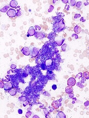
This study included 2,035 patients with MPNs, including essential thrombocythemia, polycythemia vera, myelofibrosis, and other diagnoses.
The investigators performed targeted sequencing for the full coding sequence of 69 genes and genome-wide copy number information in 1,887 patients. Whole-exome sequencing was performed in another 148 patients.
By sequencing coding exons from 69 myeloid cancer genes, the investigators were able to survey the diversity of mutations across MPN patients and identify mutation-associated clinical outcomes.
The results showed that slightly less than half (45%) of the patients had a solitary abnormality in CALR, MPL, or JAK2, while the remaining patients had additional driver mutations.
In some instances, additional mutations were numerous, particularly in older patients with advanced disease. In at least five cases, 33 genes had driver mutations.
Further analysis revealed eight genomic subgroups that could predict clinical outcomes based on shared chromosomal abnormalities and mutations.
For example, one subgroup included patients with TP53 mutations. These individuals had a “dismal prognosis” and were 15.5 times more likely to transform to acute myeloid leukemia, compared with the JAK2-heterozygous subgroup (P<0.001).
Because prognosis is “a key determinant” of MPN treatment, genomic subgrouping may one day guide clinical decision-making, the investigators concluded.
To further this cause, they have made available an online calculator of individualized patient outcomes, which can be accessed at https://cancer.sanger.ac.uk/mpn-multistage/.
This study was funded by the Wellcome Trust, the National Institute for Health Research Cambridge Biomedical Research Centre, Cancer Research UK, and others. Some study authors reported fees from Celgene, Novartis, Gilead, Shire, and others outside of the study.
New research suggests genomic characteristics of patients with myeloproliferative neoplasms (MPNs) can predict clinical outcomes.
Investigators defined eight genomic subgroups of MPNs, each with distinct clinical features, including event-free survival, risk of leukemic transformation, and blood counts.
Jacob Grinfeld, MD, of the University of Cambridge in the U.K., and his colleagues described these findings in The New England Journal of Medicine.
This study included 2,035 patients with MPNs, including essential thrombocythemia, polycythemia vera, myelofibrosis, and other diagnoses.
The investigators performed targeted sequencing for the full coding sequence of 69 genes and genome-wide copy number information in 1,887 patients. Whole-exome sequencing was performed in another 148 patients.
By sequencing coding exons from 69 myeloid cancer genes, the investigators were able to survey the diversity of mutations across MPN patients and identify mutation-associated clinical outcomes.
The results showed that slightly less than half (45%) of the patients had a solitary abnormality in CALR, MPL, or JAK2, while the remaining patients had additional driver mutations.
In some instances, additional mutations were numerous, particularly in older patients with advanced disease. In at least five cases, 33 genes had driver mutations.
Further analysis revealed eight genomic subgroups that could predict clinical outcomes based on shared chromosomal abnormalities and mutations.
For example, one subgroup included patients with TP53 mutations. These individuals had a “dismal prognosis” and were 15.5 times more likely to transform to acute myeloid leukemia, compared with the JAK2-heterozygous subgroup (P<0.001).
Because prognosis is “a key determinant” of MPN treatment, genomic subgrouping may one day guide clinical decision-making, the investigators concluded.
To further this cause, they have made available an online calculator of individualized patient outcomes, which can be accessed at https://cancer.sanger.ac.uk/mpn-multistage/.
This study was funded by the Wellcome Trust, the National Institute for Health Research Cambridge Biomedical Research Centre, Cancer Research UK, and others. Some study authors reported fees from Celgene, Novartis, Gilead, Shire, and others outside of the study.
Virus-specific T-cell infusion may resolve progressive multifocal leukoencephalopathy
, according to investigators from the University of Texas MD Anderson Cancer Center, Houston.
The infusion cleared JC virus from the cerebrospinal fluid (CSF) of two patients and reduced viral load in the third, reported lead author Muharrem Muftuoglu, MD, of MD Anderson’s department of stem cell transplantation and cellular therapy and colleagues. One of the patients completely recovered and returned to work.
“Several approaches for the treatment of PML, including the use of antiviral medications and mirtazapine, have been tested, with poor results,” the investigators wrote in the New England Journal of Medicine. Although virus-specific T-cell infusion is a novel approach to treating PML, this method has been used for other conditions.
“Several groups, including ours, have successfully used viral-specific T cells to treat BK virus infection after stem-cell transplantation,” the investigators wrote. “Because BK virus and JC virus are genetically similar to one another and share a number of immunogenic proteins with a substantial degree of sequence homology ... we hypothesized that T cells developed against BK virus may also be effective against JC virus infection.”
This hypothesis proved accurate. The investigators infused three PML patients with “cryopreserved, third-party–produced, viral-specific T cells that had been designed for the treatment of patients with BK virus infection after stem-cell transplantation.” Each patient presented with a different condition and PML-precipitating therapy. The first patient was a 32-year-old woman with high-risk acute myeloid leukemia who had received a cord-blood transplantation, the second a 73-year-old woman with JAK2-positive myeloproliferative neoplasia on ruxolitinib (Jakafi) therapy, and the third a 35-year-old man with HIV who had received highly active antiretroviral therapy.
T-cell infusions cleared JC virus from the CSF of the woman with leukemia (three infusions) and the man with HIV (four infusions). These patients recovered to different degrees: The woman had full resolution of symptoms, while the man had slurred speech and walked with a cane. Treatment reduced JC viral load in the elderly woman with myeloproliferative neoplasia (two infusions), but she did not clear the virus and died about 8 months later.
No adverse events occurred, but two patients developed immune reconstitution inflammatory syndrome. This was likely caused by the T-cell infusion, since absolute T-cell counts remained steady and white matter enhancement was detected on MRI within 4 weeks of treatment. Still, the investigators were optimistic about future potential.
“Third-party–produced, ‘off-the-shelf,’ partially HLA-matched, BK virus–specific T cells may serve as therapy for PML,” the investigators concluded. “Further study in a larger group of patients is required to determine the success rate, durability, and longer-term adverse events associated with this treatment.”
The study was funded by the MD Anderson Cancer Center Moon Shots Program and the National Institutes of Health.
SOURCE: Muftuoglu M et al. N Engl J Med. 2018 Oct 11;379:1443-51
This article was updated 3/22/19.
FROM NEW ENGLAND JOURNAL OF MEDICINE
Key clinical point: Infusion of allogeneic BK virus-specific T cells may be an effective treatment for patients with PML.
Major finding: Two of three patients cleared JC virus from cerebrospinal fluid after infusion.
Study details: A case series involving three patients with PML.
Disclosures: The study was funded by the MD Anderson Cancer Center Moon Shots Program and the National Institutes of Health.
Source: Muftuoglu M et al. N Engl J Med. 2018 Oct 11;379:1443-51.
Genomic profiling predicts outcomes in patients with MPN
Genomic characteristics of patients with myeloproliferative neoplasms (MPN) can predict clinical outcomes, a recent study found.

Eight genomic subgroups of MPN were recognized, each with distinct clinical features, including event-free survival, risk of leukemic transformation, and blood counts, according to Jacob Grinfeld, MD, of the Wellcome-MRC Cambridge (England) Stem Cell Institute and Cambridge Institute for Medical Research and his colleagues.
“Current classification schemes distinguish among the subtypes of myeloproliferative neoplasms according to clinical and laboratory features, but uncertainty clouds where and how to draw dividing lines among them,” the investigators wrote in the New England Journal of Medicine. “In blood cancers, a progressive shift is under way, from clinical and morphologic classification schemes to those that are based on genomics.”
MPNs are often driven by mutations in CALR, MPL, or JAK2 genes, but classification is not confined to just three genomic types; many patients have additional driver mutations throughout a variety of cancer genes, and it is these additional mutations that are responsible for the wide range of disease phenotypes and clinical outcomes.
This study included 2,035 patients with MPNs, including essential thrombocythemia, polycythemia vera, myelofibrosis, and other MPN diagnoses. The investigators performed targeted sequencing for the full coding sequence of 69 genes and genomewide copy-number information in 1,887 patients. Another 148 patients underwent whole-exome sequencing.
By sequencing coding exons from 69 myeloid cancer genes, the investigators were able to survey the diversity of mutations across a population of patients with MPNs and identify mutation-associated clinical outcomes.
The results showed that slightly less than half (45%) of the patients had a solitary abnormality in CALR, MPL, or JAK2, while the remaining patients had additional driver mutations. In some instances, additional mutations were numerous, particularly in older patients with advanced disease. In at least five cases, 33 genes had driver mutations.
Further analysis identified eight genomic subgroups that could predict clinical outcomes based on shared chromosomal abnormalities and mutations. For example, one subgroup included patients with TP53 mutations; these individuals had a “dismal prognosis” and were 15.5 times more likely to transform to acute myeloid leukemia (AML), compared with the JAK2-heterozygous subgroup (P less than .001).
Because prognosis is “a key determinant of the treatment of patients with MPNs,” genomic subgrouping may one day guide clinical decision making, the investigators concluded.
To further this cause, the investigators have made available an online calculator of individualized patient outcomes, which can be accessed at https://cancer.sanger.ac.uk/mpn-multistage/.
The study was funded by the Wellcome Trust, the National Institute for Health Research Cambridge Biomedical Research Centre, Cancer Research UK, and others. Some study authors reported fees from Celgene, Novartis, Gilead, Shire, and others outside of the study.
SOURCE: Grinfeld J et al. N Engl J Med. 2018;379:1416-30.
Genomic characteristics of patients with myeloproliferative neoplasms (MPN) can predict clinical outcomes, a recent study found.
Eight genomic subgroups of MPN were recognized, each with distinct clinical features, including event-free survival, risk of leukemic transformation, and blood counts, according to Jacob Grinfeld, MD, of the Wellcome-MRC Cambridge (England) Stem Cell Institute and Cambridge Institute for Medical Research and his colleagues.
“Current classification schemes distinguish among the subtypes of myeloproliferative neoplasms according to clinical and laboratory features, but uncertainty clouds where and how to draw dividing lines among them,” the investigators wrote in the New England Journal of Medicine. “In blood cancers, a progressive shift is under way, from clinical and morphologic classification schemes to those that are based on genomics.”
MPNs are often driven by mutations in CALR, MPL, or JAK2 genes, but classification is not confined to just three genomic types; many patients have additional driver mutations throughout a variety of cancer genes, and it is these additional mutations that are responsible for the wide range of disease phenotypes and clinical outcomes.
This study included 2,035 patients with MPNs, including essential thrombocythemia, polycythemia vera, myelofibrosis, and other MPN diagnoses. The investigators performed targeted sequencing for the full coding sequence of 69 genes and genomewide copy-number information in 1,887 patients. Another 148 patients underwent whole-exome sequencing.
By sequencing coding exons from 69 myeloid cancer genes, the investigators were able to survey the diversity of mutations across a population of patients with MPNs and identify mutation-associated clinical outcomes.
The results showed that slightly less than half (45%) of the patients had a solitary abnormality in CALR, MPL, or JAK2, while the remaining patients had additional driver mutations. In some instances, additional mutations were numerous, particularly in older patients with advanced disease. In at least five cases, 33 genes had driver mutations.
Further analysis identified eight genomic subgroups that could predict clinical outcomes based on shared chromosomal abnormalities and mutations. For example, one subgroup included patients with TP53 mutations; these individuals had a “dismal prognosis” and were 15.5 times more likely to transform to acute myeloid leukemia (AML), compared with the JAK2-heterozygous subgroup (P less than .001).
Because prognosis is “a key determinant of the treatment of patients with MPNs,” genomic subgrouping may one day guide clinical decision making, the investigators concluded.
To further this cause, the investigators have made available an online calculator of individualized patient outcomes, which can be accessed at https://cancer.sanger.ac.uk/mpn-multistage/.
The study was funded by the Wellcome Trust, the National Institute for Health Research Cambridge Biomedical Research Centre, Cancer Research UK, and others. Some study authors reported fees from Celgene, Novartis, Gilead, Shire, and others outside of the study.
SOURCE: Grinfeld J et al. N Engl J Med. 2018;379:1416-30.
FROM THE NEW ENGLAND JOURNAL OF MEDICINE
Key clinical point:
Major finding: Eight genomic subgroups of MPN were recognized, each with distinct clinical features, including event-free survival, risk of leukemic transformation, and blood counts.
Study details: A gene sequencing study involving 2,035 patients with MPN.
Disclosures: The study was funded by the Wellcome Trust, the National Institute for Health Research Cambridge Biomedical Research Centre, Cancer Research UK, and others. Some study authors reported fees from Celgene, Novartis, Gilead, Shire, and others outside of the study.
Source: Grinfeld J et al. N Engl J Med. 2018;379:1416-30.
Short-term NSAIDs appear safe for high-risk patients
in a retrospective, observational study.
The findings of the study challenge the Choosing Wisely campaign of the American Society of Nephrology, which recommends against NSAIDs for high-risk patients, according to lead author Zachary Bouck, MPH, of the department of medicine at Sunnybrook Health Sciences Centre in Toronto, and his coauthors.
“While these recommendations offer basic analgesics and nonpharmacological treatments as preferable alternatives, it is both possible and disconcerting that some physicians might instead prescribe opioids, which typically pose elevated risk of adverse events and dependence vs. NSAIDs,” the investigators wrote. The report is in JAMA Internal Medicine.
They sought to estimate the frequency and characteristics of NSAID prescriptions while also looking for associations with acute renal and cardiovascular complications. The retrospective, observational study involved 814,049 adults with musculoskeletal disease and 7,365 primary care physicians in Ontario, Canada. All patients were aged 65 years and older, and had been diagnosed with hypertension, chronic kidney disease, or heart failure in the past year. Instances in which a patient was prescribed an NSAID within 7 days of presentation were included.
To assess for associations between prescription NSAIDs and negative outcomes, the investigators searched for renal or cardiovascular complications within 37 days of presentation. Over-the-counter NSAID usage was not evaluated.
There were 224,825 visits. An NSAID was prescribed after 9.3% of these visits.
Renal and cardiovascular outcomes were similar between high-risk patients who received a prescription NSAID and those who did not (absolute risk reduction, .0003; P = .74).
“The similarity in risk between users and nonusers, each group primarily consisting of patients with hypertension, suggests that the short-term association of NSAIDs in high-risk patients with musculoskeletal pain may not be as dangerous as initially thought,” the authors concluded.
The investigators found that prescribing rates varied widely, ranging from 6.7% to 14.4% of different health regions, and from 0.9% to 60.3% among 688 primary care practices, with “substantial variation in use” among primary care physicians.
The authors acknowledged limitations, including the use of administrative data, but noted that their study, showing substantial variations in NSAID prescribing, “along with the identification of patient and physician characteristics associated with NSAID use, presents an opportunity for quality improvement, with some potential targets for any resulting interventions,” they wrote.
The Institute for Clinical Evaluative Sciences funded the study. The authors reported compensation from the Canadian Institute of Health Research, the department of family and community medicine at the University of Toronto, the Heart and Stroke Foundation of Canada, and Women’s College Hospital.
SOURCE: Bouck et al. JAMA Intern Med. 2018 Oct 8. doi: 10.1001/jamainternmed.2018.4273.
The study by Bouck and colleagues found that short-term prescription NSAIDs were safe for high-risk patients; however, physicians should consider the inherent limitations of observational studies before altering clinical decisions, according to Jonathan Zipursky, MD, and David N. Juurlink, MD, PhD. This is particularly important since the findings challenge the American Society of Nephrology, which recommends against NSAIDs in patients with chronic kidney disease (CKD), heart failure, or hypertension.
Among the advantages of observational studies over randomized trials is that they often include patients not eligible for randomized controlled trials, “and extended follow-up enables examination of outcomes that might not have arisen earlier in treatment,” they wrote in an editorial. “Moreover, sample sizes often greatly exceed those of RCTs, facilitating detection of less common adverse events. Consequently, population-based observational studies are critical to postmarketing surveillance and, increasingly, evidence-based prescribing recommendations.”
On the other hand, observational studies are less tightly controlled than randomized trials, with nonrandomized allocation, which “raises the possibilities of selection bias and confounding by indication,” they wrote. Furthermore, “readers are left questioning whether patients who were not prescribed NSAIDs were simply taking over-the-counter medications (including NSAIDs),” they wrote.
“Although we rely on observational studies to answer questions poorly suited to clinical trials, we have to interpret these findings with caution,” they added.
Jonathan Zipursky, MD, and Dr. Juurlink are affiliated with the department of medicine at Sunnybrook Health Sciences Centre in Toronto. These comments are adapted from their accompanying editorial (JAMA Intern Med 2018 Oct 8. doi: 10.1136/ebmed-2016-110401).
in a retrospective, observational study.
The findings of the study challenge the Choosing Wisely campaign of the American Society of Nephrology, which recommends against NSAIDs for high-risk patients, according to lead author Zachary Bouck, MPH, of the department of medicine at Sunnybrook Health Sciences Centre in Toronto, and his coauthors.
“While these recommendations offer basic analgesics and nonpharmacological treatments as preferable alternatives, it is both possible and disconcerting that some physicians might instead prescribe opioids, which typically pose elevated risk of adverse events and dependence vs. NSAIDs,” the investigators wrote. The report is in JAMA Internal Medicine.
They sought to estimate the frequency and characteristics of NSAID prescriptions while also looking for associations with acute renal and cardiovascular complications. The retrospective, observational study involved 814,049 adults with musculoskeletal disease and 7,365 primary care physicians in Ontario, Canada. All patients were aged 65 years and older, and had been diagnosed with hypertension, chronic kidney disease, or heart failure in the past year. Instances in which a patient was prescribed an NSAID within 7 days of presentation were included.
To assess for associations between prescription NSAIDs and negative outcomes, the investigators searched for renal or cardiovascular complications within 37 days of presentation. Over-the-counter NSAID usage was not evaluated.
There were 224,825 visits. An NSAID was prescribed after 9.3% of these visits.
Renal and cardiovascular outcomes were similar between high-risk patients who received a prescription NSAID and those who did not (absolute risk reduction, .0003; P = .74).
“The similarity in risk between users and nonusers, each group primarily consisting of patients with hypertension, suggests that the short-term association of NSAIDs in high-risk patients with musculoskeletal pain may not be as dangerous as initially thought,” the authors concluded.
The investigators found that prescribing rates varied widely, ranging from 6.7% to 14.4% of different health regions, and from 0.9% to 60.3% among 688 primary care practices, with “substantial variation in use” among primary care physicians.
The authors acknowledged limitations, including the use of administrative data, but noted that their study, showing substantial variations in NSAID prescribing, “along with the identification of patient and physician characteristics associated with NSAID use, presents an opportunity for quality improvement, with some potential targets for any resulting interventions,” they wrote.
The Institute for Clinical Evaluative Sciences funded the study. The authors reported compensation from the Canadian Institute of Health Research, the department of family and community medicine at the University of Toronto, the Heart and Stroke Foundation of Canada, and Women’s College Hospital.
SOURCE: Bouck et al. JAMA Intern Med. 2018 Oct 8. doi: 10.1001/jamainternmed.2018.4273.
FROM JAMA INTERNAL MEDICINE
Key clinical point: In patients with musculoskeletal disease and hypertension, chronic kidney disease, or heart failure, short-term prescription NSAIDs may be safer than once thought.
Major finding: Renal and cardiovascular outcomes were similar between high-risk patients who received a prescription NSAID and those who did not (absolute risk reduction, .0003; P = .74).
Study details: A retrospective, observational study involving 814,049 adults with musculoskeletal disease and 7,365 primary care physicians in Ontario, Canada.
Disclosures: The Institute for Clinical Evaluative Sciences funded the study. The authors reported compensation from the Canadian Institute of Health Research, the Heart and Stroke Foundation of Canada, and Women’s College Hospital.
Source: Bouck et al. JAMA Intern Med. 2018 Oct 8. doi: 10.1001/jamainternmed.2018.4273.
Allopurinol reduces risk of renal decline in gout patients
In patients with gout, at least 300 mg of allopurinol daily may reduce the risk of renal function decline, according to a new study.

Since no evidence supports allopurinol nephrotoxicity and usage does not appear to worsen chronic kidney disease (CKD), clinicians should consider other causes of declining renal function, according to lead author Ana Beatriz Vargas-Santos, MD, of the rheumatology unit at the State University of Rio de Janeiro and her colleagues.
These findings reinforce the American College of Rheumatology’s 2012 treatment recommendation that the dose of allopurinol, a urate-lowering therapy (ULT), “can be raised above 300 mg daily, even with renal impairment, as long as it is accompanied by adequate patient education and monitoring for drug toxicity may worsen renal function.”*
“Renal-dosing of allopurinol compounds the poor management of gout and adds to the perception that allopurinol may be detrimental for renal function,” the investigators wrote in JAMA Internal Medicine. “In contrast, recent studies provide support for starting allopurinol at a low dose with gradual dose escalation to serum urate target with close monitoring, even among patients with renal insufficiency, without increased risk of allopurinol hypersensitivity syndrome (AHS). Further, there is emerging evidence that ULT may be beneficial for kidney dysfunction.”
Building upon these developments, the investigators “aimed to assess the relation of allopurinol initiation to the risk of developing CKD stage 3 or higher among people with newly diagnosed gout.”
Patients for the cohort study were drawn from the Health Improvement Network (THIN), a database of records from general practitioners in the United Kingdom. Included patients were recently diagnosed with gout but did not have stage 3 or higher chronic kidney disease or ULT usage within a year prior to diagnosis. After screening, 4,760 allopurinol users were matched with 4,760 allopurinol nonusers. Overall, 71% of patients had CKD stage 2, while the remaining 29% had CKD stage 1 or normal kidney function.
The primary outcome of CKD stage 3 or higher was defined as glomerular filtration rate below 60 mL/min (recorded at least twice in 1 year with a 3-month interval between readings and GFR never exceeding 75 mL/min during the intervening period), kidney transplant, or dialysis. The mean follow-up time was 5 years for allopurinol users and 4 years for nonusers.
The investigators found that 579 allopurinol users developed CKD stage 3 or higher, compared with 623 nonusers, suggesting that allopurinol reduced risk of CKD stage 3 or higher by 13%. Allopurinol doses of at least 300 mg/day were associated with a hazard ratio of 0.87, but lower doses did not share this association (HR = 1.02).
In defense of their findings, Dr. Vargas-Santos and her associates evaluated the relevance of their study, compared with previous allopurinol studies.
“This study is one of few that have evaluated the relation of allopurinol to renal function among patients with gout and normal or near-normal kidney function at baseline,” the authors wrote, noting that most gout patients do not have severe kidney disease.
Previous studies have suggested that allopurinol worsens kidney function, but these studies were often conducted in nongout populations, with patients exhibiting CKD stage 3 or higher, they noted. Instead of allopurinol-induced kidney damage, renal decline in gout patients is likely multifactorial.
“Because people with gout have intrinsic differences compared with those with asymptomatic hyperuricemia, including higher mortality, more comorbidities, and more NSAID use, these studies’ results are not directly applicable to gout patients,” the investigators wrote.
“At minimum, allopurinol does not seem to have a detrimental effect on renal function in individuals with gout,” Dr. Vargas-Santos and her associates concluded. “Clinicians should consider evaluating other factors when faced with renal function decline in their patients with gout rather than lowering the dose of or discontinuing allopurinol, a strategy that has contributed to the ongoing suboptimal treatment of gout.”
The authors reported funding from Conselho Nacional de Desenvolvimento Científico e Tecnológico (CNPq); Ministry of Science, Technology and Innovation of Brazil; and National Institutes of Health. Dr Vargas-Santos has received speaking fees and support for international medical events from Grünenthal. No other disclosures were reported.
SOURCE: Vargas-Santos AB et al. JAMA Intern Med. 2018 Oct 8. doi: 10.1001/jamainternmed.2018.4463
*Correction, 11/5/2018: An earlier version of this story incorrectly stated the American College of Rheumatology’s 2012 gout treatment recommendation for using allopurinol in patients with renal impairment.
Physicians should consider the inherent limitations of observational studies before altering clinical decisions, according to Jonathan Zipursky, MD, and David N. Juurlink, MD, PhD. This is particularly important since the findings in this paper challenge the American College of Rheumatology, which recommends lower allopurinol doses in patients with chronic kidney disease (CKD).
On one hand, they noted, observational studies have some advantages over randomized trials.
“Observational studies frequently include patients who are ineligible for RCTs, and extended follow-up enables examination of outcomes that might not have arisen earlier in treatment,” Dr. Zipursky and Dr. Juurlink wrote in an editorial. “Moreover, sample sizes often greatly exceed those of RCTs, facilitating detection of less common adverse events. Consequently, population-based observational studies are critical to postmarketing surveillance and, increasingly, evidence-based prescribing recommendations.”
On the other hand, observational studies are less tightly controlled than randomized trials.
As “treatment allocation is nonrandom, it raises the possibilities of selection bias and confounding by indication,” Dr. Zipursky and Dr. Juurlink wrote. “Perhaps the drugs were preferentially prescribed to patients destined to tolerate them, or fare better in some other way apparent to prescribers but beyond the resolution of large databases. For example, of the nearly 43,000 patients in the study by Vargos-Santos et al, only 10% were started on 300 mg or more per day of allopurinol. This leaves readers to wonder what motivated practitioners to start such doses, or, conversely, what it was about the remaining 90% of patients that led them to receive lower doses of allopurinol or none at all.”
Along with these unanswered questions, the study compared treated patients to untreated ones, but “it is generally desirable to compare 1 drug with another used for the same indication, which can help mitigate the effect of unmeasured factors that might have influenced the decision to treat in the first place.”
Familiarity with observational studies is essential for clinicians, as Dr. Zipursky and Dr. Juurlink expect such trials will become more common in the future, and they provide useful insight if clinicians maintain an appropriate viewpoint.
“The findings will need to be contextualized and viewed with more skepticism than RCTs,” they wrote, “but in some instances, they can be thoughtfully integrated into our treatment decisions.”
Dr. Zipursky and Dr. Juurlink are with the department of medicine at Sunnybrook Health Sciences Centre, Toronto. These comments are adapted from their accompanying editorial (JAMA Intern Med. 2018 Oct 8. doi: 10.1001/jamainternmed.2018.5766).
In patients with gout, at least 300 mg of allopurinol daily may reduce the risk of renal function decline, according to a new study.
Since no evidence supports allopurinol nephrotoxicity and usage does not appear to worsen chronic kidney disease (CKD), clinicians should consider other causes of declining renal function, according to lead author Ana Beatriz Vargas-Santos, MD, of the rheumatology unit at the State University of Rio de Janeiro and her colleagues.
These findings reinforce the American College of Rheumatology’s 2012 treatment recommendation that the dose of allopurinol, a urate-lowering therapy (ULT), “can be raised above 300 mg daily, even with renal impairment, as long as it is accompanied by adequate patient education and monitoring for drug toxicity may worsen renal function.”*
“Renal-dosing of allopurinol compounds the poor management of gout and adds to the perception that allopurinol may be detrimental for renal function,” the investigators wrote in JAMA Internal Medicine. “In contrast, recent studies provide support for starting allopurinol at a low dose with gradual dose escalation to serum urate target with close monitoring, even among patients with renal insufficiency, without increased risk of allopurinol hypersensitivity syndrome (AHS). Further, there is emerging evidence that ULT may be beneficial for kidney dysfunction.”
Building upon these developments, the investigators “aimed to assess the relation of allopurinol initiation to the risk of developing CKD stage 3 or higher among people with newly diagnosed gout.”
Patients for the cohort study were drawn from the Health Improvement Network (THIN), a database of records from general practitioners in the United Kingdom. Included patients were recently diagnosed with gout but did not have stage 3 or higher chronic kidney disease or ULT usage within a year prior to diagnosis. After screening, 4,760 allopurinol users were matched with 4,760 allopurinol nonusers. Overall, 71% of patients had CKD stage 2, while the remaining 29% had CKD stage 1 or normal kidney function.
The primary outcome of CKD stage 3 or higher was defined as glomerular filtration rate below 60 mL/min (recorded at least twice in 1 year with a 3-month interval between readings and GFR never exceeding 75 mL/min during the intervening period), kidney transplant, or dialysis. The mean follow-up time was 5 years for allopurinol users and 4 years for nonusers.
The investigators found that 579 allopurinol users developed CKD stage 3 or higher, compared with 623 nonusers, suggesting that allopurinol reduced risk of CKD stage 3 or higher by 13%. Allopurinol doses of at least 300 mg/day were associated with a hazard ratio of 0.87, but lower doses did not share this association (HR = 1.02).
In defense of their findings, Dr. Vargas-Santos and her associates evaluated the relevance of their study, compared with previous allopurinol studies.
“This study is one of few that have evaluated the relation of allopurinol to renal function among patients with gout and normal or near-normal kidney function at baseline,” the authors wrote, noting that most gout patients do not have severe kidney disease.
Previous studies have suggested that allopurinol worsens kidney function, but these studies were often conducted in nongout populations, with patients exhibiting CKD stage 3 or higher, they noted. Instead of allopurinol-induced kidney damage, renal decline in gout patients is likely multifactorial.
“Because people with gout have intrinsic differences compared with those with asymptomatic hyperuricemia, including higher mortality, more comorbidities, and more NSAID use, these studies’ results are not directly applicable to gout patients,” the investigators wrote.
“At minimum, allopurinol does not seem to have a detrimental effect on renal function in individuals with gout,” Dr. Vargas-Santos and her associates concluded. “Clinicians should consider evaluating other factors when faced with renal function decline in their patients with gout rather than lowering the dose of or discontinuing allopurinol, a strategy that has contributed to the ongoing suboptimal treatment of gout.”
The authors reported funding from Conselho Nacional de Desenvolvimento Científico e Tecnológico (CNPq); Ministry of Science, Technology and Innovation of Brazil; and National Institutes of Health. Dr Vargas-Santos has received speaking fees and support for international medical events from Grünenthal. No other disclosures were reported.
SOURCE: Vargas-Santos AB et al. JAMA Intern Med. 2018 Oct 8. doi: 10.1001/jamainternmed.2018.4463
*Correction, 11/5/2018: An earlier version of this story incorrectly stated the American College of Rheumatology’s 2012 gout treatment recommendation for using allopurinol in patients with renal impairment.
FROM JAMA INTERNAL MEDICINE
Key clinical point: In patients with gout, allopurinol was associated with a reduced risk of renal function decline.
Major finding: Allopurinol doses of at least 300 mg/day reduced risk of stage-3 or higher chronic kidney disease by 13%.
Study details: A retrospective, observational study involving newly diagnosed gout patients who either started allopurinol or did not (n = 4,760 in each group).
Disclosures: The authors reported funding from Conselho Nacional de Desenvolvimento Científico e Tecnológico (CNPq); Ministry of Science, Technology and Innovation of Brazil; and National Institutes of Health. Dr Vargas-Santos has received speaking fees and support for international medical events from Grünenthal. No other disclosures were reported.
Source: Vargas-Santos AB et al. JAMA Intern Med. 2018 Oct 8. doi: 10.1001/jamainternmed.2018.4463
Weighing the costs of CAR T-cell therapy

The cost-effectiveness of tisagenlecleucel (Kymriah) depends on long-term clinical outcomes, which are presently unknown, according to investigators.
If the long-term outcomes are more modest than clinical trials suggest, then payers may be unwilling to cover the costly therapy, reported John K. Lin, MD, of Stanford University, and his colleagues.
Lowering the price or setting up an outcomes-based pricing structure may be necessary to get insurers to cover the therapy.
Tisagenlecleucel is an anti-CD19 chimeric antigen receptor (CAR) T-cell therapy that was approved by the U.S. Food and Drug Administration in August 2017 for relapsed or refractory pediatric B-cell acute lymphoblastic leukemia (ALL).
In 2018, the FDA expanded the indication for tisagenlecleucel to include adults with relapsed or refractory large B-cell lymphoma, though outcomes from lymphoma trials are not analyzed in the current study.
At a wholesale acquisition cost of $475,000 per infusion, it is the most expensive existing oncology therapy to date, and can be accompanied by expensive, potentially fatal adverse effects.
However, clinical trials suggest that tisagenlecleucel can offer years of relapse-free remission, thereby allowing patients to forgo other expensive therapies such as hematopoietic stem cell transplantation (HSCT).
“Although tisagenlecleucel-induced remission rates are promising, compared with those of established therapies (greater than 80% vs. less than 50%), only short-term follow-up data currently exist,” the investigators wrote in the Journal of Clinical Oncology.
“Given the high cost and broad applicability in other malignancies of tisagenlecleucel, a pressing question for policy makers, payers, patients, and clinicians is whether the cost of therapy represents reasonable value.”
The study used a Markov model to assess various long-term clinical outcome rates and cost thresholds of tisagenlecleucel. The lifetime cost of therapy was assessed and compared with costs of existing therapies.
The results showed that a 5-year relapse free survival rate of 40% would make the present cost ($475,000) of tisagenlecleucel economically reasonable. In this scenario, the increased life expectancy would be 12.1 years and would result in an additional 5.07 quality-adjusted life years (QALY) gained at a cost of $61,000 per QALY, compared with blinatumomab.
But if long-term outcomes are less favorable, tisagenlecleucel becomes much less cost effective. A 5-year relapse-free survival rate of 20% would drop increased life expectancy to 3.8 years, resulting in 1.80 QALYs gained and raising the cost to $151,000 per QALY.
“Our results suggest that at tisagenlecleucel’s current price and payment structure, its economic value is uncertain,” the investigators wrote.
They suggested a price drop to $200,000 or $350,000, which would allow the drug to remain cost effective even in a worse-case scenario, in which patients relapse and tisagenlecleucel is a bridge to transplant.
Another option is to move to outcomes-based pricing. Making payment conditional on 7 months of remission would make the treatment cost effective, according to the analysis.
“Price reductions of tisagenlecleucel or payment only for longer-term remissions would favorably influence cost-effectiveness, even if long-term clinical outcomes are modest,” the investigators wrote.
The study was funded by a Veterans Affairs Office of Academic Affiliations advanced fellowship in health service and research development, and a National Center for Advancing Translational Science Clinical and Translational Science Award.
One of the study coauthors reported consulting and research funding from Novartis.
Antibiotics trigger proteolytic activity that leads to chronic colitis
Antibiotics are associated with increased large intestinal proteolytic activity and gut barrier disruption, thereby raising the risk of chronic colitis in susceptible individuals, a recent study found.
Although the association between antibiotics and chronic colitis has been previously described, this is the first study to demonstrate the causative role of high proteolytic activity, reported lead author Hongsup Yoon, PhD, chair of nutrition and immunology at Technische Universität München in Freising-Weihenstephan, Germany, and colleagues. The team’s experiments support development of antiproteolytic strategies in susceptible humans.
“In the context of IBD, several clinical studies have already revealed that early and frequent antibiotic therapies, especially metronidazole or fluoroquinolone treatments, are associated with increased risk for Crohn’s disease,” the authors wrote in Cellular and Molecular Gastroenterology and Hepatology. “However, the causal role of antibiotic therapies in the disease development and the mechanisms underlying this [potentially] serious long-term adverse effect of antibiotics on the intestinal immune homeostasis remain unknown.”
Previous studies have shown that antibiotic therapy often causes high luminal proteolytic activity in the large intestine, likely because of the elimination of antiproteolytic bacteria that normally control pancreatic protease levels. Other studies have shown that exposing murine colonic mucosa to fecal supernatants with high proteolytic activity increases gut barrier permeability, which triggers chronic inflammation via translocation of luminal antigens.
“In view of these data,” the authors wrote, “we hypothesized that the antibiotic-increased proteolytic activity in the large intestine is a relevant risk factor for the development of colitis in susceptible organisms.”
The first component of the study used transwell experiments to evaluate the impact of high proteolytic activity on gut barrier integrity. High proteolytic activity was induced by several antibiotics, including fluoroquinolones with or without an imidazole (ciprofloxacin and levofloxacin plus or minus metronidazole), a beta-lactam (amoxicillin + clavulanate), cephalosporins with or without a macrolide (azithromycin and ceftriaxone plus or minus azithromycin), and a rifamycin (rifaximin).
“All tested antibiotic classes mediated a major proteolytic activity increase in some patients but not in others,” the authors wrote, “demonstrating individual-specific vulnerability of the intestinal microbiota toward antibiotic therapies, which is likely caused by the high interindividual variability of human microbial ecosystems.”
One-quarter of patients had a 400% or greater increase in large intestinal proteolytic activity following antibiotic therapy, and several had an increase greater than 900%. Analysis indicated that proteolytic activity was caused by pancreatic proteases such as chymotrypsin and trypsin.
Subsequent cell line testing showed that stool supernatants with high proteolytic activity damaged the epithelial barrier, but samples with low proteolytic activity did not. Of note, the negative impact of high proteolytic activity on epithelial cells could be mitigated by incubating stool supernatants with a serine protease inhibitor.
In analogous experiments, mice were given a combination of vancomycin and metronidazole (V/M). In contrast with the various proteolytic activity levels observed in humans, all mice had high proteolytic activity levels following treatment, suggesting that V/M eliminated almost all antiproteolytic bacteria.
The loss of antiproteolytic bacteria was clarified by cecal microbiota transplantation tests. Transplants from untreated mice were capable of normalizing proteolytic activity levels in germ-free mice (which have high proteolytic activity levels), but transplants from V/M-treated mice were ineffective, suggesting a near-total loss of antiproteolytic bacteria. The identity of these antiproteolytic bacteria remains a mystery.
“Although our data are in line with published literature suggesting specific strains of the order Bacteroidales to play a role in the physiological inactivation of pancreatic proteases,” the authors wrote, “the identity of relevant antiproteolytic species/strains remains to be elucidated.”
The next part of the study involved wild-type and interleukin (IL)-10–/– mice, the latter of which serves as a model of human colitis. Both types of mice were given V/M with or without an oral serine protease inhibitor, a potential therapy intended to limit proteolytic activity and associated intestinal barrier damage.
Although both wild-type and IL-10–/– mice had increased intestinal permeability after V/M treatment, only IL-10–/– mice showed lasting inflammation. Of note, coadministration of an oral serine protease inhibitor with V/M protected against colitis in IL-10–/– mice.
The protective benefit of an oral serine protease inhibitor in IL-10–/– mice prompts the development of antiproteolytic strategies in humans. These would target “large intestinal proteolytic activity [e.g., oral administration of encapsulated serine protease inhibitors, commensal antiproteolytic bacteria, or genetically modified bacteria expressing protease inhibitors] to protect the large intestinal mucosa from adverse effects of antibiotic-induced or diarrhea-induced high proteolytic activity,” the authors wrote.
The study was funded by the Deutscher Akademischer Austauschdienst. No conflicts of interest were reported.
SOURCE: Yoon H-S et al. Cell Mol Gastroenterol Hepatol. 2018 May 29. doi: 10.1016/j.jcmgh.2018.05.008.
FROM CELLULAR AND MOLECULAR GASTROENTEROLOGY AND HEPATOLOGY
Key clinical point: In patients susceptible to inflammatory bowel disease, antibiotics cause increased proteolytic activity in the large intestine that disrupts the gut barrier, thereby increasing risk of chronic colitis.
Major finding: One-quarter of patients had a 400% or greater increase in large intestinal proteolytic activity following antibiotic therapy.
Study details: A prospective study involving mice and humans treated with antibiotics.
Disclosures: The study was funded by the Deutscher Akademischer Austauschdienst. No conflicts of interest were reported.
Source: Yoon H et al. Cell Mol Gastroenterol Hepatol. 2018 May 29. doi: 10.1016/j.jcmgh.2018.05.008.
Autologous fecal transplant restores microbiota after allo-HSCT
For patients who undergo allogeneic hematopoietic stem cell transplant (allo-HSCT) with intensive antibiotics, a subsequent autologous fecal transplant (auto-FMT) can restore intestinal microbiota, a recent study found.

Loss of normal gut bacteria after allo-HSCT and antibiotics is a common occurrence and known risk factor for graft-versus-host disease (GVHD) and intestinal infection.
“Overall, patients who lose gut microbiota diversity at the time of hematopoietic stem cell engraftment have higher rates of transplant-related death,” reported Ying Taur, MD, of the Memorial Sloan Kettering Cancer Center in New York, and his colleagues. “We explored whether the microbiota could be restored in allo-HSCT patients through the use of auto-FMT.”
Allo-HSCT patients are immune suppressed for months after engraftment, so safety concerns led the investigators to use auto-FMT rather than a fecal transplant from another individual. The complex population of viruses, fungi, archaea, bacteria, and protozoa that inhabit the human gut remains poorly understood, as does the infectious potential of a heterologous fecal donor.
“Despite remarkable advances in recent years, current technologies are incapable of comprehensively determining fecal composition,” the authors wrote in Science Translational Medicine.
The study involved 25 patients undergoing allo-HSCT with intensive antibiotic therapy. Prior to engraftment, fecal samples were collected from all patients and analyzed for composition and diversity, measured by inverse Simpson index.
Samples were then frozen and stored. Fecal analysis also was performed after engraftment, and again after the auto-FMT time point. Auto-FMT was performed in 14 patients; 11 patients served as controls and did not receive treatment. Patients were followed for 1 year.
The investigators found that all of the patients who underwent auto-FMT recovered their pre–allo-HSCT microbiota composition and diversity, compared with none of the controls (P less than .0001). Further analysis showed that auto-FMT increased diversity (inverse Simpson index) by 64%, compared with 38% in controls.
“We have demonstrated the potential of auto-FMT as a clinical intervention to restore intestinal microbiota diversity to levels deemed safe in patients, thereby reversing the disruptive effects of broad-spectrum antibiotic treatment for patients undergoing allo-HSCT transplant,” the investigators concluded.
Study funding was provided by the Leonard Tow Foundation and the Memorial Sloan Kettering’s Center for Microbes, Inflammation, and Cancer. The authors reported financial relationships with Merck, AbbVie, Nektar Therapeutics, Novartis, and others.
If you perform fecal microbiota transplantation (FMT), learn more about how you can get involved with the AGA FMT National Registry at http://ow.ly/ke1L30m35fj.
SOURCE: Taur Y et al. Sci Transl Med. 2018 Sep 26. doi: 10.1126/scitranslmed.aap9489.
For patients who undergo allogeneic hematopoietic stem cell transplant (allo-HSCT) with intensive antibiotics, a subsequent autologous fecal transplant (auto-FMT) can restore intestinal microbiota, a recent study found.
Loss of normal gut bacteria after allo-HSCT and antibiotics is a common occurrence and known risk factor for graft-versus-host disease (GVHD) and intestinal infection.
“Overall, patients who lose gut microbiota diversity at the time of hematopoietic stem cell engraftment have higher rates of transplant-related death,” reported Ying Taur, MD, of the Memorial Sloan Kettering Cancer Center in New York, and his colleagues. “We explored whether the microbiota could be restored in allo-HSCT patients through the use of auto-FMT.”
Allo-HSCT patients are immune suppressed for months after engraftment, so safety concerns led the investigators to use auto-FMT rather than a fecal transplant from another individual. The complex population of viruses, fungi, archaea, bacteria, and protozoa that inhabit the human gut remains poorly understood, as does the infectious potential of a heterologous fecal donor.
“Despite remarkable advances in recent years, current technologies are incapable of comprehensively determining fecal composition,” the authors wrote in Science Translational Medicine.
The study involved 25 patients undergoing allo-HSCT with intensive antibiotic therapy. Prior to engraftment, fecal samples were collected from all patients and analyzed for composition and diversity, measured by inverse Simpson index.
Samples were then frozen and stored. Fecal analysis also was performed after engraftment, and again after the auto-FMT time point. Auto-FMT was performed in 14 patients; 11 patients served as controls and did not receive treatment. Patients were followed for 1 year.
The investigators found that all of the patients who underwent auto-FMT recovered their pre–allo-HSCT microbiota composition and diversity, compared with none of the controls (P less than .0001). Further analysis showed that auto-FMT increased diversity (inverse Simpson index) by 64%, compared with 38% in controls.
“We have demonstrated the potential of auto-FMT as a clinical intervention to restore intestinal microbiota diversity to levels deemed safe in patients, thereby reversing the disruptive effects of broad-spectrum antibiotic treatment for patients undergoing allo-HSCT transplant,” the investigators concluded.
Study funding was provided by the Leonard Tow Foundation and the Memorial Sloan Kettering’s Center for Microbes, Inflammation, and Cancer. The authors reported financial relationships with Merck, AbbVie, Nektar Therapeutics, Novartis, and others.
If you perform fecal microbiota transplantation (FMT), learn more about how you can get involved with the AGA FMT National Registry at http://ow.ly/ke1L30m35fj.
SOURCE: Taur Y et al. Sci Transl Med. 2018 Sep 26. doi: 10.1126/scitranslmed.aap9489.
FROM SCIENCE TRANSLATIONAL MEDICINE
Key clinical point:
Major finding: All patients who received auto-FMT regained pre–allo-HSCT microbiota composition and diversity (P less than .0001).
Study details: An open-label study involving 25 allo-HSCT patients that compared auto-FMT with no treatment.
Disclosures: Study funding was provided by the Leonard Tow Foundation and the Memorial Sloan Kettering’s Center for Microbes, Inflammation, and Cancer. The authors reported disclosures related Merck, AbbVie, Nektar Therapeutics, Novartis, and others.
Source: Taur Y et al. Sci Transl Med. 2018 Sep 26. doi: 10.1126/scitranslmed.aap9489.
First reported case of induced resistance to tisagenlecleucel
Unintentional transduction of a single leukemic B cell appears to have induced resistance to CTL019 (tisagenlecleucel, Kymriah) therapy, a recent case study suggests.

A total of 9 months after receiving a seemingly successful CD19-targeted chimeric antigen receptor (CAR) T-cell (CTL019; tisagenlecleucel) infusion, a 20-year-old man with B-cell acute lymphoblastic leukemia (B-ALL) had a frank relapse, with more than 90% bone marrow infiltration of CAR-transduced B-cell leukemia cells. Further investigation showed that the CAR gene had unintentionally been added to a solitary leukemic B cell during the CAR T-cell manufacturing process, reported Marco Ruella, MD, of the University of Pennsylvania, Philadelphia.
“The transduction of a single leukemic cell with an anti-CD19 CAR lentivirus during CTL019 manufacturing is sufficient to mediate resistance through masking of the CD19 epitope. This is a rare event, as this is the only case out of 369 patients reported worldwide at the time of publication. ... These findings illustrate the need for improved manufacturing technologies that can purge residual contaminating tumor cells from engineered T cells,” the authors wrote in Nature Medicine.
The findings also confirm the cancer stem cell hypothesis in humans, “given that clonal analysis indicated that the relapse and subsequent death of the patient were attributed to the progeny of a single leukemic blast cell with extensive replicative capacity, both in culture and in vivo,” they wrote.
Initially, “the infused CTL019 cells displayed the typical pattern of in vivo engraftment and expansion by CAR19-specific flow cytometry, followed by decline to an undetectable level in the peripheral blood” of the affected patient, the authors wrote. “The expansion and contraction phases and long-term persistence of CAR T cells were confirmed via qPCR using CAR-specific primers.” The patient was in complete remission at day 28.
However, they added, routine peripheral blood monitoring with quantitative polymerase chain reaction for CAR-specific sequences identified “the emergence of a second expansion phase of CAR cells starting at day 252, which did not correlate with re-expansion of CAR + T cells by flow cytometry.” Frank relapse soon followed.
Analysis confirmed “that the lack of detection of CD19 by flow cytometry was due to CAR19 binding in cis to CD19 on the surface of leukemic blasts, thus masking the epitope from detection by standard flow cytometry,” the authors wrote.
Study funding was provided by Bristol-Myers Squibb, Novartis, the National Institutes of Health, and others. Dr. Ruella and several of his colleagues work under a research collaboration involving the University of Pennsylvania and the Novartis Institutes of Biomedical Research and are inventors of intellectual property licensed by the University of Pennsylvania to Novartis.
SOURCE: Ruella M et al. Nat Med. 2018 Oct 1. doi: 10.1038/s41591-018-0201-9.
Unintentional transduction of a single leukemic B cell appears to have induced resistance to CTL019 (tisagenlecleucel, Kymriah) therapy, a recent case study suggests.
A total of 9 months after receiving a seemingly successful CD19-targeted chimeric antigen receptor (CAR) T-cell (CTL019; tisagenlecleucel) infusion, a 20-year-old man with B-cell acute lymphoblastic leukemia (B-ALL) had a frank relapse, with more than 90% bone marrow infiltration of CAR-transduced B-cell leukemia cells. Further investigation showed that the CAR gene had unintentionally been added to a solitary leukemic B cell during the CAR T-cell manufacturing process, reported Marco Ruella, MD, of the University of Pennsylvania, Philadelphia.
“The transduction of a single leukemic cell with an anti-CD19 CAR lentivirus during CTL019 manufacturing is sufficient to mediate resistance through masking of the CD19 epitope. This is a rare event, as this is the only case out of 369 patients reported worldwide at the time of publication. ... These findings illustrate the need for improved manufacturing technologies that can purge residual contaminating tumor cells from engineered T cells,” the authors wrote in Nature Medicine.
The findings also confirm the cancer stem cell hypothesis in humans, “given that clonal analysis indicated that the relapse and subsequent death of the patient were attributed to the progeny of a single leukemic blast cell with extensive replicative capacity, both in culture and in vivo,” they wrote.
Initially, “the infused CTL019 cells displayed the typical pattern of in vivo engraftment and expansion by CAR19-specific flow cytometry, followed by decline to an undetectable level in the peripheral blood” of the affected patient, the authors wrote. “The expansion and contraction phases and long-term persistence of CAR T cells were confirmed via qPCR using CAR-specific primers.” The patient was in complete remission at day 28.
However, they added, routine peripheral blood monitoring with quantitative polymerase chain reaction for CAR-specific sequences identified “the emergence of a second expansion phase of CAR cells starting at day 252, which did not correlate with re-expansion of CAR + T cells by flow cytometry.” Frank relapse soon followed.
Analysis confirmed “that the lack of detection of CD19 by flow cytometry was due to CAR19 binding in cis to CD19 on the surface of leukemic blasts, thus masking the epitope from detection by standard flow cytometry,” the authors wrote.
Study funding was provided by Bristol-Myers Squibb, Novartis, the National Institutes of Health, and others. Dr. Ruella and several of his colleagues work under a research collaboration involving the University of Pennsylvania and the Novartis Institutes of Biomedical Research and are inventors of intellectual property licensed by the University of Pennsylvania to Novartis.
SOURCE: Ruella M et al. Nat Med. 2018 Oct 1. doi: 10.1038/s41591-018-0201-9.
FROM NATURE MEDICINE
Key clinical point: Unintentional transduction of a single leukemic B cell induced resistance to CTL019 (tisagenlecleucel) therapy.
Major finding: A patient with B-cell acute lymphoblastic leukemia (B-ALL) had frank relapse 9 months after a CTL019 infusion, with more than 90% bone marrow infiltration of chimeric antigen receptor–transduced B-cell leukemia cells.
Study details: A case study of a 20-year-old male with B-ALL undergoing CTL019 therapy.
Disclosures: Study funding was provided by Bristol-Myers Squibb, Novartis, the National Institutes of Health, and others. Dr. Ruella and several of his colleagues work under a research collaboration involving the University of Pennsylvania and the Novartis Institutes of Biomedical Research and are inventors of intellectual property licensed by the University of Pennsylvania to Novartis.
Source: Ruella M et al. Nat Med. 2018 Oct 1. doi: 10.1038/s41591-018-0201-9.
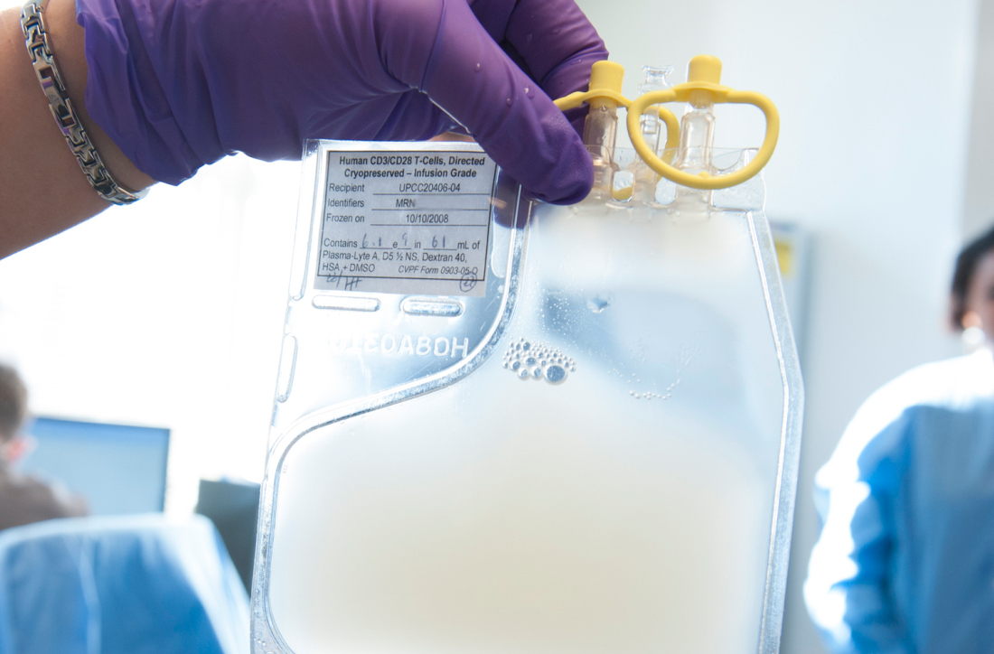
Brentuximab improves survival in older HL patients

Older patients with untreated Hodgkin lymphoma (HL) can achieve significantly improved survival by adding brentuximab vedotin to their treatment before and after standard chemotherapy, a recent study found.
In patients with low comorbidity scores, responses were even more robust, reported lead author Andrew M. Evens, DO, of the Rutgers Cancer Institute of New Jersey, and colleagues.
“Causes of poor outcomes for older patients with HL are not fully understood but have been attributed to a combination of factors, including presence of comorbidities, poorer performance status, disease and biological differences, inability to tolerate chemotherapy at the full dose, and increased treatment-related toxicities,” the authors wrote in the Journal of Clinical Oncology.
The primary goal of the study was to improve outcomes for untreated, older patients, a group that’s historically been a difficult-to-treat patient population.
The phase 2 trial included 48 HL patients with a median age of 69 (range, 60 – 88).
All patients underwent geriatric assessment for comorbidities and loss of activities of daily living.
Treatment consisted of two doses of brentuximab followed by six cycles of doxorubicin, vinblastine, and dacarbazine (AVD), then four more doses of brentuximab (consolidation doses).
The primary endpoint was complete remission at completion of AVD.
Secondary outcomes included overall response rate, 2-year progression-free survival, 2-year overall survival, and safety.
Just over half the patients (52%) completed all cycles of therapy, and almost three quarters (73%) received at least one consolidation dose of brentuximab.
Among the first 23 evaluable patients, both the complete remission rate and overall response rate were 96%. Intention-to-treat survival rates for all 48 patients were 84% for 2-year progression-free survival and 93% for 2-year overall survival.
Historical 2-year progression-free survival rates in similar older patients is poor, at 50%, so the progression-free survival rate of 84% in this study represents a significant improvement.
Of note, patients with fewer comorbidities and without loss of instrumental activities of daily living showed more robust responses.
Patients with Cumulative Illness Rating Scale for Geriatrics (CIRS-G) comorbidity scores of less than 10 had a 2-year progression-free survival rate of 100% versus 45% for those with higher scores.
Similarly, patients without loss of instrumental activities achieved a progression-free survival rate of 94% versus 25% for those who had lost some instrumental activities.
Grade 3 or 4 adverse events occurred in 42% of patients, with neutropenia being the most common (44%).
“This study represents among the best-reported outcomes to date for untreated older patients with HL,” the investigators concluded.
Seattle Genetics supported the investigator-initiated trial.