User login
Sharon Worcester is an award-winning medical journalist for MDedge News. She has been with the company since 1996, first as the Southeast Bureau Chief (1996-2009) when the company was known as International Medical News Group, then as a freelance writer (2010-2015) before returning as a reporter in 2015. She previously worked as a daily newspaper reporter covering health and local government. Sharon currently reports primarily on oncology and hematology. She has a BA from Eckerd College and an MA in Mass Communication/Print Journalism from the University of Florida. Connect with her via LinkedIn and follow her on twitter @SW_MedReporter.
“FAST” indices help to identify fibromyalgia in routine rheumatology care
CHICAGO – Fibromyalgia assessment screening tool (FAST) indices derived from the Multidimensional Health Assessment Questionnaire (MDHAQ) provide a simple and effective method for identifying fibromyalgia in routine care, according to findings from a series of more than 500 patients.
The indices are as accurate as the existing – and more complex – diagnostic criteria for fibromyalgia, Juan Schmukler, MD, reported at the annual meeting of the American College of Rheumatology.
For example, three FAST indices developed in the course of the study had better performance versus existing diagnostic criteria than did certain individual MDHAQ scales alone (area under the curve range, 0.924-0.937 vs. 0.829-0.889, respectively), said Dr. Schmukler, who conducted the research as a medical student at Rush Medical College, Chicago, and is now a rheumatology fellow at Mount Sinai Hospital in Chicago.
The findings are notable, because the two-page MDHAQ and a summary index (the RAPID3) derived from three of the MDHAQ scales are already used routinely in clinical care for diagnosing rheumatic diseases, and the new indices could easily – and with minimal work flow disruption – also be incorporated for each patient at each visit.
“Fibromyalgia is common in the general population and it is believed to be more common in patients with other rheumatic diagnoses,” Dr. Schmukler said. “Fibromyalgia may be easily recognized in many cases, but it can also be very subtle, particularly in patients who have other rheumatic diseases.”
ACR fibromyalgia classification criteria published in 1990 were based on tender point examination and non-ACR diagnostic criteria as revised in 2011 are based entirely on patient self-report.
A one-page fibromyalgia criteria questionnaire is available and is useful in clinical trials and other research, but is “rarely, if ever” used in routine care, he said, explaining that the questionnaire contains two domains: a symptom severity score (0-12 scale) and a widespread pain index (0-19 scale).
The MDHAQ/RAPID3 is informative in RA, and has also been shown to be “useful in all rheumatic diseases in which it has been studied,” and at least three prior reports have suggested that the MDHAQ may also provide clues to the presence of fibromyalgia, Dr. Schmukler said.
“And we know from prior reports that patients with fibromyalgia reported the highest RAPID3 scores, compared to other rheumatic disease,” he added, explaining that the goal of the present study was to develop FAST cumulative indices based on the routine MDHAQ scales and using the 2011 diagnostic criteria as a reference standard.
All patients with all diagnoses seen at the Rush Medical College rheumatology clinic in Chicago complete the MDHAQ at all visits in routine care, and between April and July 2017, the fibromyalgia criteria questionnaire was also administered in 502 consecutive patients.
Of those patients, 131 met the 2011 fibromyalgia diagnostic criteria.
MDHAQ scores were analyzed for agreement with the fibromyalgia criteria questionnaire according to receiver operator characteristic curves for AUC and were compiled into three different FAST indices that included various combinations of either three or four of the MDHAQ measures that had the best agreement with the diagnostic criteria questionnaire as identified by the highest AUC values. Those were the 60-symptom checklist (AUC, 0.889), painful joint count (AUC, 0.870), fatigue visual analog scale (VAS; AUC, 0.860), and pain VAS (AUC, 0.829), Dr. Schmukler said.
Proposed cut points that reflected the optimal trade-off between specificity and sensitivity for each of the scales were scores of 6 or greater for the pain VAS and fatigue VAS, and scores of 16 or greater for the symptom checklist and painful joint count measure.
In addition to the better performance of each FAST index versus the 2011 criteria, their performance was also better than that of the RAPID3 versus the 2011 criteria (AUC, 0.848), which many clinicians use in practice without using the full MDHAQ for patient assessment, he said.
Further, the “very easily calculated” FAST indices performed as well as a “very difficult to calculate” polysymptomatic distress continuous scale derived from the fibromyalgia questionnaire to assess the degree of fibromyalgia symptoms, which had an AUC of 0.929 versus the 2011 criteria. The latter index requires complex mathematical calculations for scale conversion and thus is impractical in clinical practice, he noted.
The FAST indices also performed comparably with physician diagnoses as indicated in patient charts and with diagnoses based on tender point count as shown in prior studies.
The FAST index with the greatest sensitivity (85.5% at a cut point of 2 or greater on a scale of 1-3) was the FAST3-P, which includes pain VAS score of 6 or greater, symptom checklist score of 16 or greater, and painful joint count of 16 or greater. The FAST index with the greatest specificity (90.3% at a cut point of score of 3 or greater on a scale of 1-4) was the FAST4 index, which includes a pain VAS score of 6 or greater, fatigue VAS score of 6 or greater, symptom checklist score of 16 or greater, and painful joint count of 16 or greater.
Although the findings are limited by the lack of a gold standard for fibromyalgia diagnosis, changing diagnostic criteria, and a need for physician input for a fibromyalgia diagnosis, the study provides useful real-world data and supports findings from some prior studies with respect to the benefit of using these tools in routine practice.
With minimal extra physician time – if the MDHAQ is completed by patients at the time of registration – this approach can be used in all rheumatology patients to help identify fibromyalgia, he concluded.
Dr. Schmukler reported having no disclosures. One of his coauthors at Rush, Ted Pincus, MD, receives royalties and license fees for the copyright and trademark for the MDHAQ and RAPID3, all of which go to support clinical research.
SOURCE: Schmukler J et al. Arthritis Rheumatol. 2018;70(Suppl 10), Abstract 839.
REPORTING FROM THE ACR ANNUAL MEETING
Key clinical point:
Major finding: Each fibromyalgia assessment screening tool index had better performance versus 2011 diagnostic criteria (area under the curve, 0.924-0.937) than did individual scales alone (AUCs, 0.829-0.889) and the RAPID3 summary index (AUC, 0.848).
Study details: An assessment of fibromyalgia assessment screening tool indices developed in a series of 502 patients.
Disclosures: Dr. Schmukler reported having no disclosures. One of his coauthors at Rush, Ted Pincus, MD, receives royalties and license fees for the copyright and trademark for the Multidimensional Health Assessment Questionnaire and RAPID3, all of which go to support clinical research.
Source: Schmukler J et al. Arthritis Rheumatol. 2018;70(Suppl 10), Abstract 839.
Extended AI therapy reduces breast cancer recurrence risk, ups fracture risk
SAN ANTONIO – Extending aromatase inhibitor (AI) therapy an additional 5 years reduces breast cancer recurrence risk, particularly in patients with node involvement, but the benefits vary based on prior treatment and must be weighed against the risk of bone fracture, according to findings from a meta-analysis involving more than 22,000 women.
The rate of any recurrence after 10 years in almost 7,500 women treated with 5 years of tamoxifen and then randomized to 5 additional years of AI treatment was reduced by 35%, compared with the rate in those who did not receive 5 additional years of AI therapy (recurrence rate, 10.7% vs. 7.1%, respectively; relative risk, 0.67), and the difference was “very highly significant,” Richard Gray, MD, reported at the San Antonio Breast Cancer Symposium.
The distant recurrence rate and mortality rate were also significantly improved in those who received 5 years of AI therapy (rr, 0.77 for each), but the difference in mortality was of borderline significance, Dr. Gray, professor of medical statistics at the University of Oxford, London, reported on behalf of the Early Breast Cancer Trialists’ Collaborative Group.
However, in many of the trials included in the analysis, control group patients crossed over to the treatment group, which likely reduced the effect, he noted.
In about 12,000 women who were treated with 2-3 years of tamoxifen followed by 2-3 years of an AI and who were then randomized to an additional 3-5 years of AI therapy, the effects were less pronounced, with about a 20% reduced risk of any recurrence after 10 years vs. the rate in those without extended therapy (recurrence rate, 9.2% vs. 7.1%), he said.
The differences in the rates of distant recurrence and breast cancer mortality were not statistically significant in this group, but again, follow-up was short, he said.
Similarly, in about 3,300 women treated with an AI followed by an additional 5 years of AI therapy, recurrence risk was reduced by about 25% vs. the rate in those who did not receive extended therapy, and no difference was seen in the rates of distant recurrence or breast cancer mortality.
Of note, the benefits in those who received tamoxifen were seen immediately, whereas the benefits in those receiving AIs in the first 5 years emerged after about 2 years of extended therapy, Dr. Gray said, explaining that this was likely due to “carry-over benefits” of the earlier AI therapy.
The downside with extended AI therapy was a 25% increase in fracture risk, as well as bone pain, which can reduce quality of life.
Therefore, decisions about extended therapy should involve careful risk-benefit analyses, he said, adding that the findings of this meta-analysis of 12 trials, which included postmenopausal women – 99% of whom had estrogen receptor–positive disease – provide “the most reliable assessment of the available evidence ... [for] guiding decisions about endocrine therapy.”
In this video interview, he further discussed the details and limitations of the study, the effects of nodal status on outcomes, implications of the findings for clinical practice, the need for further follow-up on all of the studies included in the analysis, and plans for incorporating new data from the AERAS trial, which were also presented at the symposium and which complement and reinforce the current findings.
Dr. Gray reported having no disclosures.
SOURCE: Gray R et al., SABCS 2018: Abstract GS3-03.
REPORTING FROM SABCS 2018
Key clinical point: Five extra years of aromatase inhibitor therapy reduces breast cancer recurrence, but increases fracture risk.
Major finding: Extending AI therapy by 5 years reduces breast cancer recurrence by 20% to 35%.
Study details: A meta-analysis of more than 22,000 women from 12 trials.
Disclosures: Dr. Gray reported having no disclosures.
Source: Gray R et al. SABCS 2018: Abstract GS3-03.
Novel SSc classification scheme aims to improve risk stratification
CHICAGO – A simple new classification scheme that combines autoantibody specificity and extent of skin involvement could improve risk stratification of patients with systemic sclerosis, according to researchers at University College London.
“The Le Roy et al. classification of SSc [systemic sclerosis] into limited and diffuse cutaneous subtype remains the most commonly used classification system for systemic sclerosis, but autoantibodies are much better predictors of organ involvement, and while more sophisticated approaches exist, this proposed simple classification using antibodies and skin subset is relevant to clinical practice and could help risk stratification,” Svetlana I. Nihtyanova, MD, said at the annual meeting of the American College of Rheumatology.
Dr. Nihtyanova, a clinical research fellow at University College London, reported how she and her colleagues at UCL divided 1,025 SSc patients into 12 subgroups based on skin subset and autoantibodies and then conducted Kaplan-Meier estimates of survival and cumulative incidence of organ complications to rank these 12 subgroups by endpoint estimates. They merged subgroups with similar ranking in multiple endpoints, ending up with seven groups in the final classification.
Group 1 comprised anti–centromere antibody–positive limited cutaneous SSc (lcSSc) patients and accounted for 29% of patients.
“This was the subgroup with the highest survival (72%) and the lowest incidence of pulmonary fibrosis (13%) and scleroderma renal crisis (no cases) at 20 years from onset,” she said, noting that the incidence of pulmonary hypertension in this group was similar to the average for the whole cohort.
Group 2 comprised all anti–RNA polymerase antibody–positive subjects and accounted for 11% of patients. This group had the highest incidence of scleroderma renal crisis (SRC; 32% at 20 years), but other organ complications and survival were similar to the cohort average.
Group 3 comprised Scl-70–positive lcSSc patients, and accounted for 11% of patients.
“Although incidence of pulmonary fibrosis in this group was the second highest (69% at 20 years), other complications were rare,” Dr. Nihtyanova said, adding that this group had the lowest incidence of pulmonary hypertension (6%) and the second lowest incidence of SRC (3%) at 20 years.
Group 4, conversely, included Scl-70–positive dcSSc patients and accounted for 11% of patients, who had a very poor prognosis; they had the highest incidence of pulmonary fibrosis (91%) and cardiac scleroderma (14%), and the worst survival (41%) at 20 years, she said.
Group 5 included all U3 RNP–positive patients, accounting for 5% of patients.
“Although survival in this group was not bad (70% at 20 years), the group had the highest pulmonary hypertension incidence (40%) and the second highest incidence of cardiac SSc (11%) at 20 years,” she noted.
Groups 6 and 7 (comprising 22% and 11% of study subjects, respectively) included lcSSc and diffuse cutaneous SSc (dcSSc) patients with other antibody specificities. Group 6 had low overall SRC and cardiac SSc risk, while other outcomes were similar to the cohort average. Group 7, however, had poor prognosis, with the second lowest survival (42% at 20 years) and above average rates of organ disease, particularly pulmonary fibrosis and SRC, she said.
Overall, estimated survival for the entire cohort was 60% at 20 years from onset, and in that time frame 44% developed significant pulmonary fibrosis, 25% pulmonary hypertension, 7% SRC, and 6% cardiac SSc. The patients had a mean age of 47 years at disease onset, and 16% were men. Diffuse cutaneous SSc was diagnosed in 35% of the subjects, she noted.
Dr. Nihtyanova reported having no disclosures.
SOURCE: Nihtyanova S et al. Arthritis Rheumatol. 2018;70(Suppl 10): Abstract 2935.
REPORTING FROM THE ACR ANNUAL MEETING
Key clinical point:
Major finding: The classification scheme for SSc risk stratification identified seven distinct SSc subgroups.
Study details: Development and testing of a novel risk classification scheme in 1,025 SSc patients.
Disclosures: Dr. Nihtyanova reported having no disclosures.
Source: Nihtyanova S et al. Arthritis Rheumatol. 2018;70(Suppl 10):Abstract 2935.
Combination immunotherapy ups survival in ILD patients with anti-MDA5–positive dermatomyositis
CHICAGO – Early treatment with combined high-dose glucocorticoids, tacrolimus, and intravenous cyclophosphamide therapy significantly improves survival vs. step-up therapy in interstitial lung disease patients with anti–melanoma differentiation–associated gene 5 (anti-MDA5)–positive dermatomyositis, according to findings from a prospective, multicenter study.
However, the combination therapy was associated with a high risk of cytomegalovirus reactivation and other opportunistic infections that warrants careful monitoring of treated patients, Hideaki Tsuji, MD, reported at the annual meeting of the American College of Rheumatology.
ILD accompanied by anti-MDA5–positive dermatomyositis (DM) is often intractable and associated with high mortality in Japanese patients. Case reports have suggested improved outcomes with combined immunosuppressive therapy, but a standard treatment has not been established, said Dr. Tsuji of Kyoto University.
“Therefore, we evaluated the efficacy and safety of combined immunosuppressive therapy for anti-MDA5–positive DM with ILD in a prospective single-arm study,” he said, adding that early administration, a short interval of intravenous cyclophosphamide, use of plasmapheresis as an additional therapy, and control of opportunistic infections may contribute to the improved outcomes seen with the regimen in this study.
The primary endpoint of 6-month survival was reached by 24 (89%) of 27 patients treated with the combination regimen for 52 weeks, compared with 5 (33%) of 15 historical controls who received high-dose glucocorticoids followed by step-wise addition of immunosuppressants. At 12 months, the survival rates were 85% and 33%, respectively, Dr. Tsuji said.
Additionally, anti-MDA5 titer, serum ferritin level, C-reactive protein level, lactate dehydrogenase, and KL-6 level gradually decreased over the 52 months, and percent vital capacity increased with combination vs. step-up therapy, he noted.
Cytomegalovirus reactivation occurred in 90% of combination regimen patients vs. 33% of controls over the 52-week study period, he said, adding that pneumocystic pneumonia and sepsis also occurred in combination regimen group patients, and were associated with death in four patients.
When the 23 surviving patients in the combination regimen group were compared with the 4 in the group who died, it was noted that the deceased patients were significantly more likely to have cutaneous ulcers (75% vs. 13%), higher mean C-reactive protein level (2.7 vs. 0.77 mg/dL), and higher creatine kinase level (644.3 vs. 219.3 IU/L), respectively, before treatment, he said.
Study subjects were Japanese adults with new-onset anti-MDA5–positive dermatomyositis with interstitial lung disease (ILD) who were enrolled between July 2014 and September 2017.
They were treated with 1 mg/kg/day of prednisolone for 4 weeks with reduced doses thereafter, 500-1,000 mg/m2 of IV cyclophosphamide every 2 weeks for six cycles then every 4 weeks for up to a total of 10-15 treatments, and 10-12 ng/mL of tacrolimus (12-hour trough). Plasmapheresis was allowed in patients who progressed and needed oxygenation after the regimen was initiated, and it was administered in nine patients (31%) in the combination regimen group vs. one (7%) of the historical controls.
Given the different frequencies of rapidly progressive ILD in Asian vs. Western countries (39%-71% vs. 22%-57%, respectively), it is unclear whether the results seen in this study can be extrapolated to patients from the United States and Europe. Therefore, it is necessary to analyze the efficacy of the regimen in those patient populations, Dr. Tsuji said, also noting that future studies should evaluate risk-based modifications of the regimen to identify the optimal treatment for individuals based on factors such as age, respiratory dysfunction, hyperferritinemia, and treatment delay.
Dr. Tsuji reported having no disclosures.
SOURCE: Tsuji H et al. Arthritis Rheumatol. 2018;70(Suppl 10), Abstract 838.
REPORTING FROM THE ACR ANNUAL MEETING
Key clinical point:
Major finding: 6-month survival was 89% vs. 33% with combination immunotherapy vs. step-up therapy.
Study details: A prospective, multicenter study of 27 patients and 15 historical controls.
Disclosures: Dr. Tsuji reported having no disclosures.
Source: Tsuji H et al. Arthritis Rheumatol. 2018;70(Suppl 10), Abstract 838.
Dr. Ingrid A. Mayer on neoadjuvant endocrine therapy: The times are changing
SAN ANTONIO – Phase 2 neoadjuvant endocrine therapy breast cancer trials can and should serve as a testing platform to inform the design of phase 3 trials with the ability to change practice, according to Ingrid A. Mayer, MD.
Correlating outcomes such as the Preoperative Endocrine Prognostic Index and Ki67 blood levels from phase 2 trials to larger, similarly designed trials could reduce costs, time, and the need for large numbers of patients, thereby promoting more efficient development of effective breast cancer therapies, she said during a plenary lecture at the San Antonio Breast Cancer Symposium.
In this video interview, Dr. Mayer, director of breast medical oncology at Vanderbilt University, Nashville, Tenn., explained that while neoadjuvant endocrine therapy is suitable for down-staging tumors in women with stage I-III breast cancer, it is not commonly used, as those patients are also good surgery candidates.
However, neoadjuvant endocrine therapy can be “an incredible platform” to test novel combinations of endocrine therapy agents with or without targeted treatments, validate new biomarkers, discover mechanisms of resistance, and predict the best combinations for moving forward in phase 3 trials, she said, adding that “this is something that all academic institutions potentially should be doing.”
Although the direct clinical benefit for patients who participate in such trials is not tremendous, participation has the potential to ultimately improve outcomes for participants and for “their friends and families and daughters,” and to help propel the science forward, Dr. Meyer noted.
“I would argue that we really should be moving forward to designing trials in the neoadjuvant endocrine setting more and more, and really utilizing this platform for that end,” she said.
Dr. Mayer reported having no relevant disclosures.
REPORTING FROM SABCS 2018
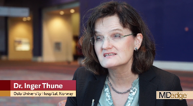
Exercise during adjuvant breast cancer therapy improves CV outcomes
SAN ANTONIO – A tailored 12-month exercise program during adjuvant breast cancer treatment appears to protect cardiovascular function, particularly in patients receiving chemotherapy, according to findings from the randomized EBBA-II trial.
The overall change in VO2max at 12 months was +0.3% in 271 patients randomized to the intervention group, compared with –8.9% in 274 patients in the “usual care” control group, Inger Thune, MD, PhD, said at the San Antonio Breast Cancer Symposium.
Among patients receiving chemotherapy, the VO2max change at 12 months was +1.6% in 120 patients in the intervention group, compared with –2.76% in 122 patients in the control group, said Dr. Thune of the Cancer Center at Oslo University Hospital.
Study participants were women aged 18-75 years (mean of 55 years at diagnosis) with stage I-II breast cancer, mean body mass index of 25 kg/m2, and a mean VO2max before surgery of 31.5 mL/kg per minute. The intervention group entered a 12-month individualized exercise program 2-3 weeks after surgery based on their own VO2max at baseline.
They met for training sessions in groups of 10-12 women for 60 minutes twice weekly over the 12-month study period, and were also told to perform at least 120 minutes of exercise at home for a total of 240 minutes of exercise weekly.
Of note, the adherence rate among participants was encouragingly high at about 90%, she said, adding that the findings strongly support tailored exercise during adjuvant breast cancer treatment, as such an intervention appears to counteract declines in cardiovascular function – particularly in those receiving chemotherapy.
In this video interview, Dr. Thune further discussed the study design, implications of the findings, and future directions.
“Cardiovascular morbidity is so important for our breast cancer patients that I think that it’s time to have physical activity [and] physical function as a main interest for all clinicians dealing with breast cancer patients,” she said.
Dr. Thune reported having no disclosures.
REPORTING FROM SABCS 2018
Key clinical point: An exercise program during adjuvant breast cancer treatment improves cardiovascular outcomes.
Major finding: The rate of VO2max change at 12 months was +0.3% in the exercise group versus –8.9% in the control group.
Study details: EBBA-II, a randomized trial of 546 women.
Disclosures: Dr. Thune reported having no disclosures.
Extended anastrozole improves DFS, distant DFS
SAN ANTONIO – Extending treatment with adjuvant anastrozole (Arimidex) to 10 years led to significantly higher rates of disease-free and distant disease-free survival in postmenopausal women with hormone receptor–positive breast cancer in the prospective, randomized, open-label phase 3 Arimidex Extended Adjuvant Randomized Study (AERAS).
After a median of 4.9 years of follow-up, the primary endpoint of disease-free survival (DFS) was 91.9% in 840 women who were randomized to continue receiving anastrozole for an additional 5 years versus 84.4% in 843 who stopped after the initial 5 years (hazard ratio, 0.548; P = .0004), Shoichiro Ohtani, MD, reported at the San Antonio Breast Cancer Symposium.
The rate of 5-year distant DFS was 97.2% vs. 94.3% in the groups, respectively (HR, 0.514; P = .0077), said Dr. Ohtani, of Hiroshima City (Japan) Hiroshima Citizens Hospital.
“As we expected, there was no difference between the two groups in overall survival,” he said; overall survival was 99.5% and 99.6% in the groups, respectively (HR, 1.389; P = .665).
Study subjects were postmenopausal patients with stages I-III hormone receptor-positive breast cancer (HR+ BC) with a median age of 64 years who were disease-free after 5 years of either anastrozole alone or tamoxifen for 2-3 years followed by anastrozole 2-3 years. They were enrolled between November 2007 and November 2012.
Treatment with an aromatase inhibitor such as anastrozole for up to 5 years either as up-front monotherapy or after 2-3 years of tamoxifen therapy is the treatment of choice for HR+ BC in postmenopausal women, but it was thought that extending aromatase inhibitor therapy to 10 years might reduce the risk of breast cancer recurrence, Dr. Ohtani explained.
Indeed, while women randomized to extended anastrozole treatment in the current study experienced more bone-related adverse events, including arthralgia (19.2% vs. 11.7%), stiff joints (11.7% vs. 4.9%), bone fractures (2.8% vs. 1.1%), and new-onset osteoporosis (33% vs. 28%) than did those in the group that stopped anastrozole at 5 years, extended treatment significantly reduced recurrence rates.
The findings show that extended adjuvant anastrozole treatment for an additional 5 years after initial treatment is safe and provides important DFS and distant DFS benefits, he concluded.
Dr. Ohtani has received speaker fees from CHUGAI, Astra Zeneca, Novartis,and Ezai.
SOURCE: Ohtani S et al. SABCS 2018, Abstract GS3-04.
REPORTING FROM SABCS 2018
Key clinical point: Extending treatment with adjuvant anastrozole to 10 years improves disease free survival and distant DFS in HR+ breast cancer.
Major finding: DFS was 91.9% in patients who continued anastrozole versus 84.4% in those who stopped anastrozole after the initial 5 years (hazard ratio, 0.548; P = .0004).
Study details: A prospective, randomized, open-label, phase 3 study of 1,683 patients.
Disclosures: Dr. Ohtani has received speaker fees from CHUGAI, Astra Zeneca, Novartis, and Ezai.
Source: Ohtani S et al. SABCS 2018, Abstract GS3-04.
TNBC survival appears better when adjuvant chemotherapy is delivered within 30 days
SAN ANTONIO – The longer the delay in initiating adjuvant chemotherapy, the worse the survival in patients with triple-negative breast cancer (TNBC), findings from a review of nearly 700 cases suggest.
Delays of more than 30 days between surgery and initiation of chemotherapy were associated with lower disease-free survival (DFS), distant recurrence–free survival (DRFS), and overall survival (OS), Zaida Morante, MD, reported at the San Antonio Breast Cancer Symposium.
In 687 women with clinical stage I, II, or III TNBC who were diagnosed at the Instituto Nacional de Enfermedades Neoplasicas in Lima, Peru, during 2000-2014 and followed for a median of 8.5 years, time to chemotherapy was less than 30 days in 189 patients (27.5%), 31-60 days in 329 patients (47.9%), 61-90 days in 115 patients (16.7%), and more than 91 days in 54 patients (7.9%), said Dr. Morante, a medical oncologist at the institute.
Overall survival at 10 years was 82% in those who received chemotherapy within 30 days of surgery, compared with 67.4%, 67.1%, and 65.1% in those treated at 31-60, 61-90, and more than 91 days after surgery, respectively, she said.
“The difference was consistent across the different periods of the evaluation,” she said during a press briefing at the symposium. “Additionally, the benefit of receiving chemotherapy within 30 days exists and is statistically significant for [nodal status] N0 and N1 (hazard ratios, 1.701 and 2.498).”
In those with N2 and N3 nodal status, there was a numerical difference, but it didn’t reach statistical significance.
DFS was also significantly worse if treated later than 30 days after surgery; those treated within 30 days had 10-year DFS of 81.4%, compared with 68.8%, 70.8%, and 68.1% in the other groups, respectively. The difference was even more pronounced for 10-year DRFS, which was 80.2%, 64.9%, 67.5%, and 58.6% in the groups, respectively.
Multivariate analyses confirmed that time to adjuvant chemotherapy was an independent prognostic factor for survival, she said, noting that compared with patients treated within 30 days of surgery, those treated at 31-60 days had 1.9-fold increased risk of death, and those treated at 61-90 days had a 2.4-fold increased risk of death.
“The difference in 10-year overall survival rates between receiving chemotherapy within 30 days after surgery and after 30 days was more than 10%,” she said. “These results represent a feasible opportunity for improving outcomes in triple-negative breast cancer patients.”
Although only 28% of patients in this review received adjuvant chemotherapy within 30 days, most patients in the United States “will fall within the 30 days and under” category, said press briefing moderator Carlos Arteaga, MD, professor and director of the Harold C. Simmons Comprehensive Cancer Center at UT Southwestern Medical Center in Dallas.
However, the findings might suggest a greater role for neoadjuvant chemotherapy in these patients.
“Because this is systemic therapy ... it’s treating the systemic disease. I wonder if this is arguing ... that we need to have an impetus to deliver the systemic therapy as soon as we can – early, even before the operation,” he said.
Indeed, while timing isn’t everything, Dr. Morante’s findings and others presented at the meeting “highlight the possibility that perhaps it is more important than we previously suspected,” discussant Joseph A. Sparano, MD, said at the meeting, adding that the findings raise questions about current paradigms for management of breast cancer.
“We now have substantial data suggesting that the timing of adjuvant chemotherapy matters in triple-negative breast cancer, and that 30 days may be optimal,” said Dr. Sparano, professor at the Albert Einstein College of Medicine, New York.
“This doesn’t mean that patients who may not be ready for the chemotherapy because of complications related to the surgery should be forced into a situation where they are at higher risk from receiving the chemotherapy, but nevertheless, the results are important,” he said.
Dr. Morante and Dr. Arteaga each reported having no relevant conflicts of interest to declare. Dr. Sparano has received consulting fees from Roche, Eli Lilly, Novartis, Celldex, AstraZeneca, Pfizer, and Adgero. He also has ownership interests with MetaStat.
SOURCE: Morante Z et al. SABCS 2018, Abstract GS2-05.
REPORTING FROM SABCS 2018
Key clinical point: Outcomes are improved with adjuvant chemotherapy within 30 days of surgery, compared with beyond 30 days, in triple-negative breast cancer.
Major finding: 10-year overall survival was 82% with chemotherapy within 30 days of surgery versus 67.4%, 67.1%, and 65.1% with chemotherapy at 31-60, 61-90, and more than 91 days after surgery, respectively.
Study details: A retrospective review of 687 cases of TNBC.
Disclosures: Dr. Morante and Dr. Arteaga each reported having no relevant conflicts of interest to declare. Dr. Sparano has received consulting fees from Roche, Eli Lilly, Novartis, Celldex, AstraZeneca, Pfizer, and Adgero. He also has ownership interests with MetaStat.
Source: Morante Z et al., SABCS 2018 Abstract GS2-05.
PD-L1 expression best predicts response to atezolizumab + nab-paclitaxel for mTNBC
SAN ANTONIO – in patients with untreated metastatic triple-negative breast cancer, according to exploratory efficacy analyses of data from the phase 3 IMpassion130 trial.
The analyses of data for the 902 patients randomized to receive the PD-L1 inhibitor atezolizumab (Tecentriq) plus nanoparticle albumin-bound (nab)–paclitaxel or placebo plus nab-palcitaxel for the study also showed consistency between local and central estrogen-receptor, progesterone-receptor, and human epidermal growth factor–receptor 2 testing, Leisha A. Emens, MD, reported at the San Antonio Breast Cancer Symposium.
“IMpassion130 is the first phase 3 study to demonstrate a benefit from [atezolizumab + nab-paclitaxel] in metastatic triple-negative breast cancer (mTNBC),” said Dr. Emens, professor of medicine in hematology/oncology, coleader of the Hillman Cancer Immunology and Immunotherapy Program, and director of translational immunotherapy for the Women’s Cancer Research Center at the University of Pittsburgh Medical Center.
She explained that progression-free survival (PFS) was significantly better in PD-L1–positive mTNBC patients treated with the atezolizumab + nab-paclitaxel, than in those who received placebo + nab-paclitaxel (hazard ratios in the intent-to-treat population, 0.8 and 0.62, respectively).
At the first interim overall survival analysis, a clinically meaningful improvement in OS was seen in PD-L1–positive patients in the treatment group (HR, 0.62; median OS improvement from 15.5 months with placebo to 25 months), she added.
In exploratory analyses, Dr. Emens and her colleagues sought to evaluate whether preexisting immune biology is associated with clinical benefit from atezolizumab + nab-paclitaxel, as has been demonstrated in studies of other agents that target the PD-1 pathway in other cancer types of cancer. They also assessed BRCA 1/2 mutation status as a biomarker for response.
“In patients enrolled on the IMpassion130 trial we found that PD-L1 in triple-negative breast cancer was expressed primarily on tumor-infiltrating immune cells,” she said. “In contrast to this, we found a very low rate of PD-L1 expression specifically on tumor cells across the patient population.”
Looking at both of those biomarkers together showed that a majority of patients with expression of PD-L1 on tumor cells were included in the PD-L1 immune cell–positive population, with only 2% having PD-L1 expression exclusively on their tumor cells.
Data previously reported at the European Society for Medical Oncology and published in the New England Journal of Medicine showed a PFS benefit, as well as a clinically meaningful improvement in OS of nearly 10 months, specifically in patients with PD-L1 immune cell–positive lesions treated with atezolizumab + nab-paclitaxel, she noted.
“In data presented for the first time today you can see that PD-L1–negative patients derive no overall survival benefit as there was no treatment effect with this therapy combination,” she said.
A trend was seen toward an association between immune cell positivity and poor prognosis, but this was not statistically significant, she said.
“Taken together, these data definitively show that PD-L1 immune cell positivity is predictive of both progression-free and overall survival benefit with atezolizumab + nab-paclitaxel,” she said.
She and her colleagues also looked at the level of PD-L1 expression in immune cells to assess whether there is a threshold that might be required.
“As long as there was a PD-L1 expression level of 1% or more in the immune cells, there was a significant progression-free and overall survival benefit for patients treated with atezolizumab + nab-paclitaxel. This suggests that this expression of over 1% will represent a threshold for identifying those patients who are likely to benefit from this combination,” she said.
Further assessment by CD8 T-cell status showed that patients who had CD8-positive T cells but who were PD-L1 immune cell negative had no benefit from atezolizumab + nab-paclitaxel, whereas those who were positive for both CD8 and PD-L1 expression on their immune cells derived significant PFS and OS benefit (HR, 0.89 and 0.77, respectively).
“So patients with CD8-positive tumors derive clinical benefit only if their tumors are also PD-L1-positive,” she said.
Similarly, no clinical benefit was seen in patients with stromal tumor-infiltrating lymphocyte (TIL)–positive tumors but who were PD-L1-negative, whereas those with stromal TIL-positive PD-L1–positive tumors derived significant PFS and OS benefit (HRs, 0.99 and 1.53, respectively), and this was also seen in the 15% of evaluable patients who had BRCA mutations.
“In patients who were BRCA mutated, but who were PD-L1 immune cell negative, there was no association of progression-free survival or an overall survival benefit [with atezolizumab + nab-paclitaxel]. In contrast, in patients who were BRCA mutated but PD-L1 immune cell positive ... there was an association with progression-free survival and a trend toward overall survival,” she said, noting that while the BRCA mutation findings are limited by small numbers, “they do show that mutations in BRCA and PD-L1 expression in immune cells are independent biomarkers; patients with BRCA1 or 2 mutations derive clinical benefit only if their tumors are also PD-L1 positive.”
“In this phase 3 IMpassion130 study, PD-L1 expression on immune cells is a predictive biomarker for selecting patients who benefit clinically during first-line treatment with atezolizumab + nab-paclitaxel for metastatic triple-negative breast cancer,” she concluded, adding that “patients with newly diagnosed metastatic and unresectable locally advanced triple-negative breast cancer should be routinely tested for their PD-L1 immune cell status to determine if they might benefit from the combination of atezolizumab + nab-paclitaxel.
IMpassion130 was sponsored by Hoffman-La Roche. Dr. Emens reported receiving royalties and consulting fees from several companies. She has contracts with Roche/Genentech, Corvus, AstraZeneca, and EMD Serono, and ownership in Molecuvax. She receives other support from DSMB and Syndax, and has received grants from Aduro Biotech, Merck, Maxcyte, and the Breast Cancer Research Foundation. She also reported serving as a member of the Food and Drug Administration Advisory Committee on Tissue, Cell, and Gene Therapies, and is a member of the board of directors for the Society of Immunotherapy for Cancer.
SOURCE: Emens L et al. SABCS 2018, Abstract GS1-04.
REPORTING FROM SABCS 2018
Key clinical point: Treatment-naive mTNBC patients should be tested for PD-L1 expression as a biomarker of potential benefit from atezolizumab + nab-paclitaxel.
Major finding: PD-L1 expression of at least 1% confers a significant PFS and OS benefit in patients treated with atezolizumab + nab-paclitaxel.
Study details: Exploratory efficacy analyses of a phase 3 study of 902 patients.
Disclosures: IMpassion130 was sponsored by Hoffman-La Roche. Dr. Emens reported receiving royalties from and consulting fees from several companies. She has contracts with Roche/Genentech, Corvus, AstraZeneca, and EMD Serono, and ownership in Molecuvax. She receives other support from DSMB and Syndax, and has received grants from Aduro Biotech, Merck, Maxcyte, and the Breast Cancer Research Foundation. She also reported serving as a member of the FDA Advisory Committee on Tissue, Cell, and Gene Therapies, and is a member of the board of directors for the Society of Immunotherapy for Cancer.
Source: Emens L et al. SABCS 2018, Abstract GS1-04.
2018: A banner year for hematology drug approvals
SAN DIEGO – It was banner year for new hematology drug approvals, according to R. Angelo de Claro, MD, of the Food and Drug Administration.
, including 12 first-time approvals, 5 new biosimilars, and 15 new indications for previously approved drugs, Dr. de Claro, clinical team leader in the FDA’s division of hematology products in Silver Spring, Md., said during an overview of the approvals at the annual meeting of the American Society of Hematology.
These include six new approvals for first-line treatment, and eight for pediatric indications, he said.
Highlights were discussed at two ASH-FDA joint symposia at the meeting, including one focused on the malignant hematology approvals, and another on the nonmalignant hematology approvals. In a video interview, Dr. de Claro provides some additional insight into their importance and about what might lie ahead.
“I think what’s exciting is that you have drug development occurring in more common conditions such as chronic lymphocytic leukemia, as well as in rare conditions, including hairy cell leukemia – and the first-ever approval in hemophagocytic lymphohistiocytosis,” he said. “It’s been very busy at the FDA; stay tuned ... the year’s not done yet. There could be more coming and we certainly anticipate more applications in the future.”
Dr. de Claro is an FDA employee. He reported having no other relevant disclosures.
REPORTING FROM ASH 2018