User login
Sharon Worcester is an award-winning medical journalist for MDedge News. She has been with the company since 1996, first as the Southeast Bureau Chief (1996-2009) when the company was known as International Medical News Group, then as a freelance writer (2010-2015) before returning as a reporter in 2015. She previously worked as a daily newspaper reporter covering health and local government. Sharon currently reports primarily on oncology and hematology. She has a BA from Eckerd College and an MA in Mass Communication/Print Journalism from the University of Florida. Connect with her via LinkedIn and follow her on twitter @SW_MedReporter.
Preliminary data show similar declines in worry, low regret with RRSO and RRS/RRO
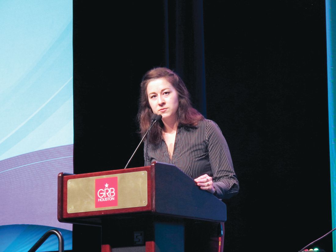
HONOLULU – Women with BRCA1/2 mutations who undergo risk-reducing salpingo-oophorectomy (RRSO) or risk-reducing salpingectomy followed by delayed risk-reducing oophorectomy (RRS/RRO) experience reduced cancer-related worry after surgery and low levels of decision regret, according to preliminary findings from the Dutch multicenter TUBA trial.
Of 384 women included to date in the ongoing trial, 51% carried a BRCA1 mutation and 49% carried a BRCA2 mutation; 72% chose RRS/RRO. A total of 289 completed the 3-month follow-up and 197 completed 1-year follow-up. At 3 and 12 months the decline on the Cancer Worry Scale was similar in the groups after adjusting for age and other baseline characteristics, with 1.9- and 1.4-point declines from baseline scores of 14.4 and 14.0 at 3 months, and 2.2- and 1.0-point declines at 12 months in the RRSO and RRS/RRO groups, respectively, Miranda P. Steenbeek, MD, reported at the Society of Gynecologic Oncology’s Annual Meeting on Women’s Cancer.
The mean scores on the 100-point Decision Regret Scale at 1-year follow-up were low at 13.4 and 13.0 after RRSO and RRS, respectively.
“But it is interesting that there is one group of women who underwent standard salpingo-oophorectomy, but weren’t using hormone replacement therapy [HRT], as these women had a higher level of decision regret [mean score, 18.8],” said Dr. Steenbeek of Radboud University Nijmegen, the Netherlands. “It is interesting to see how these results will develop over time.”
Current recommendations for preventive surgery in woman at increased risk because of BRCA1/2 mutations call for RRSO around age 40 years. However, an innovative approach using RRS followed by RRO has emerged as recent data indicate the fallopian tube, rather than the ovary, as the origin of high-grade serous ovarian cancer, she explained.
The TUBA trial was launched at 13 Dutch oncology centers in 2015, and plans to enroll 510 BRCA1/2 mutation carriers. Participants choose either standard RRSO within current guidelines (at age 35-40 years for BRCA1 carriers, age 40-45 years for BRCA2 carriers) or the innovative RRS approach followed by RRO at up to 5 years after the current guideline age (at age 40-45 years for BRCA1 carriers, age 45-50 years for BRCA2 carriers), she said.
These early findings show that baseline levels of cancer worry are higher among women choosing RRSO, which might explain the slightly larger postsurgery decline in worry in the RRSO group, Dr. Steenbeek said, noting that the higher level of regret with RRSO without HRT might be related to more severe menopausal complaints in that subset of patients.
“Based on these results we cannot conclude whether or not salpingectomy is a safe alternative to for salpingo-oophorectomy, and therefore it is very important to not perform salpingectomy with delayed oophorectomy outside the safety of a clinical and control of a clinical trial,” she said, adding that a report on the quality of life data from the study is expected in 2020.
Dr. Steenbeek reported having no disclosures.
SOURCE: Steenbeek MP et al. SGO 2019, Abstract LB2.
REPORTING FROM SGO 2019
Tumor testing cost-effective for triage to germline testing in HGSOC patients
HONOLULU – Tumor testing for the triage of women with high-grade serous ovarian cancer to confirmatory genetic testing for BRCA mutations appears feasible, according to a cost-effectiveness analysis.
In fact, based on a Markov Monte Carlo simulation model developed to compare a tumor-testing approach with a universal germline testing approach, tumor testing yields an incremental cost-effectiveness ratio (ICER) of $127,000 per year of life gained, Janice S. Kwon, MD, reported at the Society of Gynecologic Oncology’s Annual Meeting on Women’s Cancer.
“This is well in excess of [the $50,000 to $100,000 that] would be considered an acceptable threshold in the United States,” said Dr. Kwon of the University of British Columbia, Vancouver, Canada.
“We predict that tumor testing will be a cost-effective method of triage in women with high-grade serous ovarian cancer for confirmatory genetic testing to identify BRCA mutation carriers, assuming high sensitivity and acceptable cost of tumor testing,” she said.
In many areas around the world, germline testing is recommended for all women with high-grade serous ovarian cancer (HGSOC) because they have a 20% chance of carrying a BRCA 1 or 2 mutation.
“However, we all know that the referral rate for genetic testing is far from optimal, and furthermore, there are costs incurred to the healthcare system for resources utilized for genetic counseling and testing,” she said.
Tumor testing for triage is an alternative approach.
“If you consider 100 women with high-grade serous ovarian cancer and follow them through the conventional pathway in which all of them are referred for germline testing, you would expect to find 20 mutation carriers. If you take those same 100 women and apply tumor testing first, then 25 are expected to have a mutation in the tumor, Dr. Kwon said.
“If [all 25] are referred for germline testing, you would expect to find the same number of BRCA mutation carriers but with far less resource utilization.”
The remaining 75 are not expected to have a mutation in the tumor, and they may not need to be referred for confirmatory genetic testing unless there is a compelling family history or panel testing reveals a concerning mutation, she explained.
Since a randomized trial to compare these two strategies is not feasible, Dr. Kwon and her colleagues performed the current cost-effectiveness analysis.
The Markov simulation model was used to estimate the number of BRCA mutation carriers from index cases and their first degree relatives, and the number of cancer cases averted among first degree relatives, assuming they would undergo risk-reducing surgery.
“We conducted extensive sensitivity analyses to account for uncertainly around various parameters and we modeled a time horizon of 50 years,” Dr. Kwon noted. “We know that there are approximately 10,000 new [HGSOC] cases diagnosed in the United States every year, and we assumed that for every woman with [HGSOC], there was at least 1 female first-degree relative who would benefit from genetic testing.”
The model showed that applying tumor testing first would lead to a substantial reduction in the number of women undergoing germline mutation testing, but the number of BRCA mutation carriers identified would be comparable with the two strategies – assuming that the sensitivity of tumor testing is less than 100%, she said.
“As expected, the average lifetime costs associated with germline testing would be less than that for tumor testing, and even though you would expect that more first-degree relatives would be identified as BRCA mutation carriers after universal germline testing for index cases, the life expectancy gain for those first-degree relatives is averaged over the entire cohort at risk, and therefore the average incremental gain or benefit was actually quite small,” she said, noting that this yielded the ICER of $127,000 per year of life gained.
Based on this finding, tumor testing would be the preferred strategy, she added.
Sensitivity analysis around the sensitivity and specificity of tumor testing showed that tumor testing would be cost effective if its sensitivity is above 97%, and that tumor testing is cost-effective as long as it costs less than a third of the cost of germline testing – including genetic counseling.
Dr. Kwon has received research funding from AstraZeneca.
SOURCE: Kwon J et al., SGO 2019: Abstract 5.
REPORTING FROM SGO 2019
ENGOT-OV16/NOVA trial: Analysis shows improved TWiST with niraparib
HONOLULU – Patients with recurrent ovarian cancer who were treated with niraparib maintenance therapy experienced more time without symptoms or toxicities (TWiST) than did controls, according to an analysis of data from the pivotal phase 3 ENGOT-OV16/NOVA trial.
The mean 20-year TWiST benefit – defined in the current analysis as progression-free survival (PFS) without toxicity due to grade 2 or greater nausea, vomiting, or fatigue – was fourfold greater among women who were carriers of the 138 germline BRCA mutation (gBRCAmut) and received treatment with the poly(adenosine diphosphate-ribose) polymerase (PARP) 1/2 inhibitor than among the 65 similar women who received placebo. The TWiST benefit was about twofold greater among the 234 non-gBRCAmut carriers treated with niraparib than among the 116 who received placebo, Ursula A. Matulonis, MD, reported at the Society of Gynecologic Oncology’s Annual Meeting on Women’s Cancer.
The 10- and 5-year estimated mean improvement in TWiST in the groups also showed “very proportional effects that are similar to the 20-year data,” said Dr. Matulonis, chief of the division of gynecologic oncology at Dana-Farber Cancer Institute, and a professor of medicine at Harvard Medical School, Boston.
TWiSt is an “established methodology that partitions PFS ... based on time with toxicity and time without progression or toxicity." Mean TWiST is calculated as mean PFS minus mean toxicity, she explained.
In the ENGOT-OV16/NOVA study, women with recurrent ovarian cancer who received niraparib for maintenance after platinum-based therapy had significantly longer PFS, compared with patients who received placebo (21.0 vs. 5.5 months in gBRCAmut patients and 9.3 vs. 3.9 months in the non-gBRCAmut patients, respectively), she said (N Engl J Med 2016;75:2154-2164).
Quality of life (QOL) remained stable throughout niraparib treatment, according to another analysis of data from the trial published in 2018. (Lancet Oncology. Aug 1 2018;19[8]:1117-25).
The current analysis looked more closely at QOL by estimating mean TWiST in the treatment vs. control groups, she explained.
“Survival curves were used to extrapolate PFS ... over 20 years for both the gBRCAmut and non-gBRCAmut cohorts. The 20-year period was based on ovarian cancer clinical expert opinion and the biologic plausibility that patients could be on PARP inhibitors for a long period of time,” she said.
Time of toxicity was estimated based on the number of days patients experienced toxic effects due to grade 2 or higher nausea, vomiting, or fatigue during the NOVA trial.
The PFS benefit in treated vs. control subjects, respectively, was 3.23 years and 1.33 years in the gBRCAmut and non-gBRCAmut cohorts, and the mean toxicity time was 0.28 years and 0.10 years, for a mean TWiST benefit of 2.95 and 1.34 years, respectively.
“This TWiST benefit means that patients treated with niraparib experienced more progression-free time without symptoms or toxicities due to nausea, vomiting, or fatigue, compared with patients receiving placebo,” she concluded.
Dr. Matulonis is a consultant for 2X Oncology, Merck, Mersana, Fujifilm, Immunogen, and Geneos.
[email protected]
SOURCE: Matulonis U et al., SGO 2019: Abstract 1.
REPORTING FROM SGO 2019
MRD status at transplant predicts outcomes in ALL patients
HOUSTON – Acute lymphoblastic leukemia patients with measurable residual disease (MRD) negativity prior to hematopoietic cell transplantation achieve better outcomes than do those who are MRD positive, particularly when total body irradiation (TBI)–based conditioning is used, a large retrospective study suggests.
Of 2,780 ALL patients who underwent hematopoietic cell transplantation (HCT) in first or second complete remission (CR), and who were included in the study, 1,816 were MRD negative before transplantation and 964 were MRD positive.
Overall, with follow-up of 40-44 months, MRD positivity was a significant independent predictor of lower overall survival (OS; hazard ratio, 1.19), leukemia-free survival (LFS; HR, 1.26), and higher relapse incidence (RI; 1.51), Arnon Nagler, MD, reported at the Transplantation & Cellular Therapy Meetings.
Conditioning was TBI-based in 76% of the patients; when these patients were compared with those who received chemotherapy-based conditioning, they were found to have better OS, LFS, and RI (HRs, 0.75, 0.70, and 0.60, respectively), said Dr. Nagler, director of both the division of hematology and the bone marrow transplantation and cord blood bank at the Chaim Sheba Medical Center, Tel-Hashomer, and professor of medicine at Tel Aviv University, both in Israel.
“There was no significant interaction between the MRD status and the conditioning,” he said.
On multivariate analysis, MRD positivity was found to be associated with lower OS and LFS (HRs, 1.26 and 1.3), and higher RI (HR, 1.53) in the TBI group, and with higher RI (HR 1.58) in the chemotherapy group, he said. There was no significant association between MRD and other outcomes in this last cohort, he added, noting that TBI-based conditioning was associated with improved OS, LFS, and RI in both MRD-negative and MRD-positive patients.
“MRD is an extremely important prognostic factor for ALL,” he said, noting that its prognostic value in this setting has been established in multiple studies, and that MRD measured at the end of induction is increasingly used to guide further therapy.
However, although MRD detectable immediately before HCT is known to be associated with poor outcomes, it has been unclear if – or to what extent – this differs with different types of conditioning, he added.
“So the aim of this study was to explore if MRD detectable before allogeneic HCT for ALL is associated with different outcomes in adult patients receiving myeloablative conditioning, either TBI or chemotherapy based,” he said at the meeting held by the American Society for Blood and Marrow Transplantation and the Center for International Blood and Marrow Transplant Research.
At its meeting, the American Society for Blood and Marrow Transplantation announced a new name for the society: American Society for Transplantation and Cellular Therapy (ASTCT).
Patients included in the analysis had a median age of 38 years and underwent HCT between 2000 and 2017 using sibling or unrelated 9/10 or 10/10 matched donors. None received blinatumomab or inotuzumab, Dr. Nagler said, adding that more patients are likely to achieve MRD negativity with these agents.
It will be interesting to see if the prognostic value of MRD will remain as strong with the new agents, and if TBI will be “a strong factor in overall survival and disease-free survival” with modern immunotherapy, he concluded.
The study was conducted on behalf of the Acute Leukemia Working Party of the European Society for Blood and Marrow Transplantation (EBMT).
Dr. Nagler reported having no relevant financial disclosures.
SOURCE: Nagler A et al. TCT 2019, Abstract 7.
REPORTING FROM TCT 2019
Secondary AML in first remission predicts outcomes
HOUSTON – Secondary acute myeloid leukemia (sAML) predicts outcomes after stem cell transplantation in first complete remission, whereas factors such as age, cytogenetics, and performance status are more relevant predictors of outcomes in patients with de novo AML, according to a large, registry-based analysis.
Of 11,439 patients with de novo AML and 1,325 with sAML identified in the registry, 7,691 and 909, respectively, underwent a stem cell transplant (SCT) in first complete remission (CR1), Bipin Savani, MD, said at the Transplantation & Cellular Therapies Meetings.
The 3-year cumulative incidence of relapse (CIR) and nonrelapse mortality (NRM) rates in those who underwent SCT in CR1 were higher in the sAML versus de novo AML groups (35% vs. 28.5% for CIR and 23.4% vs. 16.4% for NRM, respectively), said Dr. Savani, professor of medicine, director of the Long-Term Transplant Clinic, and medical director of the Stem Cell Transplant Processing Laboratory at Vanderbilt University Medical Center & Veterans Affairs Medical Center, Nashville, Tenn.
The 3-year overall survival (OS), leukemia-free survival (LFS), and graft-versus-host disease/relapse-free survival (GRFS) were significantly lower in the sAML group versus the de novo AML group (46.7% vs. 60.8% for OS; 41.6% vs. 55.1% for LFS; and 28.4% vs. 28.6% for GRFS).
Multivariate analysis controlling for risk factors and stratified by disease stage at SCT showed that sAML in CR1 was significantly associated with higher NRM (hazard ratio, 1.32) and CIR (HR, 1.28), and with lower LFS (HR, 1.30), OS (HR, 1.32) and GRFS (HR, 1.20).
Other significant predictors of OS in the model were age, cytogenetics, patient/donor sex combination, Karnofsky performance status (KPS), and donor, he said at the meeting held by the American Society for Blood and Marrow Transplantation and the Center for International Blood and Marrow Transplant Research. At its meeting, the American Society for Blood and Marrow Transplantation announced a new name for the society: American Society for Transplantation and Cellular Therapy (ASTCT).
In the patients who underwent SCT for primary refractory AML (607 with de novo AML and 199 with sAML) or relapsed AML (1,009 with de novo AML and 124 with sAML), the outcomes were generally inferior to those seen with SCT in CR1. However, sAML in those patients did not predict outcomes, Dr. Savani said, noting that outcome in those cases were predicted by age, cytogenetics, and KPS.
In an analysis of 877 pairs matched for age, disease stage at SCT, KPS, conditioning, in vivo/ex vivo T-cell depletion, donor, donor/recipient sex and cytomegalovirus-status combination, cytogenetics, and graft source, the finding that sAML was associated with significantly higher NRM, and lower LFS, OS, and GRFS overall was confirmed.
However, stratification by stage at the time of SCT again showed that the differences between groups were only seen among those transplanted in CR1, and not in those with advanced disease at the time of transplant.
Patients included in the study were adults aged 18 years and older who underwent SCT for de novo or sAML from a matched related, unrelated, or T-cell replete haploidentical donor between 2000 and 2016.
The findings confirm the general belief that the prognosis in AML secondary to another hematologic neoplasia or malignant disease is poorer than that for de novo AML, and clarify the role of this difference for SCT, Dr. Savani said.
“These data may help to improve risk stratification and prognostic estimates after allogeneic hematopoietic cell transplantation for acute myeloid leukemia,” he concluded.
Dr. Savani reported having no financial disclosures.
SOURCE: Savani B et al. TCT 2019, Abstract 12.
REPORTING FROM TCT 2019
Haplo-HSCT bests chemotherapy for MRD-positive adult ALL
HOUSTON – Haploidentical stem cell transplantation (Haplo-HSCT) outperforms chemotherapy for the treatment of adults with acute lymphoblastic leukemia (ALL) in first complete remission, findings from a prospective multicenter trial suggest.
The 2-year leukemia-free survival (LFS) was about 70% in 49 patients in first remission who received haplo-HSCT vs. 40% in 40 patients who received chemotherapy, and 2-year overall survival (OS) was about 80% vs. 50% in the groups, respectively, Meng Lv, MD, PhD, of Peking University People’s Hospital in Beijing reported at the Transplantation & Cellular Therapy Meetings.
“This result is comparable to results of our previous reports,” he said at the meeting held by the American Society for Blood and Marrow Transplantation and the Center for International Blood and Marrow Transplant Research.
He noted that the findings also support those from other institutions.
Study subjects initially included 112 newly diagnosed standard-risk ALL patients aged 18-39 years without high-risk features who achieved complete remission (CR) after one or two cycles of induction. They were consecutively enrolled at five centers in China, including high-volume centers, between July 2014 and June 2017 and were followed for a median of 24.6 months.
Subjects without a suitable HLA-matched sibling donor (MSD) or HLA-matched unrelated donor after two cycles of consolidation with hyper-CVAD chemotherapy were eligible for haplo-HSCT or further hyper-CVAD chemotherapy.
The final analysis included 89 patients after 23 were excluded because of early relapse (6 patients) or a decision to undergo MSD HSCT (16 patients), or unrelated donor-HSCT (1 patient), Dr. Lv said, noting that landmark analysis was used when comparing the outcomes of patients receiving haplo-HSCT with those receiving chemotherapy.
Multivariate analysis with adjustment for a propensity score calculated for each patient showed that treatment (haplo-HSCT vs. chemotherapy) independently predicted LFS (hazard ratio, 0.388), OS (HR, 0.346), and cumulative incidence of relapse (CIR; HR, 0.247). Minimal residual disease (MRD) positivity after the first consolidation was an independent risk factor for LFS (HR, 2.162) and CIR (HR, 3.667). Additionally, diagnosis (T- vs. B-cell) was an independent risk factor for OS (HR, 2.267), Dr. Lv said, adding that nonrelapse mortality was similar in the groups in the propensity score–adjusted analysis.
The findings overall show that haplo-HSCT has variable impact on survival in standard-risk ALL, when compared with traditional chemotherapy, with subgroup analyses showing MRD-positive patients deriving the greatest benefit, he said. Future studies are planned to look more closely at MRD-positive disease and the possible benefits of postponing transplant until the second CR.
At its meeting, the American Society for Blood and Marrow Transplantation announced a new name for the society: American Society for Transplantation and Cellular Therapy (ASTCT).
Dr. Lv reported having no financial disclosures.
SOURCE: Lv M et al. TCT 2019, Abstract 8.
REPORTING FROM TCT 2019
Therapeutic dosing of busulfan helps reduce relapse in ASCT
HOUSTON – Compared with weight-based dosing, pharmacokinetic-directed therapeutic dose monitoring of busulfan used in combination with cyclophosphamide and etoposide reduced relapse risk in non-Hodgkin lymphoma (NHL) patients undergoing autologous stem cell transplantation (ASCT), according to a review of 336 cases.
This was particularly true in patients with less than a complete response at the time of transplant, Brian T. Hill, MD, PhD, reported at the Transplantation & Cellular Therapy Meetings.
The relapse rate at 24 months after ASCT was 19% in 78 adult NHL patients who underwent ASCT with pharmacokinetic-guided therapeutic dose monitoring (PK-TDM), compared with 38% in 258 patients who received weight-based-dosing (WBD) of busulfan with cyclophosphamide and etoposide.
Progression-free survival (PFS) improved with PK-TDM vs. WBD (69% vs. 55%) but overall survival (OS) did not differ between the groups, most likely because of subsequent therapy given at the time of relapse, said Dr. Hill, director of the lymphoid malignancies program and a staff physician at the Cleveland Clinic Taussig Cancer Institute, Ohio.
The findings are from a retrospective comparison of outcomes in patients treated between 2014 and 2017 when PK-TDM was the standard practice, and patients treated between 2007 and 2013 when fixed weight-based dosing was standard, he said at the meeting held by the American Society for Blood and Marrow Transplantation and the Center for International Blood and Marrow Transplant Research. At its meeting, the American Society for Blood and Marrow Transplantation announced a new name for the society: American Society for Transplantation and Cellular Therapy (ASTCT).
“In 2013 we began a program of therapeutic dose monitoring at our site,” Dr. Hill said, explaining that with TDM the goal is to eliminate the low and high levels seen with weight-based dosing, and “to get the maximum number of patients into the therapeutic zone.”
TDM became the preferred approach for busulfan dosing because of the drug’s “unpredictable and widely variable pharmacokinetics,” and ASBMT guidelines now call for consideration of TDM with first-line busulfan to minimize the potential complications, he noted.
“But it’s noteworthy that ... there are really no data to show that TDM can reduce the rates of relapse,” he added.
For this study, WBD busulfan dosing was 2.8 mg/kg every 24 hours on day –9 to –6 of ASCT. For PK-TDM, plasma busulfan concentration was serially determined using a previously described and externally validated in-house liquid chromatography–tandem mass spectrometry assay, he said, explaining that busulfan area under the curve (AUC) after first dose was calculated for each patient and used to adjust subsequent doses to target a daily AUC of 4,500 micromol/min.
To account for baseline differences in the two groups, including a higher number of prior chemotherapy regimens in the WBD group and a higher proportion of aggressive B-cell and T-cell lymphoma in the TDM group, two propensity-matched cohorts of 47 patients each were derived via logistic regression analysis.
“In the propensity-matched cohorts we saw a similar pattern, with therapeutic dose monitoring patients having lower relapse and improved progression-free survival, but no change in the nonrelapse mortality or the overall survival,” Dr. Hill said.
Notably, PFS did not differ between the groups when the researchers looked only at those in complete remission at transplant, but a significant improvement in PFS was seen in the TDM vs. WBD cohorts when they looked only at patients with partial remission, stable disease, or progressive disease (collectively considered as those in less than CR at transplant), he said (P = .79 vs. .08, respectively).
On multivariate analysis, less than CR status was associated with an increased risk of relapse after ASCT (hazard ratio, 2.0), and TDM vs. WBD was associated with a decreased risk of relapse (HR, 0.5).
No differences were seen between the groups with respect to changes in pulmonary or liver function from baseline, or in treatment-related mortality rates, Dr. Hill noted.
The findings support the use of PK-TDM for NHL patients undergoing ASCT with busulfan, but further study is needed, he concluded.
Dr. Hill reported having no relevant financial disclosures.
SOURCE: Hill B et al. TCT 2019, Abstract 39.
REPORTING FROM TCT 2019
Novel transplant regimen improves survival in primary immunodeficiency
HOUSTON – Allogeneic hematopoietic stem cell transplantation (allo-HCT) following a novel reduced-intensity conditioning regimen was largely successful in a heterogeneous cohort of 29 adults and children with primary immunodeficiency in a prospective clinical trial.
At 1 year after transplant, overall survival was 98% and the estimated graft failure–free and graft-versus-host disease (GVHD)–free survival was 82% among the participants, who had various underlying primary immunodeficiencies (PIDs), Dimana Dimitrova, MD, reported at the Transplantation and Cellular Therapy Meetings.
GVHD-free survival was defined in this National Institutes of Health study as the absence of steroid-refractory grade 3-4 acute GVHD and chronic GVHD, noted Dr. Dimitrova of the NIH.
All patients, including 19 adults and 10 children (median age, 25 years), received a serotherapy-free, radiation-free, reduced-intensity conditioning regimen designed to optimize immune reconstitution, minimize toxicity and GVHD, reduce the risk of infectious complications, and enable successful use of alternative donors.
The conditioning platform included pentostatin on day –11 and day –7 at 4 mg/m2 along with 8 days of low-dose cyclophosphamide and 2 days of pharmacokinetically dosed busulfan at 4,600 mmol/min. GVHD prophylaxis included posttransplantation cyclophosphamide, mycophenolate mofetil (MMF), and sirolimus.
All patients received T cell–replete bone marrow or peripheral blood stem cell allografts; 72% received alternative donor grafts, Dr. Dimitrova said.
Two patients died, including one with bacterial sepsis and invasive aspergillosis who died on day +44 and one with presumed viral encephalitis who died on day +110. The patients were high risk overall (median HCT–comorbidity index score of 3, with a range of 0-11), and the two who died had HCT-CI scores of 6 and 8, respectively.
An additional accidental death occurred at 18 months after transplant “in the setting of continued remission, good graft function, and no transplant-related complications,” she said.
Neutrophil recovery occurred at a median of 17 days after transplant; three patients experienced graft failure, including one primary failure with autologous recovery on day +14 and two secondary graft failures.
“Two patients with known underlying difficult-to-engraft diseases required second transplants using different nonmyeloabalative platforms, and nevertheless required donor lymphocyte infusions to avoid threatened secondary graft failure,” she said. “The third patient actually had sufficiently improved infectious disease control and has not needed a second transplant to date.”
Overall GVHD incidence using the novel platform has been extremely low, she said, noting that 14% of patients had grade 2-4 GVHD and 3% had grade 3-4 acute GVHD. There was no steroid-refractory GVHD or chronic GVHD.
Among the infectious complications, other than those that led to the two deaths, were cytomegalovirus reactivation in 7 of 16 patients at risk, BK virus–associated hemorrhagic cystitis in 19 of 22 patients at risk, and a suspected case of viral cardiomyopathy that ultimately resolved.
“Importantly, although many patients had Epstein-Barr virus [EBV] control issues prior to transplant, no patients received preemptive EBV-directed therapy, and no patients had EBV-PTLD [posttransplant lymphoproliferative disorder],” she said.
Additionally, blood stream infections were detected in five patients, there were two cases of confirmed aspergillosis, and one child developed cutaneous candidiasis. Other complications and toxicities appeared to relate to underlying pretransplant issues in the affected organ or exuberant immune responses to existing infection.
“Phenotype reversal was evident to some degree in all evaluable patients, even in those with mixed chimerism or unknown underlying genetic defect,” Dr. Dimitrova said.
All 10 patients with malignancy or lymphoproliferative disease as an additional indication for allo-HCT remain in remission, and most patients who required immunoglobulin replacement therapy prior to transplant have been able to discontinue it, she noted.
The findings of this study are of note, because while it has been known for decades that allo-HCT is a potentially curative therapy for patients with PIDs that arise from defects in cells of hematopoietic origin, it frequently fails because of complicating factors or is not an option, Dr. Dimitrova said.
“These patients will often enter transplant with multiple comorbidities and disease sequelae, particularly as diagnosis of PIDs increases in older children and adults following years of illness,” she explained, adding that related donor options may be limited if family members are also affected.
For this reason, and with the goal of improving access to allo-HCT to all who require it, the novel conditioning platform used in this study was developed.
The platform was well tolerated overall, Dr. Dimitrova said, emphasizing the “notably low” GVHD rates.
“Currently we are investigating reduced MMF with the goal of promoting earlier immune reconstitution, and a separate protocol has opened that includes several modifications to this platform aimed at patients with increased risk of graft failure who may not tolerate mixed chimerism early on,” she said, noting that both protocols are currently enrolling.
The meeting was held by the American Society for Blood and Marrow Transplantation and the Center for International Blood and Marrow Transplant Research. At its meeting, the American Society for Blood and Marrow Transplantation announced a new name for the society: American Society for Transplantation and Cellular Therapy (ASTCT).
Dr. Dimitrova reported having no financial disclosures.
SOURCE: Dimitrova D et al. TCT 2019, Abstract 54.
REPORTING FROM TCT 2019
Survey: CRC diagnosis often delayed or initially missed in patients under age 50
The incidence of colorectal cancer in patients aged 20-49 years is increasing, but the diagnosis is often delayed in this age group because of a failure by both patients and physicians to recognize the symptoms as related to CRC, a survey suggests.
Of 1,195 colorectal cancer (CRC) patients or survivors aged under 50 years who responded to the web-based survey, 63% waited between 3 and 12 months before visiting their doctor after experiencing symptoms, including bloating, constipation, rectal bleeding, blood in stool, abdominal pain, flatulence, fatigue, or nausea and vomiting, Ronit I. Yarden, PhD, reported during a press conference highlighting data to be presented at the upcoming American Association for Cancer Research (AACR) annual meeting in Atlanta.
More than half of the respondents (56%) had at least three symptoms and still waited at least 3 months before visiting a doctor.
“And almost one in four waited at least a year to visit their doctor or other provider,” said Dr. Yarden, director of medical affairs for the Colorectal Cancer Alliance in Washington, D.C., which conducted the survey.
When patients did seek medical care, they often were initially misdiagnosed. In fact, 67% of the respondents reported having seen at least two physicians, with some seeing more than four physicians, before being diagnosed correctly with CRC, she said, noting that among the most common misdiagnoses were hemorrhoids and inflammatory bowel disease.
The delays in treatment and correct diagnoses have life-threatening implications; data show that, while the overall incidence of CRC is declining, the incidence in younger adults has increased. According to the American Cancer Society, most CRC patients over the age of 50 years are diagnosed in the early stages of disease, whereas 71% of the young-onset survey respondents were diagnosed at stage III or IV, Dr. Yarden said.
This is important, because 5-year survival is only 70% for stage III disease, and is less than 50% for stage IV disease. One in four survey respondents was diagnosed at stage IV, she said.
The survey included young-onset patients and survivors and was administered over social media to track the self-reported clinical, psychosocial, financial, and quality of life experiences of “this often-overlooked group,” she said.
The majority of participants (57%) were diagnosed between the ages of 40 and 49 years, a third were diagnosed between the ages of 30 and 39, and about 10% were diagnosed before the age of 30.
The findings underscore the need for greater awareness that “colorectal cancer, which is one of the most preventable diseases, can happen in younger adults,” Dr. Yarden said, also noting that extended screening is needed “if we want to beat this disease.”
John D. Carpten, PhD, the AACR meeting program chair and press conference comoderator, agreed that the findings could have significant policy implications, as many screening recommendations for CRC call for screening beginning at age 50 years.
“So for those individuals who are diagnosed with colon cancer in their 30s or 40s, this raises a potentially significant problem,” he said. “Additional studies need to be done to actually identify the factors influencing these early onset cancers. ... Hopefully that will improve our ability to detect these cancers earlier and to identify the most appropriate and effective ways to treat these cancers – particularly given that they tend to be diagnosed at more advanced stages.”
Work is also needed to help ensure access to the most appropriate care for younger patients – a concern related to disparities in health care access, he said, noting that the AACR meeting with “have a strong emphasis on disparities.”
Disparities can be related to race/ethnicity, rural versus urban setting, and socioeconomic factors, but they can also be related to age – which might be a particular problem in the case of early-onset CRC, he said.
Adolescent and young adult patients, in particular, represent “sort of a new disparity group,” and “can sometimes get lost in the system,” said Dr. Carpten, professor and chair of translational genomics and director of the Institute of Translational Genomics at the University of Southern California, Los Angeles.
“So we need to find better ways to detect these cancers earlier so we can manage these better,” he said.
This study was funded by the Colorectal Cancer Alliance. Dr. Yarden reported having no conflicts of interest.
SOURCE: Yarden R et al. AACR 2019, Abstract preview.
SEER data: Abiraterone acetate may up mortality risk in prostate cancer patients with CVD
Prostate cancer patients with cardiovascular disease (CVD) who are treated with abiraterone acetate (Zytiga) have an increased risk of death within 6 months of starting therapy, compared with those without preexisting CVD, according to an analysis of Surveillance, Epidemiology, and End Results (SEER)–Medicare linked data.
Of 2,845 patients diagnosed with prostate cancer between 1991 and 2013 and treated with abiraterone acetate (AA) between 2011 and 2014, 1,924 (67.6%) had at least one serious preexisting CVD condition. Mortality within 6 months of treatment initiation in those with preexisting CVD ranged from 21.4% to 25.6%, depending on the type of condition, compared with 15.8% among those with no preexisting CVD, Grace Lu-Yao, PhD, reported during a press conference highlighting data to be presented at the upcoming American Association for Cancer Research annual meeting in Atlanta.
An additional analysis of health care utilization showed that AA treatment was associated with risks for all patients, regardless of CVD; among patients without chemotherapy and without CVD, the hospitalization rate increased by 53%, and in those with preexisting CVD the rate increased from 34% to 55%, depending on the cardiovascular condition, said Dr. Lu-Yao, associate director for population science at the Sidney Kimmel Cancer Center at Jefferson, Philadelphia.
Since patients with preexisting CVD are frequently excluded from clinical trials of AA, its effects in this population are uncertain. However, these data – though limited by the retrospective nature of the study – provide evidence that a significant proportion of patients treated in the real world differ from those in clinical trials, and therefore that the trial findings may not apply to patients who are excluded, said Dr. Lu-Yao, professor and vice chair in the department of medical Oncology at the Sidney Kimmel Medical College.
AA, which was initially approved in 2011 for use in combination with prednisone in patients with metastatic castration-resistant prostate cancer who were previously treated with docetaxel, received additional approval in 2018 for use in combination with prednisone for metastatic high-risk castration-sensitive prostate cancer.
The potential for expanded use of AA further underscores the need for improved understanding of its effects in the real-world setting, she noted.
This study was funded by a Pennsylvania CURE Program award and the National Cancer Institute. Dr. Lu-Yao has no direct conflicts to declare except that her spouse, who has no involvement with this study, is an officer of Sun Pharmaceutical Industries Inc.
SOURCE: Lu-Yao G et al. AACR 2019, Abstract preview.

