User login
Postop pain may be a predictor for readmission
PHILADELPHIA – How a patient reacts to postoperative pain and how the pain progresses or regresses can be key predictors for 30-day hospital readmissions and emergency department visits after hospital discharge, according to an analysis of more than 200,000 operations in a national database.
The study sample included 211,213 operations in the Veterans Affairs Surgical Quality Improvement Program during 2008-2014, 45% of which were orthopedic in nature, 37% general, and 18% vascular. The overall 30-day unplanned readmission rate was 10.8% and the 30-day ED utilization rate was 14.2%, Dr. Hawn said.
The study identified six distinct postoperative inpatient pain trajectories based on postop pain scores: persistently low (4.5%); mild to low (9%); persistently mild (15.3%); moderate to low (12.1%); persistently moderate (40.2%); and persistently high (18.9%). One of the study objectives, Dr. Hawn said, “was to see if we could identify patterns of postoperative pain trajectories in a surgical cohort, and to describe these subpopulations by their trajectories, and then to determine whether there was an association with postdischarge health care utilization.” The hypothesis was that inpatient postoperative pain trajectory would be associated with 30-day readmission and ED visits as well as postop complications, Dr. Hawn added.
Patients with high pain trajectories had highest rates of postdischarge readmission and ED visits, 14.4% and 16.3%, respectively, Dr. Hawn said.
“Patients in the low-pain categories were more likely to undergo general and vascular procedures compared to orthopedic procedures in the high-pain categories,” Dr. Hawn said. “In the low-pain categories, they were older and less likely to be female. They were less likely to have independent functional status whereas patients in the high-pain category had a 26% prevalence of preoperative diagnosis of depression.”
Those in the high-pain category had a 1.5 times greater risk of an unplanned readmission and a 4 times greater risk in pain-related readmission, according to an odds ratio analysis of the data.
“As for the prevalence of our outcomes, 10.7% had an unplanned readmission. A total of 1.5% from the overall cohort had a pain-related readmission, 8.6% had an emergency visit within 30 days of discharge that did not result in a readmission, and 4.4% had at least one postdischarge complication,” Dr. Hawn said.
In his discussion, Clifford Y. Ko, MD, FACS, of the University of California, Los Angeles, asked what was the take-home for surgeons managing patients postoperatively in the era of the opioid epidemic. “For these types of patients, we need to get our colleagues, the pain specialists, involved much earlier,” Dr. Hawn said. “We need to rethink that strategy of treating the pain-score number. And I think there’s been so much national attention to this that we will stop focusing on that number as a measure of quality of care and patient satisfaction. Those are some trends we’ll see in the future.”
Neither Dr. Hawn nor Dr. Ko reported any financial disclosures
The complete manuscript of this study and its presentation at the American Surgical Association’s 137th Annual Meeting, April 2017, in Philadelphia, is anticipated to be published in the Annals of Surgery pending editorial review.
AT THE ASA ANNUAL
Key clinical point: A higher trajectory of postoperative pain scores may be a predictor of 30-day readmission and emergency department utilization.
Major finding: Patients with high pain trajectories had the highest rates of post-discharge readmission and emergency department visits, 14.4% and 16.3%, respectively.
Data source: Retrospective cohort study of Veterans Affairs’ data on operations on 211,213 operations performed during 2008-2014.
Disclosures: Dr. Hawn and her coauthors reported having no financial disclosures.
Drainage may be nonfactor after distal pancreatectomy outcomes
PHILADELPHIA – A randomized multicenter trial that compared patients who had distal pancreatectomy with and without routine peritoneal drainage found no appreciable difference in the complication rates between the two groups, the lead investigator reported at the annual meeting of the American Surgical Association here.
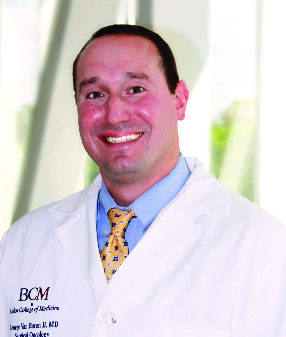
The trial randomized 202 patients to the intraperitoneal drainage group and 197 to the nondrainage group. The groups were well matched in terms of patients who dropped out because of metastatic disease or other reason, as well as demographics and operative data. “They were equally matched for laparoscopic vs. open, equally matched for vascular resection, equally matched for pancreatic texture and duct size, equally matched for method of transection, equally matched for operative time and blood loss and equally matched for surgical pathology,” Dr. Van Buren said.
The primary outcome was frequency of grade 2 complications or greater, which occurred in 44% (76) of the drainage group and 42% (72) of the nondrainage group (P = .804). “Similarly, the groups were equal for grade 3 severe complications as well as median complication severity and median number of complications,” Dr. Van Buren said. The rates of complications of grade 3 or greater were 29% (51) for the drainage group and 26% (44) for the nondrainage group (P = .477). Ninety-day mortality also was similar between the two groups: None died in the drainage group and two (1%) died in the nondrainage group (P = .24).
Drilling down into types of complications, Dr. Van Buren added that rates were similar across the board. “There was no difference in clinically relevant postoperative pancreatic fistula between the drain and no-drain group,” he said. “There was no difference in intra-abdominal abscess, no difference in the rates of postoperative imaging, no difference in the rates of postoperative percutaneous drain placements, and there was no difference in readmission or reoperation.”
One outcome that was noticeably different between the two groups was intra-abdominal fluid collections, reported in 9% (15) of the drainage group and 22% (38) of the nondrainage group (P = .0004). “However,” Dr. Van Buren said, “these were asymptomatic.”
In his discussion of the presentation, Charles Yeo, MD, FACS, of Thomas Jefferson University, Philadelphia, said that “the scope and the rigor of this study are impressive and quite laudable” but raised a number of questions, including concerns about the two deaths in the nondrainage group in the context of two other trials: a smaller multicenter trial, coauthored by Dr. Van Buren, that compared postoperative use of drains and nondrainage in pancreaticoduodenectomy but was halted because of eight deaths in the nondrainage group (Ann Surg. 2014;259:605-12); and the German PANDRA trial reported at last year’s ASA meeting that found nondrainage after pancreaticoduodenectomy to be superior to drainage in terms of reintervention and fistula rates.
Dr. Van Buren replied that the first study from Baylor – referenced by Dr. Yeo – involved “well-balanced groups, and they were equally matched and had minimal dropout throughout.” Because of that, the finding that drain placement for the pancreaticoduodenectomy “was definitive,” whereas the PANDRA trial was subject to some criticisms. The screening and randomization processes in PANDRA have been criticized because 3,200 patients were eligible for enrollment, suggesting a screening bias, Dr. Van Buren said. In addition, drains were placed in 21% of patients who were allocated to the no-drain group, suggesting surgeons deviated from the protocol in higher-risk situations, resulting in additional selection bias. This implies PANDRA was more of a selective draining trial, he said.
Dr. Van Buren and Dr. Yeo reported having no financial disclosures.
The complete manuscript of this study and its presentation at the American Surgical Association’s 137th Annual Meeting, April 2017, in Philadelphia, is to be published in Annals of Surgery pending editorial review.
AT THE ASA ANNUAL
Key clinical point: A randomized clinical trial has provided Level 1 evidence that placement of intraperitoneal drains does not alter clinical outcomes after distal pancreatectomy.
Major finding: No significant difference was found in the rate of complications between those who had drains and those who did not (44% vs. 42% of complications greater than grade 2).
Data source: Randomized trial of 399 patients who underwent distal pancreatectomy at 14 high-volume pancreas centers.
Disclosures: Dr. Van Buren reported having no financial disclosures.
Study identifies gaps in surgical trainees’ readiness
PHILADELPHIA – The question of how prepared general surgery residents are to operate independently after their training is longstanding, but clear definitions of competency and readiness have been elusive. A consortium of general surgery residencies has developed a metric for assessing surgeon readiness, but what the metric revealed may be a cause for concern for the surgical profession.
Brian C. George, MD, of the University of Michigan, Ann Arbor, reported at the annual meeting of the American Surgical Association on results of a study designed to measure the autonomy and readiness for independent practice of residents at 14 general surgery programs.
The study found that in the final 6 months of training, 96% of residents were rated competent by their observers to perform a straightforward appendectomy on their own, but only 71% were rated the same for partial colectomy, Dr. George said.
The participating general surgery attendings rated residents according to three scales (J Surg Educ. 2016;73:e118-130):
•“Performance” scale to measure readiness for independent practice, with competence defined as practice-ready and exceptional performance.
• “Zwisch” scale, named after Jay Zwischenberger, MD, FACS, of the University of Kentucky, to assess the amount of autonomy granted to a resident by the supervising surgical attending.
• “Complexity” scale to measure the patient-related complexity of the case at hand.
The study used a smartphone-based app, SIMPL, to collect data from September 2015 through December 2016. In evaluating performance, 437 observers provided 8,526 different ratings of 522 residents.
In a subset analysis, the study authors focused on the 132 operations the Surgical Council on Resident Education considers the core procedures of general surgery and found that 77% of fifth-year residents were rated as competent. Further restricting the analysis to residents in their final 6 months of training performing the five most commonly rated core procedures – appendectomy, cholecystectomy, ventral hernia repair, inguinal/femoral hernia repair, and partial colectomy – the researchers found that competency ranged from a high of 96% for appendectomy to a low of 71% for partial colectomy.
“If you combine all five procedures into one category, on average the residents are rated competent 84% of the time during the last 6 months of training,” Dr. George said. “But what’s really interesting – and I think this is probably the most important result for this study – is that looking at just the less-frequently rated core procedures and excluding the top five, residents are deemed competent in the last 6 months of training for 74% of those observed procedures.”
According to the Zwisch” scale to measure autonomy, residents in their last 6 months of training displayed what Dr. George termed “meaningful autonomy” in 77% of their observed operations for the core procedures. “Interestingly,” he added, “they were observed to have maximum autonomy, which is the supervision-only level, for 33% of all observed procedures.”
The researchers did an adjusted analysis to account for confounding factors such as the stringency of individual raters and patient complexity. “Using this type of analysis, the likelihood that a typical trainee rated by a typical attendee would be deemed competent by the end of training for a relatively straightforward laparoscopy appendectomy is 97%,” Dr. George said. “For a difficult laparoscopic appendectomy, by the end of training, they’re likely to be deemed competent 92% of time.” In the adjusted analysis, for a straightforward partial colectomy the raters predicted trainees to be competent 91.8% of time, but only 81.8% of the time for a complicated partial colectomy. “For less-frequently performed core procedures, there are many for which residents are likely to be deemed competent at a much lower level,” Dr. George added.
In discussing the study, Ara Darzi, MD, of St. Mary’s Hospital, London, called the methodology “unique and commendable,” and asked “Would you make the case to say we should increase the number of years of trainees from PG5 to PG6 or at least make your fellowships compulsory?”
Dr. George answered that 80% of general surgery residents already go into fellowships. “Whether it’s required or not is above my pay grade,” he said. “I hope not. Five years is already a big ask for a lot of medical students who are considering this profession. If we increase the training requirements, we’re going to have a supply problem that we will then need to address. We will be trading one problem for another.” He later added “The 20,000 hours of surgical residency should be enough to train a general surgeon to competence – its up to us to figure out how.”
The following organizations supported the research: American Board of Surgery, Association for Surgical Education, Association for Program Directors in Surgery, Massachusetts General Hospital, Northwestern University, Indiana University, and the members of the Procedural Learning and Safety Collaborative.
Neither Dr. George nor Dr. Darzi had any relevant financial disclosures.
The complete manuscript of this study and its presentation at the American Surgical Association’s 137th Annual Meeting, April 2017, in Philadelphia, Pennsylvania, is to be published in Annals of Surgery pending editorial review.
AT THE ASA ANNUAL MEETING
Key clinical point: General surgery residents are often but not universally ready to independently perform core surgical procedures upon completion of surgical training.
Major finding: In the last 6 months of training, residents were rated competent 84% of the time in performing the five leading core procedures and 64% of the time for less-frequently performed procedures.
Data source: Ratings of 437 of 8,526 different observations of 522 residents at 14 institutions of the Procedural Learning and Safety Collaborative.
Disclosures: Dr. George and his coauthors reported having no financial disclosures.
Antithrombotics no deterrent for emergent lap appendectomy
HOUSTON – Few studies have looked at the risk of irreversible antithrombotic therapy in patients who need emergent or urgent laparoscopic appendectomy, but a new study showed that the operation poses no significantly greater risk for such patients, compared with people who are not on antithrombotics.
The study’s findings do not apply to all patients on anticoagulation, specifically those on new novel oral anticoagulants (NOACs), said Christopher Pearcy, MD, of Methodist Dallas Medical Center.
NOAC agents include dabigatran, rivaroxaban, and apixaban.
Appendicitis is the third most common indication for abdominal surgery in the elderly, Dr. Pearcy noted, and their mortality rates are eight times greater than those of younger patients. However, these patients often proceed to operation with minimal workup, “given that laparoscopic appendectomy is a relatively benign procedure,” he said at the annual meeting of the Society of American Gastrointestinal and Endoscopic Surgeons.
The retrospective study evaluated two groups of 195 patients who had urgent or emergent laparoscopic appendectomy at three centers from 2010 to 2014. One group was on irreversible antithrombotic therapy, and the other served as controls.
The primary outcomes were blood loss, transfusion requirement, and mortality. Secondary outcomes were duration of operation, length of hospital stay, rates of infections, complications, and 30-day readmissions.
“Compared with controls, we didn’t find any significant difference in any outcome whatsoever after laparoscopic appendectomy in patients on prehospital antithrombotic therapy,” Dr. Pearcy said.
Specifically, average estimated blood loss was 18 cc in controls vs. 22 cc in patients on antithrombotics, and mortality was 0% in the former vs. 1% in the latter. Patients on antithrombotics had a lower rate of complications: 3% vs. 11%.
Dr. Pearcy discussed a case of a 70-year-old man with acute appendicitis. He had a history of coronary artery disease, hypertension, hyperlipidemia, type 2 diabetes, and stroke, and was taking clopidogrel and aspirin daily.
“Is it safe to proceed with surgery given this patient’s irreversible antithrombotic therapy? We would say yes,” he said.
Dr. Pearcy reported having no financial disclosures.
AT SAGES 2017
Key clinical point: Emergent laparoscopic appendectomy poses no significant risk for patients on irreversible antithrombotic therapy.
Major finding: Average estimated blood loss was 18 cc in controls vs. 22 cc in patients on antithrombotics, and mortality was 0% vs. 1%, respectively.
Data source: A retrospective study of 390 patients who had urgent or emergent laparoscopic appendectomy at three centers from 2010 to 2014.
Disclosures: Dr. Pearcy reported having no financial disclosures.
Series supports viability of ambulatory laparoscopic sleeve gastrectomy
HOUSTON – An ambulatory approach to laparoscopic sleeve gastrectomy is a safe and viable option to improve patient satisfaction and soften the economic blow of these procedures on patients, based on a large series at one surgery center in Cincinnati.
“With proper patient selection, utilization of enhanced recovery pathways with an overall low readmission rate and the complication profile point to the feasibility of laparoscopic sleeve gastrectomy [LSG] as a safe outpatient procedure,” said Sepehr Lalezari, MD, now a surgical fellow at Johns Hopkins University, Baltimore.
About 105,000 LSG operations were performed in the United States in 2015, representing 54% of all bariatric operations, according to the American Society for Metabolic and Bariatric Surgery.
Patient selection and strict adherence to protocols are keys to success for ambulatory LSG, Dr. Lalezari said. Suitable patients were found to be ambulatory, between ages 18 and 65 years; had a body mass index (BMI) less than 55 kg/m2 for males and less than 60 kg/m2 for females; weighed less than 500 lb; had an American Society of Anesthesiologists’ classification score less than 4; and had no significant cardiopulmonary impairment, had no history of renal failure or organ transplant, and were not on a transplant wait list.
In this series, 71% of patients (579) were female, and the average BMI was 43. The total complication rate was 2.3% (19); 17 of these patients required hospital admission.
Postoperative complications included gastric leaks (seven, 0.9%); intra-abdominal abscess requiring percutaneous drainage (four, 0.5%); dehydration, nausea, and/or vomiting (four, 0.5%); and one of each of the following: acute cholecystitis, postoperative bleeding, surgical site infection (SSI), and portal vein thrombosis/pulmonary embolism.
The two complications managed on an outpatient basis were the SSI and one intra-abdominal abscess, Dr. Lalezari said.
“The only readmissions in our series that could have been possibly prevented with an overnight stay in the hospital were the four cases of nausea, vomiting, and/or dehydration,” he said. “These only accounted for 0.5% of the total cases performed.”
The readmission rates for ambulatory LSG in this series compared favorably with large trials that did not distinguish between ambulatory and inpatient LSG procedures, Dr. Lalezari noted. A 2016 analysis of 35,655 patients in the American College of Surgeons National Surgical Quality Improvement Program database reported a readmission rate of 3.7% for LSG (Surg Endosc. 2016 Jun;30[6]:2342-50).
A larger study of 130,000 patients who had bariatric surgery reported an LSG readmission rate of 2.8% (Ann Surg. 2016 Nov 15. doi: 10.1097/SLA.0000000000002079). The most common cause for readmissions these trials reported were nausea, vomiting, and/or dehydration.
Bariatric surgeons have embraced enhanced recovery pathways and fast-track surgery, with good results, Dr. Lalezari said, citing work by Zhamak Khorgami, MD, and colleagues at the Cleveland Clinic (Surg Obes Relat Dis. 2017 Feb;13[2]:273-80).
“Looking at fast-track surgery, they found that patients discharged on postoperative day 1 vs. day 2 or 3 did not change outcomes”; those discharged later than postoperative day 1 trended toward a higher readmission rate of 2.8% vs. 3.6%, Dr. Lalezari said.
The enhanced recovery/fast track protocol Dr. Lalezari and his coauthors used involves placing intravenous lines and infusing 1 L crystalloid before starting the procedure, and administration of famotidine and metoclopramide prior to anesthesia. The protocol utilizes sequential compression devices and avoids Foley catheters and intra-abdominal drains. Patients receive dexamethasone and ondansetron during the operation. The protocol emphasizes early ambulation and resumption of oral intake.
The operation uses a 36-French bougie starting about 5 cm from the pylorus, and all staple lines are reinforced with buttress material. At the end of the surgery, all incisions are infiltrated with 30 cc of 0.5% bupivacaine with epinephrine.
Patients are ambulating about 90 minutes after surgery and are monitored for 3-4 hours. They receive a total volume of 3-4 L crystalloids. When they’re tolerating clear liquids, voiding spontaneously, and walking independently, and their pain is well controlled (pain score less than 5/10) and vital signs are within normal limits, they’re discharged.
Postoperative follow-up involves a call at 48 hours and in-clinic follow-up at weeks 1 and 4. Additional follow-up is scheduled at 3-month intervals for 1 year, then at 6 months for up to 2 years, and then yearly afterward.
“With proper patient selection and utilization of enhanced recovery pathways, the low overall readmission rate (2.1%) and complication profile (2.3%) in our series point to the feasibility of laparoscopic sleeve gastrectomy as a safe outpatient procedure,” Dr. Lalezari said.
He reported having no relevant financial disclosures.
AT SAGES 2017
Key clinical point: Laparoscopic sleeve gastrectomy is a safe outpatient procedure – with strict adherence to enhanced recovery pathways and fast-track protocols.
Major finding: This series reported an overall readmission rate of 2.1% and a complication rate of 2.3% in patients who had outpatient LSG.
Data source: A retrospective review of 821 patients who had ambulatory LSG by a single surgeon from 2011 to 2015.
Disclosures: Dr. Lalezari reported having no relevant financial disclosures.
Can public reporting improve pediatric heart surgery?
Public reporting of cardiac surgery outcomes has been a disruptive force in cardiology, and especially daunting in pediatric cardiac surgery because of low case volumes and rare mortality. To ensure that public reporting achieves its original goals – providing transparency to the patient care process, holding providers accountable, informing decision making for health care consumers, reducing costs, encouraging more efficient use of health system resources, and improving patient care and outcomes – further study that includes use of appropriate risk adjustment is needed, according to commentaries in the April issue of the Journal of Thoracic and Cardiovascular Surgery.
The journal asked two groups to provide perspective on a study Adam D. DeVore, MD, of Duke University in Durham, N.C., and his coauthors published last year (J Am Coll Cardiol. 2016 Mar 1;67:963-72). The study analyzed Medicare claims data from 2006 to 2012 for 37,829 hospitalizations for heart attack, 100,189 for heart failure (HF), and 79,076 for pneumonia. Dr. DeVore and his colleagues found readmission rates for the three conditions did not significantly improve after public reporting protocols were implemented in 2009. However, the study did show a significant decrease in ED visits and observation stays for those with HF: from 2.3% to –0.8% for the former (P = .007); and from 15% to 4% for the latter (P = .04).
• The metrics must be accurate, reliably discern hospital quality, and account for high-risk cases without penalizing hospitals. “In pediatric cardiac surgery, this can be particularly challenging, owing to the very wide heterogeneity of disease and variability in case mix and volumes across centers,” Dr. Gaynor and his coauthors wrote. While methodology for case mix and patient characteristics have improved in recent years, further improvement is needed.
• Metrics must be clearly reported and easy for stakeholders to interpret. “This is critical if the data are to be used to steer patients toward higher-performing centers and/or to provide incentives for hospitals with lower performance to make improvements,” the researchers said.
• Regional reporting or a methodology that indicates where a hospital ranks within larger categories deserve further investigation as tools to help families choose a high-performing center, “ideally based on geography and on the particular type and complexity of disease,” Dr. Gaynor and his coauthors stated (J Thorac Cardiovasc Surg. 2017 Apr;153:904-7).
• Indirect standardization, a statistical methodology used to calculate risk-adjusted performance, could help consumers to interpret hospital performance more easily. This methodology might help classify a hospital with a low-complexity population as a high performer. “Developing better methods to convey this information to consumers is vital,” according to the researchers.
The perspective acknowledged several reports of an unintended consequence of public reporting: surgeons and centers avoiding higher-risk cases to skew their performance scores higher, thus restricting access to care. However, in a separate perspective on Dr. DeVore’s study, James S. Tweddell, MD, of Cincinnati Children’s Hospital Medical Center, and his coauthors, questioned the quality of the evidence on which Dr. Gaynor and his colleagues based their conclusion of risk aversion and limited access to care: a newspaper report from the United Kingdom.
Dr. Tweddell and his coauthors noted, “The predominance of data suggest an overall beneficial impact of public reporting.” They cited a trial that showed a decrease in heart attack–related deaths after public reporting had been implemented (JAMA. 2009 Dec 2;302:2330-7); a 2012 Agency for Healthcare Research and Quality systemic review (Evidence Report No. 208) that showed that research on harm is limited, and most studies do not confirm potential harm; and a meta-analysis that found a 15% reduction in adverse events associated with public reporting (BMC Health Serv Res. 2016;16:296).
“Appropriate risk adjustment is critical to achieve effective and fair transparency, but there is little objective data of harm associated with public reporting,” Dr. Tweddell and his coauthors concluded. While examination of public reporting must continue, they said, “these efforts are likely to result in minor course changes and the effort to inform and educate our patients and their families must continue.”
Ds. Gaynor, Dr. Tweddell, and their coauthors reported having no financial disclosures.
FROM THE JOURNAL OF THORACIC AND CARDIOVASCULAR SURGERY
Key clinical point: Public reporting of outcomes in cardiac surgery in children requires further investigation but has also been associated with improved outcomes.
Major finding: Emergency department visits for patients with heart failure declined from 2.3% before public reporting to –0.8% after implementation, and observation stays declined from 15.1% to 4.1%.
Data source: Analysis of Medicare claims data from 2006 to 2012 for 271,094 patients discharged after hospitalization for heart attack, heart failure or pneumonia.
Disclosures: Dr. Gaynor and Dr. Tweddell had no financial relationships to disclose.
AATS publishes guidelines for infective endocarditis
Infective endocarditis (IE) is a devastating complication of heart valve disease that, left untreated, can be fatal. Management requires a multidisciplinary approach, and many of the respective medical societies that represent the participating specialties have developed guidelines. Now, the American Association for Thoracic Surgery has published “Consensus Guidelines for the Surgical Treatment of Infective Endocarditis” to guide thoracic and cardiovascular surgeons in making decisions of when to operate in cases of IE (J Thorac Cardiovasc Surg. 2017 Jan 24. doi: 10.1016/j.jtcvs.2016.09.093).
The rationale for developing the guidelines is a growing prevalence of IE, including in patients with normal valves and no previous diagnosis of heart disease. “These new AATS consensus guidelines primarily address questions related to active and suspected active IE affective valves and intracardiac structures,” Dr. Pettersson and his coauthors said. The AATS guidelines for infective endocarditis address complications including risk of embolism and the timing of surgery in patients with neurological complications, while acknowledging the the need for additional research into these topics.* “It is understood that surgery is beneficial only if the patient’s complications and other comorbidities do not preclude survival and meaningful recovery,” the guideline authors said.
The guidelines confirm the team approach for managing patients with IE. The team should include cardiology, infectious disease, cardiac surgery, and other specialties needed to handle IE-related complications (class of recommendation [COR] I, level of evidence [LOE] B). Before surgery, the surgeon should know the patient is on effective antimicrobial therapy (COR I, LOE B). Transesophageal echocardiography (TEE) is indicated to yield the clearest understanding of the pathology (COR I, LOE B).
Dr. Pettersson and the guideline writing team also clarified indications for surgery in patients with IE. They include when valve dysfunction causes heart failure (COR I, LOE B); when, after a full course of antibiotics, the patient has signs of heart block, annular or aortic abscess or destructive penetrating lesions (COR I, LOE B); and in the setting of recurrent emboli and persistent vegetations despite appropriate antibiotic therapy (COR IIA, LOE B).
The guideline writers acknowledged potential disagreement between the AATS guidelines and those of the American College of Cardiology/American Heart Association with regard to early surgery in IE. Debate surrounds whether to operate early or wait for symptoms of heart failure to manifest in patients with native valve endocarditis (NVE). The AATS guideline authors cite work by Duk-Hyun Kang, MD, PhD, and coauthors in South Korea (N Engl J Med. 2012;366;2466-73) and others advocating for early surgery. “For this reason, once a surgical indication is evident, surgery should not be delayed,” Dr. Pettersson and his coauthors said.
Several conditions can influence the timing of surgery. Patients with cerebral mycotic aneurysm should be managed closely with neurology or neurosurgery (COR I, LOE C). Patients with a recent intracranial hemorrhage should wait at least 3 weeks for surgery (COR IIA, LOE B), but those with nonhemorrhagic strokes could go in for urgent surgery (COR IIA, LOE B). Brain imaging is indicated for IE patients with neurological symptoms (COR I, LOE B), but anticoagulation management requires a nuanced approach that takes all risks and benefits into consideration (COR I, LOE C).
Key steps during surgery involve mandatory intraoperative TEE (COR I, LOE B), median sternotomy with few exceptions (COR I, LOE B), and radical debridement and removal of all infected and necrotic tissue (COR I, LOE B). The writers also provided four guidelines for reconstruction and valve replacement:
- Repair when possible for patients with NVE (COR I, LOE B).
- When replacement is indicated, the surgeon should base valve choice on normal criteria – age, life expectancy, comorbidities, and expected compliance with anticoagulation (COR I, LOE B).
- Avoid use of mechanical valves in patients with intracranial bleeding or who have had a major stroke (COR IIA, LOE C).
- In patients with invasive disease and deconstruction, reconstruction should depend on the involved valve, severity of destruction, and available options for cardiac reconstruction (COR I, LOE B).
The AATS guidelines also challenge conventional thinking on the practice of soaking a gel-impregnated graft with antimicrobials targeting a specific organism. “We found no evidence to support this practice,” Dr. Pettersson and his coauthors said (COR IIB, LOE B). They came to the same conclusion with regard to the use of local antimicrobials or antiseptics during irrigation after debridement and local injection of antimicrobials around the infected area (COR I, LOE C).
The guidelines provide direction on a host of other surgical issues in IE: use of aortic valve grafts; when to remove or replace noninfected grafts; when to remove pacemakers; the role of drainage; postoperative management; follow-up; and additional screening. They also shed insight into what the guideline authors call “residual controversies,” including surgery for injection drug users (use “all available resources and options for drug rehabilitation”) and dialysis patients (“it is reasonable to offer surgery when the additional burden of comorbidities is not overwhelming”). They also acknowledge seven different scenarios that lack clear evidence for intervention but require the surgeon to determine the need for surgery, ranging from timing of surgery for IE in patients with neurologic complications to how to treat patients with functional valve issues after being cured of IE.
The guideline writers acknowledged that institutional funds supported the work. Dr. Pettersson had no financial relationships to disclose.
*Correction 5/172017: It was incorrectly stated that these complications were not addressed in the guidelines due to lack of evidence
Whether they’re intended to or not, guidelines like the AATS Consensus Guidelines for the Surgical Treatment of Infected Endocarditis “can evolve into hard and fast principles, sometimes leading to incorrect decision making and even creating medicolegal problems for treating physicians,” Gus J. Vlahakes, MD, of Harvard Medical School and Massachusetts General Hospital, Boston, said in his invited commentary (J Thorac Cardiovasc Surg. 2016 Nov 3. doi: 10.1016/j.jtcvs.2016.10.041).
Guidelines cannot “integrate all the necessary considerations,” Dr. Vlahakes said, so surgeons should view them as “a set of general principles to guide decision making.” In IE, that means having an experienced cardiac surgeon who can apply the guidelines on a case-by-case basis and a multidisciplinary team that includes an infectious disease specialist, he said. The surgeon must participate in preoperative management.
Dr. Vlahakes had no relevant financial disclosures.
Infective endocarditis (IE) is a devastating complication of heart valve disease that, left untreated, can be fatal. Management requires a multidisciplinary approach, and many of the respective medical societies that represent the participating specialties have developed guidelines. Now, the American Association for Thoracic Surgery has published “Consensus Guidelines for the Surgical Treatment of Infective Endocarditis” to guide thoracic and cardiovascular surgeons in making decisions of when to operate in cases of IE (J Thorac Cardiovasc Surg. 2017 Jan 24. doi: 10.1016/j.jtcvs.2016.09.093).
The rationale for developing the guidelines is a growing prevalence of IE, including in patients with normal valves and no previous diagnosis of heart disease. “These new AATS consensus guidelines primarily address questions related to active and suspected active IE affective valves and intracardiac structures,” Dr. Pettersson and his coauthors said. The AATS guidelines for infective endocarditis address complications including risk of embolism and the timing of surgery in patients with neurological complications, while acknowledging the the need for additional research into these topics.* “It is understood that surgery is beneficial only if the patient’s complications and other comorbidities do not preclude survival and meaningful recovery,” the guideline authors said.
The guidelines confirm the team approach for managing patients with IE. The team should include cardiology, infectious disease, cardiac surgery, and other specialties needed to handle IE-related complications (class of recommendation [COR] I, level of evidence [LOE] B). Before surgery, the surgeon should know the patient is on effective antimicrobial therapy (COR I, LOE B). Transesophageal echocardiography (TEE) is indicated to yield the clearest understanding of the pathology (COR I, LOE B).
Dr. Pettersson and the guideline writing team also clarified indications for surgery in patients with IE. They include when valve dysfunction causes heart failure (COR I, LOE B); when, after a full course of antibiotics, the patient has signs of heart block, annular or aortic abscess or destructive penetrating lesions (COR I, LOE B); and in the setting of recurrent emboli and persistent vegetations despite appropriate antibiotic therapy (COR IIA, LOE B).
The guideline writers acknowledged potential disagreement between the AATS guidelines and those of the American College of Cardiology/American Heart Association with regard to early surgery in IE. Debate surrounds whether to operate early or wait for symptoms of heart failure to manifest in patients with native valve endocarditis (NVE). The AATS guideline authors cite work by Duk-Hyun Kang, MD, PhD, and coauthors in South Korea (N Engl J Med. 2012;366;2466-73) and others advocating for early surgery. “For this reason, once a surgical indication is evident, surgery should not be delayed,” Dr. Pettersson and his coauthors said.
Several conditions can influence the timing of surgery. Patients with cerebral mycotic aneurysm should be managed closely with neurology or neurosurgery (COR I, LOE C). Patients with a recent intracranial hemorrhage should wait at least 3 weeks for surgery (COR IIA, LOE B), but those with nonhemorrhagic strokes could go in for urgent surgery (COR IIA, LOE B). Brain imaging is indicated for IE patients with neurological symptoms (COR I, LOE B), but anticoagulation management requires a nuanced approach that takes all risks and benefits into consideration (COR I, LOE C).
Key steps during surgery involve mandatory intraoperative TEE (COR I, LOE B), median sternotomy with few exceptions (COR I, LOE B), and radical debridement and removal of all infected and necrotic tissue (COR I, LOE B). The writers also provided four guidelines for reconstruction and valve replacement:
- Repair when possible for patients with NVE (COR I, LOE B).
- When replacement is indicated, the surgeon should base valve choice on normal criteria – age, life expectancy, comorbidities, and expected compliance with anticoagulation (COR I, LOE B).
- Avoid use of mechanical valves in patients with intracranial bleeding or who have had a major stroke (COR IIA, LOE C).
- In patients with invasive disease and deconstruction, reconstruction should depend on the involved valve, severity of destruction, and available options for cardiac reconstruction (COR I, LOE B).
The AATS guidelines also challenge conventional thinking on the practice of soaking a gel-impregnated graft with antimicrobials targeting a specific organism. “We found no evidence to support this practice,” Dr. Pettersson and his coauthors said (COR IIB, LOE B). They came to the same conclusion with regard to the use of local antimicrobials or antiseptics during irrigation after debridement and local injection of antimicrobials around the infected area (COR I, LOE C).
The guidelines provide direction on a host of other surgical issues in IE: use of aortic valve grafts; when to remove or replace noninfected grafts; when to remove pacemakers; the role of drainage; postoperative management; follow-up; and additional screening. They also shed insight into what the guideline authors call “residual controversies,” including surgery for injection drug users (use “all available resources and options for drug rehabilitation”) and dialysis patients (“it is reasonable to offer surgery when the additional burden of comorbidities is not overwhelming”). They also acknowledge seven different scenarios that lack clear evidence for intervention but require the surgeon to determine the need for surgery, ranging from timing of surgery for IE in patients with neurologic complications to how to treat patients with functional valve issues after being cured of IE.
The guideline writers acknowledged that institutional funds supported the work. Dr. Pettersson had no financial relationships to disclose.
*Correction 5/172017: It was incorrectly stated that these complications were not addressed in the guidelines due to lack of evidence
FROM THE JOURNAL OF THORACIC AND CARDIOVASCULAR SURGERY
Key clinical point: The American Association for Thoracic Surgery charged a committee of eight members to author “Consensus Guidelines: Surgical Treatment of Infective Endocarditis.”
Major finding: Patients with infective endocarditis need early input from the responsible cardiac surgeon, who must also lead the care team in evaluation, decision- making, and ultimately carrying out surgery as needed.
Data source: The writing committee followed Institute of Medicine standards for clinical practice guidelines, invited comment from a group of 12 multidisciplinary specialists and reviewed 288 articles in drafting the guidelines.
Disclosures: Institutional funds supported the work. Dr. Pettersson and his coauthors had no financial relationships to disclose.
Time to reexamine surgery for nonlocalized bronchiectasis
Nonlocalized bronchiectasis is becoming more common in developing countries and has been difficult to treat. While surgery for localized bronchiectasis has been proven, its role in nonlocalized disease is less established; researchers from China are advocating for a reexamination of surgical resection in this disease based on results of a small cohort study at their academic center.
“Lobectomy for the predominant lesion is a safe procedure in the surgical treatment of nonlocalized bronchiectasis and leads to significant relief of symptoms with good rates of satisfaction,” Jie Dai, PhD, of Shanghai (China) Pulmonary Hospital Tongji University and his coauthors reported in the Journal of Thoracic and Cardiovascular Surgery (2017 Apr;153:979-85).
The researchers reviewed the medical records of 37 consecutive patients – 10 men and 27 women – with nonlocalized bronchiectasis who had lobectomies via thoracotomies during 2010-2013. Twenty-three patients (62.2%) were symptom free after surgery and 10 (27%) reported that their symptoms had improved. Four (10.8%) said their symptoms either did not improve or worsened, but three of them also had chronic occlusive pulmonary disease. There were no deaths, and the morbidity rate was 21.6%.
The researchers used three criteria to select candidates for surgery: persistent symptoms despite medical treatment; an identifiable predominant lesion; and cardiopulmonary function compatible with anesthetic risk. Average age was 54.5 years and more than half (19) had no smoking history.
The surgical technique involved a posterolateral thoracotomy and a double-lumen endotracheal tube to avoid contamination of the opposite side of the lung during surgery. Surgery avoided excessive bronchial dissection and preserved peribronchial tissues. Extrapleural dissection avoided spillage of lung contents into the pleural space.
Treatment of the hilum followed an order from the pulmonary artery to the pulmonary vein and then to the bronchus. An ultrasonic device helped isolate and then ligate or sever distorted bronchial arteries. A mechanical stapler was used to close the bronchial stump. Reinforcement involved an intercostal muscle flap in 16 cases and a pedicled parietal pleural flap in 21. If any sign of pleural infection appeared after hemostasis, pleural space irrigation with 0.5% neomycin (500 mg/L) was initiated. Two chest drains were placed at the bronchial stump, and the bronchial suture checked with bronchoscopy. Airway secretions were removed, and pathology confirmed bronchiectasis all specimens.
The frequency of acute infection and hemoptysis decreased significantly at 1 year postoperatively, from 5.3% to 1.8% and 4.9% to 1.1%, respectively, (P less than .01 for both). Daily sputum volume decreased an average of 26.3 mL (P less than .01) and sputum cultures became sterile in 13 (35%) of patients (P less than .01).
Previously, surgery for nonlocalized bronchiectasis had been reserved for life-threatening symptoms only, but Dr. Dai and his coauthors fashioned their study on previous studies of lobectomy that included patients with nonlocalized bronchiectasis (Br J Surg. 2005;92:836-9; Ann Thorac Surg. 2003;75:382-7).
Two major complications occurred in the Shanghai cohort: empyema and persistent air leak, both of which were managed without a reoperation and had been reported in previous series. Dr. Dai and his coauthors have adopted protocols to reduce complications, among them, requiring that patients have sputum output greater than 20 mL/day with little purulence and no engorgement or edema in the tunica mucosa bronchiorum on bronchoscopy.
But the researchers are not ready to extend surgery as a blanket indication for nonlocalized bronchiectasis. “It is worth mentioning that the surgical benefit is limited to patients who have only one predominant area of bronchiectatic disease that can be localized by CT instead of those with diffuse bronchiectasis,” they wrote. “The ideal surgical candidate has a heterogeneous distribution of diseased areas.”
The investigators pointed out that the study was limited by its small size and lack of information on etiologies of disease.
Dr. Dai and his coauthors had no financial relationships to disclose.
The study by Dr. Dai and his coauthors “is an important contribution to the literature” despite its limitations, Steven Milman, MD, and Thomas Ng, MD, of Brown University, Providence, R.I., said in their invited commentary (J Thorac Cardiovasc Surg. 2017 Apr;153:986). However, they added, “Several important points need to be stressed.”
Among those points: The researchers studied a “highly selected group” of young patients with good pulmonary functions; mean follow-up was short (15 months); the etiology of bronchiectasis was unknown; and lobectomy was not the optimal treatment for nonlocalized bronchiectasis. “It must be remembered that these patients first failed medical therapy and that the study population received lobectomy due to the extent of the dominant disease and not as routine treatment,” Dr. Milman and Dr. Ng wrote.
To validate the findings, Dr. Milman and Dr. Ng said, not only do more patients need to be studied with longer follow-up but future investigators also should study minimally invasive approaches to see if that would improve the outcomes.
Dr. Milman and Dr. Ng had no financial relationships to disclose.
Nonlocalized bronchiectasis is becoming more common in developing countries and has been difficult to treat. While surgery for localized bronchiectasis has been proven, its role in nonlocalized disease is less established; researchers from China are advocating for a reexamination of surgical resection in this disease based on results of a small cohort study at their academic center.
“Lobectomy for the predominant lesion is a safe procedure in the surgical treatment of nonlocalized bronchiectasis and leads to significant relief of symptoms with good rates of satisfaction,” Jie Dai, PhD, of Shanghai (China) Pulmonary Hospital Tongji University and his coauthors reported in the Journal of Thoracic and Cardiovascular Surgery (2017 Apr;153:979-85).
The researchers reviewed the medical records of 37 consecutive patients – 10 men and 27 women – with nonlocalized bronchiectasis who had lobectomies via thoracotomies during 2010-2013. Twenty-three patients (62.2%) were symptom free after surgery and 10 (27%) reported that their symptoms had improved. Four (10.8%) said their symptoms either did not improve or worsened, but three of them also had chronic occlusive pulmonary disease. There were no deaths, and the morbidity rate was 21.6%.
The researchers used three criteria to select candidates for surgery: persistent symptoms despite medical treatment; an identifiable predominant lesion; and cardiopulmonary function compatible with anesthetic risk. Average age was 54.5 years and more than half (19) had no smoking history.
The surgical technique involved a posterolateral thoracotomy and a double-lumen endotracheal tube to avoid contamination of the opposite side of the lung during surgery. Surgery avoided excessive bronchial dissection and preserved peribronchial tissues. Extrapleural dissection avoided spillage of lung contents into the pleural space.
Treatment of the hilum followed an order from the pulmonary artery to the pulmonary vein and then to the bronchus. An ultrasonic device helped isolate and then ligate or sever distorted bronchial arteries. A mechanical stapler was used to close the bronchial stump. Reinforcement involved an intercostal muscle flap in 16 cases and a pedicled parietal pleural flap in 21. If any sign of pleural infection appeared after hemostasis, pleural space irrigation with 0.5% neomycin (500 mg/L) was initiated. Two chest drains were placed at the bronchial stump, and the bronchial suture checked with bronchoscopy. Airway secretions were removed, and pathology confirmed bronchiectasis all specimens.
The frequency of acute infection and hemoptysis decreased significantly at 1 year postoperatively, from 5.3% to 1.8% and 4.9% to 1.1%, respectively, (P less than .01 for both). Daily sputum volume decreased an average of 26.3 mL (P less than .01) and sputum cultures became sterile in 13 (35%) of patients (P less than .01).
Previously, surgery for nonlocalized bronchiectasis had been reserved for life-threatening symptoms only, but Dr. Dai and his coauthors fashioned their study on previous studies of lobectomy that included patients with nonlocalized bronchiectasis (Br J Surg. 2005;92:836-9; Ann Thorac Surg. 2003;75:382-7).
Two major complications occurred in the Shanghai cohort: empyema and persistent air leak, both of which were managed without a reoperation and had been reported in previous series. Dr. Dai and his coauthors have adopted protocols to reduce complications, among them, requiring that patients have sputum output greater than 20 mL/day with little purulence and no engorgement or edema in the tunica mucosa bronchiorum on bronchoscopy.
But the researchers are not ready to extend surgery as a blanket indication for nonlocalized bronchiectasis. “It is worth mentioning that the surgical benefit is limited to patients who have only one predominant area of bronchiectatic disease that can be localized by CT instead of those with diffuse bronchiectasis,” they wrote. “The ideal surgical candidate has a heterogeneous distribution of diseased areas.”
The investigators pointed out that the study was limited by its small size and lack of information on etiologies of disease.
Dr. Dai and his coauthors had no financial relationships to disclose.
FROM THE JOURNAL OF THORACIC AND CARDIOVASCULAR SURGERY
Key clinical point: Lobectomy for nonlocalized bronchiectasis can improve symptoms significantly.
Major finding: Among 37 patients who had lobectomy, 62.2% were asymptomatic after surgery.
Data source: Single-center retrospective review of 37 patients who had lobectomy for nonlocalized bronchiectasis from January 2010 to December 2013.
Disclosure: Dr. Dai and his coauthors had no financial relationships to disclose.
Can high hematocrit predict early shunt thrombosis?
Shunt occlusion has been a well-documented cause of shunt failure in newborns who have had systemic to pulmonary shunt placement, and little has been known about why shunts occlude. However, researchers have reported in a small retrospective study that higher postoperative hematocrit levels immediately after surgery may be predictors of shunt occlusion.
Reporting in the April 2017 issue of The Journal of Thoracic and Cardiovascular Surgery, Brett R. Anderson, MD, of New York-Presbyterian/Morgan Stanley Children’s Hospital, Columbia University Medical Center in New York, and coauthors, found that every 5-point increase in postoperative hematocrit more than doubled an infant’s odds of having early shunt occlusion (J Thorac Cardiovasc Surg. 2017;153:947-55).
“Beginning in the latter half of 2014, we noticed an increase in the incidence of early shunt occlusions in our neonatal cardiac intensive care unit,” Dr. Anderson and coauthors said. So they conducted a retrospective chart review of 80 infants who had undergone systemic to pulmonary shunt placement from January 2010 to July 2015, hypothesizing that increased hematocrit in the early postoperative period might have caused early shunt occlusion. They investigated the association between the first postoperative hematocrit and early shunt occlusion and in-hospital mortality in these patients.
Five patients (6.3%) experienced early shunt occlusion – that is, within 24 hours of placement (actually, within 10 hours of placement). Overall, 12 infants (15%) experienced shunt occlusion. The physicians at New York-Presbyterian do not administer anticoagulation in these patients during the first 12 hours after shunt placement.
The median initial postoperative hematocrit was 41.7%, with a range of 31.7%-55.8%. The survival analysis the researchers performed found that for every 5 additional percentage points, the hazard ratio for early shunt occlusion was 2.7 (P = .007) and 1.74 for any shunt occlusion (P less than .001). Incidentally, four cases of early shunt occlusion occurred in the later study period after 2014, during which the average first postoperative hematocrit was significantly higher than in the pre-2014 study period, 45.3% vs. 41.5% (P = .21), and the odds of early shunt occlusion were 16 times higher (P less than .001). Dr. Anderson and coauthors said the possible explanation for this variation was a switch to a new point-of-care analyzer in 2013.
With regard to mortality, six infants overall (7.4%) died before discharge, and four (5%) within 30 days of shunt placement. No infants with early shunt occlusion died, although two with late shunt occlusion died. Increased inotrope score and first postoperative arterial oxygen tension were the only factors associated with increased mortality. “No significant association was identified between hematocrit and 30-day mortality,” Dr. Anderson and coauthors said.
In the first 24 hours after surgery, 11 infants (13.8%) received packed red blood cell (PRBC) transfusions, seven (8.8%) received platelets, and four (5%) received fresh frozen plasma/cryoprecipitate. Higher postoperative PRBC transfusion volumes were associated with increased odds of mortality (P = .001), but none of these factors were significantly associated with early shunt occlusion.
Dr. Anderson and coauthors acknowledged that shunt occlusion is a “vexing problem” in infants with cyanotic heart disease. While other researchers studied postoperative hematocrit levels and possible associations with outcomes, including shunt occlusion and mortality, the New York-Presbyterian investigators said this is the first study of the first postoperative hematocrit.
Dr. Anderson and coauthors said their findings raise the question about the ideal perioperative prophylactic antithrombotic therapy in these patients. These researchers initiate aspirin therapy 12 hours after surgery if hemostasis is established.
As a result of this study, Dr. Anderson and coauthors instituted a number of practice changes at their center. They include:
• Cardiac anesthesiologists have been asked not to transfuse shunted neonates with hematocrit level of greater than or equal to 35%, and hematocrits are then immediately repeated when a patient returns to the cardiac ICU.
• Patients with hematocrits greater than or equal to 55% get partial exchange transfusions.
• An individualized approach for patients with lower hematocrits who are more cyanotic than expected. This includes a diagnostic echocardiogram, nitric oxide, oxygen and heparin, escalated inotropic support if necessary and sometimes a cautious approach to transfusions if symptoms do not resolve and an acute shunt occlusion if not likely.
Dr. Anderson and coauthors acknowledged limits to their study, most notably its retrospective nature and a small population at a single center, and that large investigations are needed to validate their findings.
Dr. Anderson disclosed receiving salary support from the National Center for Advancing Translational Sciences. Coauthor Jennifer M. Duchon, MDCM, MPH, receives salary support from the National Institute of Allergy and Infectious Disease. The remaining coauthors had no financial relationships to disclose.
In her invited commentary, Nancy S. Ghanayem, MD, of the Medical College of Wisconsin, gives credit to Dr. Anderson and coauthors for introducing the notion that high initial postoperative hematocrit may increase the risk of early shunt thrombosis in newborns, but with a caveat: “we remain somewhat hesitant regarding wholesale acceptance of the validity of the conclusions for several reasons” (J Thorac Cardiovasc Surg. 2017;153:956).
Those reasons include the low number of reported events, failure to list the actual hematocrits of the five patients who experienced early shunt occlusion and the lack of hemodynamic data – the latter of which she called “a significant limitation.”
A patient with low cardiac output, especially one who is cyanotic or has a single ventricle, is at higher risk for thrombosis and more likely to be transfused, “which in this case would potentially be masked by the method of comparing transfused volumes,” Dr. Ghanayem said. The inotrope score, while useful, is not a surrogate for actual cardiac output. She asks, “Accordingly, is early shunt occlusion due predominantly to passenger (corpuscular) overload or to a slow-moving train?”
Dr. Ghanayem had no financial relationships to disclose.
Shunt occlusion has been a well-documented cause of shunt failure in newborns who have had systemic to pulmonary shunt placement, and little has been known about why shunts occlude. However, researchers have reported in a small retrospective study that higher postoperative hematocrit levels immediately after surgery may be predictors of shunt occlusion.
Reporting in the April 2017 issue of The Journal of Thoracic and Cardiovascular Surgery, Brett R. Anderson, MD, of New York-Presbyterian/Morgan Stanley Children’s Hospital, Columbia University Medical Center in New York, and coauthors, found that every 5-point increase in postoperative hematocrit more than doubled an infant’s odds of having early shunt occlusion (J Thorac Cardiovasc Surg. 2017;153:947-55).
“Beginning in the latter half of 2014, we noticed an increase in the incidence of early shunt occlusions in our neonatal cardiac intensive care unit,” Dr. Anderson and coauthors said. So they conducted a retrospective chart review of 80 infants who had undergone systemic to pulmonary shunt placement from January 2010 to July 2015, hypothesizing that increased hematocrit in the early postoperative period might have caused early shunt occlusion. They investigated the association between the first postoperative hematocrit and early shunt occlusion and in-hospital mortality in these patients.
Five patients (6.3%) experienced early shunt occlusion – that is, within 24 hours of placement (actually, within 10 hours of placement). Overall, 12 infants (15%) experienced shunt occlusion. The physicians at New York-Presbyterian do not administer anticoagulation in these patients during the first 12 hours after shunt placement.
The median initial postoperative hematocrit was 41.7%, with a range of 31.7%-55.8%. The survival analysis the researchers performed found that for every 5 additional percentage points, the hazard ratio for early shunt occlusion was 2.7 (P = .007) and 1.74 for any shunt occlusion (P less than .001). Incidentally, four cases of early shunt occlusion occurred in the later study period after 2014, during which the average first postoperative hematocrit was significantly higher than in the pre-2014 study period, 45.3% vs. 41.5% (P = .21), and the odds of early shunt occlusion were 16 times higher (P less than .001). Dr. Anderson and coauthors said the possible explanation for this variation was a switch to a new point-of-care analyzer in 2013.
With regard to mortality, six infants overall (7.4%) died before discharge, and four (5%) within 30 days of shunt placement. No infants with early shunt occlusion died, although two with late shunt occlusion died. Increased inotrope score and first postoperative arterial oxygen tension were the only factors associated with increased mortality. “No significant association was identified between hematocrit and 30-day mortality,” Dr. Anderson and coauthors said.
In the first 24 hours after surgery, 11 infants (13.8%) received packed red blood cell (PRBC) transfusions, seven (8.8%) received platelets, and four (5%) received fresh frozen plasma/cryoprecipitate. Higher postoperative PRBC transfusion volumes were associated with increased odds of mortality (P = .001), but none of these factors were significantly associated with early shunt occlusion.
Dr. Anderson and coauthors acknowledged that shunt occlusion is a “vexing problem” in infants with cyanotic heart disease. While other researchers studied postoperative hematocrit levels and possible associations with outcomes, including shunt occlusion and mortality, the New York-Presbyterian investigators said this is the first study of the first postoperative hematocrit.
Dr. Anderson and coauthors said their findings raise the question about the ideal perioperative prophylactic antithrombotic therapy in these patients. These researchers initiate aspirin therapy 12 hours after surgery if hemostasis is established.
As a result of this study, Dr. Anderson and coauthors instituted a number of practice changes at their center. They include:
• Cardiac anesthesiologists have been asked not to transfuse shunted neonates with hematocrit level of greater than or equal to 35%, and hematocrits are then immediately repeated when a patient returns to the cardiac ICU.
• Patients with hematocrits greater than or equal to 55% get partial exchange transfusions.
• An individualized approach for patients with lower hematocrits who are more cyanotic than expected. This includes a diagnostic echocardiogram, nitric oxide, oxygen and heparin, escalated inotropic support if necessary and sometimes a cautious approach to transfusions if symptoms do not resolve and an acute shunt occlusion if not likely.
Dr. Anderson and coauthors acknowledged limits to their study, most notably its retrospective nature and a small population at a single center, and that large investigations are needed to validate their findings.
Dr. Anderson disclosed receiving salary support from the National Center for Advancing Translational Sciences. Coauthor Jennifer M. Duchon, MDCM, MPH, receives salary support from the National Institute of Allergy and Infectious Disease. The remaining coauthors had no financial relationships to disclose.
FROM THE JOURNAL OF THORACIC AND CARDIOVASCULAR SURGERY
Key clinical point: Higher hematocrit levels have been associated with early shunt occlusion in newborns having systemic to pulmonary artery shunt placement.
Major finding: For every 5 additional percentage points of hematocrit, an infant’s odds of early shunt occlusion more than doubled (odds ratio, 2.70; P = .009).
Data source: Retrospective study of all newborns who underwent primary systemic to pulmonary artery shunt placement from January 2010 to July 2015 at a single center.
Disclosure: Dr. Anderson receives salary support from the National Center for Advancing Translational Sciences. Coauthor Jennifer M. Duchon, MDCM, MPH, receives salary support from the National Institute of Allergy and Infectious Disease. The remaining coauthors had no financial relationships to disclose.
Same-day discharge for lap hiatal procedures found feasible, safe
HOUSTON – Selected patients who need operations, such as paraesophageal hernia repair and Heller myotomy, for benign hiatal diseases can have laparoscopic outpatient procedures with outcomes comparable with those of inpatients, a Canadian retrospective cohort study found.
“When comparing planned day-case patients with those who had planned admissions, we found that postoperative complication was the only statistically significant different outcome. Our data show same-day discharge resulted in no postoperative mortality, and no difference in postoperative morbidity, emergency room visits, and readmissions compared to traditional inpatient care,” said Juan Carlos Molina, MD, of McGill University Health Centre in Montreal, reporting the study results at the annual meeting of the American Society of Gastrointestinal and Endoscopic Surgeons.
The McGill researchers analyzed outcomes for 261 patients who had laparoscopic hiatal procedures from April 2011 to August 2016 – 163 as inpatients, 98 as outpatients, whom the study called planned day-case patients. The outpatient cohort consisted of younger patients (aged 60 vs. 66 years), but otherwise demographics between the two cohorts were similar. Discharge requirements after same-day hiatal procedures were the same as those for other outpatient procedures, Dr. Molina said.
The procedures included primary or revisional paraesophageal hernia repair (PEHR) in 123 (47.1%) patients; Heller myotomy in 94 (36%), 9 of which also underwent resection of an epiphrenic diverticulum; and Nissen fundoplication for gastroesophageal reflux disease in 44 (16.9%), of which 20 (45.5%) had a concomitant type I hiatal hernia. Among PEHR patients, 90% were at least type III.
“We include complex cases, such as revisional surgery, massive paraesophageal hernias, patients who had previous treatments for achalasia before surgery, or concurrent epiphrenic diverticulum resection,” Dr. Molina said. “To our knowledge, this report is the first to describe successful outpatient surgery in these complex patients, with comparable outcomes to admitted patients and to those described for more well-established outpatient procedures.”
The overall success rate of planned day surgery was 81.6%. Of the 18 unplanned admissions, patient preference to stay in the hospital and pain were the most common factors. Dr. Molina-Franjola said, “We identified factors that might be predictors of unplanned admission, and we found that female [sex], intraoperative complication, postoperative complications, and procedure performed in the afternoon were significant risk factors.”
Women accounted for 94.4% of the unplanned admissions after outpatient surgery, and PEHR/fundoplication represented 72.2% of unplanned admissions. No day-case patients with unplanned admission group required a reoperation within 30 days, although one who did not have an unplanned admission did.
With time, the share of outpatient procedures increased, Dr. Molina said. “In 2011, around 10% of procedures were day-case and that increased progressively to 67% in 2016,” he said.
Dr. Molina said that patients were sent home with antinauseates and instructions to call immediately if nausea or vomiting ensued. About 5% of outpatients returned for reflux, nausea, or vomiting, he said.
Dr. Molina reported having no financial disclosures.
AT SAGES 2017
Key clinical point: Same-day surgery for benign laparoscopic hiatal procedures have outcomes comparable with those of inpatient procedures.
Major finding: Postoperative complications were 9.2% for outpatient operations vs. 19% for inpatient, and 81.7% of patients who had same-day surgery did not have an unplanned admission.
Data source: Retrospective single-center study of 261 patients who had inpatient and outpatient laparoscopic hiatal procedures from April 2011 to August 2016.
Disclosures: Dr. Molina-Franjola reported having no financial disclosures.

