User login
Who benefits most from device PFO closure after a stroke?
It has been well established that device closure has, on average, prevented stroke recurrence in people who’ve had patent foramen ovale–associated stroke, but a meta-analysis has drilled down into clinical trials to advance a potentially practice-changing principle: that, while device closure shows an overall benefit, not all patients derive a benefit and some may actually be harmed by the procedure.
What’s more, the researchers developed a scoring system that helps determine which patients are likely to benefit from device closure.
“What was unknown was how to treat individual patients because the decision to close the patent foramen ovale (PFO) is still preference sensitive because the risk of a recurrent stroke is low, and most of the strokes that recur are not terribly severe,” lead study author David M. Kent, MD, MS, said in an interview.
“On top of this,” he said, “it was still suspected that some of the PFOs, even in trials of well-selected patients, may not be causally related to stroke; the stroke may still have another occult cause, such as paroxysmal atrial fibrillation or aortic arch atheroma.” Dr. Kent is a professor of medicine at Tufts University in Boston and director of the Predictive Analytics and Comparative Effectiveness Center there.
The meta-analysis, conducted by the Systematic, Collaborative, PFO Closure Evaluation (SCOPE) consortium, analyzed data from six randomized clinical trials that compared device closure and medical therapy to medical therapy alone in 3,740 patients who had PFO-associated stroke from 2000 to 2017. It was published in JAMA.
Overall, the rate of recurrent ischemic stroke was less than half that in patients who had device closure, compared with those who were on medical therapy: 0.47% (n = 39 of 1,889) vs. 1.09% (n = 82 of 1,851).
The researchers also applied two tools designed to calculate the probability of recurrent stroke in individual patients: Risk of Paradoxical Embolism (RoPE), an index that assigns a score of 0-10 to stratify cryptogenic stroke patients with PFO by the likelihood that the stroke was associated with their PFO; and the PFO-Associated Stroke Causal Likelihood (PASCAL) classification system, which integrates the RoPE score with physiological and anatomical features – namely, the size of the PFO shunt and the presence of an atrial septal aneurysm.
“We came up with a way to more accurately identify those patients who are likely to get the most benefit from PFO closure based on mathematic modeling that estimates an individual’s probability that the PFO is causally related to the stroke,” Dr. Kent said.
Multivariate analysis determines risk
The study used a multivariate classification system that Dr. Kent had been developing to perform subgroup analyses of the clinical trials. It assigned patients to three different risk groups based on the likelihood that the PFO was causally related to their stroke: PASCAL categories of unlikely, possible, and probable.
The PASCAL unlikely group had a risk of stroke recurrence in the first 2 years of 3.4% (95% confidence interval, 1.1%-5.7%) if they were on medical therapy, and 4.1% (95% CI, 1.7%-6.4%) if they had device closure. In the PASCAL possible group, those risks were 3.6% (95% CI, 2.4%-4.9%) and 1.5% (95% CI, 0.7-2.3%), respectively. For the probable group, device closure represents “a near perfect therapy” with a 90% risk reduction, Dr. Kent said. “Moreover,” he said, “adverse events of device closure, such as atrial fibrillation, appear to be concentrated in those patients who fall into the unlikely classification, who appear to get no benefit.”
The ideal patient for device closure is age 60 years or younger and without vascular risk factors such as hypertension, diabetes, a history of smoking, or a prior stroke, but has high-risk PFO features such as a large shunt or atrial septal aneurysm, Dr. Kent said.
“We think these findings should be practice changing now,” Dr. Kent said.
Faisal M. Merchant, MD, director of cardiac electrophysiology at Emory Healthcare in Atlanta, concurred with that statement. “This is in my mind probably as good as any data we’re going to get on this,” he said in an interview. “The results support what’s been a general gestalt in the clinical world, but [also] really provide an evidence base on how to make decisions.”
He noted that guidelines, including those of the American Academy of Neurology, recommend medical therapy or device closure to prevent recurrent stroke in people who’ve had PFO-associated ischemic stroke. “But they hedge a bit,” he said of the guidelines. “We haven’t had data that’s as robust as this. I think this really solidifies those recommendations.”
He also credited the “unique” study design to extract findings from clinical trials and apply them to personalized medicine. “Clinical trial results give you an average treatment effect of the patients included, but who are ones who really benefit? Who are the ones that don’t benefit? Who are the ones who are harmed?” Dr. Merchant said. “It’s rare that you can parse out this nicely between the people who both benefit and are less likely to be harmed and the people who don’t benefit and are more likely to be harmed.”
The study received funding from the Patient-Centered Outcomes Research Institute. Dr. Kent disclosed relationships with PCORI, W.L. Gore and the Canadian Stroke Consortium. Dr. Merchant has no relevant disclosures.
FROM JAMA
Giant cell arteritis fast-track clinics pave way for greater ultrasound use in U.S.
Temporal artery biopsy has been the standard for diagnosing giant cell arteritis (GCA), but vascular ultrasound, a procedure that’s less invasive, less time-intensive, less expensive, and more convenient, has gained widespread use in Europe, and now clinics in the United States are adopting this approach and moving toward having rheumatologists take on the role of ultrasonographer.
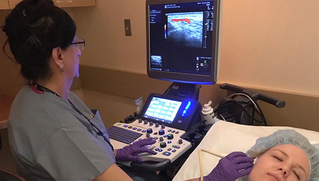
However, directors at these clinics – known as GCA fast-track clinics – caution that the bar can be high for adopting vascular ultrasound (VUS) as a tool to diagnose GCA. Ultrasonographers need specialized training to perform the task and an adequate caseload to maintain their skills. Clinics also need to be outfitted with high-definition ultrasound machines.
“It definitely takes adequate training and learning of how to adjust the settings on the ultrasound machine to be able to visualize the findings appropriately,” said Minna Kohler, MD, director of the rheumatology and musculoskeletal ultrasound program at Massachusetts General Hospital in Boston, which has its own GCA fast-track clinic.

“And the clinical context is very important,” she said. “If you have a high suspicion for someone with temporal arteritis, or GCA, and the patient has been on steroids for weeks before you see them, the ultrasound findings may not show signs of the disease. In those cases in which the imaging is equivocal, we would still pursue a biopsy.”
The idea of the fast-track clinic is as the name implies: to quickly confirm the presence of GCA in a matter of hours, or days at the most, in an outpatient setting without the hassles of a biopsy. Temporal artery biopsy (TAB), by comparison, “is more costly because it requires operating room time, a surgical consultation, and surgery time, whereas ultrasound is a very inexpensive exam since it’s done in the clinic by the rheumatologist,” Dr. Kohler said.
European experience
Use of VUS to diagnose GCA is supplanting TAB in Europe and other countries. In Denmark alone – with a population of 6 million – three outpatient fast-track clinics are operating. The United States, with a population more than 50 times larger than Denmark’s, has six.
Stavros Chrysidis, MD, PhD, chief of rheumatology at the Hospital South West Jutland, one of the fast-track clinic sites in Denmark, led a recent multicenter study, known as EUREKA, of VUS in patients with suspected GCA. He and his colleagues reported in The Lancet Rheumatology that the sensitivity and specificity of VUS was superior to TAB in confirming a diagnosis of GCA. Dr. Chrysidis has instructed U.S. rheumatologists and ultrasonographers in performing and interpreting VUS for GCA.
The study emphasizes the importance of training for ultrasonographers, said Dr. Chrysidis, who regularly performs VUS at his institution. “The most important finding is that, when we apply VUS by systematically trained ultrasonographers using appropriate equipment in appropriate settings, it has excellent diagnostic accuracy on GCA,” he told this news organization.
He noted that The Lancet Rheumatology report is the first multicenter study of VUS for diagnosing GCA in which all the ultrasonographers participated in a standardized training program, which his group developed. “Ultrasound is very operator dependent,” he said. “That’s why the training is very important.”
The training occurred over a year and included two workshops consisting of 5 days of theoretical training on VUS; supervised hands-on evaluation of healthy individuals and patients with known GCA; and evaluation of ultrasound images. Over the course of the year, trainees performed at least 50 VUS evaluations, half of which were in patients with confirmed GCA. During the training period, an external rheumatologist with broad experience in VUS made the final diagnosis.
“The equipment and settings are very important because ultrasound can be very time consuming if you are not educated well and if your equipment is not adjusted well,” Dr. Chrysidis said. The equipment must be calibrated beforehand “so you don’t spend time on adjustments.”
For diagnosing temporal artery anomalies, the ultrasound equipment must have a resolution of 0.3-0.4 mm, he said. “When you have a transducer of 10 MHz, you cannot visualize changes smaller than 5 mm.”
The EUREKA study stated that VUS could replace TAB as a first-line diagnostic tool for GCA – provided the ultrasonographers are systematically trained and the equipment and settings are appropriate. In the Jutland clinic, VUS already has replaced TAB, Dr. Chrysidis said.
“In my department since 2017, when we started the fast-track clinic after the EUREKA study was terminated, we have performed three temporal artery biopsies in the last 4 years, and we screen 60-70 patients per year because we use ultrasound as the primary imaging,” he said. In cases when the results are inconclusive, they order a PET scan. “We don’t perform biopsies anymore,” he said.
U.S. fast-track clinic models
The fast-track clinic models in the United States vary. Results of a survey of the U.S. clinics were presented as an abstract at the 2021 American College of Rheumatology annual meeting. Some centers have a vasculitis specialist obtain and interpret the imaging. At others, a vasculitis specialist refers patients to a VUS-trained rheumatologist to perform and interpret the test. Another approach is to have vasculitis specialists refer patients to ultrasound technicians trained in VUS, with a vascular surgeon interpreting the images and either a VUS-trained rheumatologist or vascular medicine specialist verifying the images.
The take-home message of that survey is that “ultrasound evaluation should be considered in the hands of experts, realizing that not everyone has that skill set, but if it is available, it’s a way to expedite diagnosis and it can be helpful in managing the GCA patient in an appropriate way, quicker than trying to schedule cross-sectional imaging,” said Massachusetts General’s Dr. Kohler, who is a coauthor of the abstract. “Certainly, cross-sectional imaging also plays an important role, but when it comes to confirming whether to continue with treatment or not for a very serious condition, ultrasound is a quick way to get the answer.”
In addition to the fast-track clinic at Massachusetts General, the survey included fast-track clinics at the University of Washington, Seattle; Brigham and Women’s Hospital, Boston; Loma Linda (Calif.) University; University of California, Los Angeles; and at a private practice, Arthritis and Rheumatism Associates in the Washington area.
Advantages of VUS vs. TAB
At Massachusetts General, some of the rheumatologists are trained to perform VUS. The rheumatologists also perform the clinical evaluation of suspected cases of GCA. The advantage of VUS, Dr. Kohler said, is that the answer is “right there”; that is, the imaging yields a diagnosis almost instantaneously whereas a biopsy must be sent to a lab for analysis.
“Since a lot of patients with suspected vasculitis may already come to us on steroid therapy, and if there’s a low probability or low suspicion for vasculitis, [VUS] actually confirms that, and we’re able to taper prednisone or steroids quickly rather than keep them on a prolonged course.”
Alison Bays, MD, MPH, of the department of rheumatology at the University of Washington, said that the advantages of avoiding biopsy aren’t to be overlooked. “Temporal artery biopsies are invasive and carry surgical risks, especially as many of our patients are elderly,” she told this news organization.
“These patients occasionally refuse biopsy, but the acceptance of ultrasound is high,” Dr. Bays said. “Scheduling surgery can be more complicated, resulting in delays to biopsy and potentially higher rates of false negatives. Additionally, ultrasound has resulted in a higher rate of diagnosis with GCA as TAB misses large-vessel involvement.” The fast-track clinic at the university has evaluated 250 patients since it opened in 2017.
Dr. Bays and colleagues published the first United States study of a fast-track protocol using vascular sonographers. “Our group has demonstrated that fast-track clinics can rapidly and effectively evaluate patients, and we demonstrated a different method of evaluation using vascular sonographers rather than rheumatologists to do the vascular ultrasound,” she said. “It utilizes the familiarity vascular sonographers already have with imaging blood vessels.”
She added that the TABUL study in the United Kingdom in 2016 demonstrated that VUS yielded a savings of $686 (£484 as reported in the study), compared with TAB. “Further studies need to be done in the United States,” she said. “Beyond direct comparison of costs, reduction in steroid burden due to quick evaluation and diagnosis may carry additional benefits.”
At Brigham and Women’s Hospital, the division of rheumatology and the vascular section of the cardiology division collaborate on the GCA evaluation, said Sara Tedeschi, MD, MPH, codirector of the fast-track clinic there. Trained vascular technologists credentialed in the procedure and specifically trained in using VUS for evaluating arteritis perform the VUS. Cardiologists with a subspecialty in vascular medicine interpret the studies.

VUS patients have a rheumatology evaluation just before they have the ultrasound. “The rheumatology evaluation is then able to incorporate information from the VUS together with laboratory data, the patient’s history, and physical examination,” Dr. Tedeschi said.
“If the rheumatologist recommends a temporal artery biopsy as a next step, we arrange this with vascular surgery,” she said. “If the rheumatologist recommends other imaging such as MRA [magnetic resonance angiography] or PET-CT, we frequently review the images together with our colleagues in cardiovascular radiology and/or nuclear medicine.”
But in the United States, it will take some time for GCA fast-track clinics to become the standard, Dr. Tedeschi said. “Temporal artery biopsy may be faster to arrange in certain practice settings if VUS is not already being employed for giant cell arteritis evaluation,” she said.
Dr. Bays recognized this limitation, saying, “We are hoping that in the future, the American College of Rheumatology will consider vascular ultrasound as a first-line diagnostic test in diagnosis as rheumatologists and vascular sonographers gain familiarity over time.”
But that would mean that centers performing VUS for GCA would have to meet rigorous standards for the procedure. “With that, standardization of a protocol, high-quality equipment, and adequate training are necessary to ensure quality and reduce the chance of false-positive or false-negative results,” she said.
Dr. Chrysidis, Dr. Bays, and Dr. Tedeschi have disclosed no relevant financial relationships. Dr. Kohler is a board member of Ultrasound School of North American Rheumatologists.
A version of this article first appeared on Medscape.com.
Temporal artery biopsy has been the standard for diagnosing giant cell arteritis (GCA), but vascular ultrasound, a procedure that’s less invasive, less time-intensive, less expensive, and more convenient, has gained widespread use in Europe, and now clinics in the United States are adopting this approach and moving toward having rheumatologists take on the role of ultrasonographer.
However, directors at these clinics – known as GCA fast-track clinics – caution that the bar can be high for adopting vascular ultrasound (VUS) as a tool to diagnose GCA. Ultrasonographers need specialized training to perform the task and an adequate caseload to maintain their skills. Clinics also need to be outfitted with high-definition ultrasound machines.
“It definitely takes adequate training and learning of how to adjust the settings on the ultrasound machine to be able to visualize the findings appropriately,” said Minna Kohler, MD, director of the rheumatology and musculoskeletal ultrasound program at Massachusetts General Hospital in Boston, which has its own GCA fast-track clinic.
“And the clinical context is very important,” she said. “If you have a high suspicion for someone with temporal arteritis, or GCA, and the patient has been on steroids for weeks before you see them, the ultrasound findings may not show signs of the disease. In those cases in which the imaging is equivocal, we would still pursue a biopsy.”
The idea of the fast-track clinic is as the name implies: to quickly confirm the presence of GCA in a matter of hours, or days at the most, in an outpatient setting without the hassles of a biopsy. Temporal artery biopsy (TAB), by comparison, “is more costly because it requires operating room time, a surgical consultation, and surgery time, whereas ultrasound is a very inexpensive exam since it’s done in the clinic by the rheumatologist,” Dr. Kohler said.
European experience
Use of VUS to diagnose GCA is supplanting TAB in Europe and other countries. In Denmark alone – with a population of 6 million – three outpatient fast-track clinics are operating. The United States, with a population more than 50 times larger than Denmark’s, has six.
Stavros Chrysidis, MD, PhD, chief of rheumatology at the Hospital South West Jutland, one of the fast-track clinic sites in Denmark, led a recent multicenter study, known as EUREKA, of VUS in patients with suspected GCA. He and his colleagues reported in The Lancet Rheumatology that the sensitivity and specificity of VUS was superior to TAB in confirming a diagnosis of GCA. Dr. Chrysidis has instructed U.S. rheumatologists and ultrasonographers in performing and interpreting VUS for GCA.
The study emphasizes the importance of training for ultrasonographers, said Dr. Chrysidis, who regularly performs VUS at his institution. “The most important finding is that, when we apply VUS by systematically trained ultrasonographers using appropriate equipment in appropriate settings, it has excellent diagnostic accuracy on GCA,” he told this news organization.
He noted that The Lancet Rheumatology report is the first multicenter study of VUS for diagnosing GCA in which all the ultrasonographers participated in a standardized training program, which his group developed. “Ultrasound is very operator dependent,” he said. “That’s why the training is very important.”
The training occurred over a year and included two workshops consisting of 5 days of theoretical training on VUS; supervised hands-on evaluation of healthy individuals and patients with known GCA; and evaluation of ultrasound images. Over the course of the year, trainees performed at least 50 VUS evaluations, half of which were in patients with confirmed GCA. During the training period, an external rheumatologist with broad experience in VUS made the final diagnosis.
“The equipment and settings are very important because ultrasound can be very time consuming if you are not educated well and if your equipment is not adjusted well,” Dr. Chrysidis said. The equipment must be calibrated beforehand “so you don’t spend time on adjustments.”
For diagnosing temporal artery anomalies, the ultrasound equipment must have a resolution of 0.3-0.4 mm, he said. “When you have a transducer of 10 MHz, you cannot visualize changes smaller than 5 mm.”
The EUREKA study stated that VUS could replace TAB as a first-line diagnostic tool for GCA – provided the ultrasonographers are systematically trained and the equipment and settings are appropriate. In the Jutland clinic, VUS already has replaced TAB, Dr. Chrysidis said.
“In my department since 2017, when we started the fast-track clinic after the EUREKA study was terminated, we have performed three temporal artery biopsies in the last 4 years, and we screen 60-70 patients per year because we use ultrasound as the primary imaging,” he said. In cases when the results are inconclusive, they order a PET scan. “We don’t perform biopsies anymore,” he said.
U.S. fast-track clinic models
The fast-track clinic models in the United States vary. Results of a survey of the U.S. clinics were presented as an abstract at the 2021 American College of Rheumatology annual meeting. Some centers have a vasculitis specialist obtain and interpret the imaging. At others, a vasculitis specialist refers patients to a VUS-trained rheumatologist to perform and interpret the test. Another approach is to have vasculitis specialists refer patients to ultrasound technicians trained in VUS, with a vascular surgeon interpreting the images and either a VUS-trained rheumatologist or vascular medicine specialist verifying the images.
The take-home message of that survey is that “ultrasound evaluation should be considered in the hands of experts, realizing that not everyone has that skill set, but if it is available, it’s a way to expedite diagnosis and it can be helpful in managing the GCA patient in an appropriate way, quicker than trying to schedule cross-sectional imaging,” said Massachusetts General’s Dr. Kohler, who is a coauthor of the abstract. “Certainly, cross-sectional imaging also plays an important role, but when it comes to confirming whether to continue with treatment or not for a very serious condition, ultrasound is a quick way to get the answer.”
In addition to the fast-track clinic at Massachusetts General, the survey included fast-track clinics at the University of Washington, Seattle; Brigham and Women’s Hospital, Boston; Loma Linda (Calif.) University; University of California, Los Angeles; and at a private practice, Arthritis and Rheumatism Associates in the Washington area.
Advantages of VUS vs. TAB
At Massachusetts General, some of the rheumatologists are trained to perform VUS. The rheumatologists also perform the clinical evaluation of suspected cases of GCA. The advantage of VUS, Dr. Kohler said, is that the answer is “right there”; that is, the imaging yields a diagnosis almost instantaneously whereas a biopsy must be sent to a lab for analysis.
“Since a lot of patients with suspected vasculitis may already come to us on steroid therapy, and if there’s a low probability or low suspicion for vasculitis, [VUS] actually confirms that, and we’re able to taper prednisone or steroids quickly rather than keep them on a prolonged course.”
Alison Bays, MD, MPH, of the department of rheumatology at the University of Washington, said that the advantages of avoiding biopsy aren’t to be overlooked. “Temporal artery biopsies are invasive and carry surgical risks, especially as many of our patients are elderly,” she told this news organization.
“These patients occasionally refuse biopsy, but the acceptance of ultrasound is high,” Dr. Bays said. “Scheduling surgery can be more complicated, resulting in delays to biopsy and potentially higher rates of false negatives. Additionally, ultrasound has resulted in a higher rate of diagnosis with GCA as TAB misses large-vessel involvement.” The fast-track clinic at the university has evaluated 250 patients since it opened in 2017.
Dr. Bays and colleagues published the first United States study of a fast-track protocol using vascular sonographers. “Our group has demonstrated that fast-track clinics can rapidly and effectively evaluate patients, and we demonstrated a different method of evaluation using vascular sonographers rather than rheumatologists to do the vascular ultrasound,” she said. “It utilizes the familiarity vascular sonographers already have with imaging blood vessels.”
She added that the TABUL study in the United Kingdom in 2016 demonstrated that VUS yielded a savings of $686 (£484 as reported in the study), compared with TAB. “Further studies need to be done in the United States,” she said. “Beyond direct comparison of costs, reduction in steroid burden due to quick evaluation and diagnosis may carry additional benefits.”
At Brigham and Women’s Hospital, the division of rheumatology and the vascular section of the cardiology division collaborate on the GCA evaluation, said Sara Tedeschi, MD, MPH, codirector of the fast-track clinic there. Trained vascular technologists credentialed in the procedure and specifically trained in using VUS for evaluating arteritis perform the VUS. Cardiologists with a subspecialty in vascular medicine interpret the studies.
VUS patients have a rheumatology evaluation just before they have the ultrasound. “The rheumatology evaluation is then able to incorporate information from the VUS together with laboratory data, the patient’s history, and physical examination,” Dr. Tedeschi said.
“If the rheumatologist recommends a temporal artery biopsy as a next step, we arrange this with vascular surgery,” she said. “If the rheumatologist recommends other imaging such as MRA [magnetic resonance angiography] or PET-CT, we frequently review the images together with our colleagues in cardiovascular radiology and/or nuclear medicine.”
But in the United States, it will take some time for GCA fast-track clinics to become the standard, Dr. Tedeschi said. “Temporal artery biopsy may be faster to arrange in certain practice settings if VUS is not already being employed for giant cell arteritis evaluation,” she said.
Dr. Bays recognized this limitation, saying, “We are hoping that in the future, the American College of Rheumatology will consider vascular ultrasound as a first-line diagnostic test in diagnosis as rheumatologists and vascular sonographers gain familiarity over time.”
But that would mean that centers performing VUS for GCA would have to meet rigorous standards for the procedure. “With that, standardization of a protocol, high-quality equipment, and adequate training are necessary to ensure quality and reduce the chance of false-positive or false-negative results,” she said.
Dr. Chrysidis, Dr. Bays, and Dr. Tedeschi have disclosed no relevant financial relationships. Dr. Kohler is a board member of Ultrasound School of North American Rheumatologists.
A version of this article first appeared on Medscape.com.
Metabolites implicated in CHD development in African Americans
Selected metabolic biomarkers may influence disease risk and progression in African American and White persons in different ways, a cohort study of the landmark Jackson Heart Study has found.

The investigators identified 22 specific metabolites that seem to influence incident CHD risk in African American patients – 13 metabolites that were also replicated in a multiethnic population and 9 novel metabolites that include N-acylamides and leucine, a branched-chain amino acid.
“To our knowledge, this is the first time that an N-acylamide as a class of molecule has been shown to be associated with incident coronary heart disease,” lead study author Daniel E. Cruz, MD, an instructor at Harvard Medical School in the division of cardiovascular medicine at Beth Israel Deaconess Medical Center in Boston, said in an interview.
The researchers analyzed targeted plasma metabolomic profiles of 2,346 participants in the Jackson Heart Study, a prospective population-based cohort study in the Mississippi city that included 5,306 African American patients evaluated over 15 years. They then performed a replication analysis of CHD-associated metabolites among 1,588 multiethnic participants in the Women’s Health Initiative, another population-based cohort study that included 161,808 postmenopausal women, also over 15 years. In all, the study, published in JAMA Cardiology, identified 46 metabolites that were associated with incident CHD up to 16 years before the incident event
Dr. Cruz said the “most interesting” findings were the roles of the N-acylamide linoleoyl ethanolamide and leucine. The former is of interest “because it is a lipid-signaling molecule that has been shown to have anti-inflammatory effects on macrophages; the influence and effects on macrophages are of particular interest because of macrophages’ central role in atherosclerosis and coronary heart disease,” he said.
Leucine draws interest because, in this study population, it was linked to a reduced risk of incident CHD. The researchers cited four previous studies in predominantly non-Hispanic White populations that found no association between branched-chain amino acids and incident CHD in Circulation, Stroke Circulation: Genomic and Precision Medicine, and Atherosclerosis. Other branched-amino acids included in the analysis trended toward a decreased risk of CHD, but those didn’t achieve the same statistical significance as that of leucine, Dr. Cruz said.
“In some of the analyses we did, there was a subset of metabolites that the associations with CHD appeared to be different between self-identified African Americans in the Jackson cohort vs. self-identified non-Hispanic Whites, and leucine was one of them,” Dr. Cruz said.
He emphasized that this study “is not a genetic analysis” because the participants self-identified their race. “So our next step is to figure out why this difference appears between these self-identified groups,” Dr. Cruz said. “We suspect environmental factors play a role – psychological stress, diet, income level, to name a few – but we are also interested to see if there are genetic causes.”
The results “are not clinically applicable,” Dr. Cruz said, but they do point to a need for more ethnically and racially diverse study populations. “The big picture is that, before we go implementing novel biomarkers into clinical practice, we need to make sure that they are accurate across different populations of people,” he said. “The only way to do this is to study different groups with the same rigor and vigor and thoughtfulness as any other group.”
These findings fall in line with other studies that found other nonmetabolomic biomarkers have countervailing effects on CHD risk in African Americans and non-Hispanic Whites, said Christie M. Ballantyne, MD, chief of cardiology at Baylor College of Medicine in Houston. For example, African Americans have been found to have lower triglyceride and HDL cholesterol levels than those of Whites.
The study “points out that there may be important biological differences in the metabolic pathways and abnormalities in the development of CHD between races,” Dr. Ballantyne said. “This further emphasizes both the importance and challenge of testing therapies in multiple racial/ethnic groups and with more even representation between men and women.”
Combining metabolomic profiling along with other biomarkers and possibly genetics may be helpful to “personalize” therapies in the future, he added.
Dr. Cruz and Dr. Ballantyne have no relevant relationships to disclose.
Selected metabolic biomarkers may influence disease risk and progression in African American and White persons in different ways, a cohort study of the landmark Jackson Heart Study has found.
The investigators identified 22 specific metabolites that seem to influence incident CHD risk in African American patients – 13 metabolites that were also replicated in a multiethnic population and 9 novel metabolites that include N-acylamides and leucine, a branched-chain amino acid.
“To our knowledge, this is the first time that an N-acylamide as a class of molecule has been shown to be associated with incident coronary heart disease,” lead study author Daniel E. Cruz, MD, an instructor at Harvard Medical School in the division of cardiovascular medicine at Beth Israel Deaconess Medical Center in Boston, said in an interview.
The researchers analyzed targeted plasma metabolomic profiles of 2,346 participants in the Jackson Heart Study, a prospective population-based cohort study in the Mississippi city that included 5,306 African American patients evaluated over 15 years. They then performed a replication analysis of CHD-associated metabolites among 1,588 multiethnic participants in the Women’s Health Initiative, another population-based cohort study that included 161,808 postmenopausal women, also over 15 years. In all, the study, published in JAMA Cardiology, identified 46 metabolites that were associated with incident CHD up to 16 years before the incident event
Dr. Cruz said the “most interesting” findings were the roles of the N-acylamide linoleoyl ethanolamide and leucine. The former is of interest “because it is a lipid-signaling molecule that has been shown to have anti-inflammatory effects on macrophages; the influence and effects on macrophages are of particular interest because of macrophages’ central role in atherosclerosis and coronary heart disease,” he said.
Leucine draws interest because, in this study population, it was linked to a reduced risk of incident CHD. The researchers cited four previous studies in predominantly non-Hispanic White populations that found no association between branched-chain amino acids and incident CHD in Circulation, Stroke Circulation: Genomic and Precision Medicine, and Atherosclerosis. Other branched-amino acids included in the analysis trended toward a decreased risk of CHD, but those didn’t achieve the same statistical significance as that of leucine, Dr. Cruz said.
“In some of the analyses we did, there was a subset of metabolites that the associations with CHD appeared to be different between self-identified African Americans in the Jackson cohort vs. self-identified non-Hispanic Whites, and leucine was one of them,” Dr. Cruz said.
He emphasized that this study “is not a genetic analysis” because the participants self-identified their race. “So our next step is to figure out why this difference appears between these self-identified groups,” Dr. Cruz said. “We suspect environmental factors play a role – psychological stress, diet, income level, to name a few – but we are also interested to see if there are genetic causes.”
The results “are not clinically applicable,” Dr. Cruz said, but they do point to a need for more ethnically and racially diverse study populations. “The big picture is that, before we go implementing novel biomarkers into clinical practice, we need to make sure that they are accurate across different populations of people,” he said. “The only way to do this is to study different groups with the same rigor and vigor and thoughtfulness as any other group.”
These findings fall in line with other studies that found other nonmetabolomic biomarkers have countervailing effects on CHD risk in African Americans and non-Hispanic Whites, said Christie M. Ballantyne, MD, chief of cardiology at Baylor College of Medicine in Houston. For example, African Americans have been found to have lower triglyceride and HDL cholesterol levels than those of Whites.
The study “points out that there may be important biological differences in the metabolic pathways and abnormalities in the development of CHD between races,” Dr. Ballantyne said. “This further emphasizes both the importance and challenge of testing therapies in multiple racial/ethnic groups and with more even representation between men and women.”
Combining metabolomic profiling along with other biomarkers and possibly genetics may be helpful to “personalize” therapies in the future, he added.
Dr. Cruz and Dr. Ballantyne have no relevant relationships to disclose.
FROM JAMA CARDIOLOGY
Sleep disorders and cancer: It’s complicated
Sleep apnea and other types of sleep disorders appear to elevate the risk for some types of cancer, specifically prostate cancer, more so than others. But the overall risk can be highly variable, and some sleep problems were found to be associated with a lower risk for cancer and cancer-related death, an analysis of a large observational cohort study of cardiovascular patients found.
Results of the analysis were published online in the journal Cancer Epidemiology. Investigators analyzed the presence of sleep apnea and insomnia and cancer risk in more than 8,500 patients in the Cardiovascular Health Study (CHS). “The fact that we observed certain sleep problems, like apneas, to be associated with elevated risk of some cancers but not others reflects the fact that cancer is a heterogeneous disease,” senior author Amanda Phipps, PhD, said in an interview. Dr. Phipps is an associate professor of epidemiology at the University of Washington and the Fred Hutchinson Cancer Research Center, both in Seattle.
Variable cancer links
The researchers assessed sleep problems in two groups in the CHS: an incident cancer group of 3,930 patients and a cancer mortality group of 4,580 patients. Within those respective groups, the investigators identified 885 first-incident cancers and 804 cancer deaths with a median follow-up of 12 and 14 years. The average age of the study population was 73 years, and 57% were women.
Sleep apnea symptoms (SAS) were associated with a lower risk for incident cancers – a 16% lower baseline risk and a 24% lower time-dependent risk. The study showed no association between cancer incidence and daytime sleepiness and apneas.
However, there was a significantly elevated risk relationship between sleep problems and prostate cancer. A time-dependent analysis of apnea showed more than double the risk (hazard ratio, 2.34), and baseline snoring carried a 69% greater risk. There was also a dose-response relationship for baseline cumulative SAS, compared with not having symptoms: an HR of 1.30 for one symptom, and 2.22 for two or more symptoms.
Risks for lymphatic or hematopoietic cancers were also associated with baseline daytime sleepiness (HR, 1.81), but not with insomnia (HR, 0.54).
With regard to cancer mortality, the study found no relationship between sleep problems and cancer death. In fact, it found an overall inverse relationship with snoring (time-dependent HR, 0.73; cumulative average HR, 0.67) and baseline apnea (HR, 0.69). Likewise, patients reporting SAS had lower risks than those having no SAS: an HR of 0.90 for one symptom and 0.75 for multiple symptoms. No relationships were found between any insomnia symptom and cancer death.
“We know the pathways that lead to prostate cancer can be very different than the pathways that lead to colorectal cancer,” Dr. Phipps said. “What we don’t yet understand is why these associations differ or what mechanisms are responsible for these cancer site-specific associations.”
Need for sleep assessment
The findings don’t change much for how clinicians should evaluate cancer risks in patients with sleep problems, Dr. Phipps said. “Other studies have clearly demonstrated the implications that sleep apnea has for a variety of other important health conditions – such as cardiovascular disease – so there are already plenty of good reasons for clinicians to ask their patients about their sleep and to connect patients with resources for the diagnosis and treatment of sleep apnea,” she added. “This study provides another possible reason.”
These findings provide context for future studies of the relationship between sleep problems and cancer. “But, given that sleep is something we all do and given that sleep problems are so pervasive, it’s important that we keep trying to better understand this relationship,” Dr. Phipps said.
“My hope is that future cancer studies will build in more detailed, longitudinal information on sleep patterns to help us fill current gaps in knowledge.”
Dr. Phipps has disclosed no relevant financial relationships.
A version of this article first appeared on Medscape.com.
FROM CANCER EPIDEMIOLOGY
Low-dose rituximab may keep RA disease activity low in responders
Rituximab doses as low as 200 mg reduced disease activity in patients with rheumatoid arthritis to an extent that’s similar to the standard 1,000-mg dose during more than 3 years of follow-up, according to results from an extension study of a clinical trial in the Netherlands.
“We could not formally statistically show that the lower doses were less effective than the higher dose,” study leader Nathan den Broeder, MSc, a PhD candidate at St. Maarten Clinic and the Radboud Institute for Health Sciences in Nijmegen, the Netherlands, said in a presentation at the virtual annual meeting of the American College of Rheumatology. “We concluded at this time that only 6% of patients needed to switch to another biologic or targeted disease-modifying antirheumatic drug and that mean disease activity remained very low,” he said of the patients treated with 200- and 500-mg doses of rituximab.
The extension study included 118 of 142 patients in the REDO trial, following them from the start of the trial in 2017-2018 through April 2021. They were randomized to three treatment arms: the standard 1,000-mg dose (24 patients), a 500-mg dose (n = 48), and the 200-mg dose (n = 46). The mean follow-up was 3.2 years.
The study evaluated disease activity by mean Disease Activity Score in 28 joints with C-reactive protein (DAS28-CRP), which during follow-up were 2.2-2.3 in the groups. Seven patients in the study cohort switched to a different DMARD, he said.
“On average, we saw a DAS28-CRP 0.15 points lower per 1,000 mg rituximab used in the past year,” Mr. den Broeder said in an interview. “For context, this is compared to a measurement error of about 0.6. A good response to a drug would be a reduction in DAS28-CRP of 1.2. This means we can just about get an effect that is bigger than the measurement error if we compare the highest dose in our study to the lowest one.”
After a year, the median yearly rituximab dose was 978 mg, with an interquartile range of 704-1,425 mg. At the end of the study, 31% of patients took 200 mg every 6 months, 40% took 500 mg every 6.2 months, and 29% took 1,000 mg every 6.4 months.
“It’s important to note, though, this is in a situation where patients are given a dose based on disease activity,” Mr. den Broeder said. “That is, we try one dose; if the patient does well, we try a lower one; if not, we might go back up to a higher dose. We could expect somewhat larger differences if all patients were to be switched to a lower dose, regardless of whether that works well for them.”
The results were achieved without a high reliance on glucocorticoids (GCs), he said. Use of comedication in the extension study population was 0.38 GC injections per patient-year and starting or increasing an oral GC occurred at a rate of only 0.05 per patient-year.
“As a result of this study, we are now implementing a strategy of rituximab dose reduction in clinical practice at our center,” Mr. den Broeder said. Patients with RA start on a 1,000-mg dose for 6 months, and if they respond well they’re put on a 500-mg dose. If they respond well after 6 months on the 500-mg dose, they’re then moved to the 200-mg dose. “With this, we hope to gain that patients have fewer side effects,” he said. “We hope to reduce the cost of treatment, and also, what we instantly gain is that the infusion time for patients is also reduced.”
Future research considerations include evaluating the 200-mg dose as a subcutaneous injection. “Another thing you might think of as well: Are even lower doses possible?” he said.
Session moderator Maya Buch, MD, professor of rheumatology and director of Experimental Medicine at the Centre for Musculoskeletal Research at the University of Manchester (England), asked if the investigators used CD19 testing to measure B-cell levels or immunoglobulin G levels to determine dose escalation.
Mr. den Broeder said that CD19 wasn’t used in clinical practice but was used in the original trial. However, it wasn’t found to have any predictive ability, while immunoglobulin G levels were measured in patients who had multiple infections. “Sporadically, a lower dose might have been initiated because of that, but not systematically,” he said.
Mr. den Broeder had no relevant relationships to disclose. Dr. Buch reported financial relationships with AbbVie, Eli Lilly, Gilead Sciences, Merck-Serono, Pfizer, Roche, Sanofi, and UCB.
FROM ACR 2021
Tofacitinib postmarketing trial data shed light on JAK inhibitor risks
Additional analyses of a postmarketing trial that was required after the Food and Drug Administration’s approval of the Janus kinase inhibitor tofacitinib (Xeljanz, Xeljanz XR) has identified characteristics of older patients with rheumatoid arthritis with at least one cardiovascular risk factor who may be at higher risk for major adverse cardiovascular events (MACE) when taking the drug.
Results from the phase 3b/4 ORAL Surveillance trial presented at the virtual annual meeting of the American College of Rheumatology show that people taking tofacitinib for RA with at least one cardiovascular (CV) risk factor had a nonsignificant higher risk for MACE than did people taking tumor necrosis factor inhibitors (TNFi), with the risk from tofacitinib more pronounced in current smokers, aspirin users, people older than 65 years, and men, compared with women.
“It is the first large, randomized safety study of active RA patients with increased CV risk comparing tofacitinib to TNF inhibition,” study author Christina Charles-Schoeman, MD, said in an interview. “These data emphasize the importance of assessing baseline CV risk when treating patients with RA.” Dr. Charles-Schoeman is chief of rheumatology at the University of California, Los Angeles.
The results shed further light on the trial’s findings, which the FDA used in September 2021 to mandate boxed warnings about the risk of MI or stroke, cancer, venous thromboembolism, and death, as well as updated indications, for tofacitinib and other JAK inhibitors baricitinib (Olumiant) and upadacitinib (Rinvoq). The FDA limited all approved uses of these three medications to patients who have not responded well to TNFi to ensure their benefits outweigh their risks.
Tofacitinib is indicated for RA, psoriatic arthritis, ulcerative colitis, and polyarticular course juvenile idiopathic arthritis. Baricitinib and upadacitinib are approved only for RA.

While the overall results of the trial results show nonsignificant increased incidence rates for MACE in tofacitinib users versus TNFI users, Katherine Liao, MD, a rheumatologist at Brigham and Women’s Hospital in Boston, noted that more information is needed to determine who is at greatest risk. “Another thing to keep in mind is, while there was evidence of an elevated relative risk for MACE, compared to TNFi, the absolute risk, based on the numbers what we know so far, is small,” she said.
The trial compared two different doses of tofacitinib – 5 mg (1,455 patients) and 10 mg (n = 1,456) twice daily – and TNFi (n = 1,451) in people with moderate to severe RA over age 50. Patient characteristics were similar across all three treatment arms, Dr. Charles-Schoeman said. All patients had inadequate response to methotrexate, and about 57% in all three treatment groups were taking corticosteroids. The 10-mg tofacitinib patients switched to the 5-mg dose in February 2019 but represent the 10-mg group in the study analysis.
ORAL Surveillance demonstrated a 24% greater risk of MACE in the 5-mg tofacitinib patients and a 43% heightened risk the 10-mg group, compared with patients who received a TNFi.
The differentiating factor for MACE incidence was MI. The higher- and lower-dose tofacitinib groups had 69% and 80% greater risk for MI. While the risk for fatal MI were similar across all three treatment groups, the risk for nonfatal MI were more than doubled in the respective tofacitinib groups: hazard ratios of 2.32 and 2.08. The incidence of stroke was similar across all three arms, Dr. Charles-Schoeman said.
The study identified a number of baseline characteristics as independent overall risk factors for MACE across all treatment groups. Current smoking and aspirin use more than doubled the risk (HR, 2.18; P < .0001 and HR, 2.11; P = .004, respectively), while age greater than 65 years and male sex approached that level (HR, 1.81; P = .0011 and HR, 1.81; P = .0015) approached that level. Other factors that elevated the risk of MACE to a lesser extent were a history of diabetes, hypertension or coronary artery procedures, and a total cholesterol to HDL ratio greater than4.
Other ORAL Surveillance subanalyses and tofacitinib real-world data reported
This was one of several analyses presented at ACR 2021 that compared adverse event risks for tofacitinib versus TNFi drugs. A separate analysis of claims data from patients with RA in two U.S. insurance databases plus Medicare found a statistically nonsignificant increased risk of adverse CV outcomes (MI or stroke) with tofacitinib, compared with TNFi users, among patients who met the same inclusion and exclusion criteria of the ORAL Surveillance trial but not in a “real-world evidence” cohort of more than 102,000 patients with RA in routine care from the databases.
Two additional ORAL Surveillance analyses presented at ACR 2021 gave details about risk factors for higher rates of malignancies and venous thromboembolic events found in patients taking tofacitinib with at least one CV risk factor. As would be expected, older age (≥65 vs. 50-64 years) and current or past smoking (vs. never smoking) were independent risk factors for higher malignancy rates across all treatment arms. Pulmonary embolism events across treatment groups were independently associated with a history of venous thromboembolism, baseline use of oral contraceptives or hormone replacement therapy, baseline body mass index of at least 30 kg/m2, age 65 or older, and history of hypertension.
The ORAL Surveillance findings are worth considering when determining treatments for RA patients with CV risk factors, Dr. Charles-Schoeman said. “Tofacitinib remains an effective RA treatment,” she said. “The choice of specific RA treatment for any patient remains an individual decision between the patient and physician, which is decided based on a number of different factors. This new study provides additional information regarding both tofacitinib as well as traditional CV risk factors for discussion with the patient.”
The ORAL Surveillance results may give rheumatologists reason to rethink use of tofacitinib in some patients with CV risk, said Dr. Liao of Brigham and Women’s Hospital in Boston. “Currently, we have limited data and are still awaiting a report of the full trial results,” she said in an interview. “Based on the data available, I can think of a few patients in my clinic where I would reconsider use of these drugs, i.e., history of heart attack with stable angina, especially if there are other options.” However, she noted that many patients on tofacitinib have already failed on older treatments.

These data emphasize the importance of addressing CV risk with patients, said Brittany N. Weber, MD, PhD, a cardio-rheumatologist at Brigham and Women’s Hospital who works with Dr. Liao. “It is also an opportunity to discuss modification of risk factors and to discuss primary prevention therapies, such as statin therapy, where appropriate,” she added. “Based on the individual’s cardiovascular risk, there may be a role for further risk stratification to further understand an individual’s risk, which can also inform primary prevention cardiovascular therapies and help guide these discussions.” Risk stratification could include cardiac CT for calcium scoring or cardiac coronary CT angiography for determining atherosclerotic burden.
The study was sponsored by Pfizer. Dr. Charles-Schoeman disclosed relationships with AbbVie, Bristol-Myers Squibb, Gilead Sciences, Pfizer, and Regeneron-Sanofi. Dr. Liao and Dr. Weber have no relevant disclosures.
Additional analyses of a postmarketing trial that was required after the Food and Drug Administration’s approval of the Janus kinase inhibitor tofacitinib (Xeljanz, Xeljanz XR) has identified characteristics of older patients with rheumatoid arthritis with at least one cardiovascular risk factor who may be at higher risk for major adverse cardiovascular events (MACE) when taking the drug.
Results from the phase 3b/4 ORAL Surveillance trial presented at the virtual annual meeting of the American College of Rheumatology show that people taking tofacitinib for RA with at least one cardiovascular (CV) risk factor had a nonsignificant higher risk for MACE than did people taking tumor necrosis factor inhibitors (TNFi), with the risk from tofacitinib more pronounced in current smokers, aspirin users, people older than 65 years, and men, compared with women.
“It is the first large, randomized safety study of active RA patients with increased CV risk comparing tofacitinib to TNF inhibition,” study author Christina Charles-Schoeman, MD, said in an interview. “These data emphasize the importance of assessing baseline CV risk when treating patients with RA.” Dr. Charles-Schoeman is chief of rheumatology at the University of California, Los Angeles.
The results shed further light on the trial’s findings, which the FDA used in September 2021 to mandate boxed warnings about the risk of MI or stroke, cancer, venous thromboembolism, and death, as well as updated indications, for tofacitinib and other JAK inhibitors baricitinib (Olumiant) and upadacitinib (Rinvoq). The FDA limited all approved uses of these three medications to patients who have not responded well to TNFi to ensure their benefits outweigh their risks.
Tofacitinib is indicated for RA, psoriatic arthritis, ulcerative colitis, and polyarticular course juvenile idiopathic arthritis. Baricitinib and upadacitinib are approved only for RA.
While the overall results of the trial results show nonsignificant increased incidence rates for MACE in tofacitinib users versus TNFI users, Katherine Liao, MD, a rheumatologist at Brigham and Women’s Hospital in Boston, noted that more information is needed to determine who is at greatest risk. “Another thing to keep in mind is, while there was evidence of an elevated relative risk for MACE, compared to TNFi, the absolute risk, based on the numbers what we know so far, is small,” she said.
The trial compared two different doses of tofacitinib – 5 mg (1,455 patients) and 10 mg (n = 1,456) twice daily – and TNFi (n = 1,451) in people with moderate to severe RA over age 50. Patient characteristics were similar across all three treatment arms, Dr. Charles-Schoeman said. All patients had inadequate response to methotrexate, and about 57% in all three treatment groups were taking corticosteroids. The 10-mg tofacitinib patients switched to the 5-mg dose in February 2019 but represent the 10-mg group in the study analysis.
ORAL Surveillance demonstrated a 24% greater risk of MACE in the 5-mg tofacitinib patients and a 43% heightened risk the 10-mg group, compared with patients who received a TNFi.
The differentiating factor for MACE incidence was MI. The higher- and lower-dose tofacitinib groups had 69% and 80% greater risk for MI. While the risk for fatal MI were similar across all three treatment groups, the risk for nonfatal MI were more than doubled in the respective tofacitinib groups: hazard ratios of 2.32 and 2.08. The incidence of stroke was similar across all three arms, Dr. Charles-Schoeman said.
The study identified a number of baseline characteristics as independent overall risk factors for MACE across all treatment groups. Current smoking and aspirin use more than doubled the risk (HR, 2.18; P < .0001 and HR, 2.11; P = .004, respectively), while age greater than 65 years and male sex approached that level (HR, 1.81; P = .0011 and HR, 1.81; P = .0015) approached that level. Other factors that elevated the risk of MACE to a lesser extent were a history of diabetes, hypertension or coronary artery procedures, and a total cholesterol to HDL ratio greater than4.
Other ORAL Surveillance subanalyses and tofacitinib real-world data reported
This was one of several analyses presented at ACR 2021 that compared adverse event risks for tofacitinib versus TNFi drugs. A separate analysis of claims data from patients with RA in two U.S. insurance databases plus Medicare found a statistically nonsignificant increased risk of adverse CV outcomes (MI or stroke) with tofacitinib, compared with TNFi users, among patients who met the same inclusion and exclusion criteria of the ORAL Surveillance trial but not in a “real-world evidence” cohort of more than 102,000 patients with RA in routine care from the databases.
Two additional ORAL Surveillance analyses presented at ACR 2021 gave details about risk factors for higher rates of malignancies and venous thromboembolic events found in patients taking tofacitinib with at least one CV risk factor. As would be expected, older age (≥65 vs. 50-64 years) and current or past smoking (vs. never smoking) were independent risk factors for higher malignancy rates across all treatment arms. Pulmonary embolism events across treatment groups were independently associated with a history of venous thromboembolism, baseline use of oral contraceptives or hormone replacement therapy, baseline body mass index of at least 30 kg/m2, age 65 or older, and history of hypertension.
The ORAL Surveillance findings are worth considering when determining treatments for RA patients with CV risk factors, Dr. Charles-Schoeman said. “Tofacitinib remains an effective RA treatment,” she said. “The choice of specific RA treatment for any patient remains an individual decision between the patient and physician, which is decided based on a number of different factors. This new study provides additional information regarding both tofacitinib as well as traditional CV risk factors for discussion with the patient.”
The ORAL Surveillance results may give rheumatologists reason to rethink use of tofacitinib in some patients with CV risk, said Dr. Liao of Brigham and Women’s Hospital in Boston. “Currently, we have limited data and are still awaiting a report of the full trial results,” she said in an interview. “Based on the data available, I can think of a few patients in my clinic where I would reconsider use of these drugs, i.e., history of heart attack with stable angina, especially if there are other options.” However, she noted that many patients on tofacitinib have already failed on older treatments.
These data emphasize the importance of addressing CV risk with patients, said Brittany N. Weber, MD, PhD, a cardio-rheumatologist at Brigham and Women’s Hospital who works with Dr. Liao. “It is also an opportunity to discuss modification of risk factors and to discuss primary prevention therapies, such as statin therapy, where appropriate,” she added. “Based on the individual’s cardiovascular risk, there may be a role for further risk stratification to further understand an individual’s risk, which can also inform primary prevention cardiovascular therapies and help guide these discussions.” Risk stratification could include cardiac CT for calcium scoring or cardiac coronary CT angiography for determining atherosclerotic burden.
The study was sponsored by Pfizer. Dr. Charles-Schoeman disclosed relationships with AbbVie, Bristol-Myers Squibb, Gilead Sciences, Pfizer, and Regeneron-Sanofi. Dr. Liao and Dr. Weber have no relevant disclosures.
FROM ACR 2021
For EGPA vasculitis, rituximab comparable with cyclophosphamide for inducing remission
Rituximab didn’t perform any worse or better at inducing remission after a year in patients with eosinophilic granulomatosis with polyangiitis (EGPA) than did conventional treatment with cyclophosphamide in the phase 3 REOVAS trial conducted in France.

The B-cell–depleting agent also had a safety profile similar to cyclophosphamide during that window of time, Benjamin Terrier, MD, PhD, said in presenting results of the trial at the virtual annual meeting of the American College of Rheumatology. He is a professor of rheumatology at Cochin Hospital in Paris, the University of Paris, and vice president of the French Vasculitis Study Group.
“So some of the major adverse events that we can consider with this treatment, especially with cyclophosphamide and specifically the fertility issues and the cancer issues, the follow-up of this study does not allow us to evaluate the potential benefit of rituximab over cyclophosphamide,” Dr. Terrier said in an interview.
“But on the short-term study of 1 year, for which the major adverse events are infections, there is almost no difference between the two treatments,” he said. A total of 11 patients taking rituximab and 9 patients taking cyclophosphamide had infections in the study period.
The REOVAS trial randomly assigned 105 adult patients with EPGA, otherwise known as Churg-Strauss syndrome, to either rituximab (52 patients) or the conventional strategy using cyclophosphamide (53). The patients had either newly diagnosed or relapsing disease. There was no significant differences in average daily glucocorticoid use between the two arms.
Patient characteristics were similar in both arms, including the percentages of patients with severe disease. Overall, 60% in each group had a Five-Factor Score (FFS) of 0, while the remainder had an FFS greater than 1. The FFS is calculated by giving 1 point for the presence of each of the following: proteinuria greater than 1 g/day, serum creatinine greater than 140 micromol/L, GI involvement, cardiomyopathy, and CNS involvement. The presence of each FFS component was similar between the groups at baseline, although 25% of the rituximab group and 30.2% of the conventional arm had myeloperoxidase-positive antineutrophilic cytoplasmic antibodies, and the absolute eosinophil count was 180 per mm3 in the former and 300 per mm3 in the latter.
Treatment outcomes at 6 months and 1 year were similar in both groups. At 1 year, remission occurred in 59.6% with rituximab and 64.2% with cyclophosphamide, Birmingham Vasculitis Activity Score equaled 0 in 85.7% with rituximab and 86.5% with cyclophosphamide, and prednisone dose was less than 7.5 mg/day in 77.1% with rituximab and 71.2% with cyclophosphamide.
The cumulative total weeks of complete remission was about 16 weeks in each group. Relapse-free survival, prednisone dosage, quality of life, and disease sequelae patterns also were similar.
Tailor treatment choice to the patient
Despite the equivocal findings between the two treatments, Dr. Terrier said there may be individual patients for whom rituximab would be preferred to cyclophosphamide. “It’s not dependent on the disease by itself but more on the patients we want to treat,” he said. “And clearly if there is a young female patient who wants to get pregnant, knowing that rituximab is not superior but does not appear to be inferior to cyclophosphamide can be interesting.”
The researchers opted to test the superiority rather than noninferiority of rituximab versus cyclophosphamide because a noninferiority design would have required a larger patient population, “which is not possible for a disease like this one,” he said.
“Ostensibly, [the trial] failed to show superiority, but given its favorable side-effect profile, it may be sufficient to show it’s more or less the same,” Michael Putman, MD, MSc, an associate professor and director of the vasculitis program at the Medical College of Wisconsin, Milwaukee, said in an interview. However, he noted that the study didn’t include some phenotypes seen in patients with EGPA, “in particular patients with neurological involvement.”
Dr. Putman added that the French REOVAS findings support recommendations in the 2021 ACR/Vasculitis Foundation guideline for using rituximab as a possible first-line agent to induce remission in severe granulomatosis with polyangiitis and microscopic polyangiitis. “I was somewhat surprised by that,” he said of the ACR/VF recommendation. “These are the best data to date comparing rituximab to cyclophosphamide in EGPA. I would feel more comfortable using rituximab in cases where previously I would have reached for cyclophosphamide.”
He also concurred with Dr. Terrier’s comment that rituximab may be preferred in people of child-bearing age, even extending that to men. “Cyclophosphamide can be quite toxic in terms of ovarian toxicity and premature ovarian failure,” he said. “Young men or women of child-bearing age are a group in whom I would strongly consider using rituximab.”
The French REOVAS investigators’ future research into rituximab for EGPA will include long-term follow-up for relapse and the induction of remission, Dr. Terrier said.
Dr. Terrier disclosed relationships with Roche, Chugai, Vifor, LFB, Grifols, GlaxoSmithKline, AstraZeneca, Bristol-Myers Squibb, Octapharma, and Janssen. Dr. Putnam has no relevant relationships to disclose
Rituximab didn’t perform any worse or better at inducing remission after a year in patients with eosinophilic granulomatosis with polyangiitis (EGPA) than did conventional treatment with cyclophosphamide in the phase 3 REOVAS trial conducted in France.
The B-cell–depleting agent also had a safety profile similar to cyclophosphamide during that window of time, Benjamin Terrier, MD, PhD, said in presenting results of the trial at the virtual annual meeting of the American College of Rheumatology. He is a professor of rheumatology at Cochin Hospital in Paris, the University of Paris, and vice president of the French Vasculitis Study Group.
“So some of the major adverse events that we can consider with this treatment, especially with cyclophosphamide and specifically the fertility issues and the cancer issues, the follow-up of this study does not allow us to evaluate the potential benefit of rituximab over cyclophosphamide,” Dr. Terrier said in an interview.
“But on the short-term study of 1 year, for which the major adverse events are infections, there is almost no difference between the two treatments,” he said. A total of 11 patients taking rituximab and 9 patients taking cyclophosphamide had infections in the study period.
The REOVAS trial randomly assigned 105 adult patients with EPGA, otherwise known as Churg-Strauss syndrome, to either rituximab (52 patients) or the conventional strategy using cyclophosphamide (53). The patients had either newly diagnosed or relapsing disease. There was no significant differences in average daily glucocorticoid use between the two arms.
Patient characteristics were similar in both arms, including the percentages of patients with severe disease. Overall, 60% in each group had a Five-Factor Score (FFS) of 0, while the remainder had an FFS greater than 1. The FFS is calculated by giving 1 point for the presence of each of the following: proteinuria greater than 1 g/day, serum creatinine greater than 140 micromol/L, GI involvement, cardiomyopathy, and CNS involvement. The presence of each FFS component was similar between the groups at baseline, although 25% of the rituximab group and 30.2% of the conventional arm had myeloperoxidase-positive antineutrophilic cytoplasmic antibodies, and the absolute eosinophil count was 180 per mm3 in the former and 300 per mm3 in the latter.
Treatment outcomes at 6 months and 1 year were similar in both groups. At 1 year, remission occurred in 59.6% with rituximab and 64.2% with cyclophosphamide, Birmingham Vasculitis Activity Score equaled 0 in 85.7% with rituximab and 86.5% with cyclophosphamide, and prednisone dose was less than 7.5 mg/day in 77.1% with rituximab and 71.2% with cyclophosphamide.
The cumulative total weeks of complete remission was about 16 weeks in each group. Relapse-free survival, prednisone dosage, quality of life, and disease sequelae patterns also were similar.
Tailor treatment choice to the patient
Despite the equivocal findings between the two treatments, Dr. Terrier said there may be individual patients for whom rituximab would be preferred to cyclophosphamide. “It’s not dependent on the disease by itself but more on the patients we want to treat,” he said. “And clearly if there is a young female patient who wants to get pregnant, knowing that rituximab is not superior but does not appear to be inferior to cyclophosphamide can be interesting.”
The researchers opted to test the superiority rather than noninferiority of rituximab versus cyclophosphamide because a noninferiority design would have required a larger patient population, “which is not possible for a disease like this one,” he said.
“Ostensibly, [the trial] failed to show superiority, but given its favorable side-effect profile, it may be sufficient to show it’s more or less the same,” Michael Putman, MD, MSc, an associate professor and director of the vasculitis program at the Medical College of Wisconsin, Milwaukee, said in an interview. However, he noted that the study didn’t include some phenotypes seen in patients with EGPA, “in particular patients with neurological involvement.”
Dr. Putman added that the French REOVAS findings support recommendations in the 2021 ACR/Vasculitis Foundation guideline for using rituximab as a possible first-line agent to induce remission in severe granulomatosis with polyangiitis and microscopic polyangiitis. “I was somewhat surprised by that,” he said of the ACR/VF recommendation. “These are the best data to date comparing rituximab to cyclophosphamide in EGPA. I would feel more comfortable using rituximab in cases where previously I would have reached for cyclophosphamide.”
He also concurred with Dr. Terrier’s comment that rituximab may be preferred in people of child-bearing age, even extending that to men. “Cyclophosphamide can be quite toxic in terms of ovarian toxicity and premature ovarian failure,” he said. “Young men or women of child-bearing age are a group in whom I would strongly consider using rituximab.”
The French REOVAS investigators’ future research into rituximab for EGPA will include long-term follow-up for relapse and the induction of remission, Dr. Terrier said.
Dr. Terrier disclosed relationships with Roche, Chugai, Vifor, LFB, Grifols, GlaxoSmithKline, AstraZeneca, Bristol-Myers Squibb, Octapharma, and Janssen. Dr. Putnam has no relevant relationships to disclose
FROM ACR 2021
mRNA COVID vaccine response found mostly robust in RA, SLE patients
Immunosuppressed patients with autoimmune diseases who received the Moderna mRNA-1273 SARS-CoV-2 two-dose vaccine series had a frequency of adverse events similar to the general population albeit with a somewhat reduced, but still significant, antibody response with no severe vaccine-related disease flares, results of a prospective, nonrandomized open-label comparative trial in Canada demonstrated.
At the same time, patients with RA who were taking rituximab and patients with systemic lupus erythematosus (SLE) who were taking mycophenolate mofetil seemed to have reduced humoral responses after receiving the vaccine, said Ines Colmegna, MD, reporting results of the COVID-19 Vaccine in Immunosuppressed Adults with Autoimmune Disease (COVIAAD) study as a late-breaking poster abstract at the virtual annual meeting of the American College of Rheumatology. Dr. Colmegna is an associate professor of rheumatology in the division of experimental medicine at McGill University, Montreal.
“The frequency of adverse events, specifically the reactogenicity in people with comorbid conditions regardless of their diagnosis, was similar to healthy controls in this study, and their frequency was similar also the initial studies in the general population,” Dr. Colmegna said.
COVIAAD prospectively enrolled 220 fully vaccinated patients, 162 with rheumatic disease (131 with RA, 23 with SLE, and 8 with other diseases) and 58 controls. Adverse events a week and a month after each dose was the primary outcome. The postvaccine presence of the IgG antibody against the SARS-CoV-2 spike protein and the receptor binding domain (IgG-RBD) was the secondary outcome. Dr. Colmegna said that the study will continue evaluating participants after they get a third dose.
The Canadian trial appears to validate the ACR’s COVID-19 vaccine guidance, the fourth version of which was issued in October, said Jeffrey Curtis, MD, MS, MPH, professor of immunology and rheumatology at the University of Alabama at Birmingham and lead of the ACR COVID-19 Vaccine Guidance Task Force. Specifically, the guidance recommends that patients on rituximab or other anti-CD20 B-cell–depleting agents discuss vaccine timing with their rheumatologist.
“A few things changed over time when there was a paucity of evidence for any vaccine, but as time has gone on, mostly we were more correct than we weren’t,” Dr. Curtis said of the task force’s work. “The evidence that now is in this poster with regard to systemic lupus erythematosus and mycophenolate mofetil is [that] you have impaired vaccine response. If you’re on a B-cell drug like rituximab, you really have impaired vaccine response.”
In the study, 100% of controls had immunogenicity in terms of anti-spike and anti-RBD levels after the first and second dose. The rate of immunogenicity after the first and second dose were 67% and 88% in all patients with RA, and 35% and 78% in patients with SLE who were taking mycophenolate mofetil. The subset of patients with RA on rituximab (n = 17) had rates of immunogenicity of 5.9% and 17.6%, respectively.
“Measured antibody response is not the only way in which people develop a response to a vaccine, and there are also similar responses that occur even in people who are on rituximab and have not developed antibodies,” Dr. Colmegna said. “That’s a very important message also that we need to convey to patients: The immune response really extends beyond antibody protection.”
Overall, disease activity in both patients with RA and SLE did not appreciably change from baseline within 7 days and 28 days of each vaccine dose.
The study raises important questions about the timing of the vaccine, particularly in patients on rituximab, Dr. Colmegna said in an interview. “In theory, there is no element to suggest that, if you would schedule the vaccine a month prior to the next dose of rituximab, the effect of the drug would have decreased the number of B cells, and that the possibility of developing antibodies in response to the vaccine might be better if you give rituximab a month later when the amount of the drug and the effect of the drug is maximal,” she said. The average interval between patients receiving rituximab and vaccines was 4.5 months, Dr. Colmegna said in answering a question after her presentation.
Dr. Curtis said that the effect of holding rituximab or the vaccine to boost antibodies “is somewhat yet unknown. We think it will help, but that’s not a guarantee,” he said. “We don’t have direct evidence that just because the drug impairs vaccine response, that holding that drug for a week or 2 is going to take care of the problem.”
The study does arm rheumatologists with more information for discussing COVID vaccines with vaccine-hesitant patients with autoimmune diseases, Dr. Curtis said.
“It gives them evidence that for most of our immunomodulatory drugs the vaccine works pretty well,” he said. “The poster provides evidence that, compared to healthy controls, the vaccine doesn’t work quite as well in some patients, but for most people it actually did work pretty well. That reinforces the message: Go get vaccinated because [you] will mount [an immune] response, even, if that response isn’t quite as brisk as it is in healthy people.”
Dr. Colmegna and Dr. Curtis have no relevant relationships to disclose. The study received funding from Health and Social Services Quebec.
FROM ACR 2021
Statins’ effects on CVD outweigh risk for diabetes in RA
The use of statins by patients with rheumatoid arthritis appears to provide an overall net benefit on cardiovascular disease outcomes that outweighs the risk of type 2 diabetes mellitus (T2DM) seen with the drugs in the general population, according to evidence from a cohort study of more than 16,000 people in the United Kingdom that was presented at the virtual annual meeting of the American College of Rheumatology.

“Our study emphasizes that RA patients should be assessed for statin initiation to improve CVD risk,” lead study author Gulsen Ozen, MD, a third-year resident at the University of Nebraska, Omaha, said in an interview. Because the risk of T2DM with statin use is no worse in patients with RA than in the general population, statin initiation “is actually a great opportunity to address the risk factors for T2DM such as activity and exercise, obesity and weight loss, and [use of glucocorticoids], which have other important health effects,” she said.
“Also, importantly, even if [patients] develop T2DM, statins still work on CVD and mortality outcomes as in patients without diabetes,” Dr. Ozen added. “Given all, the benefits of statins way outweigh the hazards.”
Dr. Ozen said this was the first large cohort study to evaluate CVD mortality and T2DM risks with statins in patients with RA, a claim with which rheumatologist Elena Myasoedova, MD, PhD, of the Mayo Clinic in Rochester, Minn., concurred.
Dr. Myasoedova, professor of rheumatology and epidemiology at Mayo, said in an interview that the study was “methodologically rigorous” using time-conditional propensity score (TCPS) matching and a prevalent new-user design, “thus addressing the immortal time bias” found in the design of studies in which patients enter a cohort but do not start a treatment before developing the outcome of interest and are assigned to the untreated group or when the period of delay from when patients enter the cohort to when they are treated is excluded from the analysis. An earlier study from the same authors did not use TCPS matching, she said.
“The study findings suggest that patients with RA can benefit from statin use in terms of CVD outcomes and mortality but physicians should use vigilance regarding increased T2DM risk and discuss this possibility with patients,” Dr. Myasoedova said. “Identifying patients who are at higher risk of developing T2DM after statin initiation would be important to personalize the approach to statin therapy.”
Study details
The study accessed records from the U.K. Clinical Practice Research Datalink and linked Hospital Episode Statistics and Office of National Statistics databases. It analyzed adult patients with RA who were diagnosed during 1989-2018 in two cohorts: One for CVD and all-cause mortality, consisting of 1,768 statin initiators and 3,528 TCPS-matched nonusers; and a T2DM cohort with 3,608 statin initiators and 7,208 TCPS-matched nonusers.
In the entire cohort, statin use was associated with a 32% reduction in CV events (composite endpoint of the nonfatal or fatal MI, stroke, hospitalized heart failure, or CVD mortality), a 54% reduction in all-cause mortality, and a 33% increase in risk for T2DM, Dr. Ozen said. Results were similar in both sexes, although CV event reduction with statins in men did not reach statistical significance, likely because of a smaller sample size, she said.
Patients with and without a history of CVD had a similar reduction in CV events and all-cause mortality, and risk for T2DM increased with statins, but the latter reached statistical significance only in patients without a history of CVD, Dr. Ozen said.
Patients with RA who are at risk for T2DM and who are taking statins require blood glucose monitoring, which is typically done in patients with RA on disease-modifying antirheumatic drugs, and hemoglobin A1c testing when glucose levels are impaired, she said. “Any concerns for T2DM would be also communicated by the primary care providers of the patients to initiate further assessment and management,” she said.
But Dr. Ozen noted that confusion exists among primary care physicians and rheumatologists about who’s responsible for prescribing statins in these patients. “I would like to remind you that instead of assigning this role to a certain specialty, just good communication could improve this care gap of statin underutilization in RA,” she said. “Also, for rheumatologists, given that all-cause mortality reduction with statins was as high as CV event reduction, statins may be reducing other causes of mortality through improving disease activity.”
Bristol-Myers Squibb provided funding for the study. Dr. Ozen and Dr. Myasoedova have no relevant disclosures.
The use of statins by patients with rheumatoid arthritis appears to provide an overall net benefit on cardiovascular disease outcomes that outweighs the risk of type 2 diabetes mellitus (T2DM) seen with the drugs in the general population, according to evidence from a cohort study of more than 16,000 people in the United Kingdom that was presented at the virtual annual meeting of the American College of Rheumatology.
“Our study emphasizes that RA patients should be assessed for statin initiation to improve CVD risk,” lead study author Gulsen Ozen, MD, a third-year resident at the University of Nebraska, Omaha, said in an interview. Because the risk of T2DM with statin use is no worse in patients with RA than in the general population, statin initiation “is actually a great opportunity to address the risk factors for T2DM such as activity and exercise, obesity and weight loss, and [use of glucocorticoids], which have other important health effects,” she said.
“Also, importantly, even if [patients] develop T2DM, statins still work on CVD and mortality outcomes as in patients without diabetes,” Dr. Ozen added. “Given all, the benefits of statins way outweigh the hazards.”
Dr. Ozen said this was the first large cohort study to evaluate CVD mortality and T2DM risks with statins in patients with RA, a claim with which rheumatologist Elena Myasoedova, MD, PhD, of the Mayo Clinic in Rochester, Minn., concurred.
Dr. Myasoedova, professor of rheumatology and epidemiology at Mayo, said in an interview that the study was “methodologically rigorous” using time-conditional propensity score (TCPS) matching and a prevalent new-user design, “thus addressing the immortal time bias” found in the design of studies in which patients enter a cohort but do not start a treatment before developing the outcome of interest and are assigned to the untreated group or when the period of delay from when patients enter the cohort to when they are treated is excluded from the analysis. An earlier study from the same authors did not use TCPS matching, she said.
“The study findings suggest that patients with RA can benefit from statin use in terms of CVD outcomes and mortality but physicians should use vigilance regarding increased T2DM risk and discuss this possibility with patients,” Dr. Myasoedova said. “Identifying patients who are at higher risk of developing T2DM after statin initiation would be important to personalize the approach to statin therapy.”
Study details
The study accessed records from the U.K. Clinical Practice Research Datalink and linked Hospital Episode Statistics and Office of National Statistics databases. It analyzed adult patients with RA who were diagnosed during 1989-2018 in two cohorts: One for CVD and all-cause mortality, consisting of 1,768 statin initiators and 3,528 TCPS-matched nonusers; and a T2DM cohort with 3,608 statin initiators and 7,208 TCPS-matched nonusers.
In the entire cohort, statin use was associated with a 32% reduction in CV events (composite endpoint of the nonfatal or fatal MI, stroke, hospitalized heart failure, or CVD mortality), a 54% reduction in all-cause mortality, and a 33% increase in risk for T2DM, Dr. Ozen said. Results were similar in both sexes, although CV event reduction with statins in men did not reach statistical significance, likely because of a smaller sample size, she said.
Patients with and without a history of CVD had a similar reduction in CV events and all-cause mortality, and risk for T2DM increased with statins, but the latter reached statistical significance only in patients without a history of CVD, Dr. Ozen said.
Patients with RA who are at risk for T2DM and who are taking statins require blood glucose monitoring, which is typically done in patients with RA on disease-modifying antirheumatic drugs, and hemoglobin A1c testing when glucose levels are impaired, she said. “Any concerns for T2DM would be also communicated by the primary care providers of the patients to initiate further assessment and management,” she said.
But Dr. Ozen noted that confusion exists among primary care physicians and rheumatologists about who’s responsible for prescribing statins in these patients. “I would like to remind you that instead of assigning this role to a certain specialty, just good communication could improve this care gap of statin underutilization in RA,” she said. “Also, for rheumatologists, given that all-cause mortality reduction with statins was as high as CV event reduction, statins may be reducing other causes of mortality through improving disease activity.”
Bristol-Myers Squibb provided funding for the study. Dr. Ozen and Dr. Myasoedova have no relevant disclosures.
FROM ACR 2021
Abatacept shows signal to delay onset of rheumatoid arthritis
Early intervention with the immunomodulator abatacept (Orencia) may enable people at risk for rheumatoid arthritis but who don’t yet manifest symptomatic inflammation to either avoid or delay the onset of full-blown, symptomatic rheumatoid arthritis, early results of a European clinical trial have shown.

Early results of the ARIAA study, presented at the virtual annual meeting of the American College of Rheumatology, showed that among patients considered at-risk for RA and having arthralgia and subclinical inflammation – considered symptomatic but not having full-blown RA – 61% of those who received a 6-month course of abatacept versus 31% of the placebo group had an improvement in MRI inflammation score (P = .0043), said Juergen Rech, MD, a rheumatologist at Friedrich-Alexander University of Erlangen-Nuremberg (Germany) and University Clinic Erlangen.
“When we actually talk about early treatment, this may be not early enough or at least could be improved,” Dr. Rech said in an interview when asked what the findings add to the evidence for treating at-risk RA patients before disease onset. “It seems as if we were in the situation of delaying the development of disease or possibly even preventing it in some patients, and in our trial this approach was safe with abatacept.”
ARIAA randomized 100 patients to abatacept or placebo at 14 study sites between November 2014 and December 2019. The goal is to treat at-risk patients for 6 months with abatacept, then follow them for 12 months to determine their progression to RA. Dr. Rech noted that 8% of patients in the treatment group and 35% in the placebo group developed arthritis (P = .0025).
He noted that the safety profile of abatacept in this patient population was similar to previous trials. “No safety issues emerged,” Dr. Rech said.
The investigators used MRI to determine the patients’ status for arthralgia and subclinical inflammation before enrollment. They had no history of clinically obvious inflammation fulfilling the criteria for RA and no previous treatment with glucocorticoids or disease-modifying antirheumatic drugs.
The results showed that abatacept is superior to placebo in improving subclinical inflammation and in inhibiting the progression to RA in at-risk patients at 6 months, Dr. Rech said, but early clinical results of patients in the study who’ve had 18 months of follow-up, which were not part of the dataset he presented, revealed that time-limited treatment with the immunomodulator has a significant sustained effect on progression to RA. That “means 6 months of treatment with abatacept will delay the development of RA after 18 months,” he said.
After the complete 18-month dataset is analyzed, the next step for investigators will be to re-evaluate the ARIAA population, perhaps for genetic markers, Dr. Rech said. What would then follow, he said, could be to conduct a larger phase 3 trial, determine the risk factors that drive RA autoimmunity, see if disease progression varies among ethnic groups and people in different geographic regions, and perhaps start a head-to-head trial with rituximab (Rituxan) or an evaluation of combined time-limited abatacept and rituximab in at-risk patients.
“We should think about new strategies, new life-quality questionnaires, new biomarkers and tools for covering and understanding these RA patients at-risk in a better way,” Dr. Rech said, noting that a European Alliance of Associations for Rheumatology task force has already addressed this topic.

John D. Isaacs, MBBS, PhD, professor of rheumatology at Newcastle (England) University, said in an interview that ARIAA is the first readout from a number of studies evaluating preemptive treatment to prevent or delay RA onset. “You have to ask a question: Is this just suppressing what’s going on?” Dr. Isaacs said. “In other words, now that the treatment has been stopped, there’s great interest in what happens over the next 12 months of this study. Have we delayed the onset of rheumatoid arthritis or have we actually prevented it? I think that’s the $10 billion dollar question of this and similar studies.”
Answering that question may be difficult without a known blood biomarker. “That’s not a criticism of the trial; we just don’t have that scientifically at the moment,” Dr. Isaacs said. “Until then, it will be difficult to say we have delayed or we have prevented rheumatoid arthritis. My feeling is, even if we delay it 6 months or even a year with safe treatment, that would be worth it.”
Bristol-Myers Squibb sponsored the trial. Dr. Rech and Dr. Isaacs disclosed having financial relationships with Bristol-Myers Squibb and other pharmaceutical companies.
Early intervention with the immunomodulator abatacept (Orencia) may enable people at risk for rheumatoid arthritis but who don’t yet manifest symptomatic inflammation to either avoid or delay the onset of full-blown, symptomatic rheumatoid arthritis, early results of a European clinical trial have shown.
Early results of the ARIAA study, presented at the virtual annual meeting of the American College of Rheumatology, showed that among patients considered at-risk for RA and having arthralgia and subclinical inflammation – considered symptomatic but not having full-blown RA – 61% of those who received a 6-month course of abatacept versus 31% of the placebo group had an improvement in MRI inflammation score (P = .0043), said Juergen Rech, MD, a rheumatologist at Friedrich-Alexander University of Erlangen-Nuremberg (Germany) and University Clinic Erlangen.
“When we actually talk about early treatment, this may be not early enough or at least could be improved,” Dr. Rech said in an interview when asked what the findings add to the evidence for treating at-risk RA patients before disease onset. “It seems as if we were in the situation of delaying the development of disease or possibly even preventing it in some patients, and in our trial this approach was safe with abatacept.”
ARIAA randomized 100 patients to abatacept or placebo at 14 study sites between November 2014 and December 2019. The goal is to treat at-risk patients for 6 months with abatacept, then follow them for 12 months to determine their progression to RA. Dr. Rech noted that 8% of patients in the treatment group and 35% in the placebo group developed arthritis (P = .0025).
He noted that the safety profile of abatacept in this patient population was similar to previous trials. “No safety issues emerged,” Dr. Rech said.
The investigators used MRI to determine the patients’ status for arthralgia and subclinical inflammation before enrollment. They had no history of clinically obvious inflammation fulfilling the criteria for RA and no previous treatment with glucocorticoids or disease-modifying antirheumatic drugs.
The results showed that abatacept is superior to placebo in improving subclinical inflammation and in inhibiting the progression to RA in at-risk patients at 6 months, Dr. Rech said, but early clinical results of patients in the study who’ve had 18 months of follow-up, which were not part of the dataset he presented, revealed that time-limited treatment with the immunomodulator has a significant sustained effect on progression to RA. That “means 6 months of treatment with abatacept will delay the development of RA after 18 months,” he said.
After the complete 18-month dataset is analyzed, the next step for investigators will be to re-evaluate the ARIAA population, perhaps for genetic markers, Dr. Rech said. What would then follow, he said, could be to conduct a larger phase 3 trial, determine the risk factors that drive RA autoimmunity, see if disease progression varies among ethnic groups and people in different geographic regions, and perhaps start a head-to-head trial with rituximab (Rituxan) or an evaluation of combined time-limited abatacept and rituximab in at-risk patients.
“We should think about new strategies, new life-quality questionnaires, new biomarkers and tools for covering and understanding these RA patients at-risk in a better way,” Dr. Rech said, noting that a European Alliance of Associations for Rheumatology task force has already addressed this topic.
John D. Isaacs, MBBS, PhD, professor of rheumatology at Newcastle (England) University, said in an interview that ARIAA is the first readout from a number of studies evaluating preemptive treatment to prevent or delay RA onset. “You have to ask a question: Is this just suppressing what’s going on?” Dr. Isaacs said. “In other words, now that the treatment has been stopped, there’s great interest in what happens over the next 12 months of this study. Have we delayed the onset of rheumatoid arthritis or have we actually prevented it? I think that’s the $10 billion dollar question of this and similar studies.”
Answering that question may be difficult without a known blood biomarker. “That’s not a criticism of the trial; we just don’t have that scientifically at the moment,” Dr. Isaacs said. “Until then, it will be difficult to say we have delayed or we have prevented rheumatoid arthritis. My feeling is, even if we delay it 6 months or even a year with safe treatment, that would be worth it.”
Bristol-Myers Squibb sponsored the trial. Dr. Rech and Dr. Isaacs disclosed having financial relationships with Bristol-Myers Squibb and other pharmaceutical companies.
FROM ACR 2021