User login
Meticulous planning, creativity key to management of EVAR infections
CHICAGO – Successful management of infected aortic endovascular grafts requires careful operative planning and execution, meticulous postoperative care, and a fair bit of creativity.
“Each patient is different, so surgeons have to tailor the reconstructions to the individual patient and with these specific infections, have to be creative,” Dr. Thomas C. Bower, chair of vascular and endovascular surgery at Mayo Clinic, Rochester, Minn., said. “I’ve found the operations to be more challenging and more difficult than explanting portions or total graft excision when the infection has occurred in a hand-sewn graft.”
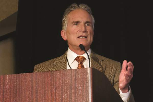
Unlike the typical bimodal distribution seen with hand-sewn graft infections, infection following endovascular repair of aortic aneurysms (EVAR) occurs from days up to 3 years after implantation. At the Mayo Clinic, a 79-year-old man presented with an infected endograft, psoas abscess, and Salmonella septicemia 4 years after EVAR.
“These infections are uncommon, but we are seeing more of them,” Dr. Bower said at a symposium on vascular surgery sponsored by Northwestern University.
Roughly two-thirds of patients will present with fever, nonspecific abdominal or back pain, malaise, weight loss or night sweats. If time permits, preoperative assessments include echocardiography for left ventricular function, arterial blood gases for pulmonary function since many patients are smokers, and renal ultrasound if creatinine is ≥ 1.5 mg/dL after rehydration. These tests are important because preoperative chronic obstructive pulmonary disease and renal dysfunction correlate with worse postoperative outcomes, he said.
Computed tomography angiography (CTA), however, stands as the single most important step of preoperative preparation, with the sine qua non of infection being air around the graft. Unlike hand-sewn grafts where infections can be localized, typically there is total graft involvement in these cases because the device is left inside the aneurysm sac. Aneurysms or pseudoaneurysms also have been seen above the infected device, including at the top end of suprarenal stents.
“This clearly has an impact on how we approach patients, but what’s become very apparent to me is that CTA often underestimates the amount of periaortic inflammation, especially at the juxta- and pararenal locations,” Dr. Bower said.
The Mayo group initially used in situ antibiotic-soaked prosthetic grafts for explanting EVAR devices, which yielded “acceptable mortality and reinfection rates, but primarily outstanding patency rates.” However, cryopreserved aortoiliac grafts have now become their first choice, Dr. Bower said. An ABO match is not imperative, preparation takes roughly 45 minutes, branch closures done in the lab are buttressed with sutures, and the graft is turned over to keep the lumbar arteries anterior, which offers an easy fix if there is bleeding, rather than having it on the posterior wall. Cryopreserved grafts, however, can dilate 40% and lengthen 10% under pressure.
“I’ve been burned more than once where the graft elongates more than I think, and I end up having to cut a small piece out to foreshorten it,” he said.
Reconstructions are tailored to patient anatomy. Surgeons should have several plans for reconstruction, including routing a graft through a remote path, remembering that CTA will underestimate the amount of periaortic inflammation. Separate bypasses of the renal or visceral arteries are performed first before the aortic clamp is applied to reduce physiologic stress. This requires knowledge of the supraceliac and pararenal aorta exposures, which really begins with the correct choice of incisions, Dr. Bower said. This is based on the aortic segment to be treated, position of the new graft, the aortic clamp site, and patient body habitus.
Most patients with EVAR infections are approached with a midline abdominal incision extended along the xiphoid process, which is the lynch pin for allowing upward and lateral retraction of the abdominal wall, he said. Choosing an incision that allows a more vertical orientation to where the new aortic anastomosis and clamp site will be, rather than operating in a keyhole, is important.
The second step is to open up the pararenal space by moving the viscera out of the way. This begins by ligating the inferior mesenteric vein and adjacent lymphatics, which allows incision of an avascular plane along the base of the left transverse colon. Retractor blades are set to allow the upward and lateral retraction of the small bowel, the left colon, and pancreas. Exposure of the suprarenal or supramesenteric aorta requires mobilization of the left renal vein after ligation and division of its branches.
“If that vein is intensely involved in inflammation, don’t ligate the branches in case you have to divide that vein at the caval confluence. Otherwise, you’ll run into some dysfunction of that left kidney,” Dr. Bower cautioned.
To have a secure place for the aortic cross clamp, the crura must be divided on either side of the diaphragm at or above the supramesenteric aorta, he added.
Key steps in total graft explantation are to drain abscesses prior to surgery to lower the bacterial burden and thus reduce the postoperative inflammatory response, bypass renal/visceral arteries first, if needed, remove the infected graft, debride the aorta to healthy tissue, place the new graft and cover it with omentum, and repair the bowel, if needed.
A piece of the proximal aortic wall should be sent to pathology to ensure the absence of bacteria or microabscesses. Organism-specific antibiotics are administered intravenously for 6-8 weeks followed by lifelong oral antibiotics, he said.
An earlier report involving 24 patients with infected aortic endografts (21 EVARs and 3 thoracic EVARs) treated at Mayo Clinic between 1997 and 2012 revealed polymicrobial infection in 11 patients, with methicillin-resistant Staphylococcus aureus being common. Potential contributors to infection were endovascular reintervention in eight, aortoenteric fistula/erosion in four, and various remote infections (J. Vasc. Surg. 2013;58:371-9).
Rifampin-soaked grafts were used in 15 patients, cryopreserved grafts in 4, femoral vein in 2, and axillofemoral grafts in 3. At a median of 14 months follow-up, patient survival, graft-related complications, and reinfection rates were 79%, 13%, and 4%, respectively, Dr. Bower said.
Dr. Bower reported having no financial disclosures.
The expert opinion from the Northwestern Vascular Symposium regarding the management of EVAR infections reminds us of the importance of appropriate patient selection, proper performance of the planned procedure, and long-term follow-up. As EVAR has become the treatment of choice for more than 80% of patients with infrarenal AAAs in the United States, the rate of patients that return with EVAR infections, although rare, is increasing and their management can be more challenging than that of a primary or aortic graft infection as suggested by Dr. Thomas C. Bower in this opinion. The planning for these cases is critical with multiple options for treatment currently available and endorsed by a variety of investigators. From an evaluation standpoint, CTA is critical for diagnosis and case planning. Air around the graft is considered the “sine qua non” of infection but if it presents in the first month after EVAR it can be due to trapped air introduced into the sac during the intervention.

|
Dr. Luis A. Sanchez |
Patients with air in the sac at the initial postprocedure evaluation should be considered for early follow-up to make sure this finding resolves. Further assessment that will change the management of the patient includes the type of EVAR device, infra- or suprarenal, since the entire removal of a suprarenal device usually requires supraceliac cross-clamping with its associated morbidity and mortality. Drainage of the infected cavity, as suggested by Dr. Bower, can help lower the bacterial burden and provide information regarding the offending organism. That information will help the vascular surgeon decide if an in-line reconstruction or an extra-anatomical one is more appropriate in the patient’s situation as more virulent organisms tend to be associated with higher reinfection and complication rates when in-line reconstructions are performed.
The different options for aortic access need to be evaluated based on the anatomy of the patient. A transabdominal approach is best for most patients as it allows access to the iliac arteries bilaterally for removal of the entire graft, debridement of the infected bed, aortic and/or visceral reconstruction, and omental coverage of the in-line graft or aortic stump if an extra-anatomical reconstruction is selected. The retroperitoneal approach should be considered for patients that will require extensive perivisceral work, as may be necessary from suprarenal or fenestrated devices, but limitations exist accessing the right iliac system and potentially intraabdominal targets for visceral or renal reconstructions.
The best configuration to reconstruct these patients remains largely undetermined based on the literature. The published experience from the Mayo Clinic (J. Vasc. Surg. 2013;58:371-9), in which some of the opinions of Dr. Bower are based, suggested excellent results in 24 patients mostly treated with rifampin-soaked in-line reconstructions with a periprocedural mortality of 4%. Cryopreserved aortic grafts “have become the conduit of choice for the group at this time,” stated Dr. Bower, to try to further decrease the reinfection rates in their patient population. There are limited data regarding the use of cryopreserved aortoiliac segments for aortic infections and less for EVAR infections. The most recent and largest series (J. Vasc. Surg. 2014;59:669-74) included 220 patients with aortic infections with a perioperative mortality of 9% and cryopreserved graft complications in another 12%-15% of patients.
In summary, aortic infections associated with EVAR are challenging problems that should be addressed in regional centers with experience. Renal and visceral reconstructions as well as supravisceral clamping are associated with significantly higher periprocedural morbidity and mortality based on the extensive experience at the Cleveland Clinic with EVAR explants (J. Vasc. Surg. 2014;59:886-93). The choice of the reconstruction and the material used should be based on the offending organism, type of EVAR device, extent of the infectious process, and the expertise of the treating physician.
Dr. Luis A. Sanchez is chief, section of vascular surgery and Gregorio A. Sicard Distinguished Professor of Surgery and Radiology, Washington University, St. Louis, and an associate medical editor for Vascular Specialist. He had no relevant disclosures.
The expert opinion from the Northwestern Vascular Symposium regarding the management of EVAR infections reminds us of the importance of appropriate patient selection, proper performance of the planned procedure, and long-term follow-up. As EVAR has become the treatment of choice for more than 80% of patients with infrarenal AAAs in the United States, the rate of patients that return with EVAR infections, although rare, is increasing and their management can be more challenging than that of a primary or aortic graft infection as suggested by Dr. Thomas C. Bower in this opinion. The planning for these cases is critical with multiple options for treatment currently available and endorsed by a variety of investigators. From an evaluation standpoint, CTA is critical for diagnosis and case planning. Air around the graft is considered the “sine qua non” of infection but if it presents in the first month after EVAR it can be due to trapped air introduced into the sac during the intervention.
|
|
Dr. Luis A. Sanchez |
Patients with air in the sac at the initial postprocedure evaluation should be considered for early follow-up to make sure this finding resolves. Further assessment that will change the management of the patient includes the type of EVAR device, infra- or suprarenal, since the entire removal of a suprarenal device usually requires supraceliac cross-clamping with its associated morbidity and mortality. Drainage of the infected cavity, as suggested by Dr. Bower, can help lower the bacterial burden and provide information regarding the offending organism. That information will help the vascular surgeon decide if an in-line reconstruction or an extra-anatomical one is more appropriate in the patient’s situation as more virulent organisms tend to be associated with higher reinfection and complication rates when in-line reconstructions are performed.
The different options for aortic access need to be evaluated based on the anatomy of the patient. A transabdominal approach is best for most patients as it allows access to the iliac arteries bilaterally for removal of the entire graft, debridement of the infected bed, aortic and/or visceral reconstruction, and omental coverage of the in-line graft or aortic stump if an extra-anatomical reconstruction is selected. The retroperitoneal approach should be considered for patients that will require extensive perivisceral work, as may be necessary from suprarenal or fenestrated devices, but limitations exist accessing the right iliac system and potentially intraabdominal targets for visceral or renal reconstructions.
The best configuration to reconstruct these patients remains largely undetermined based on the literature. The published experience from the Mayo Clinic (J. Vasc. Surg. 2013;58:371-9), in which some of the opinions of Dr. Bower are based, suggested excellent results in 24 patients mostly treated with rifampin-soaked in-line reconstructions with a periprocedural mortality of 4%. Cryopreserved aortic grafts “have become the conduit of choice for the group at this time,” stated Dr. Bower, to try to further decrease the reinfection rates in their patient population. There are limited data regarding the use of cryopreserved aortoiliac segments for aortic infections and less for EVAR infections. The most recent and largest series (J. Vasc. Surg. 2014;59:669-74) included 220 patients with aortic infections with a perioperative mortality of 9% and cryopreserved graft complications in another 12%-15% of patients.
In summary, aortic infections associated with EVAR are challenging problems that should be addressed in regional centers with experience. Renal and visceral reconstructions as well as supravisceral clamping are associated with significantly higher periprocedural morbidity and mortality based on the extensive experience at the Cleveland Clinic with EVAR explants (J. Vasc. Surg. 2014;59:886-93). The choice of the reconstruction and the material used should be based on the offending organism, type of EVAR device, extent of the infectious process, and the expertise of the treating physician.
Dr. Luis A. Sanchez is chief, section of vascular surgery and Gregorio A. Sicard Distinguished Professor of Surgery and Radiology, Washington University, St. Louis, and an associate medical editor for Vascular Specialist. He had no relevant disclosures.
CHICAGO – Successful management of infected aortic endovascular grafts requires careful operative planning and execution, meticulous postoperative care, and a fair bit of creativity.
“Each patient is different, so surgeons have to tailor the reconstructions to the individual patient and with these specific infections, have to be creative,” Dr. Thomas C. Bower, chair of vascular and endovascular surgery at Mayo Clinic, Rochester, Minn., said. “I’ve found the operations to be more challenging and more difficult than explanting portions or total graft excision when the infection has occurred in a hand-sewn graft.”
Unlike the typical bimodal distribution seen with hand-sewn graft infections, infection following endovascular repair of aortic aneurysms (EVAR) occurs from days up to 3 years after implantation. At the Mayo Clinic, a 79-year-old man presented with an infected endograft, psoas abscess, and Salmonella septicemia 4 years after EVAR.
“These infections are uncommon, but we are seeing more of them,” Dr. Bower said at a symposium on vascular surgery sponsored by Northwestern University.
Roughly two-thirds of patients will present with fever, nonspecific abdominal or back pain, malaise, weight loss or night sweats. If time permits, preoperative assessments include echocardiography for left ventricular function, arterial blood gases for pulmonary function since many patients are smokers, and renal ultrasound if creatinine is ≥ 1.5 mg/dL after rehydration. These tests are important because preoperative chronic obstructive pulmonary disease and renal dysfunction correlate with worse postoperative outcomes, he said.
Computed tomography angiography (CTA), however, stands as the single most important step of preoperative preparation, with the sine qua non of infection being air around the graft. Unlike hand-sewn grafts where infections can be localized, typically there is total graft involvement in these cases because the device is left inside the aneurysm sac. Aneurysms or pseudoaneurysms also have been seen above the infected device, including at the top end of suprarenal stents.
“This clearly has an impact on how we approach patients, but what’s become very apparent to me is that CTA often underestimates the amount of periaortic inflammation, especially at the juxta- and pararenal locations,” Dr. Bower said.
The Mayo group initially used in situ antibiotic-soaked prosthetic grafts for explanting EVAR devices, which yielded “acceptable mortality and reinfection rates, but primarily outstanding patency rates.” However, cryopreserved aortoiliac grafts have now become their first choice, Dr. Bower said. An ABO match is not imperative, preparation takes roughly 45 minutes, branch closures done in the lab are buttressed with sutures, and the graft is turned over to keep the lumbar arteries anterior, which offers an easy fix if there is bleeding, rather than having it on the posterior wall. Cryopreserved grafts, however, can dilate 40% and lengthen 10% under pressure.
“I’ve been burned more than once where the graft elongates more than I think, and I end up having to cut a small piece out to foreshorten it,” he said.
Reconstructions are tailored to patient anatomy. Surgeons should have several plans for reconstruction, including routing a graft through a remote path, remembering that CTA will underestimate the amount of periaortic inflammation. Separate bypasses of the renal or visceral arteries are performed first before the aortic clamp is applied to reduce physiologic stress. This requires knowledge of the supraceliac and pararenal aorta exposures, which really begins with the correct choice of incisions, Dr. Bower said. This is based on the aortic segment to be treated, position of the new graft, the aortic clamp site, and patient body habitus.
Most patients with EVAR infections are approached with a midline abdominal incision extended along the xiphoid process, which is the lynch pin for allowing upward and lateral retraction of the abdominal wall, he said. Choosing an incision that allows a more vertical orientation to where the new aortic anastomosis and clamp site will be, rather than operating in a keyhole, is important.
The second step is to open up the pararenal space by moving the viscera out of the way. This begins by ligating the inferior mesenteric vein and adjacent lymphatics, which allows incision of an avascular plane along the base of the left transverse colon. Retractor blades are set to allow the upward and lateral retraction of the small bowel, the left colon, and pancreas. Exposure of the suprarenal or supramesenteric aorta requires mobilization of the left renal vein after ligation and division of its branches.
“If that vein is intensely involved in inflammation, don’t ligate the branches in case you have to divide that vein at the caval confluence. Otherwise, you’ll run into some dysfunction of that left kidney,” Dr. Bower cautioned.
To have a secure place for the aortic cross clamp, the crura must be divided on either side of the diaphragm at or above the supramesenteric aorta, he added.
Key steps in total graft explantation are to drain abscesses prior to surgery to lower the bacterial burden and thus reduce the postoperative inflammatory response, bypass renal/visceral arteries first, if needed, remove the infected graft, debride the aorta to healthy tissue, place the new graft and cover it with omentum, and repair the bowel, if needed.
A piece of the proximal aortic wall should be sent to pathology to ensure the absence of bacteria or microabscesses. Organism-specific antibiotics are administered intravenously for 6-8 weeks followed by lifelong oral antibiotics, he said.
An earlier report involving 24 patients with infected aortic endografts (21 EVARs and 3 thoracic EVARs) treated at Mayo Clinic between 1997 and 2012 revealed polymicrobial infection in 11 patients, with methicillin-resistant Staphylococcus aureus being common. Potential contributors to infection were endovascular reintervention in eight, aortoenteric fistula/erosion in four, and various remote infections (J. Vasc. Surg. 2013;58:371-9).
Rifampin-soaked grafts were used in 15 patients, cryopreserved grafts in 4, femoral vein in 2, and axillofemoral grafts in 3. At a median of 14 months follow-up, patient survival, graft-related complications, and reinfection rates were 79%, 13%, and 4%, respectively, Dr. Bower said.
Dr. Bower reported having no financial disclosures.
EXPERT OPINION FROM THE NORTHWESTERN VASCULAR SYMPOSIUM

Hybrid carotid stents eyed with cautious optimism
CHICAGO – The next generation of hybrid carotid stents is slowly breathing life into the stagnant field of carotid artery stenting.
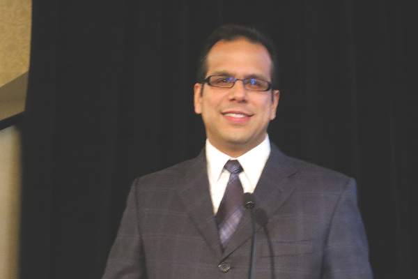
The new hybrid stents combine the flexibility of a traditional open-cell, nitinol stent with the stabilization typically offered by a closed-cell stent design. The initial clinical experience is limited, but shows promising results against embolization, Dr. Claudio Schönholz said at a symposium on vascular surgery sponsored by Northwestern University.
Last year, Dr. Schönholz and his colleagues at the Medical University of South Carolina in Charleston reported the first-in-man use of the investigational Gore Carotid Stent (W.L. Gore & Associates) (J. Endovasc. Thera. 2014 Aug;21:601-4).
As part of the Gore Carotid Stent Clinical Study for the Treatment of Carotid Artery Stenosis in Patients at Increased Risk for Adverse Events from Carotid Endarterectomy (SCAFFOLD) study, the team has successfully treated another four patients with no evidence of peri- or postprocedural neurological events. This included a case with such severe high-grade stenosis and slow flow that the external carotid artery was not even visible on imaging before the stent was placed, Dr. Schönholz said.
The Food and Drug Administration recently reviewed unreleased data for the first 100 patients enrolled in SCAFFOLD and given the green light for the multicenter, 312-patient study to resume with the start of the new year, he said.
Medtronic’s Cristallo Ideale hybrid stent is already approved outside the United States and was associated with no major neurological events and two cases of transient ischemic attack in 124 patients treated at four expert centers in Italy and Germany in the Cristallo study (J. Endovasc. Ther. 2008;15:186-92).
A more recent retrospective study revealed only one minor stroke in the perioperative period and during the first 30 days in 68 patients with symptomatic carotid stenosis treated by Turkish surgeons with the Cristallo Ideale stent and a proximal protection device (MO.MA, Invatec s.r.l., Medtronic, Italy) (Int. Angiol. 2014 Nov. 14. [Epub ahead of print]).
Better patient selection, increased operator experience, and use of embolic protection devices has reduced neurological events associated with carotid artery stenting, but embolization still occurs after protection devices are removed due to plaque protrusion through the stent struts, Dr. Schönholz said.
The unique design of the hybrid stents “may prevent plaque protrusion, eliminating peri- and postprocedural events,” he said.
The Cristallo Ideale hybrid stent is a nitinol-based stent that has a closed-cell portion at its center and an open-cell configuration on the distal and proximal sections.
In contrast, the Gore Carotid Stent has a closed-cell component throughout the entire device length that is created by placing an expanded polytetrafluoroethylene lattice with 500-micron pores over an open-cell frame. Once combined, both the stent frame and lattice are coated on all surfaces with Carmeda Bioactive Surface (CBAS) heparin. It’s action is limited only to the device surface and has no systemic anticoagulation effects, said Dr. Schönholz, who disclosed serving on Gore’s scientific advisory board.
The open-cell frame allows a high degree of flexibility and conformity to the native anatomy, while the stent lattice provides a high degree of plaque scaffolding that can reduce plaque prolapse, he said. The lattice also reduces the amount of emboli released during and after stent deployment and stabilizes the stent frame by resisting elongation as well as “fish-scaling,” or the misalignment of stent struts that protrude into the vessel wall, particularly when stents are deployed in tortuous anatomy.
When asked during a discussion whether the same results couldn’t be achieved by simply putting in another covered stent like a Viabahn, Dr. Schönholz replied that the Gore Stent isn’t a true covered stent because the 500-micron pores allow perfusion to be maintained to the external carotid artery. “It was intended to prevent plaque profusion, but at the same time allowing perfusion of the external carotid,” he said.
So far, the investigators have not incorporated intravascular ultrasound during stent placement, as it was not part of the SCAFFOLD protocol, but this will likely be used once the device is approved, he added.
Course director Dr. Mark K. Eskandari, chief of vascular surgery at Northwestern University in Chicago, said the results show that “carotid stenting isn’t dead yet and we can persevere. Advances in technology, both in regards to mechanical embolic protection devices and stent design systems, continue to improve the already great results of carotid artery stenting.”
CHICAGO – The next generation of hybrid carotid stents is slowly breathing life into the stagnant field of carotid artery stenting.
The new hybrid stents combine the flexibility of a traditional open-cell, nitinol stent with the stabilization typically offered by a closed-cell stent design. The initial clinical experience is limited, but shows promising results against embolization, Dr. Claudio Schönholz said at a symposium on vascular surgery sponsored by Northwestern University.
Last year, Dr. Schönholz and his colleagues at the Medical University of South Carolina in Charleston reported the first-in-man use of the investigational Gore Carotid Stent (W.L. Gore & Associates) (J. Endovasc. Thera. 2014 Aug;21:601-4).
As part of the Gore Carotid Stent Clinical Study for the Treatment of Carotid Artery Stenosis in Patients at Increased Risk for Adverse Events from Carotid Endarterectomy (SCAFFOLD) study, the team has successfully treated another four patients with no evidence of peri- or postprocedural neurological events. This included a case with such severe high-grade stenosis and slow flow that the external carotid artery was not even visible on imaging before the stent was placed, Dr. Schönholz said.
The Food and Drug Administration recently reviewed unreleased data for the first 100 patients enrolled in SCAFFOLD and given the green light for the multicenter, 312-patient study to resume with the start of the new year, he said.
Medtronic’s Cristallo Ideale hybrid stent is already approved outside the United States and was associated with no major neurological events and two cases of transient ischemic attack in 124 patients treated at four expert centers in Italy and Germany in the Cristallo study (J. Endovasc. Ther. 2008;15:186-92).
A more recent retrospective study revealed only one minor stroke in the perioperative period and during the first 30 days in 68 patients with symptomatic carotid stenosis treated by Turkish surgeons with the Cristallo Ideale stent and a proximal protection device (MO.MA, Invatec s.r.l., Medtronic, Italy) (Int. Angiol. 2014 Nov. 14. [Epub ahead of print]).
Better patient selection, increased operator experience, and use of embolic protection devices has reduced neurological events associated with carotid artery stenting, but embolization still occurs after protection devices are removed due to plaque protrusion through the stent struts, Dr. Schönholz said.
The unique design of the hybrid stents “may prevent plaque protrusion, eliminating peri- and postprocedural events,” he said.
The Cristallo Ideale hybrid stent is a nitinol-based stent that has a closed-cell portion at its center and an open-cell configuration on the distal and proximal sections.
In contrast, the Gore Carotid Stent has a closed-cell component throughout the entire device length that is created by placing an expanded polytetrafluoroethylene lattice with 500-micron pores over an open-cell frame. Once combined, both the stent frame and lattice are coated on all surfaces with Carmeda Bioactive Surface (CBAS) heparin. It’s action is limited only to the device surface and has no systemic anticoagulation effects, said Dr. Schönholz, who disclosed serving on Gore’s scientific advisory board.
The open-cell frame allows a high degree of flexibility and conformity to the native anatomy, while the stent lattice provides a high degree of plaque scaffolding that can reduce plaque prolapse, he said. The lattice also reduces the amount of emboli released during and after stent deployment and stabilizes the stent frame by resisting elongation as well as “fish-scaling,” or the misalignment of stent struts that protrude into the vessel wall, particularly when stents are deployed in tortuous anatomy.
When asked during a discussion whether the same results couldn’t be achieved by simply putting in another covered stent like a Viabahn, Dr. Schönholz replied that the Gore Stent isn’t a true covered stent because the 500-micron pores allow perfusion to be maintained to the external carotid artery. “It was intended to prevent plaque profusion, but at the same time allowing perfusion of the external carotid,” he said.
So far, the investigators have not incorporated intravascular ultrasound during stent placement, as it was not part of the SCAFFOLD protocol, but this will likely be used once the device is approved, he added.
Course director Dr. Mark K. Eskandari, chief of vascular surgery at Northwestern University in Chicago, said the results show that “carotid stenting isn’t dead yet and we can persevere. Advances in technology, both in regards to mechanical embolic protection devices and stent design systems, continue to improve the already great results of carotid artery stenting.”
EXPERT ANALYSIS FROM THE NORTHWESTERN VASCULAR SYMPOSIUM
FDA approves antibacterial combo drug Avycaz
The Food and Drug Administration has approved the antibacterial drug ceftazidime-avibactam (Avycaz) on Feb. 25 for complicated intra-abdominal infections in combination with metronidazole, and for complicated urinary tract infections including pyelonephritis in adults.
“It is important that the use of Avycaz be reserved for situations where there are limited or no alternative antibacterial drugs for treating a patient’s infection,” Dr. Edward Cox, director of the FDA’s Office of Antimicrobial Products in the Center for Drug Evaluation and Research, said in a statement.
Avycaz is a fixed-combination drug containing ceftazidime, a previously approved cephalosporin with in vitro activity against certain gram-negative and gram-positive bacteria, and avibactam, a beta-lactamase inhibitor.
The addition of avibactam to ceftazidime protects ceftazidime from breakdown by extended spectrum beta-lactamases, Klebsiella pneumoniae carbapenemase (KPC), and AmpC-producing pathogens, according to David Nicholson, Ph.D., executive vice president of branded research and development at Actavis, which is jointly developing the drug with AstraZeneca.
“The FDA approval of Avycaz is an important step forward in enhancing our ability to respond to serious pathogens caused by difficult-to-treat gram-negative pathogens,” he said in a statement.
The recent rise in the incidence of multidrug-resistant gram-negative pathogens poses a significant threat to patients and places a tremendous strain on the U.S. health care system, Dr. Jose Vazquez, chief of infectious disease at Georgia Regents University in Augusta, Ga., commented in the same statement.
“The increasing prevalence of KPC-producing Enterobacteriaceae in particular, has become a major therapeutic challenge for physicians managing these infections. Unfortunately, there are currently a limited number of safe and effective antimicrobials to treat these serious infections,” he said.
Avycaz was granted priority review and named a Qualified Infectious Disease Product (QIDP), a designation given to antibacterial products to treat serious or life-threatening infections.
Its efficacy was supported in part by findings of the efficacy and safety of ceftazidime for the treatment of complicated intra-abdominal infections (cIAI) and complicated urinary tract infections (cUTI). The contribution of avibactam to Avycaz was based on data from in vitro studies and animal models of infection. Avycaz was also studied in two phase II trials, one each in cIAI and cUTI.
The most common side effects are vomiting, nausea, constipation, and anxiety. The FDA advises health care professionals to inform patients of these risks and that decreased efficacy, seizures, and other neurologic events were seen in patients with renal impairment. Serious skin reactions and anaphylaxis may occur in patients with penicillin allergies.
The recommended dosage for patients with normal renal function is 2.5 g administered every 8 hours by intravenous infusion over 2 hours in adults aged 18 years and older. For patients with changing or impaired renal function (creatinine clearance < 50 mL/min), CrCL should be monitored at least daily and the dosage adjusted accordingly.
In a phase III trial of intra-abdominal infections, clinical cure rates were lower in the subgroup of patients with CrCL of 30-50 mL/min, compared with those with CrCL greater than 50 mL/min, according to the company. The reduction in cure rates was more marked in patients treated with Avycaz plus metronidazole vs. meropenem-treated patients.
Avycaz will be available in the second quarter of 2015, according to the company. Phase III studies evaluating Avycaz for the treatment of cIAI and cUTI are ongoing and targeted for completion in late 2015.
Pregnancy outcomes mixed after bariatric surgery
A history of bariatric surgery appears to both positively and negatively influence pregnancy outcomes, according to a prospective, nationwide cohort study published in the New England Journal of Medicine.
Pregnancies after bariatric surgery, as compared with control pregnancies matched for presurgery body mass index, were associated with a significantly lower risk of gestational diabetes (1.9% vs. 6.8%; odds ratio, 0.25; P < .001) and large-for-gestational age infants (8.6% vs. 22.4%; OR, 0.33; P < .001).
“However, increased surveillance during pregnancy and the neonatal period is warranted, since a history of bariatric surgery was also associated with small-for-gestational-age infants (15.6% vs. 7.6%; OR, 2.20; P < .001), shorter gestation (273 days vs. 277.5 days; P < .001), and potentially an increased risk of stillbirth or neonatal death [1.7% vs. 0.7%; OR, 2.39; P: 0.06],” study author Kari Johansson, Ph.D., from the Karolinska Institute in Stockholm, suggested in the study (N. Engl. J. Med. 2015;372:814-24 [doi:10.1056/NEJMoa1405789]).
The study, thought to be the largest to date comparing 2,952 pregnancy outcomes between women with and without a history of bariatric surgery, found no difference in the risk of congenital malformations between groups. The median time from surgery to conception was 1.1 years.
Dr. Johansson reported support from the Swedish Research Council and a young investigator award from the Obesity Society. Her coauthors reported support from the Swedish Research Council, Stockholm County Council, and consulting fees from Itrim and Strategic Health Resources.
FROM NEW ENGLAND JOURNAL OF MEDICINE
Most of the findings are consistent with those from smaller studies and meta-analyses, although an association between bariatric surgery and subsequent perinatal mortality has not been previously suggested, Dr. Aaron B. Caughey noted in an accompanying editorial (N. Engl. J. Med. 2015;372:877-8 [doi:10.1056/nejme1500230]).
If anything, a recent meta-analysis showed that higher body mass index was associated with an increased risk of stillbirth and neonatal mortality (JAMA 2014;311:1536-46).
“The current data, combined with previous reports, suggest that it may be prudent to monitor fetal growth in women who have undergone bariatric surgery, particularly in those who have had gastric bypass surgery. I would not recommend that all such women undergo antepartum fetal surveillance on the basis of the current study, since evidence to indicate such care would improve outcomes is lacking,” Dr. Caughey wrote.
Dr. Aaron B. Caughey is chair of obstetrics and gynecology and associate dean for women’s health research and policy at the Oregon Health & Science University School of Medicine in Portland. He reported having no financial disclosures.
Rethinking the ABCs of EVAR
CHICAGO – Real-world experience with novel endografts like the Ovation Prime abdominal endograft system is prompting some vascular specialists to rethink such central abdominal aortic aneurysm tenets as aortic neck dilation and minimum neck size.
“We started using this in our worst cases, patients with small caliber access vessels and very short aortic necks, to test this device, but over time we’ve pretty much made this our workhorse graft based on our outcomes,” Dr. Syed Hussain of the University of Illinois at Champaign-Urbana, said at a vascular surgery symposium sponsored by Northwestern University.

Among 67 patients with AAAs treated since the team’s first implant in November 2012, the technical success rate is 100%. At baseline, 35% of patients had access vessels < 7 mm, 45% had short aortic neck (< 15 mm), 60% had moderate to severe calcification (> 25% circumferential), and half had moderate to severe thrombus (> 25% circumferential).
The Ovation Prime (TriVascular Technologies) device is relatively quick and easy to put in, with an average procedure time of only 33 minutes, he said. Access was percutaneous in 27%, average blood loss was minimal at 60 mL, and average hospital stay was 1.7 days.
Two patients with severe comorbidities were admitted to the ICU and two patients experienced intraoperative type 1a endoleaks, both successfully treated with a Palmaz stent.
After an average follow-up of 12 months, there have been no type 1, III or IV endoleaks, graft migration, aneurysm enlargement, conversions, ruptures, limb occlusions, or secondary procedures, said Dr. Hussain, who disclosed serving as a consultant for Trivascular and national principal investigator of the PostMarket Ovation Trial. There were 12 type II endoleaks (17%) and all have been clinically irrelevant.
Because of the Ovation’s novel O-ring sealing mechanism, “you get a pretty watertight seal ring on these patients,” he said. More importantly, shear stress is distributed evenly along the entire O-ring, which creates very minimal outward stress on the aorta, “maybe 2 or 3 atmospheres at best.”
Evidence continues to build that self-expandable stents place chronic outward stress on the aorta that causes degeneration of the aortic wall, resulting in eventual aortic neck dilation and endograft migration. While it’s been argued that disease progression leads to aortic dilation, the phenomenon took off after the arrival of endovascular stents, not during decades of open AAA repair, Dr. Hussain, also of the Vein & Vascular Center at the Christie Clinic in Champaign, said.
In the Ovation approval trial, proximal neck dilation at 2 years followed a similar curve in the Ovation and open repair cohorts, compared with those for the more traditional endografts, he noted.
The Ovation Prime system was approved in 2012 and in mid-2014, the Food and Drug Administration approved changes to the indication statement that eliminated the requirement for a minimal aortic neck length.
Essentially, the Ovation device can be placed in any patient if the diameter at 13 mm below the lowest renal artery (the site of the most proximal sealing ring) is within the treatable diameter range of the device (15.8 mm-30.4 mm), Dr. Hussain said.
“The idea of having a neck length is completely starting to go away,” he said. “And even though the trial by Endologix is looking at 1 centimeter as the current requirement for enrolling patients, I think eventually it’s going to get to the point where you’re not going to need a neck for the Nellix device either. You’re going to be able to treat patients who have very short, 1 to 2 millimeter necks, basically perirenal aneurysms, and get a seal on.”
The Nellix endovascular aneurysm sealing system (Endologix) is not commercially available in the U.S., but is the being evaluated in at least three studies. It consists of dual balloon-expandable end-frames surrounded by polymer-filled endobags and is designed to completely fill and seal the aortic aneurysm sac. Anatomical requirements for patients to be enrolled in clinical studies include a nonaneurysmal aortic neck length of ≥ 10 mm, nonaneurysmal aortic neck diameter of 18 mm-32 mm, maximum aortic blood flow lumen diameter of ≤ 60 mm, and common iliac artery diameter of 8 mm-35 mm, according to the company’s website.
CHICAGO – Real-world experience with novel endografts like the Ovation Prime abdominal endograft system is prompting some vascular specialists to rethink such central abdominal aortic aneurysm tenets as aortic neck dilation and minimum neck size.
“We started using this in our worst cases, patients with small caliber access vessels and very short aortic necks, to test this device, but over time we’ve pretty much made this our workhorse graft based on our outcomes,” Dr. Syed Hussain of the University of Illinois at Champaign-Urbana, said at a vascular surgery symposium sponsored by Northwestern University.
Among 67 patients with AAAs treated since the team’s first implant in November 2012, the technical success rate is 100%. At baseline, 35% of patients had access vessels < 7 mm, 45% had short aortic neck (< 15 mm), 60% had moderate to severe calcification (> 25% circumferential), and half had moderate to severe thrombus (> 25% circumferential).
The Ovation Prime (TriVascular Technologies) device is relatively quick and easy to put in, with an average procedure time of only 33 minutes, he said. Access was percutaneous in 27%, average blood loss was minimal at 60 mL, and average hospital stay was 1.7 days.
Two patients with severe comorbidities were admitted to the ICU and two patients experienced intraoperative type 1a endoleaks, both successfully treated with a Palmaz stent.
After an average follow-up of 12 months, there have been no type 1, III or IV endoleaks, graft migration, aneurysm enlargement, conversions, ruptures, limb occlusions, or secondary procedures, said Dr. Hussain, who disclosed serving as a consultant for Trivascular and national principal investigator of the PostMarket Ovation Trial. There were 12 type II endoleaks (17%) and all have been clinically irrelevant.
Because of the Ovation’s novel O-ring sealing mechanism, “you get a pretty watertight seal ring on these patients,” he said. More importantly, shear stress is distributed evenly along the entire O-ring, which creates very minimal outward stress on the aorta, “maybe 2 or 3 atmospheres at best.”
Evidence continues to build that self-expandable stents place chronic outward stress on the aorta that causes degeneration of the aortic wall, resulting in eventual aortic neck dilation and endograft migration. While it’s been argued that disease progression leads to aortic dilation, the phenomenon took off after the arrival of endovascular stents, not during decades of open AAA repair, Dr. Hussain, also of the Vein & Vascular Center at the Christie Clinic in Champaign, said.
In the Ovation approval trial, proximal neck dilation at 2 years followed a similar curve in the Ovation and open repair cohorts, compared with those for the more traditional endografts, he noted.
The Ovation Prime system was approved in 2012 and in mid-2014, the Food and Drug Administration approved changes to the indication statement that eliminated the requirement for a minimal aortic neck length.
Essentially, the Ovation device can be placed in any patient if the diameter at 13 mm below the lowest renal artery (the site of the most proximal sealing ring) is within the treatable diameter range of the device (15.8 mm-30.4 mm), Dr. Hussain said.
“The idea of having a neck length is completely starting to go away,” he said. “And even though the trial by Endologix is looking at 1 centimeter as the current requirement for enrolling patients, I think eventually it’s going to get to the point where you’re not going to need a neck for the Nellix device either. You’re going to be able to treat patients who have very short, 1 to 2 millimeter necks, basically perirenal aneurysms, and get a seal on.”
The Nellix endovascular aneurysm sealing system (Endologix) is not commercially available in the U.S., but is the being evaluated in at least three studies. It consists of dual balloon-expandable end-frames surrounded by polymer-filled endobags and is designed to completely fill and seal the aortic aneurysm sac. Anatomical requirements for patients to be enrolled in clinical studies include a nonaneurysmal aortic neck length of ≥ 10 mm, nonaneurysmal aortic neck diameter of 18 mm-32 mm, maximum aortic blood flow lumen diameter of ≤ 60 mm, and common iliac artery diameter of 8 mm-35 mm, according to the company’s website.
AT THE NORTHWESTERN VASCULAR SYMPOSIUM
Key clinical point: Requirement for an specified aortic neck for placement diminishing for new endografts.
Major finding: No type I, III or IV endoleaks, graft migration, aneurysm enlargement, conversions, ruptures, limb occlusions, or secondary procedures occurred after 12 months follow-up.
Data source: Retrospective analysis of 67 patients with AAA treated with Ovation Prime.
Disclosures: Dr. Hussain disclosed serving as a consultant for TriVascular and a national principal investigator for the PostMarket Ovation Trial.
Tumor location portends CRC survival
Primary tumor location is an important prognostic factor in previously untreated metastatic colorectal cancer, according to a pooled analysis reported online in Journal of the National Cancer Institute.
In the prospective PROVETTA study, patients with tumors originating in the left side of the colon, distal to the splenic flexure, lived nearly twice as long as those with right-sided tumors (median 42 months vs. 24.8 months; hazard ratio, 0.44; P value < .001).

Unadjusted progression-free survival (PFS) was also significantly longer with left-sided tumors in PROVETTA (median 12.1 months vs. 9.9 months; HR, 0.52; P < .001).
A multivariable model adjusted for baseline variables confirmed that left-sided primary tumors had a lower risk of progression (HR, 0.55; P = .01) and death (HR, 0.47; P = .01), independent of BRAF mutation status or mucinous histology, study author Dr. Fotios Loupakis, of the University of Southern California Norris Comprehensive Cancer Center in Los Angeles, reported.
Subsequent analyses using data from two large phase III studies of first-line chemotherapy with or without bevacizumab (Avastin) also showed favorable outcomes in patients with left-sided tumors.
Overall survival was significantly longer in patients with left-sided vs. right-sided tumors in the NO16966 (median 23 months vs. 18 months; HR, 0.71; P < .001) and AVF2107g (median 20.4 months vs. 14.6 months; HR, 0.55; P < .001) studies.
Unadjusted median PFS was significantly longer for left-sided tumors in AVF2017 (8.5 months vs. 7.1 months; HR, 0.68; P < .001), but failed to reach statistical significance in NO16966 (median 8.9 months vs. 7.6 months; HR, 0.90; P = .12).
Multivariable analyses confirmed the independent prognostic effect of tumor location, irrespective of exposure to bevacizumab, the authors noted.
A recent retrospective analysis using two independent and nonrandomized cohorts of patients treated with capecitabine (Xeloda) and oxaliplatin (Eloxatin) with or without bevacizumab suggested that the addition of bevacizumab may primarily benefit patients with left-sided primary tumors.
“Our data do not validate those findings and reject the hypothesis of an interaction of primary tumor location with the efficacy of bevacizumab,” Dr. Loupakis wrote (J. Natl. Cancer Inst. 2015;107:dju427 [doi:10.1093/jnci/dju427]).
Another important finding according to the authors was the association of right-sided tumors with chemoresistance.
In multivariable analyses, left-sided tumor location was associated with significantly higher response rates in both AVF2107g (odds ratio, 2.48; P < .001) and NO16966 (OR, 1.49; P = .01). There was a trend toward achieving response for left-sided tumors in PROVETTA, but it did not reach statistical significance (OR, 1.23; P = .59), likely due to the limited sample size.
“These data emphasize that right-sided and left-sided CRC [colorectal cancers] have potentially important biological differences,” Dr. Loupakis observed.
The authors called for validation of the results in adjuvant and additional metastatic studies of CRC.
“This easy-to-collect dichotomous information on side of origin could be of added value in clinical decision-making and should be considered an important stratification factor for future randomized trials,” they suggested.
The study involves patients with metastatic disease and thus may not apply to those with resected primary tumors and its results may have been influenced by the lack of controls and selection bias, Dr. Howard Hochster noted in an accompanying editorial (J. Natl. Cancer Inst. 2015;107:djv011 [doi:10.1093/jnci/djv011]).
That said, “This interesting analysis gives rise to some important and testable biological hypotheses,” he observed.
The analysis may also help resolve the long-standing debate over why the AVF2107 trial, which used irinotecan-based chemotherapy and led to the first approval of bevacizumab, was markedly positive for a survival effect of bevacizumab, while the larger NO16966 study failed to show a survival benefit with the addition of bevacizumab to an oxaliplatin-based backbone.
“The present report provides an apparent explanation, as the data demonstrate a greater benefit of oxaliplatin-based chemotherapy overall and a disproportionate effect on the right-sided colon lesions, reducing the relative bevacizumab benefit. We see here, that oxaliplatin may be more effective than irinotecan for right-sided metastatic colon cancer and that bevacizumab further narrows the gap by differential treatment effect for lesions originating in the right side of the colon,” he wrote.
Dr. Hochster is an international expert in cancer clinical trials and director of GI Oncology at the Yale Cancer Center in New Haven, Conn.
Primary tumor location is an important prognostic factor in previously untreated metastatic colorectal cancer, according to a pooled analysis reported online in Journal of the National Cancer Institute.
In the prospective PROVETTA study, patients with tumors originating in the left side of the colon, distal to the splenic flexure, lived nearly twice as long as those with right-sided tumors (median 42 months vs. 24.8 months; hazard ratio, 0.44; P value < .001).
Unadjusted progression-free survival (PFS) was also significantly longer with left-sided tumors in PROVETTA (median 12.1 months vs. 9.9 months; HR, 0.52; P < .001).
A multivariable model adjusted for baseline variables confirmed that left-sided primary tumors had a lower risk of progression (HR, 0.55; P = .01) and death (HR, 0.47; P = .01), independent of BRAF mutation status or mucinous histology, study author Dr. Fotios Loupakis, of the University of Southern California Norris Comprehensive Cancer Center in Los Angeles, reported.
Subsequent analyses using data from two large phase III studies of first-line chemotherapy with or without bevacizumab (Avastin) also showed favorable outcomes in patients with left-sided tumors.
Overall survival was significantly longer in patients with left-sided vs. right-sided tumors in the NO16966 (median 23 months vs. 18 months; HR, 0.71; P < .001) and AVF2107g (median 20.4 months vs. 14.6 months; HR, 0.55; P < .001) studies.
Unadjusted median PFS was significantly longer for left-sided tumors in AVF2017 (8.5 months vs. 7.1 months; HR, 0.68; P < .001), but failed to reach statistical significance in NO16966 (median 8.9 months vs. 7.6 months; HR, 0.90; P = .12).
Multivariable analyses confirmed the independent prognostic effect of tumor location, irrespective of exposure to bevacizumab, the authors noted.
A recent retrospective analysis using two independent and nonrandomized cohorts of patients treated with capecitabine (Xeloda) and oxaliplatin (Eloxatin) with or without bevacizumab suggested that the addition of bevacizumab may primarily benefit patients with left-sided primary tumors.
“Our data do not validate those findings and reject the hypothesis of an interaction of primary tumor location with the efficacy of bevacizumab,” Dr. Loupakis wrote (J. Natl. Cancer Inst. 2015;107:dju427 [doi:10.1093/jnci/dju427]).
Another important finding according to the authors was the association of right-sided tumors with chemoresistance.
In multivariable analyses, left-sided tumor location was associated with significantly higher response rates in both AVF2107g (odds ratio, 2.48; P < .001) and NO16966 (OR, 1.49; P = .01). There was a trend toward achieving response for left-sided tumors in PROVETTA, but it did not reach statistical significance (OR, 1.23; P = .59), likely due to the limited sample size.
“These data emphasize that right-sided and left-sided CRC [colorectal cancers] have potentially important biological differences,” Dr. Loupakis observed.
The authors called for validation of the results in adjuvant and additional metastatic studies of CRC.
“This easy-to-collect dichotomous information on side of origin could be of added value in clinical decision-making and should be considered an important stratification factor for future randomized trials,” they suggested.
FROM JOURNAL OF THE NATIONAL CANCER INSTITUTE
Key clinical point: Metastatic colorectal cancer originating from the right side of the colon is associated with shorter progression-free survival and overall survival than left-sided colorectal cancers.
Major finding: Median overall survival was significantly shorter for patients with right-sided CRC tumors in the PROVETTA, AVF2107g, and NO16966 studies.
Data source: Pooled analysis from three studies in 2,027 patients with metastatic colorectal cancer.
Disclosures: The study was supported by a grant from the National Institutes of Health, the Daniel Butler Research Fund, the A.R.C.O. Foundation, Genentech, and F. Hoffmann-La Roche. Dr. Loupakis reported no conflicts of interest. Four coauthors are Roche/Genentech employees and two other coauthors have financial ties with Genentech.

Factor XI inhibitor trims DVTs after knee replacement surgery
SAN FRANCISCO – Reducing factor XI levels with the experimental antisense oligonucleotide FXI-ASO lowered venous thromboembolism rates after total knee arthroplasty without increasing bleeding in a phase II study.
Venous thromboembolism (VTE) rates were 30% among controls (21/69) on enoxaparin (Lovenox) 40 mg, compared with 27% for patients (36/134) given FXI-ASO 200 mg and 4% for those (3/71) given FXI-ASO 300 mg. Low-dose FXI-ASO was noninferior to enoxaparin (P = .59), while the high-dose regimen was superior (P < .001).

A 4% VTE rate “has never ever been seen before in patients undergoing knee surgery,” Dr. Harry Büller said during the late-breaking abstract session at the annual meeting of the American Society of Hematology.
The strategy of targeting factor XI is based on the understanding that patients with factor XI deficiency (plasma levels < 20% of normal) have a reduced risk of deep vein thrombosis (DVT). Experimental data in mice and primates also suggest that reducing factor XI attenuates thrombosis without excess bleeding.
Among the 300 patients in the open-label study, major or clinically relevant bleeding occurred in 3% of both FXI-ASO groups and 8% of the enoxaparin group (P = .09).
The findings provide the first evidence in humans that the factor XI intrinsic pathway is one of the drivers of postoperative thrombosis and support the concept that thrombosis and hemostasis can be dissociated, said Dr. Büller of the Academic Medical Center, Amsterdam.
“FXI-ASO is a promising new investigational antithrombotic agent and I believe you are witnessing the birth of a new class of antithrombotic agents,” he concluded.
During a press conference, Dr. Büller confided to reporters that he felt like a boy in a candy store, finally able to reveal the superb study findings.
Dr. Robert Flaumenhaft of Harvard Medical School, Boston, was far less effusive in an editorial that accompanied the simultaneous publication of the study in the New England Journal of Medicine.
“Do these finding prove that reduction in factor XI levels inhibits thrombosis without affecting bleeding? The conservative answer is no,” he wrote.
Dr. Flaumenhaft observed that the incidence of clinically relevant bleeding is relatively low after knee arthroplasty, even when patients receive anticoagulants, and that this safety outcome did not differ significantly between the enoxaparin and 300-mg FXI-ASO groups.
“These results also do not make a compelling case for the clinical use of the factor XI antisense oligonucleotide over anticoagulants that are currently used for prophylaxis in patients undergoing knee arthroplasty,” he wrote.
Central to this argument are issues of convenience and questions regarding reversibility. Treatment began 36 days before surgery and was associated with a high incidence of adverse events at the injection site and factor XI levels remained about 60% lower 70 days after initiation of therapy.
The half-life of FXI-ASO is about 22 days, “which in the classical setting in terms of bleeding could be seen as something of a disadvantage,” Dr. Büller told reporters. “But if we do the next study and it shows to be safe, it turns into an advantage” … because there is the possibility of giving FXI-ASO once every 3 weeks.
Dr. Flaumenhaft closed the editorial by acknowledging that the study challenges “the current paradigm” regarding the primary mechanism responsible for fibrin formation during thrombosis. “The striking observation that reducing factor XI levels prevents thrombosis after knee arthroplasty provides the best clinical evidence to date that the intrinsic pathway is essential for thrombus formation,” he wrote.
The study was conducted at 19 centers in five countries and randomly assigned 300 patients scheduled for elective primary unilateral total-knee arthroplasty to daily enoxaparin 40 mg or three doses of FXI-ASO. The protocol was amended early on to exclude a 100-mg FXI-ASO dose.
FXI-ASO 200 mg or 300 mg was given subcutaneously beginning 36 days before surgery on days 1, 3, 5, 8, 15, 22, and 29, and 6 hours postoperatively, with a final dose on day 39.
Enoxaparin 40 mg was given subcutaneously once daily, beginning the evening before or 6-8 hours after surgery, according to investigator preference, and was continued for at least 8 days postoperatively.
The primary efficacy point was a composite of asymptomatic DVT, detected by venography, and confirmed symptomatic VTE.
At baseline, the average factor XI level was 1.23 units/mL in the enoxaparin group, 1.20 U/mL in the 200-mg group, and 1.16 U/mL in the 300-mg group.
In patients with an average factor XI level of 0.2 U/mL or less, the incidence of the primary efficacy outcome was 5%.
Isis Pharmaceuticals funded the study. Dr. Büller disclosed ties with Isis, Daiichi-Sankyo, Bayer Healthcare, Pfizer, and Bristol-Myers Squibb. Dr. Flaumenhaft reported having no disclosures.
SAN FRANCISCO – Reducing factor XI levels with the experimental antisense oligonucleotide FXI-ASO lowered venous thromboembolism rates after total knee arthroplasty without increasing bleeding in a phase II study.
Venous thromboembolism (VTE) rates were 30% among controls (21/69) on enoxaparin (Lovenox) 40 mg, compared with 27% for patients (36/134) given FXI-ASO 200 mg and 4% for those (3/71) given FXI-ASO 300 mg. Low-dose FXI-ASO was noninferior to enoxaparin (P = .59), while the high-dose regimen was superior (P < .001).
A 4% VTE rate “has never ever been seen before in patients undergoing knee surgery,” Dr. Harry Büller said during the late-breaking abstract session at the annual meeting of the American Society of Hematology.
The strategy of targeting factor XI is based on the understanding that patients with factor XI deficiency (plasma levels < 20% of normal) have a reduced risk of deep vein thrombosis (DVT). Experimental data in mice and primates also suggest that reducing factor XI attenuates thrombosis without excess bleeding.
Among the 300 patients in the open-label study, major or clinically relevant bleeding occurred in 3% of both FXI-ASO groups and 8% of the enoxaparin group (P = .09).
The findings provide the first evidence in humans that the factor XI intrinsic pathway is one of the drivers of postoperative thrombosis and support the concept that thrombosis and hemostasis can be dissociated, said Dr. Büller of the Academic Medical Center, Amsterdam.
“FXI-ASO is a promising new investigational antithrombotic agent and I believe you are witnessing the birth of a new class of antithrombotic agents,” he concluded.
During a press conference, Dr. Büller confided to reporters that he felt like a boy in a candy store, finally able to reveal the superb study findings.
Dr. Robert Flaumenhaft of Harvard Medical School, Boston, was far less effusive in an editorial that accompanied the simultaneous publication of the study in the New England Journal of Medicine.
“Do these finding prove that reduction in factor XI levels inhibits thrombosis without affecting bleeding? The conservative answer is no,” he wrote.
Dr. Flaumenhaft observed that the incidence of clinically relevant bleeding is relatively low after knee arthroplasty, even when patients receive anticoagulants, and that this safety outcome did not differ significantly between the enoxaparin and 300-mg FXI-ASO groups.
“These results also do not make a compelling case for the clinical use of the factor XI antisense oligonucleotide over anticoagulants that are currently used for prophylaxis in patients undergoing knee arthroplasty,” he wrote.
Central to this argument are issues of convenience and questions regarding reversibility. Treatment began 36 days before surgery and was associated with a high incidence of adverse events at the injection site and factor XI levels remained about 60% lower 70 days after initiation of therapy.
The half-life of FXI-ASO is about 22 days, “which in the classical setting in terms of bleeding could be seen as something of a disadvantage,” Dr. Büller told reporters. “But if we do the next study and it shows to be safe, it turns into an advantage” … because there is the possibility of giving FXI-ASO once every 3 weeks.
Dr. Flaumenhaft closed the editorial by acknowledging that the study challenges “the current paradigm” regarding the primary mechanism responsible for fibrin formation during thrombosis. “The striking observation that reducing factor XI levels prevents thrombosis after knee arthroplasty provides the best clinical evidence to date that the intrinsic pathway is essential for thrombus formation,” he wrote.
The study was conducted at 19 centers in five countries and randomly assigned 300 patients scheduled for elective primary unilateral total-knee arthroplasty to daily enoxaparin 40 mg or three doses of FXI-ASO. The protocol was amended early on to exclude a 100-mg FXI-ASO dose.
FXI-ASO 200 mg or 300 mg was given subcutaneously beginning 36 days before surgery on days 1, 3, 5, 8, 15, 22, and 29, and 6 hours postoperatively, with a final dose on day 39.
Enoxaparin 40 mg was given subcutaneously once daily, beginning the evening before or 6-8 hours after surgery, according to investigator preference, and was continued for at least 8 days postoperatively.
The primary efficacy point was a composite of asymptomatic DVT, detected by venography, and confirmed symptomatic VTE.
At baseline, the average factor XI level was 1.23 units/mL in the enoxaparin group, 1.20 U/mL in the 200-mg group, and 1.16 U/mL in the 300-mg group.
In patients with an average factor XI level of 0.2 U/mL or less, the incidence of the primary efficacy outcome was 5%.
Isis Pharmaceuticals funded the study. Dr. Büller disclosed ties with Isis, Daiichi-Sankyo, Bayer Healthcare, Pfizer, and Bristol-Myers Squibb. Dr. Flaumenhaft reported having no disclosures.
AT ASH 2014
Key clinical point: Reducing factor XI levels with FXI-ASO was effective in preventing VTE in patients undergoing knee arthroplasty and appeared safe with respect to bleeding risk.
Major finding: The primary VTE endpoint occurred in 4% of patients on FXI-ASO 300 mg, 27% on FXI-ASO 200 mg, and 30% on enoxaparin.
Data source: Open-label, parallel-group phase II study of 300 patients undergoing primary unilateral total-knee arthroplasty.
Disclosures: Isis Pharmaceuticals funded the study. Dr. Büller disclosed ties with Isis, Daiichi-Sankyo, Bayer Healthcare, Pfizer, and Bristol-Myers Squibb. Dr. Flaumenhaft reported having no disclosures.
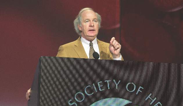
VTE risk climbs in patients on contact isolation
LAKE BUENA VISTA, FLA. – Trauma patients on contact isolation were nearly six times more likely to develop venous thromboembolism (VTE) as those who were not isolated, based on an analysis of 4,317 patients.
VTE occurred in 17.5% (44/251) of patients on contact isolation and 3.5% (141/4,066) of patients who were not isolated (P < .0001). Injury Severity Score (ISS), age, male gender, and obesity also were significantly associated with the risk of VTE.
The relationship between VTE risk and contact isolation remained significant after adjusting for gender, age, ISS, and comorbidities (odds ratio, 3.28; P < .0001), Dr. Robert Ferguson reported at the annual scientific assembly of the Eastern Association for the Surgery of Trauma.
Odds ratios also were significantly elevated for obesity (OR, 2.35; P < .006), male gender (OR, 2.1; P < .0001), ISS (OR, 1.08; P < .0001), and age (OR, 1.02; P < .0001). The presence of diabetes, dementia/Alzheimer’s, history of cerebrovascular accident, psychiatric disease, cirrhosis, cancer, or alcohol abuse was not statistically significant.
The increased risk for VTE in trauma patients on contact isolation “is likely multifactorial in nature and is related but not limited to decreased ambulation, noncompliance with prophylaxis, and restricted access by staff,” said Dr. Ferguson, a third-year resident at the Virginia Tech, Roanoke.
The risk:benefit ratio of contact isolation in the trauma population needs to be reevaluated, the researchers concluded. “We encourage hospital committees to alter protocols and supplement strategies such as staff education, dedicated ambulation areas and/or isolation wards, and eliminate contact isolation following routine methicillin-resistant Staphylococcus aureus surveillance screening.”
Dr. Ferguson and his coauthors reported having no financial disclosures.
AT THE EAST SCIENTIFIC ASSEMBLY
Key clinical point: Trauma patients on contact isolation are significantly predisposed to develop VTE.
Major finding: VTE occurred in 17.5% of patients on contact isolation and 3.5% not isolated (P < .0001).
Data source: Retrospective analysis of 4,317 trauma patients.
Disclosures: Dr. Ferguson and his coauthors reported having no financial disclosures.
Certified ACS trauma centers move the dial on patient outcomes
LAKE BUENA VISTA, FLA. – Patients undergoing emergency general surgery appear to fare better if managed at a certified acute care surgery trauma center, a nationwide analysis suggests.
“Patients managed at acute care surgery trauma centers had lower complication rates, shorter hospital length of stay, and lower hospital costs,” lead study author Dr. Mazhar Khalil said at the annual scientific assembly of the Eastern Association for the Surgery of Trauma.
Several single-institution studies have reported improved patient outcomes and system efficiencies following the 2008 creation of the American Association for the Surgery of Trauma (AAST) Acute Care Surgery fellowship. The ACS model has been endorsed by several professional organizations including EAST and the American College of Surgeons, but national outcomes have never been studied.

Dr. Khalil and his colleagues conducted a 1-year retrospective analysis of 131,410 patients who underwent emergency general surgery in 2011 in the National Inpatient Sample (NIS) database. The NIS is the largest all-payer, in-patient database in the United States and represents a stratified sample of 20% of all hospital discharges including 4,121 hospitals across 44 states.
ICD-9 codes were used to identify emergency general surgery procedures, defined as appendectomy, cholecystectomy, hernia repair, and small and large bowel resections. A total of 75,930 patients (58%) were managed at non–trauma centers (NTC), 47,753 (36%) at trauma centers (TC), and 7,727 (6%) at acute care surgery trauma centers (ACS-TC). Weekend admission rates were constant across all three groups at about 24%, as were the types of procedures performed.
In-hospital complications occurred in 18.1% of patients managed at AAST-certified acute care surgery trauma centers versus 18.7% among those at non–trauma centers and 19.4% at trauma centers (P = .04), Dr. Khalil, an international trauma fellow at the University of Arizona in Tucson, reported.
Between-group differences reached statistical significance for urinary tract infections (6.5% vs. 6.8% vs. 7.2%; P = .02), but not for the other complications of pneumonia, surgical site infection, sepsis, or reoperation.
AAST-certified ACS-TC patients had significantly shorter hospital stays than NTC or TC patients (7.2 days vs. 7.9 days vs. 8.5 days; P = .04) and lower average hospital costs ($60,000 vs. $70,000 vs. $67,000; P = .03), he said.
There was no difference in mortality across the three groups (2.1% vs. 2.4% vs. 2.2%; P = .12).
In multivariable analysis adjusted for age, gender, race, Charlson comorbidity index, type of procedure, complications, and weekend admission, patients at AAST-certified ACS-TC centers had lower odds than those at trauma centers for in-hospital complications (Odds ratio, 0.95 vs. 1.1) and hospital length of stay (OR, 0.91 vs. 1.2). Again, mortality was similar (OR, 0.98 vs. 1.07), Dr. Khalil said.
“The AAST-verified acute care surgery model should be a potential component of trauma programs practicing emergency general surgery,” he concluded.
During a discussion of the paper, concerns were raised that the results would be viewed as an overarching judgment of acute care surgery and whether the investigators could be certain the trauma centers were appropriately classified and the surgeries performed by an ACS fellow. Without correct classification, the entire premise of the paper could be undermined, it was argued.
Trauma center status was determined using the location of the center as provided in the NIS and cross-referenced with the American College of Surgeons Trauma registry, Dr. Khalil said. It was not possible to identify the centers that practice the ACS model, but are not certified by AAST.
He went on to say, “All these studies do is set up a foundation for future prospective studies. They are not definitive, authoritative answers to say that this is better than that.”
Dr. Khalil and his coauthors reported having no financial disclosures.
LAKE BUENA VISTA, FLA. – Patients undergoing emergency general surgery appear to fare better if managed at a certified acute care surgery trauma center, a nationwide analysis suggests.
“Patients managed at acute care surgery trauma centers had lower complication rates, shorter hospital length of stay, and lower hospital costs,” lead study author Dr. Mazhar Khalil said at the annual scientific assembly of the Eastern Association for the Surgery of Trauma.
Several single-institution studies have reported improved patient outcomes and system efficiencies following the 2008 creation of the American Association for the Surgery of Trauma (AAST) Acute Care Surgery fellowship. The ACS model has been endorsed by several professional organizations including EAST and the American College of Surgeons, but national outcomes have never been studied.
Dr. Khalil and his colleagues conducted a 1-year retrospective analysis of 131,410 patients who underwent emergency general surgery in 2011 in the National Inpatient Sample (NIS) database. The NIS is the largest all-payer, in-patient database in the United States and represents a stratified sample of 20% of all hospital discharges including 4,121 hospitals across 44 states.
ICD-9 codes were used to identify emergency general surgery procedures, defined as appendectomy, cholecystectomy, hernia repair, and small and large bowel resections. A total of 75,930 patients (58%) were managed at non–trauma centers (NTC), 47,753 (36%) at trauma centers (TC), and 7,727 (6%) at acute care surgery trauma centers (ACS-TC). Weekend admission rates were constant across all three groups at about 24%, as were the types of procedures performed.
In-hospital complications occurred in 18.1% of patients managed at AAST-certified acute care surgery trauma centers versus 18.7% among those at non–trauma centers and 19.4% at trauma centers (P = .04), Dr. Khalil, an international trauma fellow at the University of Arizona in Tucson, reported.
Between-group differences reached statistical significance for urinary tract infections (6.5% vs. 6.8% vs. 7.2%; P = .02), but not for the other complications of pneumonia, surgical site infection, sepsis, or reoperation.
AAST-certified ACS-TC patients had significantly shorter hospital stays than NTC or TC patients (7.2 days vs. 7.9 days vs. 8.5 days; P = .04) and lower average hospital costs ($60,000 vs. $70,000 vs. $67,000; P = .03), he said.
There was no difference in mortality across the three groups (2.1% vs. 2.4% vs. 2.2%; P = .12).
In multivariable analysis adjusted for age, gender, race, Charlson comorbidity index, type of procedure, complications, and weekend admission, patients at AAST-certified ACS-TC centers had lower odds than those at trauma centers for in-hospital complications (Odds ratio, 0.95 vs. 1.1) and hospital length of stay (OR, 0.91 vs. 1.2). Again, mortality was similar (OR, 0.98 vs. 1.07), Dr. Khalil said.
“The AAST-verified acute care surgery model should be a potential component of trauma programs practicing emergency general surgery,” he concluded.
During a discussion of the paper, concerns were raised that the results would be viewed as an overarching judgment of acute care surgery and whether the investigators could be certain the trauma centers were appropriately classified and the surgeries performed by an ACS fellow. Without correct classification, the entire premise of the paper could be undermined, it was argued.
Trauma center status was determined using the location of the center as provided in the NIS and cross-referenced with the American College of Surgeons Trauma registry, Dr. Khalil said. It was not possible to identify the centers that practice the ACS model, but are not certified by AAST.
He went on to say, “All these studies do is set up a foundation for future prospective studies. They are not definitive, authoritative answers to say that this is better than that.”
Dr. Khalil and his coauthors reported having no financial disclosures.
AT THE EAST SCIENTIFIC ASSEMBLY
Key clinical point: Patients managed at certified acute care surgery trauma centers had fewer complications, shorter hospital stays, and lower hospital costs.
Major finding: In-hospital complications rates were lower in patients at ACS trauma centers than at non–trauma centers and trauma centers (18.1% vs. 18.7% vs. 19.4%; P =.04).
Data source: Retrospective analysis of 131,410 emergency general surgery patients.
Disclosures: Dr. Khalil and his coauthors reported having no financial disclosures.
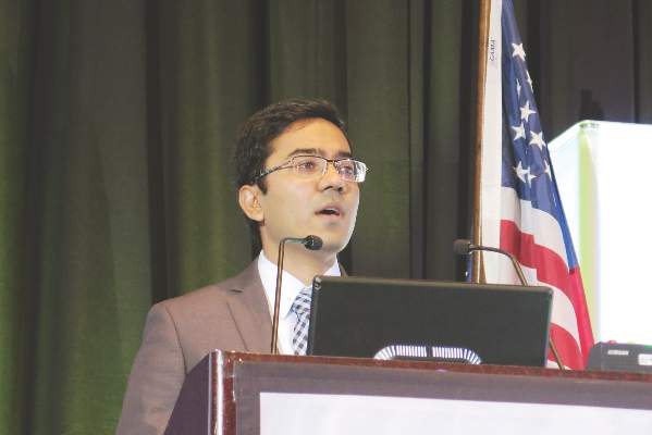
Accurate ID of nonsalvageable trauma patients improves trauma center performance metrics
LAKE BUENA VISTA, FL – When does no sign of life mean a patient is unsalvageable?
A study has found that up to 33% of patients who local providers determined had no signs of life went on to live, and 10% of patients whose heart stopped before reaching the hospital actually survived.
Historically, there’s been significant variation across trauma centers and registries of how unsalvageable patients are identified. This presents a problem when measuring trauma center performance, particularly when you consider that 25% of deaths occur within 15 minutes of arrival at high-volume trauma centers, Dr. James P. Byrne, with the University of Toronto, explained.
“Variation within inclusion and exclusion criteria can lead to big differences in risk-adjusted trauma center mortality. Therefore, there’s a need for the adoption of a single-best definition for unsalvageable patients,” he said at the annual scientific assembly of the Eastern Association for the Surgery of Trauma (EAST).
To that end, three case definitions of the unsalvageable patient were proposed based on data from the 2012-2013 American College of Surgeons Trauma Quality Improvement Program (ACS TQIP) database. They were no signs of life as determined by local providers (NSOL), prehospital cardiac arrest (PHCA) as entered into local trauma registries, and a PROXY for death established by the ACS TQIP and defined as an emergency department heart rate of 0 and an ED systolic blood pressure of 0 and a Glasgow Coma Scale motor component of 1.
Over the study period, 223,643 patients from 192 trauma centers met the inclusion criteria of at least 16 years of age, blunt or penetrating mechanism of injury, and known hospital discharge status. In-hospital mortality was 7.2%.
NSOL and PHCA, had positive predictive values low enough (66.58% and 89.71%) such that 33% and 10% of patients meeting these criteria went on to survive.
The PROXY had excellent predictive utility for death (PPV 99.09%), with just 0.9% of PROXY patients going on to survive (22/2,424), he said.
To test its validity, the investigators looked more closely at the 2,424 patients who met PROXY criteria (15% of all deaths). PROXY patients mostly fell into two distinct groups: severe multisystem blunt injury caused by motor vehicle collision (MVC) and penetrating trauma to the head or chest caused by firearm, Dr. Byrne said. The median time to death was 8 minutes, with 87% dying in the ED.
Among the 22 unexpected PROXY survivors, 77% had penetrating trauma. Most had isolated injuries to the heart, lung, or large blood vessels and underwent thoracotomy (71%) or open cardiac massage (35%).
“We feel these patients are adequately explained for the most part, even though they come in without vital signs, as patients that have some chance for survival with prompt hemorrhage control surgery or cardiac repair,” Dr. Byrne said.
PROXY patients with penetrating injury rather than blunt injury were more likely to be male (90% vs. 73%), younger (34 years vs. 44 years), and fall victim to firearms or stabbing than an MVC (88% & 12% vs. 59%).
Overall, patients with penetrating injury were 10 times more likely to meet PROXY criteria than those with blunt injury (5.7% vs. 0.6%; P < .001).
“The characteristics of the PROXY patients, as well as the significant association with penetrating trauma, is something that we know to be true for patients who die early from trauma,” Dr. Byrne said.
In contrast, the 13,659 patients who died without meeting PROXY criteria were older (mean age, 60 years), 72% had severe head injury from falls or MVC, and their median time to death was 52 hours.
“We feel these patients represent alternative trajectories to death that are not predicted by presenting characteristics,” he said. “This actually lends construct validity to the PROXY definition since it was able to isolate patients who died early, while excluding those patients who died later and might have a chance for a modifiable outcome.”
Finally, a hierarchical linear model that calculated risk-adjusted mortality was used to look at the influence of including nonsalvageable patients on trauma center performance. Based on the model, 36 trauma centers (19%) were below-average performers and 29 (15%) were above-average performers.
After excluding PROXY patients, 64% of trauma centers changed rank, 17% by three or more positions, but only two centers changed outlier status. The latter suggests that inclusion of unsalvageable PROXY patients would have a minor impact on risk-adjusted mortality used for peer-to-peer benchmarking. However, their inclusion could have a big impact on benchmarking at centers that receive unsalvageable patients more frequently and therefore, PROXY should be used to exclude them from registries, Dr. Byrne said.
Poster discussion comoderator Christopher J. Dente from Emory University in Atlanta, said the reason the PROXY model performs so well is that it is something that could easily translate from the bedside into a registry and from a registry to a national database, whereas measures like “no signs of life” have to translate from the field to the bedside to the registry and then TQIP.
“The same is true for prehospital cardiac arrest, which you’d think would be a little more tangible, but isn’t necessarily,” Dr. Dente said. “This is incredibly important work.”
Dr. Byrne and his coauthors reported having no financial disclosures.
Byrne et al.’s study evaluating three criteria for identifying unsalvageable trauma patients demonstrates the critical role that high-quality data can play in quality improvement efforts. Using data from the American College of Surgeons Trauma Quality Improvement Program (ACS TQIP), they developed a proxy measure that resulted in less than 1% of patients being incorrectly classified as unsalvageable.
Having an accurate, easy-to-calculate model for predicting survival is essential in applying these findings at the point of care, as the decision of whether or not to perform a resuscitative thoracotomy must be made expediently. An accurate model also allows resources to be concentrated on those who might derive the most benefit and minimizes the known harms, particularly to health care providers, and costs associated with resuscitative thoracotomies. Finally, an accurate model is necessary to ensure that benchmarking accurately reflects quality of care rather than case mix, and that efforts are appropriately directed toward those centers whose adjusted mortality is above expected, whether or not they are outliers.
Future efforts should be directed toward evaluating whether implementation of this proxy measure into clinical practice improves not just survival but survival with good functional status at a longer term end point.
Dr. Lillian S. Kao is an ACS Fellow and associate professor of surgery at the University of Texas Health Science Center at Houston.
LAKE BUENA VISTA, FL – When does no sign of life mean a patient is unsalvageable?
A study has found that up to 33% of patients who local providers determined had no signs of life went on to live, and 10% of patients whose heart stopped before reaching the hospital actually survived.
Historically, there’s been significant variation across trauma centers and registries of how unsalvageable patients are identified. This presents a problem when measuring trauma center performance, particularly when you consider that 25% of deaths occur within 15 minutes of arrival at high-volume trauma centers, Dr. James P. Byrne, with the University of Toronto, explained.
“Variation within inclusion and exclusion criteria can lead to big differences in risk-adjusted trauma center mortality. Therefore, there’s a need for the adoption of a single-best definition for unsalvageable patients,” he said at the annual scientific assembly of the Eastern Association for the Surgery of Trauma (EAST).
To that end, three case definitions of the unsalvageable patient were proposed based on data from the 2012-2013 American College of Surgeons Trauma Quality Improvement Program (ACS TQIP) database. They were no signs of life as determined by local providers (NSOL), prehospital cardiac arrest (PHCA) as entered into local trauma registries, and a PROXY for death established by the ACS TQIP and defined as an emergency department heart rate of 0 and an ED systolic blood pressure of 0 and a Glasgow Coma Scale motor component of 1.
Over the study period, 223,643 patients from 192 trauma centers met the inclusion criteria of at least 16 years of age, blunt or penetrating mechanism of injury, and known hospital discharge status. In-hospital mortality was 7.2%.
NSOL and PHCA, had positive predictive values low enough (66.58% and 89.71%) such that 33% and 10% of patients meeting these criteria went on to survive.
The PROXY had excellent predictive utility for death (PPV 99.09%), with just 0.9% of PROXY patients going on to survive (22/2,424), he said.
To test its validity, the investigators looked more closely at the 2,424 patients who met PROXY criteria (15% of all deaths). PROXY patients mostly fell into two distinct groups: severe multisystem blunt injury caused by motor vehicle collision (MVC) and penetrating trauma to the head or chest caused by firearm, Dr. Byrne said. The median time to death was 8 minutes, with 87% dying in the ED.
Among the 22 unexpected PROXY survivors, 77% had penetrating trauma. Most had isolated injuries to the heart, lung, or large blood vessels and underwent thoracotomy (71%) or open cardiac massage (35%).
“We feel these patients are adequately explained for the most part, even though they come in without vital signs, as patients that have some chance for survival with prompt hemorrhage control surgery or cardiac repair,” Dr. Byrne said.
PROXY patients with penetrating injury rather than blunt injury were more likely to be male (90% vs. 73%), younger (34 years vs. 44 years), and fall victim to firearms or stabbing than an MVC (88% & 12% vs. 59%).
Overall, patients with penetrating injury were 10 times more likely to meet PROXY criteria than those with blunt injury (5.7% vs. 0.6%; P < .001).
“The characteristics of the PROXY patients, as well as the significant association with penetrating trauma, is something that we know to be true for patients who die early from trauma,” Dr. Byrne said.
In contrast, the 13,659 patients who died without meeting PROXY criteria were older (mean age, 60 years), 72% had severe head injury from falls or MVC, and their median time to death was 52 hours.
“We feel these patients represent alternative trajectories to death that are not predicted by presenting characteristics,” he said. “This actually lends construct validity to the PROXY definition since it was able to isolate patients who died early, while excluding those patients who died later and might have a chance for a modifiable outcome.”
Finally, a hierarchical linear model that calculated risk-adjusted mortality was used to look at the influence of including nonsalvageable patients on trauma center performance. Based on the model, 36 trauma centers (19%) were below-average performers and 29 (15%) were above-average performers.
After excluding PROXY patients, 64% of trauma centers changed rank, 17% by three or more positions, but only two centers changed outlier status. The latter suggests that inclusion of unsalvageable PROXY patients would have a minor impact on risk-adjusted mortality used for peer-to-peer benchmarking. However, their inclusion could have a big impact on benchmarking at centers that receive unsalvageable patients more frequently and therefore, PROXY should be used to exclude them from registries, Dr. Byrne said.
Poster discussion comoderator Christopher J. Dente from Emory University in Atlanta, said the reason the PROXY model performs so well is that it is something that could easily translate from the bedside into a registry and from a registry to a national database, whereas measures like “no signs of life” have to translate from the field to the bedside to the registry and then TQIP.
“The same is true for prehospital cardiac arrest, which you’d think would be a little more tangible, but isn’t necessarily,” Dr. Dente said. “This is incredibly important work.”
Dr. Byrne and his coauthors reported having no financial disclosures.
AT THE EAST SCIENTIFIC ASSEMBLY 2015
Key clinical point: The ACS TQIP proxy definition of DOA should be used to exclude unsalvageable patients from peer-to-peer benchmarking and performance improvement efforts.
Major finding: Just 0.9% of patients identified as being unsalvageable by the PROXY criteria went on to survive.
Data source: Retrospective analysis of 223,643 trauma patients in the ACS TQIP database.
Disclosures: Dr. Byrne and his coauthors reported having no financial disclosures.