User login
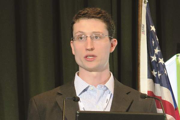
New scoring system for small bowel–obstruction severity
LAKE BUENA VISTA, FLA. – A novel three-item scoring system reliably categorizes severity of small bowel obstruction and is more strongly associated with in-hospital mortality than the American Association for the Surgery of Trauma anatomic score alone.
The AAST developed a scoring system to standardize the severity of small-bowel obstruction (SBO) based on anatomic criteria. Its authors have subsequently recommended, however, that other parametersare needed that would take into consideration the entirety of the patient’s clinical situation (J. Trauma Acute Care Surg. 2014;77:705-8 and J. Trauma Acute Care Surg. 2014;76:884-7).
To that end, investigators at the Mayo Clinic in Rochester, Minn., created the Acute General Emergency Surgical Severity-Small Bowel Obstruction (AGESS-SBO) system that incorporates presenting physiology and pre-existing comorbidities with anatomic criteria.
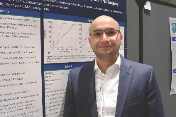
“It’s evident that the complications and patient outcomes clearly depend on the extent of the involvement of the diseased organ, but also depend on the hosting environment, which means the patient’s physiology and pre-existing conditions,” Dr. Yaser Baghdadi explained at the annual scientific assembly of the Eastern Association for the Surgery of Trauma.
He reported a cohort study involving 377 patients who were treated for SBO at the Mayo Clinic between 2009 and 2012 and evaluated using anatomic criteria and the AGESS-SBO, which uses a 5-point scoring system for each of its three scales.
Most patients (57%) received a score of 1 on the AGESS-SBO anatomic involvement scale for a partial SBO without need of operation, while only 1% had a score of 5, indicating strangulation and perforation with diffuse peritoneal contamination.
On the physiology scale, 58.6% had no physiologic derangement or a score of 0, 36% had a score of 1 because of systemic inflammatory response syndrome, and only 1.1% had a score of 5 for multiple organ dysfunction syndrome.
A Charlson comorbid score of 1 or 2 earned 32% of patients 1 point on the comorbidity scale, while 4% had a score of 5 because of a Charlson score of 9 or more.
In all, 215 patients (57%) had nonoperative treatment and 162 patients (43%) underwent surgical exploration. The median overall AGESS-SBO score was 6 points (interquartile range [IQR], 3-13 points).
The median length of stay (LOS) was 5 days (IQR, 3-9.5 days), with 94 patients (25%) having a stay exceeding 9.5 days, Dr. Baghdadi said in the poster presentation. In-hospital complications occurred in 82 patients (22%) and eight patients (2%) died during their hospital stay.
Comparison of the areas under receiver operative characteristic curves revealed a statistically significant greater association between the AGESS-SBO score and in-hospital mortality than the AAST anatomic score (AUC, 0.79 vs. 0.55, P value = .015), reported Dr. Baghdadi, a research fellow in the Mayo Clinic’s trauma division.
The two scoring systems had comparable ability to predict in-hospital complications (AUC, 0.72 vs. 0.69; P = .42) and extended LOS (AUC, 0.72 vs. 0.74; P = .47). The lack of statistical significance favoring the AGESS-SBO may be because these outcomes would be more likely in patients requiring surgery and the analysis combined patients who did and did not require operative care, he said in an interview.
“The AGESS-SBO system is a useful tool to classify the disease severity among SBO patients compared to the AAST anatomic score alone. We are planning to run a prospective study to validate what we have found,” he added.
Dr. Baghdadi and his coauthors reported having no financial disclosures.
AT THE EAST SCIENTIFIC ASSEMBLY
Key clinical point: Adding presenting physiology and comorbidities to anatomic criteria provides a reliable tool to categorize severity of small-bowel obstruction.
Major finding: The AGESS-SBO score was significantly associated with in-hospital mortality, versus the AAST anatomic score (area under ROC curves: 0.79 vs. 0.55; P = .015).
Data source: A cohort study of 377 patients treated for small-bowel obstruction.
Disclosures: Dr. Baghdadi and his coauthors reported having no financial disclosures.
Base deficit and lactate vary with resuscitation fluid type
LAKE BUENA VISTA, FLA. – Base deficit and lactate after resuscitation were measurably different based on the type of crystalloid solution used in a class I hemorrhage model.
Further, an award-winning prospective study found that, compared with lactated Ringer’s or no intravenous fluid, normal saline results in significantly higher postresuscitation sodium and chloride levels and significantly lower ionized calcium and bicarbonate.
Taken together, these derangements are important because blood gases are one of the first objective measurements performed in acute trauma patients, Dr. Samuel Wade Ross said at the annual scientific assembly of the Eastern Association for the Surgery of Trauma (EAST).
“We might actually be overestimating the amount of shock due specifically to the iatrogenic cause of crystalloid solutions,” he said. “Additionally, this goes beyond just trauma because all medicine – surgery, anesthesia, and critical care – use crystalloid. And it should be in the back of our minds when using normal saline that this contributes to acidosis and that lactated Ringer’s can falsely elevate lactate levels.”
The analysis involved 157 voluntary blood donors, who donated 0.5 L and were then randomly assigned to normal saline, lactated Ringer’s (LR), or no IV fluid. The percentage of total blood volume lost was about 11%, which is consistent with a class I hemorrhage model, said Dr. Ross of the Carolinas Medical Center in Charlotte, N.C.
Base deficit, which is used to guide the volume of fluid needed for trauma patients’ resuscitation, was similar before administration of normal saline, lactated Ringer’s, or no IV fluid (–0.24 vs. 0.33 vs. 0.04).
After fluid administration, however, the normal saline group had almost five times the base deficit of the no IV fluid group (–3.06 vs. –0.65) and almost 10 times the base deficit of the LR group (–3.06 vs. –0.34). The differences were statistically significant, even using a conservative statistical correction with a P value cutoff of 0.0167, he said.
Preresuscitation lactate levels also were similar in the LR, normal saline, and no IV groups (1.05 mmol/L vs. 1.12 mmol/L vs. 1.10 mmol/L).
Postresuscitation, however, lactate increased by roughly 50% in the LR group vs. the normal saline group (1.54 mmol/L vs. 1.0 mmol/L) and was elevated compared with no IV fluid (1.54 mmol/L vs. 1.36 mmol/L). Both findings were statistically significant (P < .0167).
This is the first time these differences have been quantified and runs contrary to the dogma that serum lactate does not increase with the use of LR because of enzymatic clearance of the molecule in the liver, said Dr. Ross, the EAST 2015 Raymond Alexander Residents Paper Competition winner.
“With ongoing shock, lactate rises and clinicians use that as a guide for further fluid resuscitation. Thus, lactate levels could be falsely elevated with LR use, and drive further decisions for unnecessary and potentially harmful additional resuscitation and procedures,” he said in an interview.
As noted above, use of normal saline rather than LR or no IV fluid resulted in significantly higher postresuscitation sodium (141.7 mmol/L vs. 139.8 mmol/L vs. 139.8 mmol/L) and chloride (107.3 mmol/L vs. 102.3 mmol/L vs. 102.9 mmol/L), and significantly lower ionized calcium (1.15 vs. 1.22 vs. 1.24), pH (7.32 vs. 7.34 vs. 7.36), and bicarbonate (23 mmol/L vs. 25.3 mmol/L vs. 24.6 mmol/L; all P values < .001).
“The two recommended isotonic crystalloid fluids used for hemorrhagic and other forms of shock – normal saline and lactated Ringer’s – have been in use since the 19th century and early 20th century,” senior author Dr. Ronald F. Sing said in an interview. “Despite the tremendous advances in shock and resuscitation, we have identified, and actually confirmed, potentially confounding factors related to both LR and [normal saline] for resuscitation. Our next goal is to examine other crystalloid solutions and their impacts not only on shock markers, but inflammatory markers,” he added.
Future studies also will use animal models to look at class II-IV hemorrhage and increased follow-up time.
The study was supported by the Carolinas Trauma Network. Dr. Ross and his coauthors reported having no financial disclosures.
AT THE EAST SCIENTIFIC ASSEMBLY
Key clinical point: Quantifiable differences exist in base deficit and lactate based on the type of resuscitation fluid used in a class I hemorrhage model.
Major finding: Base deficit was dramatically lower after normal saline vs. no intravenous fluid or lactated Ringer’s (–3.06 vs. –0.65 vs. –0.34; P <.001).
Data source: Prospective study in 157 blood donors.
Disclosures: The study was supported by the Carolinas Trauma Network. Dr. Ross and his coauthors reported having no financial disclosures.
VIDEO: Cast a wider net for intimate partner and sexual violence
LAKE BUENA VISTA, FLA. – Screening for intimate partner and sexual violence occurs sporadically in trauma centers, typically among women and when there is a suspicious mechanism of injury.
In hopes of reaching a broader population, investigators at the Ryder Trauma Center at the University of Miami conducted a prospective pilot study to determine the feasibility of universal screening for intimate partner and sexual violence in all patients presenting at the level I trauma center.
In all, 399 consecutive patients were eligible, with 40% screened using a four-item questionnaire that asks patients how often their partner physically hurt, insulted, threatened with harm, and screamed at them (HITS) and the SAVE (screen, ask, validate, evaluate) method developed in Florida to screen for sexual violence. Both instruments were available in English, Spanish, and Haitian French.
Over a 4-month period, 14% of patients scored positive for physical and psychological abuse – and even more surprising, 8% scored positive for sexual violence, lead study author Dr. Tanya Zakrison reported at the annual scientific assembly of the Eastern Association for the Surgery of Trauma (EAST).
“Interestingly, there was no significant difference between men and women. They were all scoring at an equal rate,” Dr. Zakrison said. Patients who scored positive on HITS or SAVE were also found across all ethnicities and all age groups.
“The last thing that was quite surprising for us to find was that only 14% of patients who were HITS positive were actually admitted as a result of intimate partner or interpersonal violence, and none of the patients who scored positive for SAVE were admitted to trauma because of this,” she added. “Again, they’re coming in for the motor vehicle collisions, falls from standing, or other mechanisms of injury.”
To learn more about the intervention and its impact at Ryder Trauma, watch our video interview.
Dr. Zakrison and her coauthors reported no relevant financial conflicts.
The video associated with this article is no longer available on this site. Please view all of our videos on the MDedge YouTube channel
AT THE EAST SCIENTIFIC ASSEMBLY
Botox benefits in primary fascial closure questioned
LAKE BUENA VISTA, FLA. – Contrary to prior results, use of onabotulinumtoxinA did not improve primary fascial closure rates after damage control laparotomy in a multicenter, prospective study.
“Botox injections were safe, but they did not have any effect on our primary or secondary endpoints,” Dr. Martin D. Zielinski said at the annual scientific assembly of the Eastern Association for the Surgery of Trauma (EAST).
Dr. Zielinski and his colleagues at Mayo Clinic, Rochester, Minn., previously published retrospective data showing that use of Botox-induced paralysis of the abdominal wall musculature resulted in a primary fascial closure rate of 83% among 18 patients with open abdomen (OA) and 89% if injected within 24 hours of the initial OA procedure (Hernia 2013;17:101-7).
In contrast, the primary closure rate was just 66% in a prospective American Association for the Surgery of Trauma study involving 572 patients requiring OA management after damage control laparotomy (J. Trauma Acute Care Surg. 2013;74:113-20).
Though negative pressure dressings and use of patches have been shown to increase primary closure by facilitating the midline tension, up to 30% of patients will not achieve primary fascial closure. Botox injections block the release of acetylcholine, thereby preventing a rush of calcium to the abdomen and inducing flaccid paralysis of the lateral abdominal wall muscles, Dr. Zielinski explained.
To test their hypothesis that Botox would improve rates of primary fascial closure, decrease hospital stay, and enhance pain control, 46 patients who had undergone damage control laparotomy were randomly assigned to six separate injections of their external, oblique, internal oblique, and transverse abdominal muscles with 150 cc/injection of Botox A or sodium chloride 0.9%.
The two groups were well matched, with the exception of Botox patients having a significantly lower body mass index (BMI) than did controls (30 kg/m2vs. 26.3 kg/m2) and receiving more intraoperative packed red blood cells (8 units vs. 4.8 units).
Primary fascial closure rates were unexpectedly high, but did not differ between the Botox and control groups (96% vs. 93%; hazard ratio, 1.0), Dr. Zielinski said.
Secondary endpoints were also similar between the Botox and control groups including average time until closure (5 days vs. 2 days; P = .15), hospital stay (24 days vs. 19 days; P = .19), median ICU duration (8 days vs. 6 days; P = 32), and wound infection rate (8% vs. 0%; P > .99).
Given the potential pain effects of Botox, the investigators anticipated a difference in pain medication use, but morphine equivalents were actually slightly higher in the Botox group on postoperative day 1 (120 mg vs. 81.8 mg; P = .27) through day 5 (57.3 vs. 49.0 mg; P = .47), he said.
In trying to explain why the injections failed to prove beneficial, Dr. Zielinski said, “The question that crosses my mind is did we exclude the wrong patients?”
A total of 181 trauma or emergency general surgery patients, aged 18 years and older, with a damage-control laparotomy per the surgeon’s discretion were eligible for the double-blind study, but 87 were excluded because they had one or more exclusion criterion: a body mass index ≥ 50, hemodynamic instability, complicated chronic obstructive pulmonary disease, impaired neuromuscular transmission, aminoglycoside use, international normalized ratio < 1.5, trunk necrotizing fasciitis, metastatic malignancy, or were pregnant or a prisoner.
Discussant Michael Rotondo of the University of Rochester (New York) Medical Center, applauded the authors for presenting a negative study and called it “impeccably designed” and “a great piece of work.”
Dr. Rotondo went on to ask whether there was any relationship between degree of advancement, abdominal breach, or lateral diameter that predicts success with Botox.
Despite the use of surface wave elastography to measure abdominal tension in eight patients and use of a handheld durometer, Dr. Zielinski said they were unable to accurately measure degree of advancement. Future work may look at the relationship between these factors and will include long-term follow-up to evaluate hernia rates and quality of life. In the one patient with full elastography measurements, abdominal tension trended lower for 2 days after the Botox injection before increasing after primary closure, he said.abdominal tension trended lower for 2 days after the Botox injection before increasing after primary closure, he said.
The study was funded by the EAST Scholar program. Dr. Zielinski, his coauthors, and Dr. Rotondo reported having no financial disclosures.
AT THE EAST SCIENTIFIC ASSEMBLY
Key clinical point: The use of Botox to induce flaccid paralysis of the abdominal wall muscles is of questionable value in achieving primary facial closure.
Major finding: Primary fascial closure rates did not differ between the Botox and control groups (96% vs. 93%; HR, 1.0).
Data source: Prospective randomized trial in 46 patients who had undergone damage control laparotomy.
Disclosures: The study was funded by the EAST Scholar program. Dr. Zielinski, his coauthors, and Dr. Rotondo reported having no financial disclosures.
Peer pressure moves dial on restricting RBC transfusions
LAKE BUENA VISTA, FLA. – A multimodal intervention founded on prompt peer-to-peer review increased adherence to restrictive red blood cell transfusion guidelines without increasing mortality.
During the intervention, if patients were transfused outside of established hospital guidelines, all clinicians taking care of the patient received an e-mail notification within 72 hours of transfusion. This included the ICU staff, primary team, intern, resident, fellow, nurse practitioner, and attending, said Dr. Daniel Yeh, a trauma and critical care surgeon at Massachusetts General Hospital in Boston whose signature appears on the e-mail along with the endorsement of the surgeon-in-chief, anesthetist-in-chief, director of the MGH Critical Care Center, codirector of blood transfusion services, and the urology chief.
The e-mail blast was coupled with a 1-hour educational lecture during surgery grand rounds on the potential harms of and indications for blood transfusion, a surgical ICU didactic lecture, and monthly division-wide reports.
Prior to the intervention, providers felt they were probably doing pretty well in terms of using a more restrictive transfusion strategy, Dr. Yeh said. After all, there are strong guidelines in all the professional societies and 15 years of high-quality, level 1 evidence from the pivotal 1999 TRICC (Transfusion Requirements in Critical Care) trial to last year’s data in traumatic brain injury (TBI) patients (JAMA 2014;312:36-47) showing that a lower transfusion threshold is noninferior, if not superior, to a higher transfusion threshold.
“Yet, anecdotally in my own practice and objectively in observational data, we see what can only be euphemistically described as a knowledge-practice gap,” he said at the annual scientific assembly of the Eastern Association for the Surgery of Trauma (EAST). “Basically, we weren’t practicing what we were preaching.”
A record review of 144 patients from January to June 2013 found that fully 25% of all transfusions in stable, low-risk ICU patients had a pretransfusion hemoglobin (Hb) trigger > 8.0 g/dL.
Further, 5% of patients received the old standby 2-unit transfusion without an intervening hemoglobin measurement, which has been recommended against by all major medical societies, Dr. Yeh said.
The overtransfusion rate, defined as a post-transfusion level > 10 g/dL, was 11%. Most of this was accounted for by these 2-unit patients, he added.
Stable, low-risk patients comprised 33% of all transfusions during the review period and were identified using liberal exclusion criteria including anyone with visible bleeding or suspected internal bleeding, hemodynamic instability, ischemia (myocardial, intestinal, or peripheral vascular), or who were high risk (defined by a history of coronary artery disease, coronary artery bypass grafting, or congestive heart failure).
After the October 2013 to March 2014 intervention, the percentage of stable, low-risk patients with a Hb trigger of > 8.0 g/dL declined from 25% to 2% (P < .001), with a 1-month audit of 15 patients showing a rebound up to 17% 6 months after the intervention ended, Dr. Yeh said.
“There was no difference in the patients who got a single transfusion; however, the ones who got multiple transfusions during their hospital stay decreased, so that is where we believe most of the decline came from,” he explained.
Among the 137 postintervention patients, the average pretransfusion trigger decreased significantly (7.6 g/dL vs. 7.1 g/dL; P < .001), before rising to 7.3 g/dL 6 months post intervention.
The number of monthly transfusions declined 35% from 47 units to 31 units (P = NS) and the overtransfusion rate fell from 11% to 3% (P = .004).
There were no significant differences in maximum lactate, maximum troponin, median ICU or hospital length of stay, although ICU discharge Hb (8.6 g/dL vs. 8.2 g/dL; P = .087) and hospital discharge Hb (9.0 g/dL vs. 8.6 g/dL; P = .006) were lower in the intervention period, Dr. Yeh said.
No significant differences were seen post intervention in 30-day readmission (23% vs. 16%; NS) or overall mortality (4% vs. 9%; NS).
“Transfusions were not really temporally related to the death nor did we attribute any of these deaths to symptomatic anemia,” he said.
Limitations of the intervention include the possibility of a Hawthorne effect, use of crude clinical outcomes, and the lack of concomitant control in the ICU, although the partial regression seen 6 months after the intervention suggests something about the intervention was working, Dr. Yeh observed.
Discussant Dr. Laura J. Moore of the University of Texas Health Science Center at Houston, congratulated the authors on an interesting and pertinent study, but questioned why they utilized a Hb threshold of 8 g/dL when the transfusion trigger was set at 7 g/dL in TRICC and in the Villanueva et al. study in acute upper gastrointestinal bleeders (N. Engl. J. Med. 2013;368:11-21).
“Certainly in my institution, we utilize a trigger of 7 [g/dL] and some of us might even push that down to 6 [g/dL],” she said.
Some of the landmark studies including FOCUS and TRACS used a trigger of 8 g/dL and the investigators wanted to give the clinicians a bit of a benefit of doubt, Dr. Yeh responded.
Results of the intervention were met with surprise, but were quickly reinforced with the publication of the TBI and Villanueva studies and an accompanying editorial (N. Engl. J. Med. 2014;371:1459-61) arguing that a transfusion threshold of 7 g/dL is the new normal, he noted.
“We’ve continued on daily rounds to really focus on that [trigger]. We’ve become empowered as the ICU team to say ‘No,’ to argue and at least put up roadblocks when the primary teams are requesting transfusions and ask them to justify why patients need the additional oxygen carrying capacity when they’re waiting on the floor for 3 days and doing totally fine,” Dr. Yeh said.
As for how they deal with offending providers or outliers, electronic records identify who signed each transfusion order, even if it’s in the dead of night, and simply showing physicians where they stack up with their peers can be the biggest driver of practice change. The team no longer performs monthly audits due to time constraints, but hopes to resume e-mail interventions once MGH’s transition to a new electronic medical system is complete, Dr. Yeh said in an interview.
AT THE EAST SCIENTIFIC ASSEMBLY
Key clinical point: Peer-to-peer review improves the use of restrictive transfusion guidelines without increasing mortality.
Major finding: The percentage of stable, low-risk patients with a hemoglobin trigger > 8 g/dL decreased from 25% to 2% post intervention (P < .001).
Data source: Prospective interventional study of 137 stable, low-risk patients with 144 retrospective controls.
Disclosures: Dr. Yeh, his coauthors, and Dr. Moore reported having no disclosures.
Trio of risk factors predict gangrenous cholecystitis
LAKE BUENA VISTA, FL. – Older age, diabetes, and elevated bilirubin were significant risk factors for acute gangrenous cholecystitis in a retrospective study of 489 patients undergoing cholecystectomy.
Patients with acute gangrenous cholecystitis (AGC) were on average 15 years older than those with cholecystitis without necrosis (CN) were (55.8 vs. 40.8 years; P value ≤ .001), almost five times more likely to have comorbid diabetes (32% vs. 6.7%; P≤ .05), and had significantly higher bilirubin levels (1.96 mg/dL vs. 0.89 mg/dL; P≤ .001).
The findings are consistent with previous studies showing that all three risk factors are strongly associated with gangrenous cholecystitis, Seda Bourikian reported at the annual scientific assembly of the Eastern Association for the Surgery of Trauma (EAST).
“Future studies may explore how the pathophysiology of diabetes, or the duration of illness in each patient, plays a role in the development of AGC,” the authors suggested in the poster presentation.
The chart review included 489 patients admitted to an emergency general surgery service who underwent cholecystectomy between January 2009 and April 2014. Retrospectively evaluated pathological specimen reports identified 464 patients with CN and 25 patients with AGC.
Male patients had a significantly higher incidence of AGC than CN (56% vs. 26%; P≤ .05), whereas women were less likely to have AGC (44% vs. 74%; NS), Ms. Bourikian and her colleagues at Virginia Commonwealth University in Richmond wrote.
Previous studies also have shown that acute cholecystitis is more common in men and patients over the age of 50 years.
Notably, lactate, obesity, and systolic blood pressure below 100 mm Hg were not different between groups.
As expected, patients with AGC were significantly more likely to die than their counterparts with cholecystitis without necrosis (16% vs. 0.86%; P≤ .05), the authors reported.
People with diabetes with AGC were almost five times more likely to die than were diabetics with CN (32% vs. 6.7%; P≤ .05). according to the authors.
Mortality, however, was nearly identical between AGC and CN patients with a systolic BP ≤ 100 mm Hg (0% vs. 0.02%; NS).
Logistic regression analysis showed that increased age (P≤ .001) and male gender (P≤ .05) were strongly associated with the development of AGC. The failure of more risk factors to pan out in logistic regression is likely because of the small number of patients with gangrenous cholecystitis, senior author and colleague Dr. Paula Ferrada of Virginia Commonwealth University suggested.
“This is not a common disease,” she said in an interview. “That’s why it’s so hard to diagnose and triage. Clinicians need to have a higher suspicion” of AGC.
AT THE EAST SCIENTIFIC ASSEMBLY
Key clinical point: Older age, diabetes and elevated bilirubin were risk factors for acute gangrenous cholecystitis.
Major finding: Patients with acute gangrenous cholecystitis vs. cholecystitis without necrosis were older (55.8 vs. 40.8 years; P value ≤ .001), more likely to have diabetes (32% vs. 6.7%; P≤ .05) and an elevated bilirubin (1.96 mg/dL vs. 0.89 mg/dL; P≤ .001).
Data source: Retrospective analysis of 489 patients undergoing cholecystectomy.
Disclosures: The authors reported having no relevant financial disclosures.
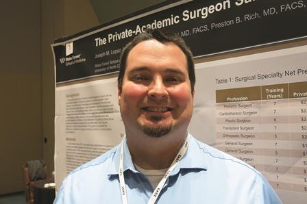
The private-academic surgeon salary gap: Would you pick academia if you stood to lose $1.3 million?
LAKE BUENA VISTA, FLA. – Academic surgeons earn an average of 10% or $1.3 million less in gross income across their lifetime than surgeons in private practice, an analysis shows.
Some surgical specialties fare better than others, with academic neurosurgeons having the largest reduction in gross income at $4.2 million (-24.2%), while academic pediatric surgeons earn $238,376 more (1.53%) than their private practice counterparts. They were the only ones to do so.
Several academic surgical specialties did not make the 10% average including trauma surgeons whose lifetime earnings were down 12% or $2.4 million, vascular surgeons at 13.8% or $1.7 million, and surgical oncologists at 12.2% or $1.3 million.
“The concern that we have is that the academic surgeons are where the education of the future lies,” lead study author Dr. Joseph Martin Lopez said at the annual scientific assembly of the Eastern Association for the Surgery of Trauma (EAST).
Every year a new class of surgeons is faced with the question of academic practice or private practice, but they are also struggling with increasing student loan debt and longer training as more surgical residents elect to enter fellowship rather than general practice. This growing financial liability coupled with declining physician reimbursement could rapidly shift physician practices and thus threaten the fiscal viability of certain surgical fields or academic surgical careers.
“The more financially irresponsible you make it to become an academic surgeon, the more we put at risk our current mode of training,” Dr. Lopez of Wake Forest University in Winston-Salem, N.C., said.
To account for additional factors outside gross income, the investigators ran the numbers using a second analysis, a net present value calculation, however, and came up with roughly the same salary gap to contend with.
Net present value (NPV) calculations are commonly used in business to calculate the profitability of an investment and also have been used in the medical field to gauge return on investment for various careers. The NPV calculation accounts for positive and negative cash flows over the entire length of a career, using in this case, a 5% discount rate and adjusting for inflation, Dr. Lopez explained.
Both the lifetime gross income and 5% NPV calculation used data from the Medical Group Management Association’s 2012 physician salary report, the 2012 Association of American Medical Colleges physician salary report, and the AAMC database for residency and fellow salary.
The NPV assumed a career length of 37-39 years, based on a retirement age of 65 years for all specialties. Positive cash flows included annual salary less federal income tax. Negative cash flows included the average principal for student loans, according to the AAMC, and interest at 5%, the average for the three largest student loan lenders in 2014, he said. Student loan repayment was calculated for a fixed-rate loan to be paid over 25 years beginning after residency or any required fellowship.
The average reduction in 5% NPV across surgical specialties for an academic surgeon versus a privately employed surgeon was 12.8% or $246,499, Dr. Lopez said.
Once again, academic neurosurgeons had the largest reduction in 5% NPV at 25.5% or a loss of $619,681, followed closely by trauma surgeons (23% or $381,179) and surgical oncologists (16.3% or $256,373). Academic pediatric surgeons had the smallest reduction in 5% NPV at 4.2% or $88,827.
During a discussion of the provocative poster, attendees questioned whether it was fair to say that private surgeons make more money without acknowledging the risk they face, compared with surgeons employed in an academic setting.
Dr. Lopez countered that increasingly, even private surgeons are no longer truly private surgeons.
“More and more surgical groups are being bought up by hospitals, and even the private surgical groups are being bought up by hospitals, which does stabilize your income to some extent,” he said. “We all still have RVU goals to meet and RVU incentives that make it so you can get paid a little more, but it’s something that’s a consideration. It is a risk-reward to be a private surgeon. Depending on how your contract is structured or how your group decides to pay the partners, it may be that if you don’t take very much call or take that many cases, you’ll end up on the short end of the stick.”
Dr. Ben L. Zarzaur, a general surgeon at Indiana University in Indianapolis who comoderated the poster discussion, pointed out that market pressures unaccounted for in the model can dramatically influence a surgeon’s salary over a lifetime.
Dr. Lopez agreed, citing how the increasing number of stent placements by cardiologists, for example, has impacted the bottom line of cardiothoracic surgeons. The NPV calculation was specifically used, however, because it gets at market forces such as inflation and return on investment, not addressed by gross income figures alone.
Finally, Dr. Zarzaur turned and asked the relatively young crowd what they would do if offered $600,000 a year, but had to work 110 hours a week or could get $250,000 and work only 40 hours a week. Most responded that they’d choose the former to repay their student loans and then switch to the lower-paying position. Responders made much of job satisfaction, work-life balance, and the ability of surgeons in academic practice to take time away from clinical work to conduct research, their ready access to continuing medical education, and their ability to educate the next generation of surgeons.
“Any time we see this academic-private disparity, you have to think about these secondary gains,” Dr. Zarzaur said. “This is really interesting work. It gets into why we choose what we do, why we’d take $600,000, work 110 hours a week, and get our rear ends kicked. The flip side is, if I saw this, why would you ever go into academics? But people still choose to do it. I’m in academics so there’s a bias, but we choose to do it anyway up to a point. I don’t know where that point is, but up to a point we do.”
AT THE EAST SCIENTIFIC ASSEMBLY
Key clinical point: Whether calculated as gross lifetime income or 5% net present value, a salary disparity exists between academic and private practice surgeons.
Major finding: Academic surgeons earn an average of 10% or $1.3 million less in gross lifetime income than surgeons in private practice.
Data source: Salary analysis and net present value calculation.
Disclosures: Dr. Lopez and his coauthors reported having no financial disclosures. Dr. Zarzaur disclosed honorarium from and serving as an advisor for Merck.
Regionalized trauma care trims 30-day mortality
LAKE BUENA VISTA, FLA. – Regionalized trauma care significantly reduces long-term mortality and maintains similar functional outcomes in patients with severe traumatic brain injury, a study showed.
Regionalization of trauma care is a health care strategy that attempts to improve outcomes for trauma patients by setting up a tiered, integrated system that aims to match the injured patient with the appropriate health care facility in a timely fashion. The Northern Ohio Trauma System (NOTS) was created in 2010 to manage trauma patients using the regionalization model.
In this study of outcomes in NOTS patients, longer-term follow-up of 3,496 severe traumatic brain injury (TBI) admissions showed 30-day mortality fell from 21% to 16% after regionalization (24% relative reduction; P value < .0001) and 6-month mortality declined from 24% to 20% (17% relative reduction; P = .004).
Multivariable logistic regression only strengthened the effect of regionalization on the primary outcome, lead study author Dr. Michael Kelly said at the annual scientific assembly of the Eastern Association for the Surgery of Trauma (EAST).
The odds ratio for 30-day mortality was 0.74, representing a 26% relative mortality reduction, and 0.82 for 6-month mortality, representing an 18% relative reduction.
At last year’s EAST meeting, Dr. Kelly and his colleagues reported that hospital mortality declined from 19% (262/1,359 patients) to 14% (302/2,137) in patients with severe TBI after the creation of the NOTS in 2010.
Despite bucking the current trend of rising mortality in hospitalized TBI patients, particularly those with severe brain injuries, their previous results were criticized by some as incomplete because hospital mortality was used without functional status measures or long-term mortality, he said.
To bridge the knowledge gap, the investigators identified all TBI patients older than 14 years with a Head Abbreviated Injury Scale (AIS) ≥ 3 from 2008 to 2012 in the NOTS database and matched them to the Ohio death index and the regional TBI rehabilitation database. Overall Functional Independence Measure (FIM) scores and FIM score gains were compared in 414 patients who were discharged to the regional TBI rehabilitation unit.
As a general rule of thumb, an overall FIM score of 60 is equivalent to 4 hours of personal care assistance in a nursing facility–type setting, a score of 80 equals 2 hours of personalized care in a nursing facility, more than 80 means the patient is able to receive family care at home, and ≥ 100 means minimal burdens in personal care, said Dr. Kelly of the MetroHealth Medical Center, Cleveland, and Cleveland Clinic.
A gain of 22 points is considered a minimal clinically important difference (MCID) for the overall FIM score. The MCID is 17 points for a FIM motor subscale gain, 3 for a FIM cognitive subscale gain, and has not been established for the FIM social subscale.
Overall FIM scores were similar before and after regionalization of trauma care (RT) at admission (54 vs. 48; P = .2) and at discharge (92 vs. 89; P = .1), he said.
FIM scores were similar in both groups at admission and discharge on the cognitive and social subscale domains, but were significantly lower post RT on the motor subscale at admission (38 vs. 31; P = .02) and discharge (68 vs. 65; P = .03). These differences were not clinically significant, according to Dr. Kelly and senior study author Dr. Jeffrey Claridge, NOTS medical director.
Pre- and post-RT patients had similar overall FIM score gains (37 vs. 36; P = .6), motor subscale gains (both groups 29), and social subscale gains (both groups 1). FIM cognitive subscale gains were significantly lower post RT (6 vs. 5; P = .01), but this difference was also not clinically significant.
Notably, discharges to regional TBI rehabilitation increased from 9% before RT to 14% after RT, Dr. Kelly said. The percentage of patients who were discharged to a skilled nurse facility or long-term care facility remained stable at about 30%, as did the percentage discharged home at about 40%.
“Regionalization improves long-term survival and maintains similar functional outcomes for patients with severe traumatic brain injury,” he concluded.
Discussant Dr. Jeffrey Coughenour of the University of Missouri Health System in Columbia said it appears regionalization is working, but added, “While we are saving more lives, what kind of lives are we saving? A question that has ever increasing implications for patients and payers evaluating the care we provide.”
He praised the investigators for using the FIM scale rather than the Glasgow Outcome Scale to try and answer this question, but said more information is needed on whether FIM scores improved in more challenging patients such as those with an AIS score of 4 or 5 or those entering rehabilitation after discharge to a skilled nursing or long-term care facility.
Data were not broken down for these more challenging subsets, Dr. Kelly said. The question of quality of life post regionalization was asked after the first study and that functional status was shown to be maintained in TBI patients in the follow-up study.
“Since no major changes in the hospital-based care or rehabilitation care of these TBI patients occurred, we weren’t surprised to see that functional outcomes did not improve,” he said in an interview. “The regionalization protocols were designed primarily to improve survival.”
During a discussion of the results, audience members questioned whether the investigators could be certain the results could be attributed to regionalization and not improvements in treatment of concurrent injuries or improvements in TBI treatment already underway at the time of policy change.
For the most part, these patients had isolated TBIs and no major changes in personnel or TBI care occurred during the study period, Dr. Kelly said.
Under NOTS, region-wide initiatives included use of the Centers for Disease Control and Prevention guidelines for field triage, a transfer line and transfer protocols, and a research database shared between two large hospital systems comprising the level I MetroHealth Medical Center trauma center, two level II trauma centers, and 12 nontrauma hospitals.
Dr. Kelly, his coauthors, and Dr. Coughenour reported no financial disclosures.
AT THE EAST SCIENTIFIC ASSEMBLY
Key clinical point: Regionalized trauma care reduces long-term mortality and maintains functional outcomes in patients with severe TBI.
Major finding: RT reduced 30-day mortality from 21% to 16% (P < .0001) and 6-month mortality from 24% to 20% (P = .004).
Data source: Analysis of 3,496 patients with severe TBI.
Disclosures: Dr. Kelly, his coauthors, and Dr. Coughenour reported no financial disclosures.
Checklist dramatically improves safety of bedside tracheostomy
LAKE BUENA VISTA, FLA. – Use of a multidisciplinary safety checklist prior to all bedside bronchoscopy-guided percutaneous tracheostomies was independently associated with a dramatic reduction in adverse procedural events, a study showed.
After adjustment for age, vitals, tracheostomy risk factors, and ICU duration after the procedure, multivariable logistic regression determined that no other factor – not age, baseline heart rate, baseline oxygen saturation, or difficult airway status – was significant against adverse procedural events except use of the checklist (odds ratio, 5.85; P = .022
Overall, in the postchecklist group, 3.2% experienced adverse events, compared with 14.7% in the prechecklist group.
“We feel this checklist could be used by other institutions with similar success and also could be modified for other invasive bedside procedures,” lead study author Dr. Joshua Hazelton said at the annual scientific assembly of the Eastern Association for the Surgery of Trauma (EAST).
Bedside bronchoscopy-guided percutaneous tracheostomies (BBPT) are safe, yet like many bedside procedures are seldom subject to the same safety checklists and timeout processes that have become standard in the operating room, he observed. When asked specifically how the checklist improved safety, Dr. Hazelton said the multidisciplinary aspect of the checklist distills a sense of ownership in every team member. A significant amount of evidence published on the benefits of World Health Organization checklists also shows checklists improve communication and empower members to speak up if something doesn’t feel right.
“Most of all, the checklist helped to focus the team on a procedure that really has very little margin of error,” he said. “As we do more of these procedures, I think the tendency for the entire team can be to look at these procedures as almost routine, and certainly with an airway procedure, I don’t think you should look at it as a routine thing.”
The safety checklist was created by a team of surgeons, critical care nurses, respiratory therapists, and trauma technicians at Cooper University Hospital in Camden, N.J., and focused on preprocedural setup, procedural steps, and a group timeout process.
In the equipment setup phase, the bedside nurse, respiratory therapist, and trauma technician must assemble and test all necessary equipment.
During the timeout process, team members are introduced, the nurse confirms the patient, the planned procedure, positioning of the patient, medications, and equipment, and the surgeon discusses any critical steps or steps that may be unique to the patient. Finally, each team member is given an opportunity to discuss any concerns or ask any questions prior to proceeding.
“The idea of this checklist is to bring the team together, so if there is one member of the team who doesn’t feel right about the procedure or has concerns, the procedure is stopped,” said Dr. Hazelton of the department of surgery at Cooper.
The last phase of the checklist is a postprocedural assessment to make sure the tracheostomy is properly secured and the patient is stable.
To assess its impact, the investigators prospectively analyzed all 63 patients who underwent bedside tracheostomy at the level I trauma center from checklist implementation July 1, 2013 to June 30, 2014 and compared them with 184 historical controls who underwent bedside tracheostomy without the checklist from July 1, 2011 to June 30, 2013. All tracheostomies were performed using the Blue Rhino Percutaneous Tracheostomy Kit (Cook Medical).
The pre- and postchecklist groups had similar numbers of patients with potentially difficult airways including those with cervical spine fracture (30 patients vs. 4 patients; P = .056), cervical ligamentous injury (3 vs. 0; not significant), occipital condyle fracture (3 vs. 0; NS), cervical spinal cord injury (4 vs. 2; NS), mandible fracture (8 vs. 2; NS), LeFort III fracture (5 vs. 1; NS), or oral cavity injury (5 vs. 1; NS).
Prechecklist patients were younger than postchecklist patients (48 years vs. 57 years; P = .001), but all other baseline characteristics were well matched including gender, body mass index, duration of mechanical ventilation, heart rate, mean arterial pressure, and oxygen saturation.
The pre- and postchecklist groups had the same occurrence of individual adverse procedural events including loss of airway (2 vs. 0; P = 1.0), deterioration in respiratory parameters during procedure (7 vs. 2; P = 1.0), hemodynamic compromise (19 vs. 2; P = .079), need for vasopressors or antiarrhythmics are (1 vs. 0; P = 1.0), and conversion to open procedure (5 vs. 0; P = .333). But overall, the postchecklist group experienced significantly fewer adverse events (3.2% vs. 14.7% prechecklist; P = .014), Dr. Hazelton said.
Discussant Dr. Bradley Dennis of Vanderbilt University Medical Center in Nashville, Tenn., said, “I don’t think we even need P values to identify this as a successful intervention, so I commend you for your excellent work.”
As bedside surgical procedures become more prevalent, it only stands to reason that the same patient safety measures used in the OR would be extended into the ICU, he said. Indeed, Vanderbilt has employed a preprocedure checklist for all procedures in the surgical trauma ICU since 2007, but unfortunately did not measure the outcomes prospectively.
Dr. Dennis went on to ask whether there were any patients in whom the checklist identified something that caused the team to abort the procedure.
Early in the process, there were several near-misses that were difficult to categorize, but typically revolved around not having a specific piece of rescue equipment in the room such as an airway box, Dr. Hazelton said. One particular case also raised concerns on the part of the respiratory therapist because of the patient’s large head, short neck, and body habitus, and after discussion, the procedure was halted.
“It was done in the operating room the next day and my colleague who performed that procedure told me it was somewhat challenging, a difficult cut down, a T-bar wing,” he said. “But certainly, that level of trust in your other colleagues, nursing, technicians, respiratory, is necessary for success of this kind of checklist.”
Dr. Hazelton and his coauthors reported having no financial disclosures.
AT THE EAST SCIENTIFIC ASSEMBLY
Key clinical point: A multidisciplinary checklist before percutaneous bedside tracheostomy can improve adverse event rates and overall safety.
Major finding: The overall adverse procedural event rate declined from 14.7% to 3.2% post checklist (P = .014).
Data source: Analysis of 247 patients who underwent percutaneous tracheostomy.
Disclosures: Dr. Hazelton and his coauthors reported having no financial disclosures.
Transsplenic TIPS procedure catching on for portal vein thrombosis
CHICAGO – Portal vein recanalization using a transsplenic approach can be utilized to improve transplant candidacy in patients with cirrhosis and chronic portal vein thrombosis, according to Dr. Bartley Thornburg.
“It’s a safe and effective procedure that allows for end-to-end anastomoses at transplant in patients who otherwise would not be able to have them or would require thrombectomy without transplant, and we know that end-to-end anastomoses are associated with decreased morbidity and mortality,” said Dr. Thornburg of Northwestern University Medical Center in Chicago.
Historically, and at his institution, portal vein thrombosis (PVT) is a relative contraindication to liver transplant. The American Association for the Study of Liver Diseases also recommends that transjugular intrahepatic portosystemic shunt (TIPS) placement be avoided in patients with a Model for End-State Liver Disease (MELD) score >18.
In 2013, colleague Dr. Riad Salem demonstrated the efficacy of portal vein recanalization during TIPS using a transhepatic approach in 44 patients, with only one technical failure and three cases of rethrombosis. Among six patients with a baseline MELD score >18, four went on to successful liver transplant, one is awaiting transplant, and one died as a result of bleeding 45 days post procedure despite an improvement in MELD score.
The last three patients in the series underwent TIPS with a transsplenic approach, and not only were the results equally good, but the approach was technically easier, Dr. Thornburg said at a symposium on vascular surgery sponsored by Northwestern University.
Since then, this increasingly common alternative approach has been assessed in another 11 consecutive patients with cirrhosis, portal hypertension, and chronic PVT. All patients had been denied listing for transplant because of their PVT, and four had a baseline MELD score >18.
At the end of the procedure, thrombus persisted in 45% (5 of 11 patients). On a 1-month follow-up venogram, however, three of the five patients had complete resolution of the thrombus without any added anticoagulation, one had persistent partial thrombus that was smaller than at stent placement, and one went on to transplant, Dr. Thornburg said.
All six of the patients with portal vein (PV) patency post procedure have retained patency after a median follow-up of 6.4 months.
“What we’ve learned from doing these cases is that complete elimination of the portal vein thrombus at the time of TIPS placement isn’t necessary,” he said. “It would be easy to get carried away and do suction or AngioJet [mechanical thrombectomy], but what we’ve found is that because of how much flow there is in the portal vein once it’s recanalized and the TIPS is in place, basically establishing a flow allowed for clot clearance by 1 month in all patients, except one.”
The procedure starts like a typical TIPS, with access achieved by advancing a 21-guage needle into the peripheral splenic vein or hilum under ultrasound guidance. A 5-French sheath is then placed through the parenchyma to the origin of splenic vein or the clot and an intrahepatic venogram performed to confirm occlusion, Dr. Thornburg said.
A 5-French Kumpe catheter and glide wire are used to recanalize the thrombosed portal vein, with a 10-mm gooseneck snare placed through the Kumpe in the peripheral portal vein as a target for the TIPS needle.
“Then, we basically get through-and-through access from the IJ [internal jugular] through this splenic access, and that gives us the workability to get our sheath across the portal vein and place our TIPS,” he said.
The remainder of the procedure is similar to that of the transhepatic approach. Angioplasty of the thrombosed PV is performed with an 8-by-40-mm balloon, followed by deployment of a Viatorr stent graft. The stent and PV are dilated with a 10-by-40-mm balloon and the splenic tract embolized with a couple of 4-by-14-cm Nester coils.
Based on their experience, short TIPS are always placed to maximize the amount of portal vein that is available at transplant for the end-to-end anastomoses, Dr. Thornburg said.
All patients who went on to transplant have received end-to-end anastomoses on what transplant surgeons have described as “totally normal” walled portal veins, including one patient who underwent transplant just 1 week post TIPS, he added.
There have been no major bleeding events with the transsplenic approach and only two adverse events: one case of transient encephalopathy and one low-grade fever.
Dr. Thornburg and Dr. Salem reported having no relevant financial disclosures.
AT THE NORTHWESTERN VASCULAR SYMPOSIUM
