User login
Mitchel is a reporter for MDedge based in the Philadelphia area. He started with the company in 1992, when it was International Medical News Group (IMNG), and has since covered a range of medical specialties. Mitchel trained as a virologist at Roswell Park Memorial Institute in Buffalo, and then worked briefly as a researcher at Boston Children's Hospital before pivoting to journalism as a AAAS Mass Media Fellow in 1980. His first reporting job was with Science Digest magazine, and from the mid-1980s to early-1990s he was a reporter with Medical World News. @mitchelzoler
Statins, ACE inhibitors linked to fetal cardiac anomalies
ORLANDO – Women exposed to statin treatment during the first trimester of pregnancy had a doubled rate of delivering neonates with a cardiac anomaly, and a nearly 400% increased rate of delivering a baby with a ventricular septal defect, compared with infants born to unexposed women in a case-control review of nearly 400,000 U.S. births during 2003-2014.
A similar, parallel analysis of the same cohort also showed that exposure to an ACE inhibitor at any time during pregnancy linked with roughly tripled rates of premature delivery, low birth weight, and neonatal cardiac anomaly, compared with unexposed women, Ming-Sum Lee, MD, and her associates reported in two posters presented at the annual meeting of the American College of Cardiology.

For the statin analysis the researchers reviewed data collected from 379,238 singleton pregnancies delivered during January 2003-December 2014 to women who received their health care from Kaiser Permanente of Southern California. The cohort included 280 women who filled at least one prescription for a statin during their first trimester of pregnancy. Half the women received simvastatin, and 37% received lovastatin. The researchers used propensity score matching to identify 1,160 women with no statin exposure who closely matched 279 of the women with statin exposure.
The review showed a 2.1% incidence of fetal cardiac anomalies in the infants born to the unexposed women and a 5.0% rate among the exposed women; the hazard ratio was 2.5, which was statistically significant. More detailed analysis showed that the increased incidence of cardiac anomalies was primarily caused by ventricular septal defects, which occurred at a 4.3% rate among the infants born to exposed mothers, a rate 370% higher than among the unexposed pregnancies. No other types of cardiac anomaly examined showed a significant increase among the exposed infants.
Assessment of links with ACE-inhibitor use focused on 404 women who had exposure to the drug class at any time during pregnancy. The most commonly used drug was lisinopril, by 98% of the women. The researchers compared these links against all the other women with exposure to an ACE inhibitor who delivered in the database without propensity score matching or in general any adjustment for clinical features or comorbidities. The analysis showed premature birth (less than 37 weeks’ gestational age) occurred at a 24% rate among the ACE inhibitor–exposed infants and 8% of the unexposed; low birth weight (less than 2,500 g) occurred in 15% of the exposed infants and in 5% of those not exposed, and any type of cardiac anomaly occurred in 4.5% of the exposed neonates and in 1.4% of the unexposed.
Dr. Lee and her associates reported the results of one adjusted analysis that factored maternal comorbidities into the calculation of the relative risk for delivering a neonate with any cardiac anomaly. After adjustment, the incremental risk linked with ACE inhibitor exposure any time during gestation was a statistically significant 80% increase.
Dr. Lee had no disclosures.
SOURCES: Hekimian A et al. ACC 18, Poster 1124-366. Chintamaneni S et al. ACC 18, Poster 1124-365.
ORLANDO – Women exposed to statin treatment during the first trimester of pregnancy had a doubled rate of delivering neonates with a cardiac anomaly, and a nearly 400% increased rate of delivering a baby with a ventricular septal defect, compared with infants born to unexposed women in a case-control review of nearly 400,000 U.S. births during 2003-2014.
A similar, parallel analysis of the same cohort also showed that exposure to an ACE inhibitor at any time during pregnancy linked with roughly tripled rates of premature delivery, low birth weight, and neonatal cardiac anomaly, compared with unexposed women, Ming-Sum Lee, MD, and her associates reported in two posters presented at the annual meeting of the American College of Cardiology.
For the statin analysis the researchers reviewed data collected from 379,238 singleton pregnancies delivered during January 2003-December 2014 to women who received their health care from Kaiser Permanente of Southern California. The cohort included 280 women who filled at least one prescription for a statin during their first trimester of pregnancy. Half the women received simvastatin, and 37% received lovastatin. The researchers used propensity score matching to identify 1,160 women with no statin exposure who closely matched 279 of the women with statin exposure.
The review showed a 2.1% incidence of fetal cardiac anomalies in the infants born to the unexposed women and a 5.0% rate among the exposed women; the hazard ratio was 2.5, which was statistically significant. More detailed analysis showed that the increased incidence of cardiac anomalies was primarily caused by ventricular septal defects, which occurred at a 4.3% rate among the infants born to exposed mothers, a rate 370% higher than among the unexposed pregnancies. No other types of cardiac anomaly examined showed a significant increase among the exposed infants.
Assessment of links with ACE-inhibitor use focused on 404 women who had exposure to the drug class at any time during pregnancy. The most commonly used drug was lisinopril, by 98% of the women. The researchers compared these links against all the other women with exposure to an ACE inhibitor who delivered in the database without propensity score matching or in general any adjustment for clinical features or comorbidities. The analysis showed premature birth (less than 37 weeks’ gestational age) occurred at a 24% rate among the ACE inhibitor–exposed infants and 8% of the unexposed; low birth weight (less than 2,500 g) occurred in 15% of the exposed infants and in 5% of those not exposed, and any type of cardiac anomaly occurred in 4.5% of the exposed neonates and in 1.4% of the unexposed.
Dr. Lee and her associates reported the results of one adjusted analysis that factored maternal comorbidities into the calculation of the relative risk for delivering a neonate with any cardiac anomaly. After adjustment, the incremental risk linked with ACE inhibitor exposure any time during gestation was a statistically significant 80% increase.
Dr. Lee had no disclosures.
SOURCES: Hekimian A et al. ACC 18, Poster 1124-366. Chintamaneni S et al. ACC 18, Poster 1124-365.
REPORTING FROM ACC 18
Key clinical point: Fetal exposures to statins or ACE inhibitors link to cardiac anomalies.
Major finding: Ventricular septal defects occurred 370% more often among infants born after first-trimester statin exposure.
Study details: A retrospective review of 379,238 singleton pregnancies delivered at Kaiser Permanente of Southern California during 2003-2014.
Disclosures: Dr. Lee had no disclosures.
Sources: Hekimian A et al. ACC 18, Poster 1124-366. Chintamaneni S et al. ACC 18, Poster 1124-365.
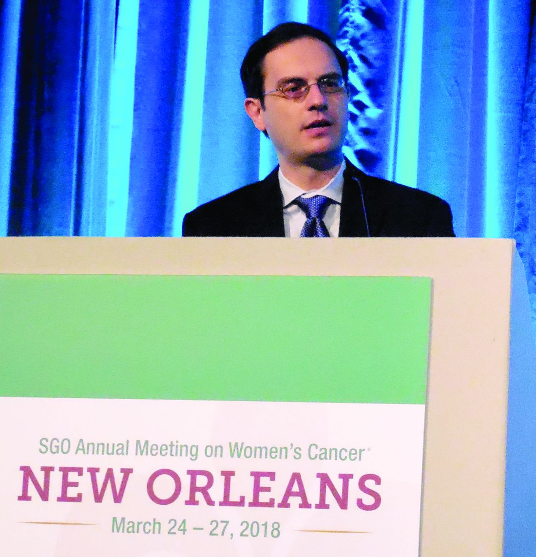
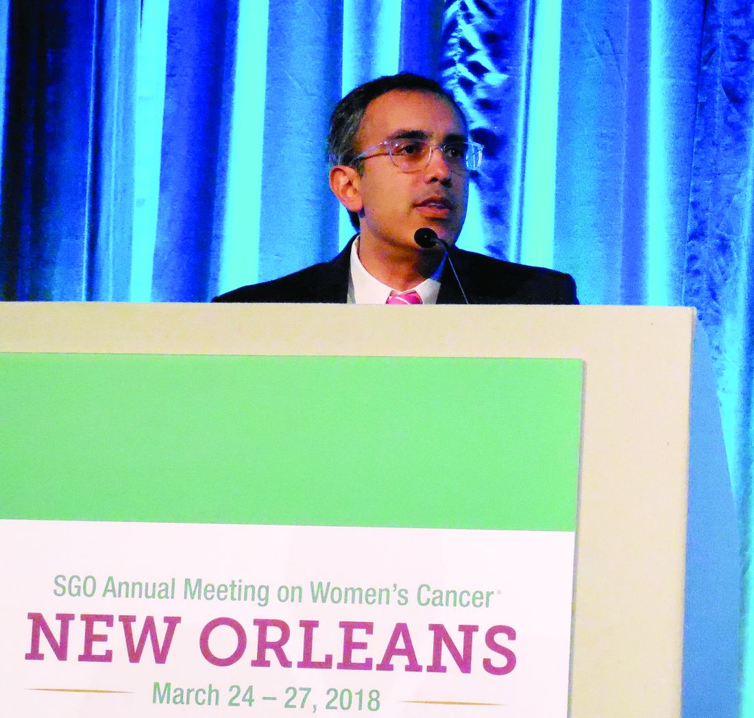
Combined PARP, checkpoint inhibitor regimens show ovarian cancer activity
NEW ORLEANS – Results from the first two trials to assess the efficacy of combined treatment with a PARP inhibitor plus an immune checkpoint inhibitor in patients with ovarian cancer showed promising results in both phase 2.
The second trial treated 34 patients with platinum-sensitive ovarian cancer with a “chemotherapy-sparing” combination of the PARP inhibitor olaparib (Lynparza) and the immune checkpoint inhibitor durvalumab (Imfinzi) and showed an objective response rate of 72% and a disease control rate of 81%, reported Yvette Drew, MD, of the Northern Institute for Cancer Research in Newcastle upon Tyne, England.
These responses in the two uncontrolled series appeared to exceed historical rates on monotherapy with a PARP inhibitor or immune checkpoint inhibitor, leading both Dr. Konstantinopoulos and Dr. Drew to suggest possible synergy between the two drug classes. The response rates “were higher than previously reported” with either drug alone in platinum-resistant or -refractory ovarian cancer patients, noted Dr. Konstantinopoulos, the director of translational research in the Gynecologic Oncology Program at the Dana-Farber Cancer Institute in Boston.
In both phase 2 studies, the drugs were generally well tolerated, with grade 3 or higher adverse effects roughly matching what’s been reported using monotherapy with each of the tested drugs. The most common was anemia, in 12%-19% of patients. The data on niraparib plus pembrolizumab that Dr. Konstantinopoulos reported also included nine patients in a phase 1 study that had compared the dosages used in the phase 2 study against a second regimen that used a higher niraparib dosage.
The TOPACIO (Niraparib in Combination With Pembrolizumab in Patients With Triple-Negative Breast Cancer or Ovarian Cancer) trial enrolled ovarian cancer patients who had been on a platinum regimen for at least 6 months and had received no more than five prior lines of treatment. The phase 1/2 study MEDIOLA (MEDI4736 in Combination With Olaparib in Patients With Advanced Solid Tumors) enrolled only patients with a BRCA mutation and at least two prior lines of treatment. The substantial differences in response and control rates between the two studies can be at least partially attributed to differences in their enrollment criteria and other study details, commented Amir A. Jazaeri, MD, director of the Gynecologic Cancer Immunotherapy Program at the University of Texas MD Anderson Cancer Center in Houston.
TOPACIO was funded by Tesaro and Merck. MEDIOLA was funded by AstraZeneca. Dr. Konstantinopoulos is an adviser to AstraZeneca, Merck, and Pfizer. Dr. Drew is an adviser to AstraZeneca and Clovis. Dr. Jazaeri is an adviser to Areva, has received travel support from AstraZeneca and Genentech as well as research funding from AstraZeneca, Bristol-Myers Squibb, Iovance, and Pfizer.
SOURCE: Konstantinopoulos PA and Drew Y. SGO 2018.
REPORTING FROM SGO 2018
Key clinical point:
Major finding: Disease control rate was 67% in the TOPACIO study and 81% in the MEDIOLA study.
Study details: TOPACIO was a multicenter phase 1/2 study with 62 total patients. MEDIOLA was a multicenter phase 2 study with 34 patients.
Disclosures: TOPACIO was funded by Tesaro and Merck. MEDIOLA was funded by AstraZeneca. Dr. Konstantinopoulos is an adviser to AstraZeneca, Merck, and Pfizer. Dr. Drew is an adviser to AstraZeneca and Clovis. Dr. Jazaeri is an adviser to Areva, has received travel support from AstraZeneca and Genentech as well as research funding from AstraZeneca, Bristol-Myers Squibb, Iovance, and Pfizer.
Source: Konstantinopoulos PA and Drew Y. SGO 2018.
Opinions clash over private equity’s effect on dermatology
SAN DIEGO – Although practices that has been snowballing for a little under a decade is often bemoaned by dermatologists, at least some physicians in the field may see it as a positive development.
“I think private equity investment is good for dermatology,” Clifford Perlis, MD, said at the annual meeting of the American Academy of Dermatology. “Dermatologists should be thrilled that private equity is here.” Investment by private equity companies into dermatology practices “adds value to practices, creates more practice options, enhances advocacy, and better manages the complexity” of practices, claimed Dr. Perlis, a dermatologist and Mohs surgeon who practices in King of Prussia, Pa. The debate on this topic was presented during a forum on dermatoethics at the meeting, structured as a pro and con conversation. The organizers, including Dr. Perlis and Jane Grant-Kels, MD., chose the topics and assigned positions to each presenter, which do not necessarily represent their personal views.*

“The challenges to solo practice have nothing to do with private equity investment. It’s narrower insurance networks” that are squeezing out solo practices. He also expressed skepticism that private equity creates a barrier that interferes with the physician-patient relationship.
“Insurers and administrators already do that,” Dr. Perlis said in an interview. It’s “misguided” to place the blame on the financing. Rejection of private equity investment “clings to an outdated image of how medicine is practiced” that had already been disappearing for several decades before entry of private equity into dermatology began, he noted.
But other dermatologists have voiced concern about the changes brought by private equity investment.
“I’m frightened for the future of dermatology,” Jane M. Grant-Kels, MD, said at the meeting. “It shifts control [of patient care] away from dermatologists and into the hands of business and investors who are only interested in profit and not in quality of care,” she added.

“Have we learned nothing from dermatopathology, which has been ruined” by private equity, she asked. “The only ones who benefit are those making the private equity investments.” She cited similar, recently published views from other dermatologists, such as a recently published viewpoint article written by Jack S. Resneck Jr., MD, professor of dermatology, University of California, San Francisco (JAMA Dermatol. 2018 Jan 1;154[1]:13-4).
Dr. Perlis and Dr. Grant-Kels had no disclosures. They spoke during a session on dermatoethics at the AAD annual meeting. In an interview, Dr. Perlis said that his own personal views on this topic are far more nuanced.*
*This article was updated on April 4, 2018.
SAN DIEGO – Although practices that has been snowballing for a little under a decade is often bemoaned by dermatologists, at least some physicians in the field may see it as a positive development.
“I think private equity investment is good for dermatology,” Clifford Perlis, MD, said at the annual meeting of the American Academy of Dermatology. “Dermatologists should be thrilled that private equity is here.” Investment by private equity companies into dermatology practices “adds value to practices, creates more practice options, enhances advocacy, and better manages the complexity” of practices, claimed Dr. Perlis, a dermatologist and Mohs surgeon who practices in King of Prussia, Pa. The debate on this topic was presented during a forum on dermatoethics at the meeting, structured as a pro and con conversation. The organizers, including Dr. Perlis and Jane Grant-Kels, MD., chose the topics and assigned positions to each presenter, which do not necessarily represent their personal views.*
“The challenges to solo practice have nothing to do with private equity investment. It’s narrower insurance networks” that are squeezing out solo practices. He also expressed skepticism that private equity creates a barrier that interferes with the physician-patient relationship.
“Insurers and administrators already do that,” Dr. Perlis said in an interview. It’s “misguided” to place the blame on the financing. Rejection of private equity investment “clings to an outdated image of how medicine is practiced” that had already been disappearing for several decades before entry of private equity into dermatology began, he noted.
But other dermatologists have voiced concern about the changes brought by private equity investment.
“I’m frightened for the future of dermatology,” Jane M. Grant-Kels, MD, said at the meeting. “It shifts control [of patient care] away from dermatologists and into the hands of business and investors who are only interested in profit and not in quality of care,” she added.
“Have we learned nothing from dermatopathology, which has been ruined” by private equity, she asked. “The only ones who benefit are those making the private equity investments.” She cited similar, recently published views from other dermatologists, such as a recently published viewpoint article written by Jack S. Resneck Jr., MD, professor of dermatology, University of California, San Francisco (JAMA Dermatol. 2018 Jan 1;154[1]:13-4).
Dr. Perlis and Dr. Grant-Kels had no disclosures. They spoke during a session on dermatoethics at the AAD annual meeting. In an interview, Dr. Perlis said that his own personal views on this topic are far more nuanced.*
*This article was updated on April 4, 2018.
EXPERT ANALYSIS FROM AAD 18
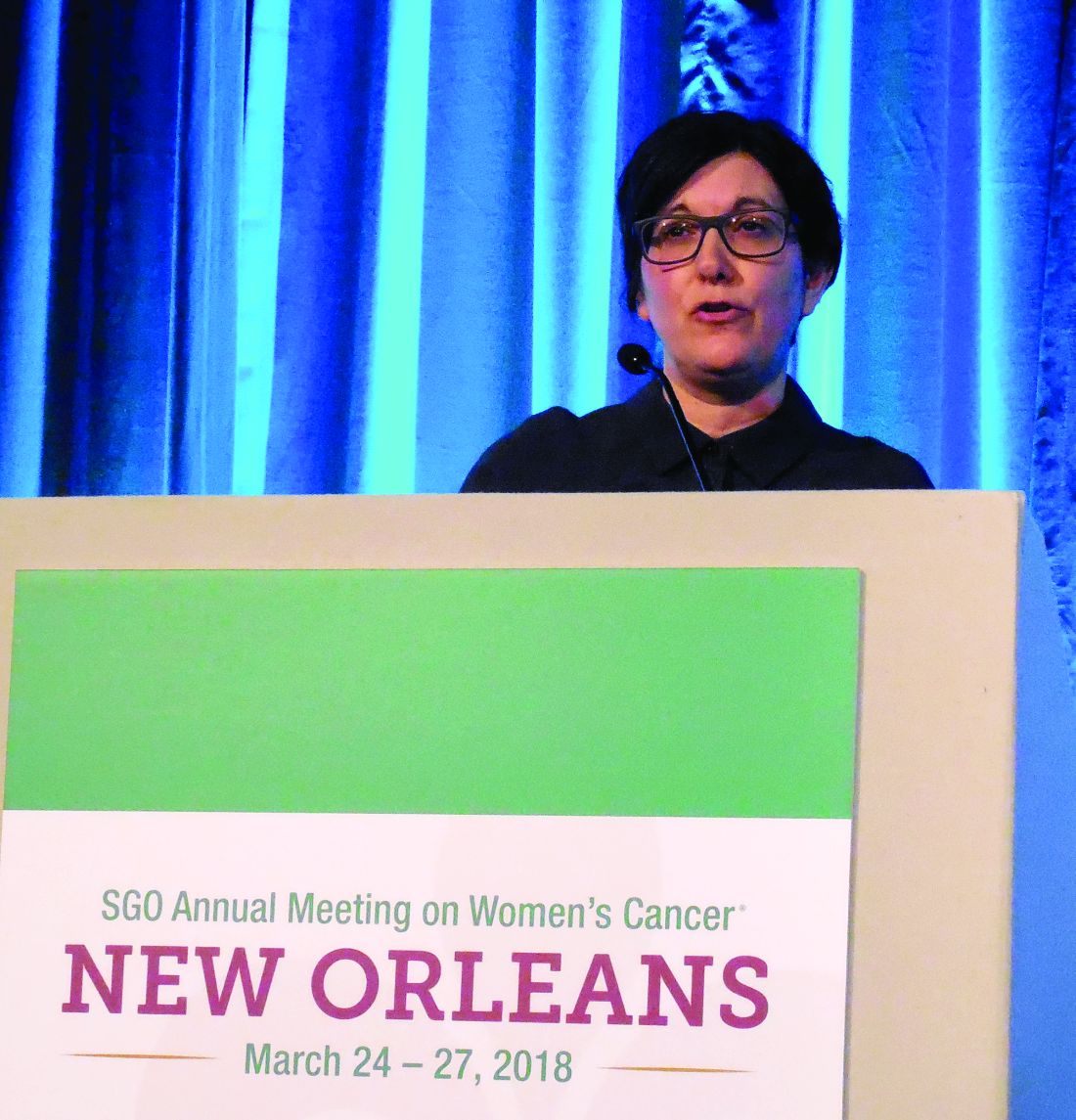
VIDEO: Everolimus/letrozole promising for recurrent endometrial cancer
NEW ORLEANS – Combined treatment for 28 days with the mammalian target of rapamycin inhibitor everolimus plus the aromatase inhibitor letrozole in 37 women with recurrent endometrial cancer produced an overall objective response rate of 24% and an average progression-free survival rate of 6.3 months in a randomized phase 2 study with a total of 74 patients. The treatment was also relatively well tolerated, with more serious adverse vents of anemia and hyperglycemia.
The video associated with this article is no longer available on this site. Please view all of our videos on the MDedge YouTube channel
But the most attention-grabbing finding of this study was that, in the subset of 15 women who had received no prior chemotherapy, the objective response rate on this regimen was 53% and median progression-free survival was 21.6 months, Brian M. Slomovitz, MD, said at the annual meeting of the Society of Gynecologic Oncology.
This level of response in the chemotherapy-naive subgroup was “very high, and not what we expected,” Dr. Slomovitz, , professor of ob.gyn. and human genetics and director of gynecologic oncology at the University of Miami, said in a video interview. “This is something we need to further investigate to see if we can make this part of standard care.”
Although he conceded that the data from this study were too limited to warrant a regulatory indication, he suggested that it might be enough to gain the everolimus plus letrozole combination used in the study citation as a treatment option in clinical guidelines. The next step should be a phase 3 trial that compares the everolimus plus letrozole combination with the traditional chemotherapy regimen of carboplatin plus paclitaxel, Dr. Slomovitz added.
The Everolimus and Letrozole or Hormonal Therapy to Treat Endometrial Cancer phase 2 trial, initiated by the Gynecologic Oncology Group, ran at 26 U.S. centers, and randomized patients with stage III or IV recurrent, advanced, or persistent endometrial cancer who had either no or at most one prior course of chemotherapy. The study design also excluded patients who had previously been treated with an mammalian target of rapamycin inhibitor or hormonal therapy for their endometrial cancer. The control arm placed 37 patients on a standard hormonal therapy regimen of tamoxifen plus medroxyprogesterone, with 36 of patients in this subgroup evaluable.
The overall results in the two treatment arms were roughly similar, except for the striking benefit seen with everolimus(Afinitor) plus letrozole(Femara) in the chemotherapy-naive patents; 15 patients in each arm had not received any chemotherapy before entering the study. In this subgroup, among the patients who received conventional hormonal therapy, the objective response rate was 43% and progression-free survival was 6.6 months.
The two arms also differed by their pattern of grade 3 or 4 adverse events. Among the patients on everolimus plus letrozole, the most common were anemia (24%) and hyperglycemia (14%), both expected consequences of the regimen. The hormonal therapy arm led to an 8% incidence of thromboembolic events, which did not occur in the everolimus plus letrozole arm.
Another attraction of the everolimus and letrozole regimen is that it is oral and avoids the need for drug infusions, Dr. Slomovitz noted.
The investigator-initiated study received funding from Novartis, the company that markets everolimus and letrozole. Dr. Slomovitz has been an advisor to Advaxis, AstraZeneca, Clovis, Genmab, Jannsen, and Tesaro, and he has received research funding from Novartis.
SOURCE: Slomovitz BM et al. SGO 2018, abstract 1.
The results from this study are exciting, with fairly compelling response rates. The rate of progression-free survival with everolimus and letrozole treatment is very impressive, compared with traditional chemotherapy using carboplatin and paclitaxel. The median progression-free survival rate of 21.6 months seen among the chemotherapy naive patients who received everolimus and letrozole showed a 7-month edge over the 14-month median progression-free survival previously reported with chemotherapy. This difference is very provocative and could be practice changing, but it of course needs further evaluation.

Paola A. Gehrig, MD , is professor of ob.gyn. and director of gynecologic oncology at the University of North Carolina in Chapel Hill. She made these comments as designated discussant for the study. She had no disclosures.
The results from this study are exciting, with fairly compelling response rates. The rate of progression-free survival with everolimus and letrozole treatment is very impressive, compared with traditional chemotherapy using carboplatin and paclitaxel. The median progression-free survival rate of 21.6 months seen among the chemotherapy naive patients who received everolimus and letrozole showed a 7-month edge over the 14-month median progression-free survival previously reported with chemotherapy. This difference is very provocative and could be practice changing, but it of course needs further evaluation.
Paola A. Gehrig, MD , is professor of ob.gyn. and director of gynecologic oncology at the University of North Carolina in Chapel Hill. She made these comments as designated discussant for the study. She had no disclosures.
NEW ORLEANS – Combined treatment for 28 days with the mammalian target of rapamycin inhibitor everolimus plus the aromatase inhibitor letrozole in 37 women with recurrent endometrial cancer produced an overall objective response rate of 24% and an average progression-free survival rate of 6.3 months in a randomized phase 2 study with a total of 74 patients. The treatment was also relatively well tolerated, with more serious adverse vents of anemia and hyperglycemia.
The video associated with this article is no longer available on this site. Please view all of our videos on the MDedge YouTube channel
But the most attention-grabbing finding of this study was that, in the subset of 15 women who had received no prior chemotherapy, the objective response rate on this regimen was 53% and median progression-free survival was 21.6 months, Brian M. Slomovitz, MD, said at the annual meeting of the Society of Gynecologic Oncology.
This level of response in the chemotherapy-naive subgroup was “very high, and not what we expected,” Dr. Slomovitz, , professor of ob.gyn. and human genetics and director of gynecologic oncology at the University of Miami, said in a video interview. “This is something we need to further investigate to see if we can make this part of standard care.”
Although he conceded that the data from this study were too limited to warrant a regulatory indication, he suggested that it might be enough to gain the everolimus plus letrozole combination used in the study citation as a treatment option in clinical guidelines. The next step should be a phase 3 trial that compares the everolimus plus letrozole combination with the traditional chemotherapy regimen of carboplatin plus paclitaxel, Dr. Slomovitz added.
The Everolimus and Letrozole or Hormonal Therapy to Treat Endometrial Cancer phase 2 trial, initiated by the Gynecologic Oncology Group, ran at 26 U.S. centers, and randomized patients with stage III or IV recurrent, advanced, or persistent endometrial cancer who had either no or at most one prior course of chemotherapy. The study design also excluded patients who had previously been treated with an mammalian target of rapamycin inhibitor or hormonal therapy for their endometrial cancer. The control arm placed 37 patients on a standard hormonal therapy regimen of tamoxifen plus medroxyprogesterone, with 36 of patients in this subgroup evaluable.
The overall results in the two treatment arms were roughly similar, except for the striking benefit seen with everolimus(Afinitor) plus letrozole(Femara) in the chemotherapy-naive patents; 15 patients in each arm had not received any chemotherapy before entering the study. In this subgroup, among the patients who received conventional hormonal therapy, the objective response rate was 43% and progression-free survival was 6.6 months.
The two arms also differed by their pattern of grade 3 or 4 adverse events. Among the patients on everolimus plus letrozole, the most common were anemia (24%) and hyperglycemia (14%), both expected consequences of the regimen. The hormonal therapy arm led to an 8% incidence of thromboembolic events, which did not occur in the everolimus plus letrozole arm.
Another attraction of the everolimus and letrozole regimen is that it is oral and avoids the need for drug infusions, Dr. Slomovitz noted.
The investigator-initiated study received funding from Novartis, the company that markets everolimus and letrozole. Dr. Slomovitz has been an advisor to Advaxis, AstraZeneca, Clovis, Genmab, Jannsen, and Tesaro, and he has received research funding from Novartis.
SOURCE: Slomovitz BM et al. SGO 2018, abstract 1.
REPORTING FROM SGO 2018
Key clinical point: Treatment of recurrent endometrial cancer with everolimus and letrozole shows promise.
Major finding: Among 15 chemotherapy-naive patients, objective responses occurred in 53% and median progression-free survival was 21.6 months.
Study details: A multicenter, phase 2 randomized study in 74 patients.
Disclosures: The investigator-initiated study received funding from Novartis, the company that markets everolimus(Afinitor) and letrozole(Femara). Dr. Slomovitz has been an advisor to Advaxis, AstraZeneca, Clovis, Genmab, Jannsen, and Tesaro, and he has received research funding from Novartis.
Source: Slomovitz BM et al. SGO 2018, abstract 1.
VIDEO: Case for rivaroxaban & aspirin for PAD gets stronger
ORLANDO – Combination treatment with aspirin and a low dosage of the anticoagulant rivaroxaban had a broader benefit for reducing adverse events in patients with peripheral artery disease than initially reported from the COMPASS trial, which included more than 7,000 patients whose primary diagnosis at study entry was stable PAD.
Secondary analysis of data from the PAD patients enrolled in the COMPASS (Rivaroxaban for the Prevention of Major Cardiovascular Events in Coronary or Peripheral Artery Disease) trial showed that, compared with aspirin alone, treatment with 100 mg aspirin daily plus a low dosage of the anticoagulant rivaroxaban (2.5 mg b.i.d.) resulted in a statistically significant, 24% relative cut in the combined rate of vascular interventions: acute or chronic limb ischemia, vascular amputations, peripheral vascular bypass or percutaneous intervention, and vascular hospitalizations, Sonia S. Anand, MD, said at the annual meeting of the American College of Cardiology.
This finding plus the results in a prior report from COMPASS that rivaroxaban plus aspirin cut major adverse limb events (MALE) by 33%, compared with aspirin alone (Lancet. 2018 Jan 20;391:219-9), together show that rivaroxaban plus aspirin represent a new, effective regimen for a majority of PAD patients (in addition to treating with a statin and an ACE inhibitor) to cut adverse outcomes in a high-risk but historically undertreated patient population, Dr. Anand said in a video interview.
Additional analysis that Dr. Anand reported in her talk showed that patients with PAD who had an index MALE during follow-up had a very high rate of subsequent events. Among the 128 PAD patients in COMPASS who had an index MALE (major vascular amputation or severe limb ischemia that resulted in an intervention) and, compared with the PAD patients who did not have a MALE, the rate of subsequent death was more than three times higher, the rate of hospitalization increased more than 7-fold, and the rate of amputation increased nearly 200-fold.
Concurrently with Dr. Anand’s report at the meeting the results appeared in an article online in the Journal of the American College of Cardiology.

The analysis of post-MALE outcomes, as well as the expanded vascular-outcomes analysis, focused on 6,391 of the COMPASS patients who had lower-extremity PAD and were randomized to either 100 mg aspirin daily or the low-dosage rivaroxaban plus aspirin regimen. COMPASS also randomized patients to a higher-dosage rivaroxaban-only regimen administered at 5 mg b.i.d., but this arm did not perform as well as the lower-dosage regimen, with an efficacy that equaled aspirin only and with more bleeding. The primary efficacy and safety data from COMPASS for all 27,395 enrolled patients, which included many patients with stable coronary artery disease and without diagnosed PAD, appeared in 2017 (N Engl J Med. Oct 5;377[14]:1319-30).
Dr. Anand also reported on a comparison of the clinical and demographic profiles of the 128 PAD patients who developed MALE during follow-up and the 6,263 who did not, a 2% incidence during almost 2 years.
Multivariate analysis identified four significant factors that closely linked with MALE incidence: a history of peripheral surgery or angioplasty, prior limb amputation, baseline Fontaine classification of stage III or IV, and treatment in COMPASS with aspirin alone and not with rivaroxaban plus aspirin.
The new, additional analyses Dr. Anand reported also showed total peripheral vascular outcomes during follow-up in COMPASS in 8.0% of patients on aspirin only and 6.2% of patients on aspirin plus low-dosage rivaroxaban, a 24% relative risk reduction, and vascular interventions in 7.1% of aspirin-only patients and in 5.5% of the combined-regimen patients, also a 24% relative risk reduction. MALE occurred in 2.6% of the aspirin-only patients and in 1.5% of patients on both drugs, a 33% relative risk reduction. All three of these relative risk reductions were statistically significant, said Dr. Anand, a cardiologist and professor of medicine at McMaster University in Hamilton, Ont.
She estimated that about two-thirds of the PAD patients she sees in routine practice would qualify for treatment with aspirin plus low-dosage rivaroxaban once the 2.5 mg formulation becomes approved by regulators. The companies that jointly market rivaroxaban (Xarelto)have an application pending with the Food and Drug Administration to market a 2.5-mg pill based on the COMPASS results. Patients with stable PAD who are not good candidates for the COMPASS regimen are those with a history of a major bleed, those who require full-dose anticoagulation for a comorbidity such as atrial fibrillation or a mechanical heart valve, and patients with newly diagnosed, stable PAD without concurrent coronary artery disease who might receive adequate protection from aspirin alone, Dr. Anand said.
COMPASS was sponsored by Bayer, the company that along with Janssen markets rivaroxaban. Dr. Anand has been a consultant to Bayer and Novartis. Dr. Beckman has been a consultant to Janssen and other pharmaceutical companies.
SOURCE: Anand SS et al. J Amer Coll Cardiol. 2018 Mar 11. doi: 10.1016/j.jacc.2018.03.008.
ORLANDO – Combination treatment with aspirin and a low dosage of the anticoagulant rivaroxaban had a broader benefit for reducing adverse events in patients with peripheral artery disease than initially reported from the COMPASS trial, which included more than 7,000 patients whose primary diagnosis at study entry was stable PAD.
Secondary analysis of data from the PAD patients enrolled in the COMPASS (Rivaroxaban for the Prevention of Major Cardiovascular Events in Coronary or Peripheral Artery Disease) trial showed that, compared with aspirin alone, treatment with 100 mg aspirin daily plus a low dosage of the anticoagulant rivaroxaban (2.5 mg b.i.d.) resulted in a statistically significant, 24% relative cut in the combined rate of vascular interventions: acute or chronic limb ischemia, vascular amputations, peripheral vascular bypass or percutaneous intervention, and vascular hospitalizations, Sonia S. Anand, MD, said at the annual meeting of the American College of Cardiology.
This finding plus the results in a prior report from COMPASS that rivaroxaban plus aspirin cut major adverse limb events (MALE) by 33%, compared with aspirin alone (Lancet. 2018 Jan 20;391:219-9), together show that rivaroxaban plus aspirin represent a new, effective regimen for a majority of PAD patients (in addition to treating with a statin and an ACE inhibitor) to cut adverse outcomes in a high-risk but historically undertreated patient population, Dr. Anand said in a video interview.
Additional analysis that Dr. Anand reported in her talk showed that patients with PAD who had an index MALE during follow-up had a very high rate of subsequent events. Among the 128 PAD patients in COMPASS who had an index MALE (major vascular amputation or severe limb ischemia that resulted in an intervention) and, compared with the PAD patients who did not have a MALE, the rate of subsequent death was more than three times higher, the rate of hospitalization increased more than 7-fold, and the rate of amputation increased nearly 200-fold.
Concurrently with Dr. Anand’s report at the meeting the results appeared in an article online in the Journal of the American College of Cardiology.
The analysis of post-MALE outcomes, as well as the expanded vascular-outcomes analysis, focused on 6,391 of the COMPASS patients who had lower-extremity PAD and were randomized to either 100 mg aspirin daily or the low-dosage rivaroxaban plus aspirin regimen. COMPASS also randomized patients to a higher-dosage rivaroxaban-only regimen administered at 5 mg b.i.d., but this arm did not perform as well as the lower-dosage regimen, with an efficacy that equaled aspirin only and with more bleeding. The primary efficacy and safety data from COMPASS for all 27,395 enrolled patients, which included many patients with stable coronary artery disease and without diagnosed PAD, appeared in 2017 (N Engl J Med. Oct 5;377[14]:1319-30).
Dr. Anand also reported on a comparison of the clinical and demographic profiles of the 128 PAD patients who developed MALE during follow-up and the 6,263 who did not, a 2% incidence during almost 2 years.
Multivariate analysis identified four significant factors that closely linked with MALE incidence: a history of peripheral surgery or angioplasty, prior limb amputation, baseline Fontaine classification of stage III or IV, and treatment in COMPASS with aspirin alone and not with rivaroxaban plus aspirin.
The new, additional analyses Dr. Anand reported also showed total peripheral vascular outcomes during follow-up in COMPASS in 8.0% of patients on aspirin only and 6.2% of patients on aspirin plus low-dosage rivaroxaban, a 24% relative risk reduction, and vascular interventions in 7.1% of aspirin-only patients and in 5.5% of the combined-regimen patients, also a 24% relative risk reduction. MALE occurred in 2.6% of the aspirin-only patients and in 1.5% of patients on both drugs, a 33% relative risk reduction. All three of these relative risk reductions were statistically significant, said Dr. Anand, a cardiologist and professor of medicine at McMaster University in Hamilton, Ont.
She estimated that about two-thirds of the PAD patients she sees in routine practice would qualify for treatment with aspirin plus low-dosage rivaroxaban once the 2.5 mg formulation becomes approved by regulators. The companies that jointly market rivaroxaban (Xarelto)have an application pending with the Food and Drug Administration to market a 2.5-mg pill based on the COMPASS results. Patients with stable PAD who are not good candidates for the COMPASS regimen are those with a history of a major bleed, those who require full-dose anticoagulation for a comorbidity such as atrial fibrillation or a mechanical heart valve, and patients with newly diagnosed, stable PAD without concurrent coronary artery disease who might receive adequate protection from aspirin alone, Dr. Anand said.
COMPASS was sponsored by Bayer, the company that along with Janssen markets rivaroxaban. Dr. Anand has been a consultant to Bayer and Novartis. Dr. Beckman has been a consultant to Janssen and other pharmaceutical companies.
SOURCE: Anand SS et al. J Amer Coll Cardiol. 2018 Mar 11. doi: 10.1016/j.jacc.2018.03.008.
REPORTING FROM ACC 18
Key clinical point: PAD patients on rivaroxaban plus aspirin had significantly fewer adverse peripheral vascular outcomes.
Major finding: Dual therapy cut total adverse peripheral vascular outcomes by a relative 24%, compared with aspirin alone, during 23-month follow-up.
Study details: Secondary analysis of data from COMPASS, a multicenter, randomized trial with 6,391 patients in the new analysis.
Disclosures: COMPASS was sponsored by Bayer, the company that, along with Janssen markets rivaroxaban. Dr. Anand has been a consultant to Bayer and Novartis. Dr. Beckman has been a consultant to Janssen and other pharmaceutical companies.
Source: Anand SS et al. J Amer Coll Cardiol. 2018 Mar 11. doi: 10.1016/j.jacc.2018.03.008.

VIDEO: Andexanet alfa effectively reverses factor Xa anticoagulant
ORLANDO – Andexanet alfa, a new agent that reverses the anticoagulant effect of direct factor Xa inhibitors, showed an acceptable level of efficacy and safety in 227 patients who received the drug in the agent’s pivotal trial.
These results, which placed andexanet in the same ballpark for efficacy and safety as idarucizumab (Praxbind), approved in 2015 for reversing the anticoagulant dabigatran (Pradaxa), suggest that andexanet is likely on track for its own Food and Drug Administration marketing approval, Stuart Connolly, MD, said at the annual meeting of the American College of Cardiology.

Portola Pharmaceuticals, the company developing andexanet alfa (AndexXa) previously announced that it expected Food and Drug Administration action on its marketing application by May 2018.
Andexanet reversal “has similar efficacy and safety as seen with other reversal agents” for other types of anticoagulants, said Dr. Connolly, a professor of medicine and an electrophysiologist at McMaster University in Hamilton, Ont. In the trial results he reported, andexanet treatment of patients who were bleeding while on treatment with a direct factor Xa inhibitor had an 83% rate of hemostatic efficacy and an 11% rate of thrombotic events. By comparison, idarucizumab, the FDA-approved reversal agent for the anticoagulant dabigatran, produced a 68% hemostatic efficacy and a 6% rate of thrombotic events in the idarucizumab pivotal trial, RE-VERSE AD (N Engl J Med. 2015 Aug 6;373[6]:511-20).
The Prospective, Open-Label Study of Andexanet Alfa in Patients Receiving a Factor Xa Inhibitor Who Have Acute Major Bleeding (ANNEXA-4) enrolled 227 patients at any of 60 centers, with efficacy data available from 132 of the patients. About 60% of the patients had an intracranial bleed, and about 30% had a gastrointestinal bleed, and their average age was 77 years. Roughly three-quarters of patients were on an anticoagulant for atrial fibrillation, with the rest treated for venous thromboembolism, with 4% having both conditions. The most commonly used direct factor Xa inhbitors in these patients were apixaban (Eliquis) in 105 and rivaroxaban (Xarelto) in 75. The ANNEXA-4 study has not enrolled patients treated with a direct factor Xa inhibitor anticoagulant and undergoing surgery, a setting that will be the subject of a future study, Dr. Connolly said.
Clinicians administered andexanet alfa as a bolus followed by a 2-hour continuous infusion, with hemostatic efficacy assessed 12 hours after the start of treatment. The results showed that factor Xa inhibition fell by about 75%-90% within minutes of starting the bolus and remained depressed at that level during the infusion but then began recovering by 2 hours after the stop of infusion. Andexanet is a factor Xa “decoy” molecule that acts by latching onto the inhibitor molecules and thereby preventing them from interacting with actual factor Xa, but andexanet also has a short half life and hence the effect quickly reduces once treatment stops.
“There is no doubt that andexanet rapidly decreases anti–factor Xa activity,” he said.
Adjudicated efficacy results were available for 132 patients and showed good or excellent hemostasis achieved on andexanet in 109 patients (83%), Dr. Connolly reported. The effect on hemostasis was consistent regardless of patient age, sex, bleeding site, type of anticoagulant, and dosage tested.
Thrombotic events during the 30 days following treatment occurred in 24 of 227 patients (11%) who received andexanet and were evaluable for safety. Notably, no clustering of thrombotic events occurred early, even among the 129 patients who restarted on an anticoagulant during the 30 days after treatment. Among the 129 patients who restarted on an anticoagulant, 9 (7%) had a thrombotic event during the 30-day follow-up, compared with 15 events among 98 patients (15%) who did not restart on an anticoagulant.
Dr. Connolly acknowledged that a limitation of the ANNEXA-4 study is the absence of a control group, but he added that he and his associates believed randomizing patients with a serious bleed to placebo control would not have been “practical, feasible, or ethical.”
ANNEXA-4 is sponsored by Portola Pharmaceuticals, the company developing andexanet alfa (AndexXa). Dr. Connolly has been a consultant to Portola, and also to Bayer, Boehringer-Ingelheim, Bristol-Myers Squibb, and Sanofi-Aventis. Dr. Kirtane has received research support from several device manufacturers.
SOURCE: Connolly S. ACC 2018.
Treatment with andexanet alfa produced good or excellent hemostasis in 83% of patients in the ANNEXA-4 study, which is what matters when patients are bleeding. Clinicians want to know that you can restore coagulation to a level where you can stop bleeding, and that’s what the results show.
The lack of a reversal agent until now for direct-acting factor Xa inhibitor drugs has probably been a modest but real obstacle to widespread adoption of these agents. We can look at the example of another new oral anticoagulant, dabigatran (Pradaxa), which works by a different mechanism, specifically by inhibiting thrombin. After a reversal agent for dabigatran, idarucizumab (Praxbind) received Food and Drug Administration approval and became available in late 2015, an uptick in dabigatran prescriptions occurred. That experience shows that patients and providers want the safety net of a reversal agent. They want to know that, if there is bleeding or need for urgent surgery, there is a way to facilitate restoration of hemostasis.
It’s the same with direct factor Xa inhibitors: Some patients are concerned about the lack of a reversal agent, and having such an agent may help increase access to these agents for such patients. I think that, once andexanet becomes available for routine U.S. practice, we’ll see an uptick in prescribing of direct factor Xa inhibitors. Also, some patients who have opted for treatment with warfarin will switch to a safer class of drugs, the direct factor X a inhibitors. A myth exists that reversal agents can easily negate the anticoagulant effect of warfarin. The reality is that, despite having treatments that reverse warfarin’s effect, this is often not an easy process in actual practice.
On the safety side, there was no indication in the ANNEXA-4 results of rebound thrombosis with andexanet alfa treatment. Patients receiving a direct factor Xa inhibitor are prothrombotic – that’s why they are on an anticoagulant – so their risk for a thrombotic event is always there, especially when they are not fully anticoagulated, such as when a reversal agent is administered. We need to look to restarting treatment with an anticoagulant because these patients have a high thrombotic risk.
Gregory Piazza, MD , is a cardiologist at Brigham and Women’s Hospital in Boston. He has been an advisor to Portola Pharmaceuticals, the company developing andexanet alfa, as well as to Bayer and Pfizer, and he has received research funding from Bristol-Myers Squibb, Janssen, and Daiichi Sankyo. He made these comments in an interview .
ORLANDO – Andexanet alfa, a new agent that reverses the anticoagulant effect of direct factor Xa inhibitors, showed an acceptable level of efficacy and safety in 227 patients who received the drug in the agent’s pivotal trial.
These results, which placed andexanet in the same ballpark for efficacy and safety as idarucizumab (Praxbind), approved in 2015 for reversing the anticoagulant dabigatran (Pradaxa), suggest that andexanet is likely on track for its own Food and Drug Administration marketing approval, Stuart Connolly, MD, said at the annual meeting of the American College of Cardiology.
Portola Pharmaceuticals, the company developing andexanet alfa (AndexXa) previously announced that it expected Food and Drug Administration action on its marketing application by May 2018.
Andexanet reversal “has similar efficacy and safety as seen with other reversal agents” for other types of anticoagulants, said Dr. Connolly, a professor of medicine and an electrophysiologist at McMaster University in Hamilton, Ont. In the trial results he reported, andexanet treatment of patients who were bleeding while on treatment with a direct factor Xa inhibitor had an 83% rate of hemostatic efficacy and an 11% rate of thrombotic events. By comparison, idarucizumab, the FDA-approved reversal agent for the anticoagulant dabigatran, produced a 68% hemostatic efficacy and a 6% rate of thrombotic events in the idarucizumab pivotal trial, RE-VERSE AD (N Engl J Med. 2015 Aug 6;373[6]:511-20).
The Prospective, Open-Label Study of Andexanet Alfa in Patients Receiving a Factor Xa Inhibitor Who Have Acute Major Bleeding (ANNEXA-4) enrolled 227 patients at any of 60 centers, with efficacy data available from 132 of the patients. About 60% of the patients had an intracranial bleed, and about 30% had a gastrointestinal bleed, and their average age was 77 years. Roughly three-quarters of patients were on an anticoagulant for atrial fibrillation, with the rest treated for venous thromboembolism, with 4% having both conditions. The most commonly used direct factor Xa inhbitors in these patients were apixaban (Eliquis) in 105 and rivaroxaban (Xarelto) in 75. The ANNEXA-4 study has not enrolled patients treated with a direct factor Xa inhibitor anticoagulant and undergoing surgery, a setting that will be the subject of a future study, Dr. Connolly said.
Clinicians administered andexanet alfa as a bolus followed by a 2-hour continuous infusion, with hemostatic efficacy assessed 12 hours after the start of treatment. The results showed that factor Xa inhibition fell by about 75%-90% within minutes of starting the bolus and remained depressed at that level during the infusion but then began recovering by 2 hours after the stop of infusion. Andexanet is a factor Xa “decoy” molecule that acts by latching onto the inhibitor molecules and thereby preventing them from interacting with actual factor Xa, but andexanet also has a short half life and hence the effect quickly reduces once treatment stops.
“There is no doubt that andexanet rapidly decreases anti–factor Xa activity,” he said.
Adjudicated efficacy results were available for 132 patients and showed good or excellent hemostasis achieved on andexanet in 109 patients (83%), Dr. Connolly reported. The effect on hemostasis was consistent regardless of patient age, sex, bleeding site, type of anticoagulant, and dosage tested.
Thrombotic events during the 30 days following treatment occurred in 24 of 227 patients (11%) who received andexanet and were evaluable for safety. Notably, no clustering of thrombotic events occurred early, even among the 129 patients who restarted on an anticoagulant during the 30 days after treatment. Among the 129 patients who restarted on an anticoagulant, 9 (7%) had a thrombotic event during the 30-day follow-up, compared with 15 events among 98 patients (15%) who did not restart on an anticoagulant.
Dr. Connolly acknowledged that a limitation of the ANNEXA-4 study is the absence of a control group, but he added that he and his associates believed randomizing patients with a serious bleed to placebo control would not have been “practical, feasible, or ethical.”
ANNEXA-4 is sponsored by Portola Pharmaceuticals, the company developing andexanet alfa (AndexXa). Dr. Connolly has been a consultant to Portola, and also to Bayer, Boehringer-Ingelheim, Bristol-Myers Squibb, and Sanofi-Aventis. Dr. Kirtane has received research support from several device manufacturers.
SOURCE: Connolly S. ACC 2018.
REPORTING FROM ACC 18
Key clinical point:
Major finding: Hemostatic efficacy of andexanet alfa was 83%, and thrombotic events occurred in 11%.
Study details: ANNEXA-4, a single arm cohort study with 227 patients.
Disclosures: ANNEXA-4 is sponsored by Portola Pharmaceuticals, the company developing andexanet alfa (AndexXa). Dr. Connolly has been a consultant to Portola and also to Bayer, Boehringer-Ingelheim, Bristol-Myers Squibb, and Sanofi-Aventis.
Source: Connolly S. ACC 2018.
Transporting stroke patients directly to thrombectomy boosts outcomes
LOS ANGELES – Evidence continues to mount that in the new era of thrombectomy treatment for selected acute ischemic stroke patients outcomes are better when patients go directly to the closest comprehensive stroke center that offers intravascular procedures rather than first being taken to a closer hospital and then needing transfer.
Nils H. Mueller-Kronast, MD, presented a modeled analysis of data collected in a registry on 236 real-world U.S. patients who underwent mechanical thrombectomy for an acute, large-vessel occlusion stroke following transfer from a hospital that could perform thrombolysis but couldn’t offer thrombectomy. The analysis showed that if the patients had instead gone directly to the closest thrombectomy center the result would have been a 16-percentage-point increase in patients with a modified Rankin Scale (mRS) score of 0-1 after 90 days, and a 9-percentage-point increase in mRS 0-2 outcomes, Dr. Mueller-Kronast said at the International Stroke Conference, sponsored by the American Heart Association.

The analysis he presented used data from the Systematic Evaluation of Patients Treated With Stroke Devices for Acute Ischemic Stroke (STRATIS) registry, which included 984 acute ischemic stroke patients who underwent mechanical thrombectomy at any one of 55 participating U.S. sites (Stroke. 2017 Oct;48[10]:2760-8). A previously-reported analysis of the STRATIS data showed that the 55% of patients taken directly to a center that performed thrombectomy had a 60% rate of mRS score 0-2 after 90 days, compared with 52% of patients taken first to a hospital unable to perform thrombectomy and then transferred (Circulation. 2017 Dec 12;136[24]:2311-21).
The current analysis focused on 236 of the transferred patients with complete information on their location at the time of their stroke and subsequent time intervals during their transport and treatment, including 117 patients with ground transfer from their first hospital to the thrombectomy site, 114 with air transfer, and 5 with an unreported means of transport.
Dr. Mueller-Kronast and his associates calculated the time it would have taken each of the 117 ground transported patients to have gone directly to the closest thrombectomy center (adjusted by traffic conditions at the time of the stroke), and modeled the likely outcomes of these patients based on the data collected in the registry. This projected a 47% rate of mRS scores 0-1 (good outcomes) after 90 days, and a 60% rate of mRS 0-2 scores with a direct-to-thrombectomy strategy, compared with actual rates of 31% and 51%, respectively, among the patients who were transferred from their initial hospital.
“Bypass to an endovascular-capable center may be an option to improve rapid access to mechanical thrombectomy,” he concluded.
The STRATIS registry is sponsored by Medtronic. Dr. Mueller-Kronast has been a consultant to Medtronic.
SOURCE: Mueller-Kronast N et al. Abstract LB12.
LOS ANGELES – Evidence continues to mount that in the new era of thrombectomy treatment for selected acute ischemic stroke patients outcomes are better when patients go directly to the closest comprehensive stroke center that offers intravascular procedures rather than first being taken to a closer hospital and then needing transfer.
Nils H. Mueller-Kronast, MD, presented a modeled analysis of data collected in a registry on 236 real-world U.S. patients who underwent mechanical thrombectomy for an acute, large-vessel occlusion stroke following transfer from a hospital that could perform thrombolysis but couldn’t offer thrombectomy. The analysis showed that if the patients had instead gone directly to the closest thrombectomy center the result would have been a 16-percentage-point increase in patients with a modified Rankin Scale (mRS) score of 0-1 after 90 days, and a 9-percentage-point increase in mRS 0-2 outcomes, Dr. Mueller-Kronast said at the International Stroke Conference, sponsored by the American Heart Association.
The analysis he presented used data from the Systematic Evaluation of Patients Treated With Stroke Devices for Acute Ischemic Stroke (STRATIS) registry, which included 984 acute ischemic stroke patients who underwent mechanical thrombectomy at any one of 55 participating U.S. sites (Stroke. 2017 Oct;48[10]:2760-8). A previously-reported analysis of the STRATIS data showed that the 55% of patients taken directly to a center that performed thrombectomy had a 60% rate of mRS score 0-2 after 90 days, compared with 52% of patients taken first to a hospital unable to perform thrombectomy and then transferred (Circulation. 2017 Dec 12;136[24]:2311-21).
The current analysis focused on 236 of the transferred patients with complete information on their location at the time of their stroke and subsequent time intervals during their transport and treatment, including 117 patients with ground transfer from their first hospital to the thrombectomy site, 114 with air transfer, and 5 with an unreported means of transport.
Dr. Mueller-Kronast and his associates calculated the time it would have taken each of the 117 ground transported patients to have gone directly to the closest thrombectomy center (adjusted by traffic conditions at the time of the stroke), and modeled the likely outcomes of these patients based on the data collected in the registry. This projected a 47% rate of mRS scores 0-1 (good outcomes) after 90 days, and a 60% rate of mRS 0-2 scores with a direct-to-thrombectomy strategy, compared with actual rates of 31% and 51%, respectively, among the patients who were transferred from their initial hospital.
“Bypass to an endovascular-capable center may be an option to improve rapid access to mechanical thrombectomy,” he concluded.
The STRATIS registry is sponsored by Medtronic. Dr. Mueller-Kronast has been a consultant to Medtronic.
SOURCE: Mueller-Kronast N et al. Abstract LB12.
REPORTING FROM ISC 2018
Key clinical point: A direct-to-thrombectomy strategy maximizes good stroke outcomes.
Major finding: Modeling showed a 47% rate of good 90-day outcomes by taking patients to the closest thrombectomy center, compared with an actual 31% rate with transfers.
Study details: A simulation-model analysis of data collected by the STRATIS registry of acute stroke thrombectomies.
Disclosures: The STRATIS registry is sponsored by Medtronic. Dr. Mueller-Kronast has been a consultant to Medtronic.
Source: Mueller-Kronast N et al. Abstract LB12.
Wearable defibrillator cuts mortality in post-MI patients
ORLANDO – Wearable cardioverter defibrillator vests failed to significantly cut the rate of arrhythmic death in at-risk post-MI patients but succeeded in significantly dropping total mortality during a median of 84 days of use in the first randomized trial of nonimplanted defibrillators in such patients.
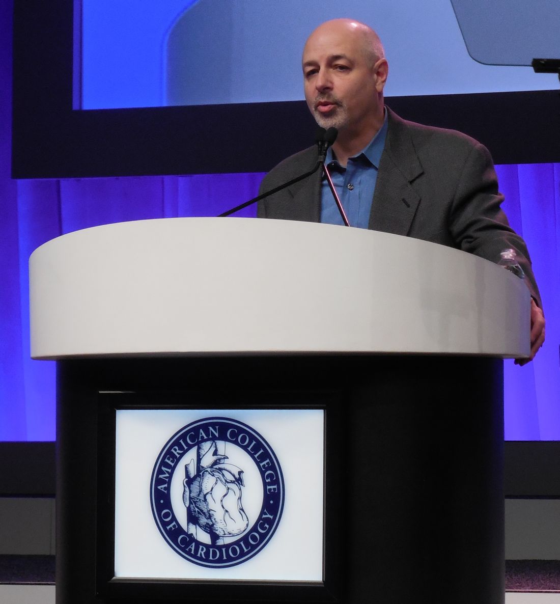
But despite this overall mortality benefit, the 1,524 patients randomized to the WCD group failed to show a significant improvement in the rate of sudden and ventricular tachycardia death, the primary endpoint for the study, said Dr. Olgin, chief of cardiology at the University of California, San Francisco. Total mortality was a secondary endpoint in the study. Based on the total mortality benefit observed and the “totality of evidence” from prior, uncontrolled observational studies, Dr. Olgin concluded that it is now “reasonable” to protect post-MI patients with ejection fractions of 35% or less during the first 40-90 days following an MI when patients can then be assessed for receiving an implantable cardioverter defibrillator.
That would be an upgrade from the current American College of Cardiology/American Heart Association guidelines on managing ventricular arrhythmias and preventing sudden cardiac death, issued in 2017, that classified WCDs as a class IIb recommendations – “may be reasonable” – for post-MI patients with a reduced left ventricular ejection fraction (Circulation. 2017 Oct 30;doi:10.1161/CIR.0000000000000549).
WCDs are currently approved for routine prescribing by U.S. physicians, but their use is very variable in post-MI patients. Just before Dr. Olgin delivered his report at the meeting, a poll of the several thousand meeting attendees who heard his talk showed that roughly a third reported routinely prescribing WCDs, with the other two thirds saying they did not.
Several electrophysiologists who heard the report agreed that further research needs to better tease out which post-MI patients get the most benefit from this treatment.

The patients enrolled in the study “were not a sick population; they had a low event rate,” commented Sana M. Al-Khatib, MD, professor of medicine at Duke University in Durham, N.C. and chair of the panel that wrote the 2017 ventricular arrhythmia guidelines. She suggested testing the efficacy of WCDs in post-MI patients with lower ejection fractions or those with a greater history of heart disease prior to their index MI. Nearly half of the patients enrolled in the study had New York Heart Association class I symptoms, indicating that they had mild heart disease, she noted in an interview. Another issue left unresolved by the results Dr. Olgin reported was how much of the mortality benefit was attributable to the shocks delivered by the tested WCDs and how much derived from the arrhythmia monitoring that the WCDs provided.

Another way to better target WCDs to post-MI patients who could derive the most benefit might be to focus on patients with frequent premature ventricular contractions and nonsustained ventricular tachycardia, suggested Dhanunjaya Lakkireddy, MD, professor of medicine and director of the Center for Excellence in AF and Complex Arrhythmias at the University of Kansas Medical Center in Kansas City. But Dr. Lakkireddy acknowledged that currently left ventricular ejection fraction is the primary surrogate marker cardiologists rely on to identify post-MI patients who are at increased risk for ventricular arrhythmia.
Dr. Olgin countered that the total mortality rate seen among the control, usual care patients in his study, 4.9% during the median 84 day follow-up, closely matched the 5% rate reported in prior trials of at-risk patients who received implantable cardioverter defibrillators.

During follow-up, total mortality occurred in 3.1% of the patients randomized to WCD use and 4.9% among the control patients.
The results also showed that 19% of the patients randomized to the WCD arm failed to ever use the device, and that over the course of follow-up the usage rate fell below 50%. Patients who used the device generally wore it as directed, however, with an average 22 hours a day of use at the start of treatment that subsequently dipped to 21 hours a day near the end of the 90-day treatment period, Dr. Olgin reported.
The most likely explanation for the disparity between the significant effect on total mortality and the insignificant effect on arrhythmia mortality is misclassification of some deaths. “Any small number of misclassified sudden deaths would have dramatically reduced our power to see a difference” in arrhythmia deaths, Dr. Olgin noted.
VEST was sponsored by Zoll, the company that markets the tested device. Dr. Olgin has no personal disclosures. Dr. Al-Khatib and Dr. Lakkireddy had no disclosures. Dr. Wilber is a consultant to Biosense Webster and Medtronic.
[email protected]
SOURCE: Olgin J et al. ACC 18.
ORLANDO – Wearable cardioverter defibrillator vests failed to significantly cut the rate of arrhythmic death in at-risk post-MI patients but succeeded in significantly dropping total mortality during a median of 84 days of use in the first randomized trial of nonimplanted defibrillators in such patients.
But despite this overall mortality benefit, the 1,524 patients randomized to the WCD group failed to show a significant improvement in the rate of sudden and ventricular tachycardia death, the primary endpoint for the study, said Dr. Olgin, chief of cardiology at the University of California, San Francisco. Total mortality was a secondary endpoint in the study. Based on the total mortality benefit observed and the “totality of evidence” from prior, uncontrolled observational studies, Dr. Olgin concluded that it is now “reasonable” to protect post-MI patients with ejection fractions of 35% or less during the first 40-90 days following an MI when patients can then be assessed for receiving an implantable cardioverter defibrillator.
That would be an upgrade from the current American College of Cardiology/American Heart Association guidelines on managing ventricular arrhythmias and preventing sudden cardiac death, issued in 2017, that classified WCDs as a class IIb recommendations – “may be reasonable” – for post-MI patients with a reduced left ventricular ejection fraction (Circulation. 2017 Oct 30;doi:10.1161/CIR.0000000000000549).
WCDs are currently approved for routine prescribing by U.S. physicians, but their use is very variable in post-MI patients. Just before Dr. Olgin delivered his report at the meeting, a poll of the several thousand meeting attendees who heard his talk showed that roughly a third reported routinely prescribing WCDs, with the other two thirds saying they did not.
Several electrophysiologists who heard the report agreed that further research needs to better tease out which post-MI patients get the most benefit from this treatment.
The patients enrolled in the study “were not a sick population; they had a low event rate,” commented Sana M. Al-Khatib, MD, professor of medicine at Duke University in Durham, N.C. and chair of the panel that wrote the 2017 ventricular arrhythmia guidelines. She suggested testing the efficacy of WCDs in post-MI patients with lower ejection fractions or those with a greater history of heart disease prior to their index MI. Nearly half of the patients enrolled in the study had New York Heart Association class I symptoms, indicating that they had mild heart disease, she noted in an interview. Another issue left unresolved by the results Dr. Olgin reported was how much of the mortality benefit was attributable to the shocks delivered by the tested WCDs and how much derived from the arrhythmia monitoring that the WCDs provided.
Another way to better target WCDs to post-MI patients who could derive the most benefit might be to focus on patients with frequent premature ventricular contractions and nonsustained ventricular tachycardia, suggested Dhanunjaya Lakkireddy, MD, professor of medicine and director of the Center for Excellence in AF and Complex Arrhythmias at the University of Kansas Medical Center in Kansas City. But Dr. Lakkireddy acknowledged that currently left ventricular ejection fraction is the primary surrogate marker cardiologists rely on to identify post-MI patients who are at increased risk for ventricular arrhythmia.
Dr. Olgin countered that the total mortality rate seen among the control, usual care patients in his study, 4.9% during the median 84 day follow-up, closely matched the 5% rate reported in prior trials of at-risk patients who received implantable cardioverter defibrillators.
During follow-up, total mortality occurred in 3.1% of the patients randomized to WCD use and 4.9% among the control patients.
The results also showed that 19% of the patients randomized to the WCD arm failed to ever use the device, and that over the course of follow-up the usage rate fell below 50%. Patients who used the device generally wore it as directed, however, with an average 22 hours a day of use at the start of treatment that subsequently dipped to 21 hours a day near the end of the 90-day treatment period, Dr. Olgin reported.
The most likely explanation for the disparity between the significant effect on total mortality and the insignificant effect on arrhythmia mortality is misclassification of some deaths. “Any small number of misclassified sudden deaths would have dramatically reduced our power to see a difference” in arrhythmia deaths, Dr. Olgin noted.
VEST was sponsored by Zoll, the company that markets the tested device. Dr. Olgin has no personal disclosures. Dr. Al-Khatib and Dr. Lakkireddy had no disclosures. Dr. Wilber is a consultant to Biosense Webster and Medtronic.
[email protected]
SOURCE: Olgin J et al. ACC 18.
REPORTING FROM ACC18
Key clinical point: The first RCT of a wearable defibrillator in post-MI patients showed reduced total mortality.
Major finding: Total mortality was 3.2% in patients treated with a wearable cardioverter defibrillator and 4.9% in controls after a median of 84 days.
Study details: VEST, a multicenter, randomized trial with 2,302 patients.
Disclosures: VEST was sponsored by Zoll, the company that markets the tested device. Dr. Olgin has no personal disclosures.
Source: Olgin J. ACC 18.

VEST: Closer tailoring might boost wearable cardioverter defibrillator’s benefit
ORLANDO – , the findings failed to show a statistically significant reduction in sudden death or death from ventricular tachycardia. This suggests better targeting of the device is needed, commented Dhanunjaya Lakkireddy, MD, in a video interview at the annual meeting of the American College of Cardiology.
The Vest Prevention of Early Sudden Death Trial (VEST) randomized 2,302 patients within the first 7 days following an acute MI who also had a left ventricular ejection fraction of 35% or less to either 90 days of treatment with a wearable cardioverter defibrillator (WCD) or usual care. After a median of 84 days, the results showed no statistically significant reduction from WCD use in the primary endpoint of sudden death or death from ventricular tachyarrhythmias, but a statistically significant reduction in the secondary endpoint of all-cause death: 3.1% in the patients randomized to WCD use and 4.9% among the control patients, reported Jeffrey Olgin, MD, chief of cardiology at the University of California, San Francisco.
The results suggest that a more robust benefit might occur in post-MI, low ejection fraction patients who undergo additional selection based on having frequent premature ventricular contractions and nonsustained ventricular tachycardia, suggested Dr. Lakkireddy, professor of medicine and director of the Center for Excellence in AF and Complex Arrhythmias at the University of Kansas Medical Center in Kansas City.
VEST was sponsored by Zoll, a company that markets a WCD. Dr. Lakkireddy had no relevant disclosures.
[email protected]
On Twitter @mitchelzoler
Source: Olgin J and Lakkireddy D ACC 18.
EXPERT ANALYSIS FROM ACC 18

U.S. adolescent malignant melanoma nearly halved during 2000-2014
SAN DIEGO – based on information from a National Cancer Institute database.
The substantial drop in new cases of malignant melanoma in Americans aged 10-19 years over the most recent 15-year period with data available contrasts with a stable rate among children aged 0-9 years, and a steadily rising rate among adults during the same period, Ryan C. Kelm said at the annual meeting of the American Academy of Dermatology.

Mr. Kelm and his associates studied U.S. data compiled from 2000 to 2014 by the SEER Program, maintained by the National Cancer Institute. They identified 1,796 patients aged 0-19 years diagnosed with malignant melanoma (218 children and 1,578 adolescents). The overall incidence rate for the entire 15-year period was just over 1 case per million among children and just under 9 cases per million among adolescents. In contrast, the adult U.S. incidence rate estimates for 2018 are pegged at 260 per million among non-Hispanic whites, 40 per million among Hispanics, and 10 per million among black Americans, according to the American Cancer Society.
An additional analysis showed a notable difference in incidence rates over the 15-year period studied, depending on age. In children aged 0-9 years, the annual incidence rate held roughly steady at just under 2 cases per million throughout the 15 years. But among adolescents, the rate fell over time, from about 10-12 cases per million during 2000-2004 to about 5-7 cases per million during 2010-2014. In 2001, the rate was about 11 cases per million, and in 2013, the rate was about 6 cases per million. This contrasts with the adult rate, which has “risen rapidly over the past 30 years,” according to the American Cancer Society’s 2018 report.
The SEER data also showed that distribution of melanoma histologic types differed by age. Among adolescents the most common identified form was “superficial spreading,” in 32%, with nodular in 6%, mixed epithelioid and spindle cell in 2%, and “not otherwise specified” in 54%. In children, the most commonly identified form was mixed epithelioid and spindle cell, in 10%, followed by nodular in 9%, and superficial spreading in 9%, with 63% not otherwise specified.
SOURCE: Kelm RC et al. AAD 18, Abstract 6722.
SAN DIEGO – based on information from a National Cancer Institute database.
The substantial drop in new cases of malignant melanoma in Americans aged 10-19 years over the most recent 15-year period with data available contrasts with a stable rate among children aged 0-9 years, and a steadily rising rate among adults during the same period, Ryan C. Kelm said at the annual meeting of the American Academy of Dermatology.
Mr. Kelm and his associates studied U.S. data compiled from 2000 to 2014 by the SEER Program, maintained by the National Cancer Institute. They identified 1,796 patients aged 0-19 years diagnosed with malignant melanoma (218 children and 1,578 adolescents). The overall incidence rate for the entire 15-year period was just over 1 case per million among children and just under 9 cases per million among adolescents. In contrast, the adult U.S. incidence rate estimates for 2018 are pegged at 260 per million among non-Hispanic whites, 40 per million among Hispanics, and 10 per million among black Americans, according to the American Cancer Society.
An additional analysis showed a notable difference in incidence rates over the 15-year period studied, depending on age. In children aged 0-9 years, the annual incidence rate held roughly steady at just under 2 cases per million throughout the 15 years. But among adolescents, the rate fell over time, from about 10-12 cases per million during 2000-2004 to about 5-7 cases per million during 2010-2014. In 2001, the rate was about 11 cases per million, and in 2013, the rate was about 6 cases per million. This contrasts with the adult rate, which has “risen rapidly over the past 30 years,” according to the American Cancer Society’s 2018 report.
The SEER data also showed that distribution of melanoma histologic types differed by age. Among adolescents the most common identified form was “superficial spreading,” in 32%, with nodular in 6%, mixed epithelioid and spindle cell in 2%, and “not otherwise specified” in 54%. In children, the most commonly identified form was mixed epithelioid and spindle cell, in 10%, followed by nodular in 9%, and superficial spreading in 9%, with 63% not otherwise specified.
SOURCE: Kelm RC et al. AAD 18, Abstract 6722.
REPORTING FROM AAD 18
Key clinical point: U.S. incident malignant melanoma in adolescents dropped by nearly 50% during 2000-2014.
Major finding: Malignant melanoma occurred in about 11 adolescents per million in 2001 and about 6 per million in 2013.
Study details: Review of data collected in the SEER database of the National Cancer Institute.
Disclosures: Mr. Kelm had no disclosures.
Source: Kelm RC et al. AAD 18, Abstract 6722.