User login
Doug Brunk is a San Diego-based award-winning reporter who began covering health care in 1991. Before joining the company, he wrote for the health sciences division of Columbia University and was an associate editor at Contemporary Long Term Care magazine when it won a Jesse H. Neal Award. His work has been syndicated by the Los Angeles Times and he is the author of two books related to the University of Kentucky Wildcats men's basketball program. Doug has a master’s degree in magazine journalism from the S.I. Newhouse School of Public Communications at Syracuse University. Follow him on Twitter @dougbrunk.
Three distinct scenarios for treating facial redness with lasers and light
SAN DIEGO – In the clinical experience of J. Stuart Nelson, MD, PhD,

“There’s the patient with telangiectasia without diffuse redness, the patient who has telangiectasia with diffuse redness, and the patient who has diffuse redness,” Dr. Nelson said at the annual Masters of Aesthetics Symposium. “Because the vessel sizes are different, your approach to the clinical management of each one of these patients is going to be very different.”
For patients with telangiectasia without the redness, using pulsed dye lasers with a wavelength of 585-600 nm can be effective. “If someone has a single isolated telangiectasia, it’s the simplest thing you’ll do that day in your office,” said Dr. Nelson, professor of surgery and biomedical engineering at the Beckman Laser Institute and Medical Clinic at the University of California, Irvine. “It’s like Tiger Woods putting for a 2-foot birdie. Similarly, with the millisecond green devices, you can focus the laser beam onto the spot and you will see the blood vessels go away in real time.”
Treating patients who have telangiectasia and diffuse redness requires two steps. First, treat the larger telangiectasia with pulse durations of 20 ms, he said, and then treat the global background redness with shorter pulse durations (of 3 ms and 6 ms). “You can do this with pulse dye lasers and with green millisecond devices,” he noted.
For patients who present with diffuse global redness, “you don’t have to worry about the larger blood vessels, so you’re not going to be using the long pulse durations of the laser exposure,” said Dr. Nelson, past president of the American Society for Laser Medicine and Surgery. “You’re going to be using much shorter pulse durations, because you’re targeting blood vessels that are much smaller. You’re trying to tease out that background redness.”
If you’re concerned about how a particular patient will fare, consider performing a test spot. “This allows you to check for any unusual tissue reaction and to gauge the potential success of the laser treatment you’re doing,” he said. “It allows the patient to sort of experience the swelling and healing process they’re going to be going through.”
Dr. Nelson advised against applying a “cookbook” approach to using lasers and light sources in dermatology. “Don’t memorize treatment parameters,” he said. “What you really need to do is look for the clinical endpoints. What is the tissue response you want to see? You also want to exercise caution in patients who are tanned. The epidermal melanin absorption by tanned patients can be significant, even with some of the cooling technologies we have.”
Dr. Nelson reported having intellectual property rights with Syneron/Candela.
SAN DIEGO – In the clinical experience of J. Stuart Nelson, MD, PhD,
“There’s the patient with telangiectasia without diffuse redness, the patient who has telangiectasia with diffuse redness, and the patient who has diffuse redness,” Dr. Nelson said at the annual Masters of Aesthetics Symposium. “Because the vessel sizes are different, your approach to the clinical management of each one of these patients is going to be very different.”
For patients with telangiectasia without the redness, using pulsed dye lasers with a wavelength of 585-600 nm can be effective. “If someone has a single isolated telangiectasia, it’s the simplest thing you’ll do that day in your office,” said Dr. Nelson, professor of surgery and biomedical engineering at the Beckman Laser Institute and Medical Clinic at the University of California, Irvine. “It’s like Tiger Woods putting for a 2-foot birdie. Similarly, with the millisecond green devices, you can focus the laser beam onto the spot and you will see the blood vessels go away in real time.”
Treating patients who have telangiectasia and diffuse redness requires two steps. First, treat the larger telangiectasia with pulse durations of 20 ms, he said, and then treat the global background redness with shorter pulse durations (of 3 ms and 6 ms). “You can do this with pulse dye lasers and with green millisecond devices,” he noted.
For patients who present with diffuse global redness, “you don’t have to worry about the larger blood vessels, so you’re not going to be using the long pulse durations of the laser exposure,” said Dr. Nelson, past president of the American Society for Laser Medicine and Surgery. “You’re going to be using much shorter pulse durations, because you’re targeting blood vessels that are much smaller. You’re trying to tease out that background redness.”
If you’re concerned about how a particular patient will fare, consider performing a test spot. “This allows you to check for any unusual tissue reaction and to gauge the potential success of the laser treatment you’re doing,” he said. “It allows the patient to sort of experience the swelling and healing process they’re going to be going through.”
Dr. Nelson advised against applying a “cookbook” approach to using lasers and light sources in dermatology. “Don’t memorize treatment parameters,” he said. “What you really need to do is look for the clinical endpoints. What is the tissue response you want to see? You also want to exercise caution in patients who are tanned. The epidermal melanin absorption by tanned patients can be significant, even with some of the cooling technologies we have.”
Dr. Nelson reported having intellectual property rights with Syneron/Candela.
EXPERT ANALYSIS FROM MOA 2019
Hypoattenuated leaflet thickening often present in bioprosthetic valves
SAN FRANCISCO – The was 10% at 30 days and increased to 24% at 1 year, results from a PARTNER 3 substudy demonstrated.
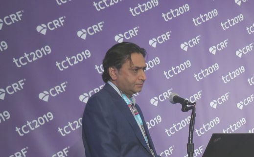
However, the lack of a clear association with serious clinical events such as death, MI, and stroke “does not justify the routine prophylactic use of anticoagulation [following TAVR] in all patients,” lead study investigator Raj R. Makkar, MD, said during a press briefing at the Transcatheter Cardiovascular Therapeutics annual meeting.
“Subclinical leaflet thrombosis characterized by hypoattenuated leaflet thickening and reduced leaflet motion has been frequently observed in transcatheter and surgical aortic bioprosthetic valves,” said Dr. Makkar, director of the interventional cardiology division, Cedars-Sinai Medical Center, Los Angeles. “Thrombus on bioprosthetic valves can present as a spectrum: HALT with relatively normal leaflet motion, HALT with reduced leaflet motion but normal gradients, and clinical valve thrombosis with elevated gradients.”
The primary objective of the current Food and Drug Administration–mandated study, known as the PARTNER 3 Low-Risk Computed Tomography Sub-study, was to evaluate HALT and reduced leaflet motion in terms of differences in transcatheter and surgical bioprosthetic aortic valves among patients enrolled in the randomized PARTNER 3 cohort, to understand the natural history of HALT and reduced leaflet motion in the absence of anticoagulation, and to understand its impact on valve hemodynamics and clinical outcomes. Patients underwent specialized serial CTs at 30 days and at 1 year post TAVR or surgical aortic valve replacement (SAVR). All scans were analyzed by a CT core lab blinded to patient information or time of the scans, and the treating investigators were blinded to the results of the 30-day and 1-year CT scans. A clinical events committee adjudicated key clinical events.
Dr. Makkar reported outcomes from 408 patients: 213 who underwent TAVR and 195 who underwent surgery. There were 348 evaluable serial CT scans at 30 days and 312 at 1 year. The incidence of HALT at 30 days was 13.3% in the TAVR group and 5% in the surgery group, a difference that reached statistical significance (P = .03). At 1 year, however, the difference was not significant (27.5% vs. 20.2%, respectively; P = .19). In the overall cohort, he said, the incidence of HALT was 10% at 30 days and increased to 24% at 1 year.
The researchers also found that HALT was dynamic and spontaneously resolved in 56% of patients in the absence of anticoagulation at 30 days, while new HALT appeared in 21% of patients at 1 year.
“In terms of its impact on valve gradient, the impact was minimal,” Dr. Makkar said. “There was an increase of 1-2 mm Hg in patients who had HALT and in patients who had reduced leaflet motion.”
As for impact on clinical outcomes, the researchers observed no deaths or any myocardial infarction at any time point in patients who had HALT. “There were four cases of valve thrombosis, three of which occurred in patients who had HALT,” Dr. Makkar said at the meeting, sponsored by the Cardiovascular Research Foundation. “One stroke occurred in each group. TIA [transient ischemic attack] occurred in 1 patient out of 35 in the HALT group and 3 out of 311 in the no-HALT group. There was one case of retinal artery embolism in each group.”
In a pooled analysis of clinical events, he and his colleagues observed a numerical increase in death, stroke, TIA, and thrombotic events in patients who had HALT at 30 days, compared with those who did not (8.6% vs. 2.9%, respectively), but the difference did not reach statistical significance (P = .11). “However, given the low total number of events, the data are inconclusive and only hypothesis generating,” he said. “A longer-term follow-up and [a] larger data set will further clarify the impact on clinical outcomes.”
Dr. Makkar emphasized that routine post–TAVR/SAVR CT scans outside of research protocols are not indicated. “CTs should be prompted by increased gradients or thromboembolic events,” he said.
One of the discussants at the briefing, Michael J. Mack, MD, chair of the cardiovascular service line at Baylor Scott and White Health in Dallas and primary author of the PARTNER 3 study, said that prior to the substudy results, “I’ve always thought that the incidence of valve thrombosis would be higher with TAVR than with surgery. So the fact that it was higher at 30 days didn’t surprise me. One of the reasons is that you lose the backwashing effect by changing flow dynamics in the aortic route. What did surprise me is the percent that resolved without anticoagulation.”
He added, “The impact of all this is that we are not justified recommending routine anticoagulation [after bioprosthetic aortic valve replacement surgery]. I think it does call into question the guidelines for surgical valves, because we did that based on smaller observational studies. Now that we have routine surveillance of surgical valves, I think it calls into question the class IIa recommendation for 3 months of anticoagulation. It’s what we’ve always done, and we’ll probably stop doing it on the basis of this. The other shoe that hasn’t dropped is its effect on long-term structural valve deterioration. I do think that early HALT does explain premature structural valve deterioration.”
The trial was sponsored by Edwards Lifesciences. Dr. Makkar disclosed that he is a consultant for and has received research grants from Edwards Lifesciences, Abbott, Medtronic, and Boston Scientific. Dr. Mack is a consultant to Gore and an investigator for Abbott, Edwards Lifesciences, and Medtronic.
SAN FRANCISCO – The was 10% at 30 days and increased to 24% at 1 year, results from a PARTNER 3 substudy demonstrated.
However, the lack of a clear association with serious clinical events such as death, MI, and stroke “does not justify the routine prophylactic use of anticoagulation [following TAVR] in all patients,” lead study investigator Raj R. Makkar, MD, said during a press briefing at the Transcatheter Cardiovascular Therapeutics annual meeting.
“Subclinical leaflet thrombosis characterized by hypoattenuated leaflet thickening and reduced leaflet motion has been frequently observed in transcatheter and surgical aortic bioprosthetic valves,” said Dr. Makkar, director of the interventional cardiology division, Cedars-Sinai Medical Center, Los Angeles. “Thrombus on bioprosthetic valves can present as a spectrum: HALT with relatively normal leaflet motion, HALT with reduced leaflet motion but normal gradients, and clinical valve thrombosis with elevated gradients.”
The primary objective of the current Food and Drug Administration–mandated study, known as the PARTNER 3 Low-Risk Computed Tomography Sub-study, was to evaluate HALT and reduced leaflet motion in terms of differences in transcatheter and surgical bioprosthetic aortic valves among patients enrolled in the randomized PARTNER 3 cohort, to understand the natural history of HALT and reduced leaflet motion in the absence of anticoagulation, and to understand its impact on valve hemodynamics and clinical outcomes. Patients underwent specialized serial CTs at 30 days and at 1 year post TAVR or surgical aortic valve replacement (SAVR). All scans were analyzed by a CT core lab blinded to patient information or time of the scans, and the treating investigators were blinded to the results of the 30-day and 1-year CT scans. A clinical events committee adjudicated key clinical events.
Dr. Makkar reported outcomes from 408 patients: 213 who underwent TAVR and 195 who underwent surgery. There were 348 evaluable serial CT scans at 30 days and 312 at 1 year. The incidence of HALT at 30 days was 13.3% in the TAVR group and 5% in the surgery group, a difference that reached statistical significance (P = .03). At 1 year, however, the difference was not significant (27.5% vs. 20.2%, respectively; P = .19). In the overall cohort, he said, the incidence of HALT was 10% at 30 days and increased to 24% at 1 year.
The researchers also found that HALT was dynamic and spontaneously resolved in 56% of patients in the absence of anticoagulation at 30 days, while new HALT appeared in 21% of patients at 1 year.
“In terms of its impact on valve gradient, the impact was minimal,” Dr. Makkar said. “There was an increase of 1-2 mm Hg in patients who had HALT and in patients who had reduced leaflet motion.”
As for impact on clinical outcomes, the researchers observed no deaths or any myocardial infarction at any time point in patients who had HALT. “There were four cases of valve thrombosis, three of which occurred in patients who had HALT,” Dr. Makkar said at the meeting, sponsored by the Cardiovascular Research Foundation. “One stroke occurred in each group. TIA [transient ischemic attack] occurred in 1 patient out of 35 in the HALT group and 3 out of 311 in the no-HALT group. There was one case of retinal artery embolism in each group.”
In a pooled analysis of clinical events, he and his colleagues observed a numerical increase in death, stroke, TIA, and thrombotic events in patients who had HALT at 30 days, compared with those who did not (8.6% vs. 2.9%, respectively), but the difference did not reach statistical significance (P = .11). “However, given the low total number of events, the data are inconclusive and only hypothesis generating,” he said. “A longer-term follow-up and [a] larger data set will further clarify the impact on clinical outcomes.”
Dr. Makkar emphasized that routine post–TAVR/SAVR CT scans outside of research protocols are not indicated. “CTs should be prompted by increased gradients or thromboembolic events,” he said.
One of the discussants at the briefing, Michael J. Mack, MD, chair of the cardiovascular service line at Baylor Scott and White Health in Dallas and primary author of the PARTNER 3 study, said that prior to the substudy results, “I’ve always thought that the incidence of valve thrombosis would be higher with TAVR than with surgery. So the fact that it was higher at 30 days didn’t surprise me. One of the reasons is that you lose the backwashing effect by changing flow dynamics in the aortic route. What did surprise me is the percent that resolved without anticoagulation.”
He added, “The impact of all this is that we are not justified recommending routine anticoagulation [after bioprosthetic aortic valve replacement surgery]. I think it does call into question the guidelines for surgical valves, because we did that based on smaller observational studies. Now that we have routine surveillance of surgical valves, I think it calls into question the class IIa recommendation for 3 months of anticoagulation. It’s what we’ve always done, and we’ll probably stop doing it on the basis of this. The other shoe that hasn’t dropped is its effect on long-term structural valve deterioration. I do think that early HALT does explain premature structural valve deterioration.”
The trial was sponsored by Edwards Lifesciences. Dr. Makkar disclosed that he is a consultant for and has received research grants from Edwards Lifesciences, Abbott, Medtronic, and Boston Scientific. Dr. Mack is a consultant to Gore and an investigator for Abbott, Edwards Lifesciences, and Medtronic.
REPORTING FROM TCT 2019
Key clinical point: Hypo-attenuated leaflet thickening (HALT) and reduced leaflet motion resulted in a minimal increase in valve gradients, which can be considered clinically insignificant.
Major finding: The incidence of HALT at 30 days was 13.3% in the TAVR group and 5% in the surgery group, a difference that reached statistical significance (P = .03). At 1 year, however, the difference did not differ significantly (27.5% vs. 20.2%, respectively; P = .19).
Study details: An analysis of 408 patients in the PARTNER 3 Low-Risk Computed Tomography Sub-study.
Disclosures: The trial was sponsored by Edwards Lifesciences. Dr. Makkar disclosed that he is a consultant for and has received research grants from Edwards Lifesciences, Abbott, Medtronic, and Boston Scientific. Dr. Mack is a consultant to Gore and an investigator for Abbott, Edwards Lifesciences, and Medtronic.
Source: Makkar R et al. TCT 2019.
European postmarket trial confirms findings of Disrupt CAD I
SAN FRANCISCO – A postmarket analysis of the coronary intravascular lithotripsy (IVL) system validated the safety and utility of the procedure first described in the Disrupt CAD I study.
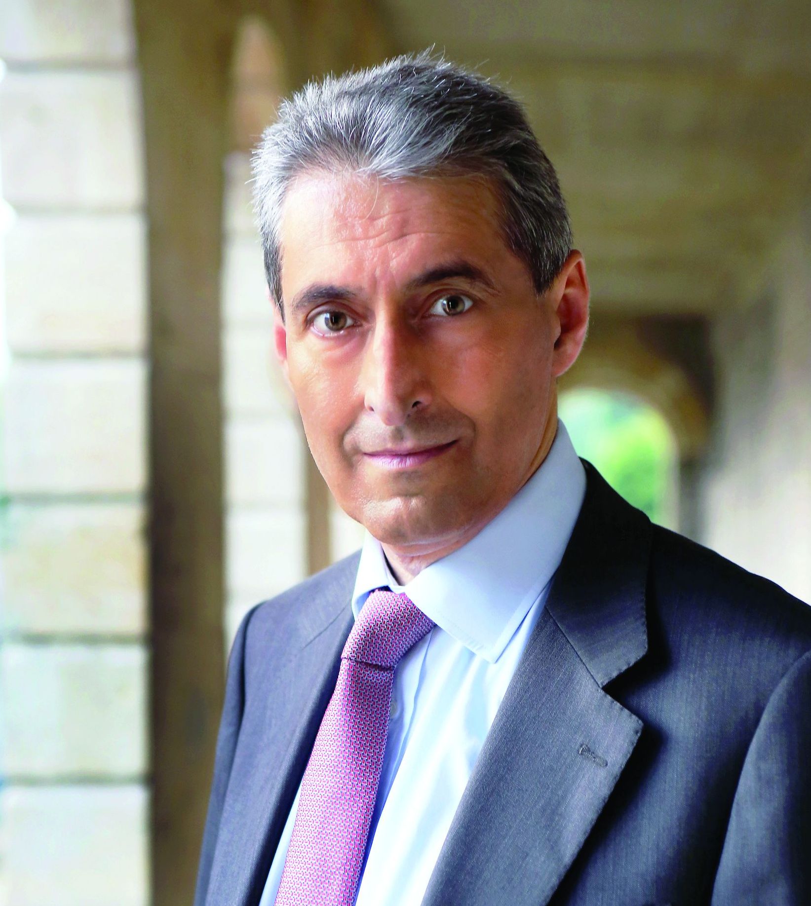
“We now have an efficient technology to treat calcium that is safe to use, simple to learn, and able to achieve 100% success in delivering stents, with low final residual stenosis (7.8%),” Carlo Di Mario, MD, said in an interview in advance of the Transcatheter Cardiovascular Therapeutics annual meeting. “This was accomplished with a low rate of complications.”
Developed by Shockwave Medical, coronary IVL is an innovative lesion preparation tool designed to fracture challenging calcium using sonic pressure waves in order to facilitate stent delivery, deployment, and optimal expansion. In a feasibility study known as DISRUPT CAD I, Dr. Di Mario, director of structural interventional cardiology at Careggi University Hospital in Florence, Italy, and his colleagues performed the procedure in 60 patients (Circulation 2019;139:834-6). Clinical success, defined as residual stenosis of less than 50% post PCI with no evidence of in-hospital major adverse cardiac events (MACE), was 95%, while device success, defined as successful delivery and IVL treatment at target lesion, reached 98.3%.
In an effort to ensure that results of DISRUPT CAD 1 were generalizable to a broader population, Dr. Di Mario and his colleagues enrolled 120 subjects at 15 sites in nine European countries into DISRUPT CAD II, a postmarket, single-arm study. They underwent vessel preparation for stent implantation with IVL, and the primary endpoint was in-hospital MACE, defined as cardiac death, myocardial infarction, or target vessel revascularization. The researchers also performed an optical coherence tomography (OCT) substudy to evaluate the mechanism of action of IVL and to quantify coronary artery calcium characteristics and calcium plaque fracture.
The mean age of the 120 patients was 72 years, 78% were male, 80% had hypertension, 72% had hyperlipidemia, 32% had diabetes, and 65% had class I or II angina. Most of the patients (94%) had severe calcification, with a mean lesion length of about 26 mm. The lesions were concentric in 70% of cases, and 30% had side-branch involvement.
Dr. Di Mario and his associates reported that IVL was delivered successfully in all cases. It also delivered stents successfully in all cases, with a high acute luminal gain (a mean of 1.7 mm2), and low residual stenosis (7.8%). The primary endpoint occurred in 5.8% of patients, consisting of seven non–Q-wave myocardial infarctions. The IVL mechanism of action was shown to be intraplaque calcium fracture, which occurred in about 80% of lesions analyzed by OCT.
“Based on my previous experience with IVL, I was confident that it could modify the calcium, but the results of the OCT substudy of the Disrupt CAD II utilizing OCT to evaluate the mechanism of action was clearly more positive than expected,” said Dr. Di Mario, who was a coprincipal investigator for the trial. “It demonstrated that IVL creates visible calcium fractures in the majority of cases and confirmed that full stent expansion secondary to the circumferential calcium modification is achievable, despite that nearly all the patients (94%) had severe coronary artery calcification. We know from previous work that full stent expansion is required to minimize complications and improve clinical outcomes, which was not always achievable before IVL.”
He acknowledged certain limitations of the study, including the fact that it lacked a concurrent control group, “but it was run very carefully with complete monitoring of events and core lab and CEC [clinical endpoint committee] adjudication,” he said. “Also, the study was used to confirm short-term safety, and as such, did not include long term follow-up.”
In an interview at the meeting, Ajay J. Kirtane, MD, an interventional cardiologist at Columbia University Medical Center, New York, called the findings “reassuring,” but said that he looks forward to results from the trial of the system currently under way in the United States known as the DISRUPT CAD III IDE Study. “The technology is accessible to many physicians because it’s a balloon-based technology,” he said. “Yet in terms of performance, we need to not only evaluate short-term outcomes, we need to see long-term outcomes as well, to make sure there are no untoward effects.”
Shockwave C2 Coronary IVL catheters are commercially available for the treatment of de novo coronary artery disease in Europe and other select countries; in the United States they are limited to investigational use within the DISRUPT CAD III IDE Study.
At the meeting, Ziad A. Ali, MD, an interventional cardiologist at Columbia University/New York–Presbyterian Hospital, presented results from Disrupt CAD II, and the content was published online at the time of presentation. The meeting was sponsored by the Cardiovascular Research Foundation. Dr. Di Mario disclosed that Shockwave Medical provided a grant for the study to Careggi University Hospital. Dr. Ali reported having personal equity and fees from Shockwave Medical, as well as grants from other companies outside the scope of the study.
SOURCE: Di Mario C et al. Circ Cardiovasc Interv. 2019 Sep 25 doi: 10.1161/CIRCINTERVENTIONS.119.008434.
SAN FRANCISCO – A postmarket analysis of the coronary intravascular lithotripsy (IVL) system validated the safety and utility of the procedure first described in the Disrupt CAD I study.
“We now have an efficient technology to treat calcium that is safe to use, simple to learn, and able to achieve 100% success in delivering stents, with low final residual stenosis (7.8%),” Carlo Di Mario, MD, said in an interview in advance of the Transcatheter Cardiovascular Therapeutics annual meeting. “This was accomplished with a low rate of complications.”
Developed by Shockwave Medical, coronary IVL is an innovative lesion preparation tool designed to fracture challenging calcium using sonic pressure waves in order to facilitate stent delivery, deployment, and optimal expansion. In a feasibility study known as DISRUPT CAD I, Dr. Di Mario, director of structural interventional cardiology at Careggi University Hospital in Florence, Italy, and his colleagues performed the procedure in 60 patients (Circulation 2019;139:834-6). Clinical success, defined as residual stenosis of less than 50% post PCI with no evidence of in-hospital major adverse cardiac events (MACE), was 95%, while device success, defined as successful delivery and IVL treatment at target lesion, reached 98.3%.
In an effort to ensure that results of DISRUPT CAD 1 were generalizable to a broader population, Dr. Di Mario and his colleagues enrolled 120 subjects at 15 sites in nine European countries into DISRUPT CAD II, a postmarket, single-arm study. They underwent vessel preparation for stent implantation with IVL, and the primary endpoint was in-hospital MACE, defined as cardiac death, myocardial infarction, or target vessel revascularization. The researchers also performed an optical coherence tomography (OCT) substudy to evaluate the mechanism of action of IVL and to quantify coronary artery calcium characteristics and calcium plaque fracture.
The mean age of the 120 patients was 72 years, 78% were male, 80% had hypertension, 72% had hyperlipidemia, 32% had diabetes, and 65% had class I or II angina. Most of the patients (94%) had severe calcification, with a mean lesion length of about 26 mm. The lesions were concentric in 70% of cases, and 30% had side-branch involvement.
Dr. Di Mario and his associates reported that IVL was delivered successfully in all cases. It also delivered stents successfully in all cases, with a high acute luminal gain (a mean of 1.7 mm2), and low residual stenosis (7.8%). The primary endpoint occurred in 5.8% of patients, consisting of seven non–Q-wave myocardial infarctions. The IVL mechanism of action was shown to be intraplaque calcium fracture, which occurred in about 80% of lesions analyzed by OCT.
“Based on my previous experience with IVL, I was confident that it could modify the calcium, but the results of the OCT substudy of the Disrupt CAD II utilizing OCT to evaluate the mechanism of action was clearly more positive than expected,” said Dr. Di Mario, who was a coprincipal investigator for the trial. “It demonstrated that IVL creates visible calcium fractures in the majority of cases and confirmed that full stent expansion secondary to the circumferential calcium modification is achievable, despite that nearly all the patients (94%) had severe coronary artery calcification. We know from previous work that full stent expansion is required to minimize complications and improve clinical outcomes, which was not always achievable before IVL.”
He acknowledged certain limitations of the study, including the fact that it lacked a concurrent control group, “but it was run very carefully with complete monitoring of events and core lab and CEC [clinical endpoint committee] adjudication,” he said. “Also, the study was used to confirm short-term safety, and as such, did not include long term follow-up.”
In an interview at the meeting, Ajay J. Kirtane, MD, an interventional cardiologist at Columbia University Medical Center, New York, called the findings “reassuring,” but said that he looks forward to results from the trial of the system currently under way in the United States known as the DISRUPT CAD III IDE Study. “The technology is accessible to many physicians because it’s a balloon-based technology,” he said. “Yet in terms of performance, we need to not only evaluate short-term outcomes, we need to see long-term outcomes as well, to make sure there are no untoward effects.”
Shockwave C2 Coronary IVL catheters are commercially available for the treatment of de novo coronary artery disease in Europe and other select countries; in the United States they are limited to investigational use within the DISRUPT CAD III IDE Study.
At the meeting, Ziad A. Ali, MD, an interventional cardiologist at Columbia University/New York–Presbyterian Hospital, presented results from Disrupt CAD II, and the content was published online at the time of presentation. The meeting was sponsored by the Cardiovascular Research Foundation. Dr. Di Mario disclosed that Shockwave Medical provided a grant for the study to Careggi University Hospital. Dr. Ali reported having personal equity and fees from Shockwave Medical, as well as grants from other companies outside the scope of the study.
SOURCE: Di Mario C et al. Circ Cardiovasc Interv. 2019 Sep 25 doi: 10.1161/CIRCINTERVENTIONS.119.008434.
REPORTING FROM TCT 2019

Dual therapy best for AFib with ACS no matter the treatment strategy
SAN FRANCISCO – Anticoagulation with apixaban and a P2Y12 inhibitor without aspirin provides superior safety and similar efficacy in patients with atrial fibrillation who have an acute coronary syndrome, compared with regimens that include vitamin K antagonists, aspirin, or both.

The findings come from a prespecified analysis of data from the AUGUSTUS trial presented by Stephan Windecker, MD, at the Transcatheter Cardiovascular Therapeutics annual meeting.
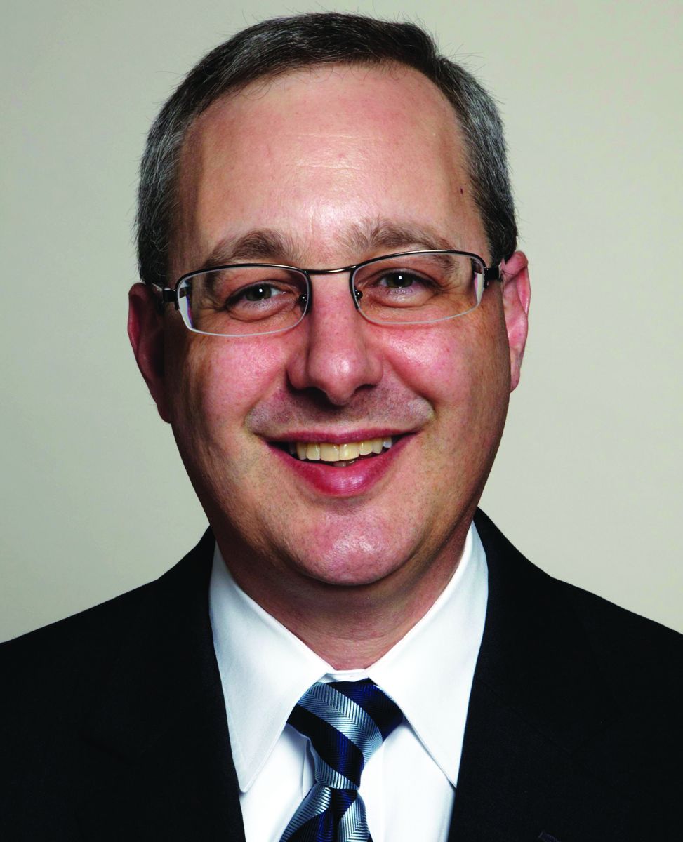
“This study adds very important information [to the notion] that triple therapy in the setting of atrial fibrillation and PCI [percutaneous coronary intervention] is really not the way to go,” Ori Ben-Yehuda, MD, FACC, executive director of the Cardiovascular Research Foundation’s Clinical Trials Center, said during a media briefing.
In the recent multicenter AUGUSTUS trial, Dr. Windecker, of the department of cardiology at Bern University Hospital, Switzerland, and colleagues found that among 4,614 patients with atrial fibrillation and a recent acute coronary syndrome or PCI treated with a P2Y12 inhibitor, apixaban without aspirin resulted in less bleeding, fewer hospitalizations, and no significant differences in ischemic events compared with regimens that included a vitamin K antagonist (VKA), aspirin, or both (N Engl J Med. 2019;380:1509-24). For this prespecified analysis, the researchers used a 2×2 factorial design to compare apixaban with VKA and aspirin with placebo in the AUGUSTUS trial participants with ACS treated medically (group 1; 1,097 patients, or 24%), those with ACS treated with PCI (group 2; 1,714 patients, or 37%), and those undergoing elective PCI (group 3; 1,784 patients, or 39%). The outcomes of interest were bleeding, death, and hospitalization as well as death and ischemic events by antithrombotic strategy in the study participants. This marks the only trial in the field that included patients with ACS managed medically, Dr. Windecker said.
At baseline, the median age of patients was 71 years, 30% were female, 36% had diabetes, and 45% had heart failure. Patients managed medically were younger (a median age of 70) and more frequently female; 57% presented with heart failure. The groups had identical CHA2DS2VASc scores (4), and very similar HAS-BLED scores (2 in groups 1 and 2, and 3 in group 3).
Apixaban compared with VKA showed lower International Society on Thrombosis and Haemostasis–defined major or clinically relevant nonmajor bleeding among patients in group 1 (HR, 0.44), group 2 (HR, 0.68), and group 3 (HR, 0.82) (P for interaction = .052). Apixaban compared with VKA reduced death or hospitalization among patients in group 1 (HR, 0.71), group 2 (HR 0.88), and group 3 (HR, 0.87) (P for interaction = .345). Compared with VKA, apixaban resulted in a similar effect on death and ischemic events among patients in all three treatment groups (P for interaction = .356).
Compared with placebo, aspirin had a higher rate of bleeding among patients in group 1 (HR, 1.49), group 2 (HR, 2.02) and group 3 (HR, 1.91) (P for interaction = .479). For the same comparison, there was no difference in outcomes among the three groups for the composite of death or hospitalization and death and ischemic events.
“The overall results of the AUGUSTUS trial are consistent across the three clinically important subgroups,” Dr. Windecker said. The reasons why patients received medical therapy remain unclear, “because it was at the physician’s discretion as to whether they were treated medically or underwent PCI,” he said. “The proportion very much reflects our clinical practice, where 20%-25% of patients are treated medically. What was surprising for me is that I would have anticipated there would be more elderly patients with comorbidities, but I did anticipate that there would be more female patients (in this subgroup).”
Robert A. Harrington, MD, an interventional cardiologist at Stanford (Calif.) University who served on the Data Safety and Monitoring Board for the trial, noted that the patients with atrial fibrillation represent 7%-10% of all ACS patients, “so it’s a big population,” he said. “What’s been disappointing is that none of the trials have been big enough to uncouple the bleeding vs. ischemic issue. We don’t know the answer for how long do you need the triple therapy versus when you can switch to the double therapy.”
Dr. Windecker said that the optimal duration of short-term aspirin remains unclear in this patient population. “Whether there is a benefit of giving aspirin for 2 weeks or 4 weeks remains unanswered,” he said. “Triple therapy is not the way to go, but we need to fine-tune, and probably individualize, which patients may benefit from a certain duration of aspirin.”
The study results were published online at the time of presentation (Circulation 2019 Sep 26. doi: 10.1161/CIRCULATIONAHA.119.043308.
AUGUSTUS was funded by Bristol-Myers Squibb and Pfizer Inc. Dr. Windecker reported having received institutional research and educational grants to Bern University Hospital from Abbott, Amgen, Bayer, BMS, CSL Behring, Boston Scientific, Biotronik, Edwards Lifesciences, Medtronic, Polares, and Sinomed. His coauthors reported having numerous financial ties to the pharmaceutical and device industries.
SOURCE: Windecker S. TCT 2019, Late-Breaking Trials 1 session.
SAN FRANCISCO – Anticoagulation with apixaban and a P2Y12 inhibitor without aspirin provides superior safety and similar efficacy in patients with atrial fibrillation who have an acute coronary syndrome, compared with regimens that include vitamin K antagonists, aspirin, or both.
The findings come from a prespecified analysis of data from the AUGUSTUS trial presented by Stephan Windecker, MD, at the Transcatheter Cardiovascular Therapeutics annual meeting.
“This study adds very important information [to the notion] that triple therapy in the setting of atrial fibrillation and PCI [percutaneous coronary intervention] is really not the way to go,” Ori Ben-Yehuda, MD, FACC, executive director of the Cardiovascular Research Foundation’s Clinical Trials Center, said during a media briefing.
In the recent multicenter AUGUSTUS trial, Dr. Windecker, of the department of cardiology at Bern University Hospital, Switzerland, and colleagues found that among 4,614 patients with atrial fibrillation and a recent acute coronary syndrome or PCI treated with a P2Y12 inhibitor, apixaban without aspirin resulted in less bleeding, fewer hospitalizations, and no significant differences in ischemic events compared with regimens that included a vitamin K antagonist (VKA), aspirin, or both (N Engl J Med. 2019;380:1509-24). For this prespecified analysis, the researchers used a 2×2 factorial design to compare apixaban with VKA and aspirin with placebo in the AUGUSTUS trial participants with ACS treated medically (group 1; 1,097 patients, or 24%), those with ACS treated with PCI (group 2; 1,714 patients, or 37%), and those undergoing elective PCI (group 3; 1,784 patients, or 39%). The outcomes of interest were bleeding, death, and hospitalization as well as death and ischemic events by antithrombotic strategy in the study participants. This marks the only trial in the field that included patients with ACS managed medically, Dr. Windecker said.
At baseline, the median age of patients was 71 years, 30% were female, 36% had diabetes, and 45% had heart failure. Patients managed medically were younger (a median age of 70) and more frequently female; 57% presented with heart failure. The groups had identical CHA2DS2VASc scores (4), and very similar HAS-BLED scores (2 in groups 1 and 2, and 3 in group 3).
Apixaban compared with VKA showed lower International Society on Thrombosis and Haemostasis–defined major or clinically relevant nonmajor bleeding among patients in group 1 (HR, 0.44), group 2 (HR, 0.68), and group 3 (HR, 0.82) (P for interaction = .052). Apixaban compared with VKA reduced death or hospitalization among patients in group 1 (HR, 0.71), group 2 (HR 0.88), and group 3 (HR, 0.87) (P for interaction = .345). Compared with VKA, apixaban resulted in a similar effect on death and ischemic events among patients in all three treatment groups (P for interaction = .356).
Compared with placebo, aspirin had a higher rate of bleeding among patients in group 1 (HR, 1.49), group 2 (HR, 2.02) and group 3 (HR, 1.91) (P for interaction = .479). For the same comparison, there was no difference in outcomes among the three groups for the composite of death or hospitalization and death and ischemic events.
“The overall results of the AUGUSTUS trial are consistent across the three clinically important subgroups,” Dr. Windecker said. The reasons why patients received medical therapy remain unclear, “because it was at the physician’s discretion as to whether they were treated medically or underwent PCI,” he said. “The proportion very much reflects our clinical practice, where 20%-25% of patients are treated medically. What was surprising for me is that I would have anticipated there would be more elderly patients with comorbidities, but I did anticipate that there would be more female patients (in this subgroup).”
Robert A. Harrington, MD, an interventional cardiologist at Stanford (Calif.) University who served on the Data Safety and Monitoring Board for the trial, noted that the patients with atrial fibrillation represent 7%-10% of all ACS patients, “so it’s a big population,” he said. “What’s been disappointing is that none of the trials have been big enough to uncouple the bleeding vs. ischemic issue. We don’t know the answer for how long do you need the triple therapy versus when you can switch to the double therapy.”
Dr. Windecker said that the optimal duration of short-term aspirin remains unclear in this patient population. “Whether there is a benefit of giving aspirin for 2 weeks or 4 weeks remains unanswered,” he said. “Triple therapy is not the way to go, but we need to fine-tune, and probably individualize, which patients may benefit from a certain duration of aspirin.”
The study results were published online at the time of presentation (Circulation 2019 Sep 26. doi: 10.1161/CIRCULATIONAHA.119.043308.
AUGUSTUS was funded by Bristol-Myers Squibb and Pfizer Inc. Dr. Windecker reported having received institutional research and educational grants to Bern University Hospital from Abbott, Amgen, Bayer, BMS, CSL Behring, Boston Scientific, Biotronik, Edwards Lifesciences, Medtronic, Polares, and Sinomed. His coauthors reported having numerous financial ties to the pharmaceutical and device industries.
SOURCE: Windecker S. TCT 2019, Late-Breaking Trials 1 session.
AT TCT 2019
Biologics beyond anti-TNF therapies show promise for ulcerative colitis
In one of two such trials published in The New England Journal of Medicine on Sept. 26, 2019, researchers led by Bruce E. Sands, MD, of the Icahn School of Medicine at Mount Sinai, New York, and associates evaluated ustekinumab as an 8-week induction therapy and a 44-week maintenance therapy in patients with moderate to severe ulcerative colitis (N Engl J Med 2019 Sep 25 doi: 10/1056/NEJMoa1900750). For the phase 3 trial, known as UNIFI, researchers randomly assigned 961 patients to receive an intravenous induction dose of ustekinumab over the course of 8 weeks (320 to a dose of 130 mg and 322 to a weight-range–based dose that approximated 6 mg per kilogram of body weight), while the remaining 319 received placebo. Patients who responded to induction therapy were randomly assigned to a 44-week maintenance phase in which they received subcutaneous maintenance injections of 90 mg of ustekinumab (172 to injections every 12 weeks, 176 to injections every 8 weeks, and 175 to placebo). The primary endpoint for both phases of the trial was clinical remission, defined as a total score of 2 or lower on the Mayo scale, and no subscore greater than 1 on any of the four Mayo scale components.
Dr. Sands and his colleagues found that at week 8, clinical remission was achieved in 15.6% of patients in the 130-mg ustekinumab infusion group, compared with 15.5% in the 6-mg per kg of body weight group, and 5.3% of those in the placebo group. “The percentages of patients who met major secondary endpoints or had histo-endoscopic mucosal healing were significantly higher in both ustekinumab groups than in the placebo group,” the researchers wrote. “Through week 8, the median changes from baseline in the IBDQ [Inflammatory Bowel Disease Questionnaire] score were significantly greater in both ustekinumab groups than in the placebo group.”
Meanwhile, at week 44, clinical remission was achieved in 38.4% of patients in the group receiving 90-mg subcutaneous ustekinumab every 12 weeks, compared with 43.8% of those in the group receiving 90 mg every 8 weeks, and 24% of those in the placebo group. “The percentages of patients with maintenance of clinical response through week 44, endoscopic improvement at week 44, or corticosteroid-free clinical remission (with either definition of clinical remission) at week 44 were significantly higher in both ustekinumab groups than in the placebo group,” the researchers wrote.
When they evaluated other endpoints, Dr. Sands and his colleagues observed that improvements in partial Mayo scores and reductions in serum and fecal concentrations of inflammatory biomarkers that occurred with induction were sustained through week 44. “Although our findings suggest that ustekinumab was effective in patients with or without previous treatment failure with biologics for both induction and maintenance therapy, the percentages of patients in whom each endpoint was achieved were lower across groups with previous treatment failure with biologics,” they wrote.
In the second study, known as VARSITY, researchers led by Dr. Sands conducted a randomized, phase 3b, head-to-head trial comparing vedolizumab with adalimumab in 769 adults with moderate to severe ulcerative colitis, in an effort to determine if vedolizumab is superior after 52 weeks of treatment (N Engl J Med 2019 Sep 25. doi: 10/1056/NEJMoa1905725). They assigned patients to receive IV infusions of 300 mg of vedolizumab on day 1 and at weeks 2, 6, 14, 22, 30, 38, and 46 (plus injections of placebo) or subcutaneous injections of 40 mg of adalimumab, with a total dose of 160 mg at week 1, 80 mg at week 2, and 40 mg every 2 weeks thereafter until week 50 (plus infusions of placebo). The primary endpoint was clinical remission, defined as a total score of 2 or lower on the Mayo scale, and no subscore greater than 1 on any of the four Mayo scale components.
At week 52, the researchers found that 31.3% of patients in the vedolizumab group achieved clinical remission, compared with 22.5% of those in the adalimumab group, while a higher percentage of patients in the vedolizumab group demonstrated endoscopic involvement (the first secondary outcome), compared with their counterparts in the adalimumab group (39.7% vs. 27.7%, respectively). Treatment effects were more pronounced in patients who had not previously used a TNF inhibitor.
In contrast, Dr. Sands and colleagues reported that at week 52, corticosteroid-free clinical remission was observed in 12.6% of patients in the vedolizumab group, compared with 21.8% of their counterparts in the adalimumab group. “It is difficult to explain the inconsistency of the results between this secondary remission outcome and the primary remission outcome,” they wrote. “The trial did not require a specific schedule for corticosteroid tapering, which can vary among practitioners. However, this limitation should not have resulted in differential effects in the two treatment groups.”
They noted that dosing regimens used in VARSITY were based on a conservative approach and use according to U.S. labels. “Real-world studies have shown improved efficacy outcomes after dose intensification in both adalimumab and vedolizumab therapies,” they wrote. “Data from ongoing trials of adalimumab (NCT02065622) and vedolizumab (NCT03029143) may further characterize the effect of higher doses on efficacy outcomes.”
UNIFI was funded by Janssen Research and Development. Dr. Sands disclosed that the Icahn School of Medicine received an institutional grant from Janssen to conduct the study. VARSITY was funded with support from Takeda. Dr. Sands reported that he received grant support and consulting fees from Takeda. Dr. Sands and coauthors reported having financial ties to many other companies in the pharmaceutical and biotechnology industries.
SOURCE: Sands B et al. N Engl J Med. 2019 Sep 25 doi: 10/1056/NEJMoa1900750; N Engl J Med. 2019 Sep 25 doi:.10/1056/NEJMoa1905725.
Long-term rates of colectomy for ulcerative colitis have not declined over a 10-year period, a fact that highlights the need for new biologic therapies and strategies.
Although the VARSITY trial presents a head-to-head comparison of biologics for inflammatory bowel disease and aims to determine the first-line biologic therapy for ulcerative colitis, any clinical superiority of vedolizumab should be balanced against the significant cost advantages of a subcutaneous regimen of adalimumab. In many respects, the ideal trial to assess whether vedolizumab should supplant anti-TNF therapies would involve a head-to-head comparison of infliximab infusions with vedolizumab infusions in patients who have not previously received anti-TNF therapies.
The UNIFI trial assessed the combination of a single induction infusion followed by a maintenance subcutaneous regimen in patients with ulcerative colitis and may lead to the assessment of similar regimens in future trials of biologics in an effort to reduce our dependence on expensive, completely infusion-based biologic regimens, not to mention to relieve pressure on our increasingly busy infusion units. Indeed, the landscape of biologic therapies for ulcerative colitis has changed so dramatically over the past decade with the widespread introduction of less-expensive infliximab and adalimumab biosimilars, as well as vedolizumab, oral Janus kinase inhibitors (tofacitinib), and now ustekinumab, that biologics rather than hospitalization or colectomy are now the main driver of health care costs in the management of inflammatory bowel disease.
The findings in both these trials by Sands et al. highlight the importance of alternative biologic treatments and regimens for ulcerative colitis in patients who are not able to receive anti-TNF therapies because of unacceptable side effects or who have disease that is refractory to anti-TNF therapies. The cost-effectiveness of all biologics will have to come into sharper focus in future trials and longitudinal studies of biologics to help determine not only their eventual place in the treatment algorithm for moderate to severe ulcerative colitis but also the true effect of existing and newer biologics on disease course and rates of colectomy.
This text was extracted from an editorial by Richard J. Farrell, MD, that appeared online Sep. 25, 2019, in The New England Journal of Medicine. Dr. Farrell is with Connolly Hospital and Royal College of Surgeons in Dublin, Ireland.
In one of two such trials published in The New England Journal of Medicine on Sept. 26, 2019, researchers led by Bruce E. Sands, MD, of the Icahn School of Medicine at Mount Sinai, New York, and associates evaluated ustekinumab as an 8-week induction therapy and a 44-week maintenance therapy in patients with moderate to severe ulcerative colitis (N Engl J Med 2019 Sep 25 doi: 10/1056/NEJMoa1900750). For the phase 3 trial, known as UNIFI, researchers randomly assigned 961 patients to receive an intravenous induction dose of ustekinumab over the course of 8 weeks (320 to a dose of 130 mg and 322 to a weight-range–based dose that approximated 6 mg per kilogram of body weight), while the remaining 319 received placebo. Patients who responded to induction therapy were randomly assigned to a 44-week maintenance phase in which they received subcutaneous maintenance injections of 90 mg of ustekinumab (172 to injections every 12 weeks, 176 to injections every 8 weeks, and 175 to placebo). The primary endpoint for both phases of the trial was clinical remission, defined as a total score of 2 or lower on the Mayo scale, and no subscore greater than 1 on any of the four Mayo scale components.
Dr. Sands and his colleagues found that at week 8, clinical remission was achieved in 15.6% of patients in the 130-mg ustekinumab infusion group, compared with 15.5% in the 6-mg per kg of body weight group, and 5.3% of those in the placebo group. “The percentages of patients who met major secondary endpoints or had histo-endoscopic mucosal healing were significantly higher in both ustekinumab groups than in the placebo group,” the researchers wrote. “Through week 8, the median changes from baseline in the IBDQ [Inflammatory Bowel Disease Questionnaire] score were significantly greater in both ustekinumab groups than in the placebo group.”
Meanwhile, at week 44, clinical remission was achieved in 38.4% of patients in the group receiving 90-mg subcutaneous ustekinumab every 12 weeks, compared with 43.8% of those in the group receiving 90 mg every 8 weeks, and 24% of those in the placebo group. “The percentages of patients with maintenance of clinical response through week 44, endoscopic improvement at week 44, or corticosteroid-free clinical remission (with either definition of clinical remission) at week 44 were significantly higher in both ustekinumab groups than in the placebo group,” the researchers wrote.
When they evaluated other endpoints, Dr. Sands and his colleagues observed that improvements in partial Mayo scores and reductions in serum and fecal concentrations of inflammatory biomarkers that occurred with induction were sustained through week 44. “Although our findings suggest that ustekinumab was effective in patients with or without previous treatment failure with biologics for both induction and maintenance therapy, the percentages of patients in whom each endpoint was achieved were lower across groups with previous treatment failure with biologics,” they wrote.
In the second study, known as VARSITY, researchers led by Dr. Sands conducted a randomized, phase 3b, head-to-head trial comparing vedolizumab with adalimumab in 769 adults with moderate to severe ulcerative colitis, in an effort to determine if vedolizumab is superior after 52 weeks of treatment (N Engl J Med 2019 Sep 25. doi: 10/1056/NEJMoa1905725). They assigned patients to receive IV infusions of 300 mg of vedolizumab on day 1 and at weeks 2, 6, 14, 22, 30, 38, and 46 (plus injections of placebo) or subcutaneous injections of 40 mg of adalimumab, with a total dose of 160 mg at week 1, 80 mg at week 2, and 40 mg every 2 weeks thereafter until week 50 (plus infusions of placebo). The primary endpoint was clinical remission, defined as a total score of 2 or lower on the Mayo scale, and no subscore greater than 1 on any of the four Mayo scale components.
At week 52, the researchers found that 31.3% of patients in the vedolizumab group achieved clinical remission, compared with 22.5% of those in the adalimumab group, while a higher percentage of patients in the vedolizumab group demonstrated endoscopic involvement (the first secondary outcome), compared with their counterparts in the adalimumab group (39.7% vs. 27.7%, respectively). Treatment effects were more pronounced in patients who had not previously used a TNF inhibitor.
In contrast, Dr. Sands and colleagues reported that at week 52, corticosteroid-free clinical remission was observed in 12.6% of patients in the vedolizumab group, compared with 21.8% of their counterparts in the adalimumab group. “It is difficult to explain the inconsistency of the results between this secondary remission outcome and the primary remission outcome,” they wrote. “The trial did not require a specific schedule for corticosteroid tapering, which can vary among practitioners. However, this limitation should not have resulted in differential effects in the two treatment groups.”
They noted that dosing regimens used in VARSITY were based on a conservative approach and use according to U.S. labels. “Real-world studies have shown improved efficacy outcomes after dose intensification in both adalimumab and vedolizumab therapies,” they wrote. “Data from ongoing trials of adalimumab (NCT02065622) and vedolizumab (NCT03029143) may further characterize the effect of higher doses on efficacy outcomes.”
UNIFI was funded by Janssen Research and Development. Dr. Sands disclosed that the Icahn School of Medicine received an institutional grant from Janssen to conduct the study. VARSITY was funded with support from Takeda. Dr. Sands reported that he received grant support and consulting fees from Takeda. Dr. Sands and coauthors reported having financial ties to many other companies in the pharmaceutical and biotechnology industries.
SOURCE: Sands B et al. N Engl J Med. 2019 Sep 25 doi: 10/1056/NEJMoa1900750; N Engl J Med. 2019 Sep 25 doi:.10/1056/NEJMoa1905725.
FROM THE NEW ENGLAND JOURNAL OF MEDICINE
Key clinical point: New biologic therapies and strategies for ulcerative colitis patients continue to evolve.
Major finding: In UNIFI, a higher percentage of patients who received a single infusion of ustekinumab at 130 mg or 6 mg per kg of body weight achieved clinical remission at 8 weeks, compared with placebo (15.6% and 15.5%, vs. 5.3%, respectively. In VARSITY, a higher percentage of patients who received vedolizumab achieved clinical remission at 52 weeks, compared with those who received adalimumab (31.3% vs. 22.5%, respectively).
Study details: UNIFI, a phase 3 trial, included an 8-week randomized induction trial of 961 patients and a 44-week randomized-withdrawal maintenance trial. VARSITY was a randomized, phase 3b, head-to-head trial comparing vedolizumab with adalimumab in 769 adults with moderate to severe ulcerative colitis.
Disclosures: UNIFI was funded by Janssen Research and Development. Dr. Sands disclosed that the Icahn School of Medicine received an institutional grant from Janssen to conduct the study. VARSITY was funded with support from Takeda. Dr. Sands reported that he received grant support and consulting fees from Takeda. Dr. Sands and coauthors reported having financial ties to many other companies in the pharmaceutical and biotechnology industries.
Sources: Sands B et al. N Engl J Med. 2019 Sep 25. doi: 10/1056/NEJMoa1900750; N Engl J Med. 2019 Sep 25. doi: 10/1056/NEJMoa1905725.
Don’t let legal considerations drive your decision making about a new job offer
SAN DIEGO – When weighing an offer to join a dermatology practice, don’t allow legal considerations to drive your decision making, Mathew M. Avram, MD, JD, advised at the annual Masters of Aesthetics Symposium.

“Determine your professional interests and follow them accordingly,” said Dr. Avram, director of laser, cosmetics, and dermatologic surgery at Massachusetts General Hospital in Boston. “Your decision as to whether to join a particular practice should not be based upon a contract. Make certain to determine the business end of your deal with any new employer first: the compensation, the work hours, etc. Decide that upfront before you get into the legal details.”
The way he sees it, your relationship with your employer will always be paramount. “This is very important,” said Dr. Avram, who practiced law before he became a dermatologist. “A good contract with a bad employer will not save you from an unhappy employment experience. Few contracts ever end up in litigation, and you cannot sue your way to a happier work experience. Trust your intuition about people; trust your underlying interest in potentially working with this person, and work from there.”
Whether you choose to work in an academic practice, a private practice, or a hybrid, the key issues to address include compensation, benefits, work hours, job responsibilities, and future partnership possibilities. “As an employee, you have the greatest leverage prior to signing an agreement,” he said. “This is the time to ask for what you want.” This may include special requests such as asking the employer to purchase lasers or other special equipment for your office, or to set aside dedicated clinical time for cosmetic procedures, so that you can build a cosmetic practice.
If you’re mulling over a job offer in academics, consider asking for an academic title, what the scope of your authority is, and about your ability to hire or prevent the hiring of others. “Let’s say you’re starting a laser center in your academic center,” said Dr. Avram, who is a past president of the American Society for Laser Medicine and Surgery. “You may not want to find out after you’ve signed your contract that they’ve hired five other people to do the same thing.”
If you’re considering a job offer from a private practice, ask for specifics about bonuses, partnership track, scope of practice, and device purchases. “You want to have that decided before you sign,” he said. “But these are business issues, not legal issues. An agreement needs to be made between you and your employer as to these issues. Once they’ve been agreed upon, it’s time to proceed with a contract. This is where legal advice becomes helpful.” The trick is to negotiate in good faith while maintaining your relationship with your new employer. “Do not destroy your relationship over legal points, but do not cave on crucial issues for fear of upsetting your new employer,” he said.
In law school, Dr. Avram learned that there is no such thing as a standard employment contract. Any contract can be amended, no matter how large or small the institution. “You can amend a mortgage agreement, so you can certainly amend a physician employment contract,” he said. “If the employer is not going to put their commitments in writing, they are probably not going to honor that commitment to you. Written agreements supersede all preceding oral agreements. Each key term needs to be stated explicitly. If not, you have lost all leverage to enforce your initial agreement.”
Key provisions are restrictive covenants and at-will employment. Dr. Avram defined restrictive covenants as contractual agreements that attempt to restrict an employee so as to limit that person’s ability to compete. “This can include noncompete clauses, nonsolicitation agreements, and confidentiality agreements,” he said. “A noncompete agreement prohibits a doctor from competing against their former practice within a specific geographic area for a period of time after the employment has ended. The nonsolicitation agreement restricts the manner and time during which a physician can solicit patients or employees from a practice after termination of employment. A confidentiality agreement is usually indefinite as to time and it restricts the employee from disclosing confidential practice information.”
State laws, he continued, govern restrictive covenants. Some states prohibit them. States that allow them limit their scope to prevent undue burden on the employee’s ability to make a living after termination. “Restrictive covenants in cities need to be narrower than those in rural areas,” he said. “Overly broad restrictive covenants may be unenforceable. Courts can ‘blue line’ covenants to make them more reasonable in scope.”
Next, Dr. Avram discussed involuntary termination in the workplace. Termination with cause means that termination can only result from violation of policy or ethics code violation or significantly poor performance. “In the absence of these circumstances, termination cannot legally proceed,” he said. “This protects the employee from an arbitrary termination.”
On the other hand, at-will employees can be terminated without cause. “It’s a much easier standard to terminate an at-will employee, as long as the reason is not illegal,” he said. “In academic centers, your employment as a physician may require a for-cause termination, whereas your administrative title may be without cause.”
One way to avoid potential legal trouble is to abide by your employer’s rules governing a physician’s interactions with industry. “It is foolish to break those rules,” Dr. Avram said. “Transparency ahead of time is always your best policy. Remember: Your day job is far more valuable to you than part-time work with industry. Also, know that pharma payments are publicly reported by the Centers for Medicare & Medicaid Services.” When it comes to establishing contracts with industry, “read them carefully and have your academic center approve them, if applicable,” he said. “Watch out for noncompete clauses, and some contracts require that you do not speak negatively about your findings. That’s something you should try to avoid.”
Dr. Avram also advised clinicians not to discuss intellectual property with industry representatives. “Do not give away your intellectual property to industry when you’re consulting with them,” he said. “It is easier to do than you might think. If you have a great idea, keep it to yourself or get it protected. This is much easier for an institution where they want you to do these things. They take ownership of a lot of it, but they make it easier to do. Otherwise, you have to go through the expensive process of getting a patent.”
Dr. Avram reported that he has received consulting fees from Allergan, Merz Pharma, Sciton, Soliton, and Zalea. He also reported having ownership and/or shareholder interest in Cytrellis Biosystems, InMode, and Zalea, and intellectual property rights with Cytrellis.
SAN DIEGO – When weighing an offer to join a dermatology practice, don’t allow legal considerations to drive your decision making, Mathew M. Avram, MD, JD, advised at the annual Masters of Aesthetics Symposium.
“Determine your professional interests and follow them accordingly,” said Dr. Avram, director of laser, cosmetics, and dermatologic surgery at Massachusetts General Hospital in Boston. “Your decision as to whether to join a particular practice should not be based upon a contract. Make certain to determine the business end of your deal with any new employer first: the compensation, the work hours, etc. Decide that upfront before you get into the legal details.”
The way he sees it, your relationship with your employer will always be paramount. “This is very important,” said Dr. Avram, who practiced law before he became a dermatologist. “A good contract with a bad employer will not save you from an unhappy employment experience. Few contracts ever end up in litigation, and you cannot sue your way to a happier work experience. Trust your intuition about people; trust your underlying interest in potentially working with this person, and work from there.”
Whether you choose to work in an academic practice, a private practice, or a hybrid, the key issues to address include compensation, benefits, work hours, job responsibilities, and future partnership possibilities. “As an employee, you have the greatest leverage prior to signing an agreement,” he said. “This is the time to ask for what you want.” This may include special requests such as asking the employer to purchase lasers or other special equipment for your office, or to set aside dedicated clinical time for cosmetic procedures, so that you can build a cosmetic practice.
If you’re mulling over a job offer in academics, consider asking for an academic title, what the scope of your authority is, and about your ability to hire or prevent the hiring of others. “Let’s say you’re starting a laser center in your academic center,” said Dr. Avram, who is a past president of the American Society for Laser Medicine and Surgery. “You may not want to find out after you’ve signed your contract that they’ve hired five other people to do the same thing.”
If you’re considering a job offer from a private practice, ask for specifics about bonuses, partnership track, scope of practice, and device purchases. “You want to have that decided before you sign,” he said. “But these are business issues, not legal issues. An agreement needs to be made between you and your employer as to these issues. Once they’ve been agreed upon, it’s time to proceed with a contract. This is where legal advice becomes helpful.” The trick is to negotiate in good faith while maintaining your relationship with your new employer. “Do not destroy your relationship over legal points, but do not cave on crucial issues for fear of upsetting your new employer,” he said.
In law school, Dr. Avram learned that there is no such thing as a standard employment contract. Any contract can be amended, no matter how large or small the institution. “You can amend a mortgage agreement, so you can certainly amend a physician employment contract,” he said. “If the employer is not going to put their commitments in writing, they are probably not going to honor that commitment to you. Written agreements supersede all preceding oral agreements. Each key term needs to be stated explicitly. If not, you have lost all leverage to enforce your initial agreement.”
Key provisions are restrictive covenants and at-will employment. Dr. Avram defined restrictive covenants as contractual agreements that attempt to restrict an employee so as to limit that person’s ability to compete. “This can include noncompete clauses, nonsolicitation agreements, and confidentiality agreements,” he said. “A noncompete agreement prohibits a doctor from competing against their former practice within a specific geographic area for a period of time after the employment has ended. The nonsolicitation agreement restricts the manner and time during which a physician can solicit patients or employees from a practice after termination of employment. A confidentiality agreement is usually indefinite as to time and it restricts the employee from disclosing confidential practice information.”
State laws, he continued, govern restrictive covenants. Some states prohibit them. States that allow them limit their scope to prevent undue burden on the employee’s ability to make a living after termination. “Restrictive covenants in cities need to be narrower than those in rural areas,” he said. “Overly broad restrictive covenants may be unenforceable. Courts can ‘blue line’ covenants to make them more reasonable in scope.”
Next, Dr. Avram discussed involuntary termination in the workplace. Termination with cause means that termination can only result from violation of policy or ethics code violation or significantly poor performance. “In the absence of these circumstances, termination cannot legally proceed,” he said. “This protects the employee from an arbitrary termination.”
On the other hand, at-will employees can be terminated without cause. “It’s a much easier standard to terminate an at-will employee, as long as the reason is not illegal,” he said. “In academic centers, your employment as a physician may require a for-cause termination, whereas your administrative title may be without cause.”
One way to avoid potential legal trouble is to abide by your employer’s rules governing a physician’s interactions with industry. “It is foolish to break those rules,” Dr. Avram said. “Transparency ahead of time is always your best policy. Remember: Your day job is far more valuable to you than part-time work with industry. Also, know that pharma payments are publicly reported by the Centers for Medicare & Medicaid Services.” When it comes to establishing contracts with industry, “read them carefully and have your academic center approve them, if applicable,” he said. “Watch out for noncompete clauses, and some contracts require that you do not speak negatively about your findings. That’s something you should try to avoid.”
Dr. Avram also advised clinicians not to discuss intellectual property with industry representatives. “Do not give away your intellectual property to industry when you’re consulting with them,” he said. “It is easier to do than you might think. If you have a great idea, keep it to yourself or get it protected. This is much easier for an institution where they want you to do these things. They take ownership of a lot of it, but they make it easier to do. Otherwise, you have to go through the expensive process of getting a patent.”
Dr. Avram reported that he has received consulting fees from Allergan, Merz Pharma, Sciton, Soliton, and Zalea. He also reported having ownership and/or shareholder interest in Cytrellis Biosystems, InMode, and Zalea, and intellectual property rights with Cytrellis.
EXPERT ANALYSIS FROM MOAS 2019
Study aims to define symptoms of Sjögren’s syndrome secondary to SLE
Sjögren’s syndrome secondary to systemic lupus erythematosus rises in frequency with age, affects nearly one-quarter of all people with SLE, and is marked by a systemic inflammatory state with high levels of proinflammatory cytokines.
Those are key findings from a Swedish study that set out to evaluate the subjective and objective symptoms of secondary Sjögren’s syndrome (sSS) from a large cohort of SLE patients and matched controls.
“The diagnosis SS is a clinical entity, based on dryness of eyes and mouth due to destructive inflammation in the exocrine glands, especially tear and salivary glands,” researchers led by Guillermo Ruacho, DMD, and Marika Kvarnström, MD, PhD, of the Karolinska Institute, wrote in a study published in the Journal of Rheumatology (doi: 10.3899/jrheum.190250). “SS can exist [as] isolated, primary SS (pSS) or together with other rheumatic diseases, referred to as secondary SS (sSS). A major difference according to the 2002 Revised American-European Consensus Criteria (AECC) is the classification where the serologic item (SSA/SSB antibodies) is included for pSS, but not for sSS (Ann Rheum Dis. 2002;61:554-8). In SLE, these autoantibodies are common, usually stable over time, and they appear early, even several years before disease onset.”
The researchers evaluated 504 consecutive SLE patients and 319 controls from the general population, who were matched for age and gender to the first 319 SLE patients. They used AECC to define SLE-sSS and conducted a thorough clinical investigation of all patients, including analysis of autoantibodies and 20 selected cytokines.
The researchers found that SLE-sSS occurred in 23% of the SLE patients. In comparison with SLE patients who did not have sSS, those in the SLE-sSS group were an average of 9 years older, more likely to be female (96% vs. 84%, respectively), and more likely to have leukopenia (57% vs. 45%), yet less likely to have nephritis (32% vs. 43%). Of 20 proinflammatory cytokines investigated, 6 were higher in the SLE-sSS group: TNF-alpha, IL-6, MCP-4, MIP-1beta, IL-12/IL-23p40, and IP-10. Other clinical measures higher in the SLE-sSS group were total IgG, anti-SSA/Ro52, anti-SSA/Ro60, anti-SSB/La antibodies, and rheumatoid factor (IgM and IgA; P less than .05 for all comparisons).
“To our knowledge this is the first study to investigate if systemic inflammation, as measured by cytokine levels, differs between SLE-sSS and SLE-nonsSS,” the researchers wrote. “In clinical practice, it is often difficult to delineate pSS from SLE-sSS. Organ manifestations commonly reported in pSS are fever, lymphadenopathy, parotid gland enlargement, Raynaud’s phenomenon, interstitial lung disease, peripheral neuropathy, and vasculitis. All these clinical features, except parotid gland enlargement, were investigated in the present study, but only peripheral neuropathy differed and was more frequent in SLE-sSS than in SLE-nonsSS.”
They acknowledged certain limitations of the study, including the fact that they did not measure saliva and tear production in controls without sicca symptoms.
The study was supported by funds from Swedish local and national governments, medical societies, foundations, and patient advocacy groups, One author is an employee at AstraZeneca, which provided reagents for the cytokine analyses but had no impact on the analyses, the authors said.
SOURCE: Ruacho G et al. J Rheumatol. 2019 Sep 1. doi: 10.3899/jrheum.190250.
FROM THE JOURNAL OF RHEUMATOLOGY
Longer-lasting neuromodulators coming down the pike
SAN DIEGO – In the coming years, expect to see an increasing number of neuromodulators hit the market, Joel L. Cohen, MD, predicted at the annual Masters of Aesthetics Symposium.
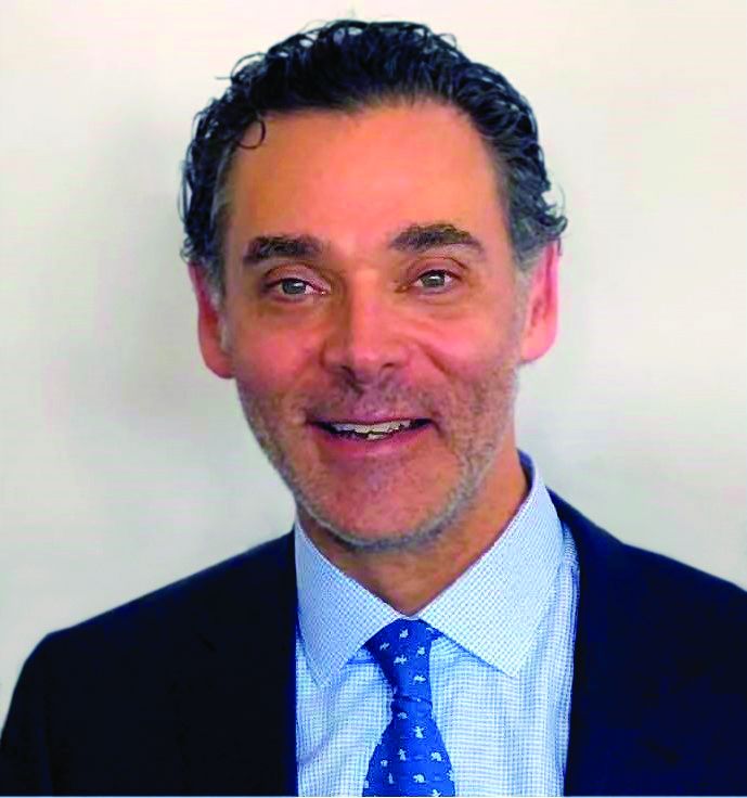
One such product, DaxibotulinumtoxinA (Daxi), formerly known as RT002, contains a proprietary peptide that may contribute to extending its duration of action beyond currently available neuromodulator products. “Another difference for Daxi is that it does not contain human serum albumin,” said Dr. Cohen, who’s in private practice in Greenwood Village and Lone Tree, both in Colo.
In trials of the agent conducted by Revance, the manufacturer, for the treatment of moderate to severe glabellar lines, DaxibotulinumtoxinA achieved a 1-point change in results from baseline in a median of 24 weeks, while the return to baseline wrinkle severity occurred in a median of 28 weeks. According to the Revance web site, DaxibotulinumtoxinA is up for possible Food and Drug Administration approval in 2020.
Though current neuromodulators on the market may be most effective for 3-4 months, the reality is that patients often don’t come in for longer stretches of time – as there is still some degree of efficacy. Dr. Cohen shared interim data from an ongoing study that showed that at 6 months 69% of patients remain satisfied with the result of their last injection. “With Dysport, for example, even though we know the durability is to 3-4 months, we have patients who may still be happy with the results at 6 months,” he said.
Another trend he discussed is the increasing interest in QM1114, a novel, ready-to-use type A botulinum toxin formulation being developed by Galderma for the aesthetic treatment of glabellar lines. Unlike Botox, Dysport, Xeomin, and Jueveau, QM1114 is a liquid and thus does not require reconstitution.
“Myobloc is also a liquid but it is a type B botulinum toxin,” Dr. Cohen said. “It’s always been formulated as a liquid toxin, but it’s not something we can use commonly in our aesthetic practices [unless a patient is suspected of having extremely rare type A antibodies] for many reasons beyond simply it not being approved for aesthetic use. Though Myobloc kicks in faster, it spreads more, it hurts more, and it doesn’t last as long.”
In a phase 2 study presented at the 2019 World Congress of Dermatology, investigators, including Dr. Cohen, evaluated the safety and efficacy of QM1114 for the treatment of glabellar lines in 359 patients aged 23-79 years. Patients were randomly assigned to one of three single-treatment groups – 35 units, 45 units, or 60 units – or to placebo. Two weeks post treatment, wrinkle severity improved by at least two grades based on the assessment of investigators (a range from 83%-91%) and by that of treated subjects (a range from 73%-86%), compared with 6% and 8%, respectively, in the placebo group. In addition, 90%-98% of subjects rated themselves as “very satisfied” or “satisfied” with the treatment at month 1, compared with 72%-80% of subjects at month 6. Treatment-related adverse events occurred in little more than 1% of subjects in any QM1114 group and presented as mild to moderate injection-site pain, headache, eyelid ptosis, injection-site pruritus, injection-site swelling, and eyelid edema.
Dr. Cohen reported having research and financial ties to numerous pharmaceutical and device companies including Merz, Galderma, Allergan, Revance, Evolus, and Croma.
SAN DIEGO – In the coming years, expect to see an increasing number of neuromodulators hit the market, Joel L. Cohen, MD, predicted at the annual Masters of Aesthetics Symposium.
One such product, DaxibotulinumtoxinA (Daxi), formerly known as RT002, contains a proprietary peptide that may contribute to extending its duration of action beyond currently available neuromodulator products. “Another difference for Daxi is that it does not contain human serum albumin,” said Dr. Cohen, who’s in private practice in Greenwood Village and Lone Tree, both in Colo.
In trials of the agent conducted by Revance, the manufacturer, for the treatment of moderate to severe glabellar lines, DaxibotulinumtoxinA achieved a 1-point change in results from baseline in a median of 24 weeks, while the return to baseline wrinkle severity occurred in a median of 28 weeks. According to the Revance web site, DaxibotulinumtoxinA is up for possible Food and Drug Administration approval in 2020.
Though current neuromodulators on the market may be most effective for 3-4 months, the reality is that patients often don’t come in for longer stretches of time – as there is still some degree of efficacy. Dr. Cohen shared interim data from an ongoing study that showed that at 6 months 69% of patients remain satisfied with the result of their last injection. “With Dysport, for example, even though we know the durability is to 3-4 months, we have patients who may still be happy with the results at 6 months,” he said.
Another trend he discussed is the increasing interest in QM1114, a novel, ready-to-use type A botulinum toxin formulation being developed by Galderma for the aesthetic treatment of glabellar lines. Unlike Botox, Dysport, Xeomin, and Jueveau, QM1114 is a liquid and thus does not require reconstitution.
“Myobloc is also a liquid but it is a type B botulinum toxin,” Dr. Cohen said. “It’s always been formulated as a liquid toxin, but it’s not something we can use commonly in our aesthetic practices [unless a patient is suspected of having extremely rare type A antibodies] for many reasons beyond simply it not being approved for aesthetic use. Though Myobloc kicks in faster, it spreads more, it hurts more, and it doesn’t last as long.”
In a phase 2 study presented at the 2019 World Congress of Dermatology, investigators, including Dr. Cohen, evaluated the safety and efficacy of QM1114 for the treatment of glabellar lines in 359 patients aged 23-79 years. Patients were randomly assigned to one of three single-treatment groups – 35 units, 45 units, or 60 units – or to placebo. Two weeks post treatment, wrinkle severity improved by at least two grades based on the assessment of investigators (a range from 83%-91%) and by that of treated subjects (a range from 73%-86%), compared with 6% and 8%, respectively, in the placebo group. In addition, 90%-98% of subjects rated themselves as “very satisfied” or “satisfied” with the treatment at month 1, compared with 72%-80% of subjects at month 6. Treatment-related adverse events occurred in little more than 1% of subjects in any QM1114 group and presented as mild to moderate injection-site pain, headache, eyelid ptosis, injection-site pruritus, injection-site swelling, and eyelid edema.
Dr. Cohen reported having research and financial ties to numerous pharmaceutical and device companies including Merz, Galderma, Allergan, Revance, Evolus, and Croma.
EXPERT ANALYSIS FROM MOA 2019
Early infusion of mononuclear cells may benefit stroke patients
, results from a single-arm, phase I trial demonstrated. Unlike autologous mesenchymal stem cells, mononuclear cells (MNCs) do not require passage in culture, which allows for testing in the early poststroke time therapy window.
Bone marrow MNCs are attractive in regenerative medicine studies because they can be rapidly isolated; are enriched with hematopoietic, mesenchymal, and endothelial progenitor cells; and permit autologous applications. “The regenerative potential of bone marrow–derived MNCs is attributed to various mechanisms that impact stroke recovery,” researchers led by Sean I. Savitz, MD, wrote in a study published online Sept. 17 in Stem Cells. “These cells migrate to the site of injury, release cytokines and other trophic factors, decrease proinflammatory and upregulate anti-inflammatory pathways, and enhance angiogenesis, neurogenesis, and synaptogenesis.”
For the trial, Dr. Savitz, MD, director of the Institute for Stroke and Cerebrovascular Disease at UTHealth, Houston, and colleagues recruited 25 patients to receive an IV dose of their own bone marrow mononuclear cells within 72 hours after stroke onset, a time frame supported by previous preclinical studies. They followed the patients for 1 year and compared the results with a control group of 185 patients who received conventional poststroke treatment. Primary outcomes were study-related serious adverse events and the proportion of patients successfully completing study intervention.
The researchers reported results from 25 patients who received bone marrow MNCs. The mean age of patients in the MNC and control groups were 61 and 63 years, respectively, 53% were female, and 69% were white. No study-related adverse events were observed in the MNC group, but three (12%) had infarct expansion between enrollment and harvest and underwent elective hemicraniectomy after cell infusion.
Advanced magnetic resonance imaging revealed that the average mean fractional anisotropy (FA), a measure of structural integrity and directional coherence of axonal fibers, within the ipsilesional pons was decreased between 1 and 3 months after stroke, “which translated to a relative FA [rFA] comparable with prior reports at this time point,” the researchers wrote. “However, by 6 months, mean rFA began to increase and by 2 years it was significantly higher than at 1 month. This increasing trend in rFA may imply an increase in axonal and fiber coherence as well as thickness in myelin sheets, suggesting microstructural repair. However, without a comparable group of stroke patients not treated with MNCs, we cannot directly ascribe the white matter changes to MNC treatment.”
In light of the findings, the researchers concluded that MNCs “pose no additional harm in ischemic stroke patients when given during the acute phase, doses up to 10 million cells per kilogram are tolerated, and it is feasible to perform a bone marrow harvest and reinfusion of MNCs for a wide range of stroke patients. Well-designed RCTs are needed to further assess safety and efficacy of this novel investigational approach to enhance stroke recovery.”
The study was supported by grants from the National Institutes of Health. Dr. Savitz and many of his coauthors disclosed having numerous financial ties to the pharmaceutical and biotechnology industries.
FROM STEM CELLS
Subcutaneous vedolizumab effective for maintenance in subset of UC patients
, results from a phase 3, double-blind trial demonstrated.

“The route of drug administration can be an important determinant of a patient’s treatment experience, particularly for chronic diseases such as UC [ulcerative colitis],” investigators led by William J. Sandborn, MD, of the division of gastroenterology and hepatology at the University of California, San Diego, wrote in a study published online in Gastroenterology (doi: 10/1053/j.gastro.2019.08.027). “Intravenous administration of a biologic treatment requires the patient to set time aside and travel to a treatment center for an infusion. In addition, the greater use of a health care facility increases the direct costs of care. Some studies show that even with the option of self-injection some patients may still prefer an IV route of administration for the reassurance provided by the opportunity for interacting with a health care professional or because they are averse to self-injection. The availability of both an SC and IV injection of vedolizumab will enable patients to choose the route of administration for maintenance treatment.”
Between Dec. 18, 2015, and Aug. 21, 2018, Dr. Sandborn and colleagues at 141 sites in 29 countries enrolled 353 patients with moderate to severely active UC to receive IV vedolizumab 300 mg at weeks 0 and 2. At week 6, 216 patients who demonstrated clinical response were randomly assigned to maintenance treatment: 106 to SC vedolizumab 108 mg every 2 weeks, 54 to IV vedolizumab 300 mg every 8 weeks, and 56 to placebo. The study’s primary endpoint was clinical remission at week 52, which was defined as a total Mayo score of 2 or lower and no subscore greater than 1.
The mean age of patients was 40 years and 60% were male, and they had UC for a mean of 8 years. At week 52, the researchers found that clinical remission was achieved by 46.2% of patients in the SC vedolizumab group, compared with 42.6% of patients in the IV vedolizumab group and 14.3% of patients in the placebo group. In addition, patients in the SC vedolizumab group experienced significantly greater rates of endoscopic improvement and durable clinical response compared with those in the placebo group (P less than .001).
In terms of safety, injection-site reactions were noted by 10.4% of patients in the SC vedolizumab group (mostly rash, swelling, erythema, and pruritus), compared with 1.9% of patients in the IV vedolizumab group and in no patients in the placebo group. “No serious cases were reported for the AEs of special interest: hypersensitivity (including injection site reactions or infusion-related AEs), malignancies, and liver injury,” the researchers wrote. “There were no cases of PML [progressive multifocal leukoencephalopathy] and no deaths.” They acknowledged that the study’s sample size was smaller than the previous GEMINI pivotal trial for vedolizumab IV in ulcerative colitis (N Engl J Med 2013;369:699-710). “This limitation may have contributed to the findings of numerically greater but not statistically significant differences between treatment arms for some secondary endpoints such as durable clinical remission and corticosteroid-free clinical remission,” they wrote.
Takeda sponsored the study. Dr. Sandborn and coauthors reported having numerous financial ties to industry.
SOURCE: Sandborn WJ et al. Gastroenterol 2019 Aug. 27. doi: 10/1053/j.gastro.2019.08.027.
, results from a phase 3, double-blind trial demonstrated.
“The route of drug administration can be an important determinant of a patient’s treatment experience, particularly for chronic diseases such as UC [ulcerative colitis],” investigators led by William J. Sandborn, MD, of the division of gastroenterology and hepatology at the University of California, San Diego, wrote in a study published online in Gastroenterology (doi: 10/1053/j.gastro.2019.08.027). “Intravenous administration of a biologic treatment requires the patient to set time aside and travel to a treatment center for an infusion. In addition, the greater use of a health care facility increases the direct costs of care. Some studies show that even with the option of self-injection some patients may still prefer an IV route of administration for the reassurance provided by the opportunity for interacting with a health care professional or because they are averse to self-injection. The availability of both an SC and IV injection of vedolizumab will enable patients to choose the route of administration for maintenance treatment.”
Between Dec. 18, 2015, and Aug. 21, 2018, Dr. Sandborn and colleagues at 141 sites in 29 countries enrolled 353 patients with moderate to severely active UC to receive IV vedolizumab 300 mg at weeks 0 and 2. At week 6, 216 patients who demonstrated clinical response were randomly assigned to maintenance treatment: 106 to SC vedolizumab 108 mg every 2 weeks, 54 to IV vedolizumab 300 mg every 8 weeks, and 56 to placebo. The study’s primary endpoint was clinical remission at week 52, which was defined as a total Mayo score of 2 or lower and no subscore greater than 1.
The mean age of patients was 40 years and 60% were male, and they had UC for a mean of 8 years. At week 52, the researchers found that clinical remission was achieved by 46.2% of patients in the SC vedolizumab group, compared with 42.6% of patients in the IV vedolizumab group and 14.3% of patients in the placebo group. In addition, patients in the SC vedolizumab group experienced significantly greater rates of endoscopic improvement and durable clinical response compared with those in the placebo group (P less than .001).
In terms of safety, injection-site reactions were noted by 10.4% of patients in the SC vedolizumab group (mostly rash, swelling, erythema, and pruritus), compared with 1.9% of patients in the IV vedolizumab group and in no patients in the placebo group. “No serious cases were reported for the AEs of special interest: hypersensitivity (including injection site reactions or infusion-related AEs), malignancies, and liver injury,” the researchers wrote. “There were no cases of PML [progressive multifocal leukoencephalopathy] and no deaths.” They acknowledged that the study’s sample size was smaller than the previous GEMINI pivotal trial for vedolizumab IV in ulcerative colitis (N Engl J Med 2013;369:699-710). “This limitation may have contributed to the findings of numerically greater but not statistically significant differences between treatment arms for some secondary endpoints such as durable clinical remission and corticosteroid-free clinical remission,” they wrote.
Takeda sponsored the study. Dr. Sandborn and coauthors reported having numerous financial ties to industry.
SOURCE: Sandborn WJ et al. Gastroenterol 2019 Aug. 27. doi: 10/1053/j.gastro.2019.08.027.
FROM GASTROENTEROLOGY