User login
Doug Brunk is a San Diego-based award-winning reporter who began covering health care in 1991. Before joining the company, he wrote for the health sciences division of Columbia University and was an associate editor at Contemporary Long Term Care magazine when it won a Jesse H. Neal Award. His work has been syndicated by the Los Angeles Times and he is the author of two books related to the University of Kentucky Wildcats men's basketball program. Doug has a master’s degree in magazine journalism from the S.I. Newhouse School of Public Communications at Syracuse University. Follow him on Twitter @dougbrunk.
PLA testing brings nuance to the diagnosis of early-stage melanoma
BOSTON – Although
One such test, the Pigmented Lesional Assay (PLA) uses adhesive patches applied to lesions of concern at the bedside to extract RNA from the stratum corneum to help determine the risk for melanoma.

At the annual meeting of the American Academy of Dermatology, Caroline C. Kim, MD, director of melanoma and pigmented lesion clinics at Newton Wellesley Dermatology, Wellesley Hills, Mass., and Tufts Medical Center, Boston, spoke about the PLA, which uses genetic expression profiling to measure the expression level of specific genes that are associated with melanoma: PRAME (preferentially expressed antigen in melanoma) and LINC00518 (LINC). There are four possible results of the test: Aberrant expression of both LINC and PRAME (high risk); aberrant expression of a single gene (moderate risk); aberrant expression of neither gene (low risk); or inconclusive.
Validation data have shown a sensitivity of 91% and a specificity of 69% for the PLA, with a 99% negative predictive value; so a lesion that tested negative by PLA has a less than 1% chance of being melanoma. In addition, a study published in 2020 found that the addition of TERT (telomerase reverse transcriptase) mutation analyses increased the sensitivity of the PLA to 97%.
While the high negative predictive value is helpful to consider in clinical scenarios to rule-out melanoma for borderline lesions, one must consider the positive predictive value as well and how this may impact clinical care, Dr. Kim said. In a study examining outcomes of 381 lesions, 51 were PLA positive (single or double) and were biopsied, of which 19 (37%) revealed a melanoma diagnosis. In a large U.S. registry study of 3,418 lesions, 324 lesions that were PLA double positive were biopsied, with 18.7% revealing a melanoma diagnosis.
“No test is perfect, and this applies to PLA, even if you get a double-positive or double-negative test result,” Dr. Kim said. “You want to make sure that patients are aware of false positives and negatives. However, PLA could be an additional piece of data to inform your decision to proceed with biopsy on select borderline suspicious pigmented lesions. More studies are needed to better understand the approach to single- and double-positive PLA results.”
The PLA kit contains adhesive patches and supplies and a FedEx envelope for return to DermTech, the test’s manufacturer, for processing. The patches can be applied to lesions at least 4 mm in diameter; multiple kits are recommended for those greater than 16 mm in diameter. The test is not validated for lesions located on mucous membranes, palms, soles, nails, or on ulcerated or bleeding lesions, nor for those that have been previously biopsied. It is also not validated for use in pediatric patients or in those with skin types IV or higher. Results are returned in 2-3 days. If insurance does not cover the test, the cost to the patient is approximately $50 per lesion or a maximum of $150, according to Dr. Kim.
Use in clinical practice
In Dr. Kim’s clinical experience, the PLA can be considered for suspicious pigmented lesions on cosmetically sensitive areas and for suspicious lesions in areas difficult to biopsy or excise. For example, she discussed the case of a 72-year-old woman with a family history of melanoma, who presented to her clinic with a longstanding pigmented lesion on her right upper and lower eyelids that had previously been treated with laser. She had undergone multiple prior biopsies over 12 years, which caused mild to moderate atypical melanocytic proliferation. The PLA result was double negative for PRAME and LINC in her upper and lower eyelid, “which provided reassurance to the patient,” Dr. Kim said. The patient continues to be followed closely for any clinical changes.
Another patient, a 67-year-old woman, was referred to Dr. Kim from out of state for a teledermatology visit early in the COVID-19 pandemic. The patient had a lesion on her right calf that was hard, raised, and pink, did not resemble other lesions on her body, and had been present for a few weeks. “Her husband had recently passed away from brain cancer and she was very concerned about melanoma,” Dr. Kim recalled. “She lived alone, and the adult son was with her during the teledermatology call to assist. The patient asked about the PLA test, and given her difficulty going to a medical office at the time, we agreed to help her with this test.” The patient and her son arranged another teledermatology visit with Dr. Kim after receiving the kit in the mail from DermTech, and Dr. Kim coached them on how to properly administer the test. The results came back as PRAME negative and LINC positive. A biopsy with a local provider was recommended and the pathology results showed an inflamed seborrheic keratosis.
“This case exemplifies a false-positive result. We should be sure to make patients aware of this possibility,” Dr. Kim said.
Incorporating PLA into clinical practice requires certain workflow considerations, with paperwork to fill out in addition to performing the adhesive test, collection of insurance information, mailing the kit via FedEx, retrieving the results, and following up with the patient, said Dr. Kim. “For select borderline pigmented lesions, I discuss the rationale of the test with patients, the possibility of false-positive and false-negative results and the need to return for a biopsy when there is positive result. Clinical follow-up is recommended for negative results. There is also the possibility of charge to the patient if the test is not covered by their insurance.”
Skin biopsy still the gold standard
Despite the availability of the PLA as an assessment tool, Dr. Kim emphasized that skin biopsy remains the gold standard for diagnosing melanoma. “Future prospective randomized clinical trials are needed to examine the role of genetic expression profiling in staging and managing patients,” she said.
In 2019, she and her colleagues surveyed 42 pigmented lesion experts in the United States about why they ordered one of three molecular tests on the market or not and how results affected patient treatment. The proportion of clinicians who ordered the tests ranged from 21% to 29%. The top 2 reasons respondents chose for not ordering the PLA test specifically were: “Feel that further validation studies are necessary” (20%) and “do not feel it would be useful in my practice” (18%).
Results of a larger follow-up survey on usage patterns of PLA of both pigmented lesion experts and general clinicians on this topic are expected to be published shortly.
Dr. Kim reported having no disclosures related to her presentation.
BOSTON – Although
One such test, the Pigmented Lesional Assay (PLA) uses adhesive patches applied to lesions of concern at the bedside to extract RNA from the stratum corneum to help determine the risk for melanoma.
At the annual meeting of the American Academy of Dermatology, Caroline C. Kim, MD, director of melanoma and pigmented lesion clinics at Newton Wellesley Dermatology, Wellesley Hills, Mass., and Tufts Medical Center, Boston, spoke about the PLA, which uses genetic expression profiling to measure the expression level of specific genes that are associated with melanoma: PRAME (preferentially expressed antigen in melanoma) and LINC00518 (LINC). There are four possible results of the test: Aberrant expression of both LINC and PRAME (high risk); aberrant expression of a single gene (moderate risk); aberrant expression of neither gene (low risk); or inconclusive.
Validation data have shown a sensitivity of 91% and a specificity of 69% for the PLA, with a 99% negative predictive value; so a lesion that tested negative by PLA has a less than 1% chance of being melanoma. In addition, a study published in 2020 found that the addition of TERT (telomerase reverse transcriptase) mutation analyses increased the sensitivity of the PLA to 97%.
While the high negative predictive value is helpful to consider in clinical scenarios to rule-out melanoma for borderline lesions, one must consider the positive predictive value as well and how this may impact clinical care, Dr. Kim said. In a study examining outcomes of 381 lesions, 51 were PLA positive (single or double) and were biopsied, of which 19 (37%) revealed a melanoma diagnosis. In a large U.S. registry study of 3,418 lesions, 324 lesions that were PLA double positive were biopsied, with 18.7% revealing a melanoma diagnosis.
“No test is perfect, and this applies to PLA, even if you get a double-positive or double-negative test result,” Dr. Kim said. “You want to make sure that patients are aware of false positives and negatives. However, PLA could be an additional piece of data to inform your decision to proceed with biopsy on select borderline suspicious pigmented lesions. More studies are needed to better understand the approach to single- and double-positive PLA results.”
The PLA kit contains adhesive patches and supplies and a FedEx envelope for return to DermTech, the test’s manufacturer, for processing. The patches can be applied to lesions at least 4 mm in diameter; multiple kits are recommended for those greater than 16 mm in diameter. The test is not validated for lesions located on mucous membranes, palms, soles, nails, or on ulcerated or bleeding lesions, nor for those that have been previously biopsied. It is also not validated for use in pediatric patients or in those with skin types IV or higher. Results are returned in 2-3 days. If insurance does not cover the test, the cost to the patient is approximately $50 per lesion or a maximum of $150, according to Dr. Kim.
Use in clinical practice
In Dr. Kim’s clinical experience, the PLA can be considered for suspicious pigmented lesions on cosmetically sensitive areas and for suspicious lesions in areas difficult to biopsy or excise. For example, she discussed the case of a 72-year-old woman with a family history of melanoma, who presented to her clinic with a longstanding pigmented lesion on her right upper and lower eyelids that had previously been treated with laser. She had undergone multiple prior biopsies over 12 years, which caused mild to moderate atypical melanocytic proliferation. The PLA result was double negative for PRAME and LINC in her upper and lower eyelid, “which provided reassurance to the patient,” Dr. Kim said. The patient continues to be followed closely for any clinical changes.
Another patient, a 67-year-old woman, was referred to Dr. Kim from out of state for a teledermatology visit early in the COVID-19 pandemic. The patient had a lesion on her right calf that was hard, raised, and pink, did not resemble other lesions on her body, and had been present for a few weeks. “Her husband had recently passed away from brain cancer and she was very concerned about melanoma,” Dr. Kim recalled. “She lived alone, and the adult son was with her during the teledermatology call to assist. The patient asked about the PLA test, and given her difficulty going to a medical office at the time, we agreed to help her with this test.” The patient and her son arranged another teledermatology visit with Dr. Kim after receiving the kit in the mail from DermTech, and Dr. Kim coached them on how to properly administer the test. The results came back as PRAME negative and LINC positive. A biopsy with a local provider was recommended and the pathology results showed an inflamed seborrheic keratosis.
“This case exemplifies a false-positive result. We should be sure to make patients aware of this possibility,” Dr. Kim said.
Incorporating PLA into clinical practice requires certain workflow considerations, with paperwork to fill out in addition to performing the adhesive test, collection of insurance information, mailing the kit via FedEx, retrieving the results, and following up with the patient, said Dr. Kim. “For select borderline pigmented lesions, I discuss the rationale of the test with patients, the possibility of false-positive and false-negative results and the need to return for a biopsy when there is positive result. Clinical follow-up is recommended for negative results. There is also the possibility of charge to the patient if the test is not covered by their insurance.”
Skin biopsy still the gold standard
Despite the availability of the PLA as an assessment tool, Dr. Kim emphasized that skin biopsy remains the gold standard for diagnosing melanoma. “Future prospective randomized clinical trials are needed to examine the role of genetic expression profiling in staging and managing patients,” she said.
In 2019, she and her colleagues surveyed 42 pigmented lesion experts in the United States about why they ordered one of three molecular tests on the market or not and how results affected patient treatment. The proportion of clinicians who ordered the tests ranged from 21% to 29%. The top 2 reasons respondents chose for not ordering the PLA test specifically were: “Feel that further validation studies are necessary” (20%) and “do not feel it would be useful in my practice” (18%).
Results of a larger follow-up survey on usage patterns of PLA of both pigmented lesion experts and general clinicians on this topic are expected to be published shortly.
Dr. Kim reported having no disclosures related to her presentation.
AT AAD 22
Photoprotection strategies for melasma are increasing
BOSTON – Untinted chemical sunscreens on the market are not sufficient to protect the skin from the effects of visible light, complicating sun protection efforts for patients with melasma and other conditions aggravated by sun exposure, according to Henry W. Lim, MD.
A , Dr. Lim, former chair of the department of dermatology at Henry Ford Health, Detroit, said at the annual meeting of the American Academy of Dermatology. Tinted sunscreens contain iron oxides; some also contain pigmentary titanium dioxide.

“Black, red, and yellow iron oxide all reflect visible light,” he added, noting that currently, there are no regulations as to how tinted sunscreens are marketed, making it difficult for practicing clinicians to advise patients about what products to choose. However, he said, “unlike ‘SPF’ and ‘broad spectrum’ labeling, there is no specific guidance on tinted sunscreens. “ ‘Universal’ shade is a good start but might not be ideal for users with very fair or deep skin tones,” he noted.
In December 2021, a guide to tinted sunscreens, written by Dr. Lim and colleagues, was published, recommending that consumers choose a product that contains iron oxides, is labeled as broad spectrum, and has an SPF of at least 30.
A comprehensive list of 54 tinted sunscreens with an SPF of 30 or greater that contain iron oxide is also available . The authors of the guide contributed to this resource, which lists sunscreens by average price per ounce.
At the meeting, Dr. Lim highlighted tinted sunscreens that cost about $20 or less per ounce. They include Supergoop 100% Mineral CC Cream (SPF 50); Bare Republic Mineral Tinted Face Sunscreen Lotion (SPF 30); CeraVe Hydrating Sunscreen with Sheer Tint (SPF 30); Tizo Ultra Zinc Body & Face Sunscreen (SPF 40); Vichy Capital Soleil Tinted Face Mineral Sunscreen (SPF 60); EltaMD UV Elements Tinted (SPF 44); La Roche-Posay Anthelios Ultra-Light Tinted Mineral (SPF 50), SkinMedica Essential Defense Mineral Shield (SPF 32), ISDIN Eryfotona Ageless Ultralight Tinted Mineral Sunscreen (SPF 50), and SkinCeuticals Physical Fusion UV Defense (SPF 50).
Sunscreens with antioxidants
Sunscreens with biologically active antioxidants may be another option for patients with melasma. A proof-of-concept study that Dr. Lim and colleagues conducted in 20 patients found that application of a blend of topical antioxidants (2%) was associated with less erythema at the application sites among those with skin phototypes I-III and less pigmentation at the application sites among those with skin phototypes IV-VI after exposure to visible light and UVA-1, compared with controls.
Certain antioxidants have been added to sunscreens currently on the market, including niacinamide (vitamin B3), licochalcone A, carotenoids (beta-carotene), vitamin E, vitamin C, glycyrrhetinic acid, and diethylhexyl syringylidenemalonate.
A recently published paper on the role of antioxidants and free radical quenchers in protecting skin from visible light referred to unpublished data from Dr. Lim (the first author) and colleagues, which demonstrated a significant reduction in visual light–induced hyperpigmentation on skin with sunscreen that contained the antioxidants vitamin E, vitamin C, diethylhexyl syringylidenemalonate, licochalcone A, and a glycyrrhetinic acid, compared with sunscreen that had no antioxidants.
Novel filters
Another emerging option is sunscreen with new filters that cover UVA-1 and visible light. In a randomized, controlled trial of 19 patients, researchers evaluated the addition of methoxypropylamino cyclohexenylidene ethoxyethylcyanoacetate (MCE) absorber, a new UVA-1 filter known as Mexoryl 400, which has a peak absorption of 385 nm, to a sunscreen formulation.
“Currently, peak absorption in the U.S. is with avobenzone, which peaks at about 357 nm,” but MCE “covers a longer spectrum of UVA-1,” Dr. Lim said. The researchers found that the addition of MCE reduced UVA-1-induced dermal and epidermal alterations at cellular, biochemical, and molecular levels; and decreased UVA-1-induced pigmentation.
Another relatively new filter, phenylene bis-diphenyltriazine (also known as TriAsorB) not only protects against UVA but it extends into the blue light portion of visible light, according to a recently published paper. According to a press release from Pierre Fabre, which has developed the filter, studies have shown that TriAsorB is not toxic for three key species of marine biodiversity: a coral species, a phytoplankton species, and a zooplankton.
This filter and MCE are available in Europe but not in the United States.
Dr. Lim reported that he is an investigator for Incyte, L’Oréal, Pfizer, and the Patient-Centered Outcomes Research Institute.
BOSTON – Untinted chemical sunscreens on the market are not sufficient to protect the skin from the effects of visible light, complicating sun protection efforts for patients with melasma and other conditions aggravated by sun exposure, according to Henry W. Lim, MD.
A , Dr. Lim, former chair of the department of dermatology at Henry Ford Health, Detroit, said at the annual meeting of the American Academy of Dermatology. Tinted sunscreens contain iron oxides; some also contain pigmentary titanium dioxide.
“Black, red, and yellow iron oxide all reflect visible light,” he added, noting that currently, there are no regulations as to how tinted sunscreens are marketed, making it difficult for practicing clinicians to advise patients about what products to choose. However, he said, “unlike ‘SPF’ and ‘broad spectrum’ labeling, there is no specific guidance on tinted sunscreens. “ ‘Universal’ shade is a good start but might not be ideal for users with very fair or deep skin tones,” he noted.
In December 2021, a guide to tinted sunscreens, written by Dr. Lim and colleagues, was published, recommending that consumers choose a product that contains iron oxides, is labeled as broad spectrum, and has an SPF of at least 30.
A comprehensive list of 54 tinted sunscreens with an SPF of 30 or greater that contain iron oxide is also available . The authors of the guide contributed to this resource, which lists sunscreens by average price per ounce.
At the meeting, Dr. Lim highlighted tinted sunscreens that cost about $20 or less per ounce. They include Supergoop 100% Mineral CC Cream (SPF 50); Bare Republic Mineral Tinted Face Sunscreen Lotion (SPF 30); CeraVe Hydrating Sunscreen with Sheer Tint (SPF 30); Tizo Ultra Zinc Body & Face Sunscreen (SPF 40); Vichy Capital Soleil Tinted Face Mineral Sunscreen (SPF 60); EltaMD UV Elements Tinted (SPF 44); La Roche-Posay Anthelios Ultra-Light Tinted Mineral (SPF 50), SkinMedica Essential Defense Mineral Shield (SPF 32), ISDIN Eryfotona Ageless Ultralight Tinted Mineral Sunscreen (SPF 50), and SkinCeuticals Physical Fusion UV Defense (SPF 50).
Sunscreens with antioxidants
Sunscreens with biologically active antioxidants may be another option for patients with melasma. A proof-of-concept study that Dr. Lim and colleagues conducted in 20 patients found that application of a blend of topical antioxidants (2%) was associated with less erythema at the application sites among those with skin phototypes I-III and less pigmentation at the application sites among those with skin phototypes IV-VI after exposure to visible light and UVA-1, compared with controls.
Certain antioxidants have been added to sunscreens currently on the market, including niacinamide (vitamin B3), licochalcone A, carotenoids (beta-carotene), vitamin E, vitamin C, glycyrrhetinic acid, and diethylhexyl syringylidenemalonate.
A recently published paper on the role of antioxidants and free radical quenchers in protecting skin from visible light referred to unpublished data from Dr. Lim (the first author) and colleagues, which demonstrated a significant reduction in visual light–induced hyperpigmentation on skin with sunscreen that contained the antioxidants vitamin E, vitamin C, diethylhexyl syringylidenemalonate, licochalcone A, and a glycyrrhetinic acid, compared with sunscreen that had no antioxidants.
Novel filters
Another emerging option is sunscreen with new filters that cover UVA-1 and visible light. In a randomized, controlled trial of 19 patients, researchers evaluated the addition of methoxypropylamino cyclohexenylidene ethoxyethylcyanoacetate (MCE) absorber, a new UVA-1 filter known as Mexoryl 400, which has a peak absorption of 385 nm, to a sunscreen formulation.
“Currently, peak absorption in the U.S. is with avobenzone, which peaks at about 357 nm,” but MCE “covers a longer spectrum of UVA-1,” Dr. Lim said. The researchers found that the addition of MCE reduced UVA-1-induced dermal and epidermal alterations at cellular, biochemical, and molecular levels; and decreased UVA-1-induced pigmentation.
Another relatively new filter, phenylene bis-diphenyltriazine (also known as TriAsorB) not only protects against UVA but it extends into the blue light portion of visible light, according to a recently published paper. According to a press release from Pierre Fabre, which has developed the filter, studies have shown that TriAsorB is not toxic for three key species of marine biodiversity: a coral species, a phytoplankton species, and a zooplankton.
This filter and MCE are available in Europe but not in the United States.
Dr. Lim reported that he is an investigator for Incyte, L’Oréal, Pfizer, and the Patient-Centered Outcomes Research Institute.
AT AAD 22
Global registry tracks COVID-19 outcomes in atopic dermatitis patients
BOSTON – , results from a global registry demonstrated.
Moreover, combination systemic treatment, especially those that included systemic corticosteroids, was associated with the highest risk of COVID-19–related hospitalization.
“Patients with inflammatory skin diseases such as AD may be at higher risk of COVID-19,” Annelie H. Musters, MD, said during a late-breaking abstract session at the annual meeting of the American Academy of Dermatology. “Another factor to consider is that AD patients are often treated with systemic immunomodulatory therapy, including systemic corticosteroids and nonsteroidal immunosuppressants such as methotrexate, cyclosporin, biologics, and Janus kinase inhibitors. Different mechanisms of action and levels of immunosuppression may impart variable risks of serious infections.”
On the other hand, some degree of immunomodulation may have beneficial effects on the course of COVID-19 in AD patients, said Dr. Musters, of the department of dermatology at Academic Medical Center, University of Amsterdam. Targeting of specific immune pathways could reduce the development of a hyperinflammatory state in severe COVID-19. Dual blockade of interleukin (IL)-4 and IL-13 with dupilumab may have a protective effect in the context of COVID-19 infection, because expression of Th2 cytokines, including IL-4 and IL-13, may be increased during COVID-19.
“At the start of the pandemic, many of us were faced with important questions, like do systemic immunomodulatory treatments influence outcomes of COVID-19 in patients with AD?” she said. “Do patients on dupilumab or other novel systemics fare better than those on conventional systemic treatment?”
To answer these questions, she and her colleagues launched a web-based registry in April 2020 to investigate COVID-19 outcomes in patients with AD treated with or without systemic immunomodulatory treatments. For the registry, known as Surveillance Epidemiology of Coronavirus Under Research Exclusion for Atopic Dermatitis (SECURE-AD), clinicians in 27 countries used a web-based form to enter anonymized data after patients had fully recovered from COVID-19. Eligibility criteria included having proven or highly suspected COVID-19, and there were no restrictions on age nor the type of AD treatment they were receiving.
Dr. Musters reported results from 442 patients who were recruited between April 2, 2020, and Oct. 31, 2021. Their mean age was 35.6 years, their median body mass index was 23.7 kg/m2, and there was an even sex distribution. Most patients were White and were recruited from Italy. Of the 442 patients, 216 (48.8%) received dupilumab monotherapy, 131 (29.6%) received topical treatments, and 14 (3.16%) received combination systemic treatments, including systemic corticosteroids. About 12% presented to the emergency department and 6% were hospitalized. Of those hospitalized, 2% required intensive care and/or ventilation, and no deaths have occurred in the registry to date.
By treatment group, hospitalization rates were highest among those on combination treatments (35.7%), followed by systemic corticosteroids (14.3%), topical treatments only (9.9%), other conventional systemics (3.6%), methotrexate (3.3%), and dupilumab (2.3%).
To further explore the differences between hospitalization rates in treatment groups, the researchers performed a multivariable logistic regression analysis, adjusted for age, sex, ethnicity, and comorbidity score. Compared with those who received dupilumab, the adjusted odds ratios (ORs) for hospitalization were highest among those who received topical treatments (OR, 4.95), followed by those who received systemic corticosteroids (OR, 2.81), and those who received other conventional systemic treatments (OR, 2.36).
Dr. Musters and colleagues also found that compared with patients on nonsteroidal immunosuppressive therapy, patients on combination systemic therapy had a significantly higher odds of hospitalization, specifically an OR of 45.75 for those on combination treatment including corticosteroids, an OR of 37.57 for those on combination treatment not including steroids, and an OR of 1.87 for those on systemic corticosteroids as monotherapy.
“Overall, the risk of COVID-19 complications appears to be low in patients with AD, even when treated with systemic immunomodulatory agents,” Dr. Musters concluded. “Dupilumab monotherapy was associated with lower odds of hospitalizations compared with other therapies. Moreover, combination systemic treatment, especially combinations including systemic corticosteroids, was associated with the highest risk of severe COVID-19.”
She added that other population-based study designs are more suitable to answer other important questions, such as whether the overall risk of COVID-19 in patients with AD is higher or lower compared to healthy controls.
Amy S. Paller, MD, professor and chair of the department of dermatology at Northwestern University, Chicago, who was asked to comment on the study, characterized the results as reassuring. In this patient population, “we expected that dupilumab would not cause any problems,” she said. “We wouldn’t necessarily expect it to [confer] a benefit, but I think it’s because the patients who need a systemic medication are going on something that’s very targeted (dupilumab) rather than something that has a broader immunosuppressing function. It was interesting but not surprising that those on systemic steroids had more of a problem. Get them on something that’s very targeted if you can and don’t suppress the immune systems that might be handling COVID-19.”
Dr. Musters reported having no disclosures. Dr. Paller disclosed that she is consultant to and/or an investigator for many pharmaceutical companies.
AT AAD 22
Novel isotretinoin ointment for congenital ichthyosis shows promise
BOSTON – , results from a phase 2b study demonstrated.
“Patients with these deficiencies have generally had very limited treatment options, including lifelong use of emollients and keratolytics, and in severe cases, systemic retinoids,” Christopher G. Bunick, MD, PhD, associate professor of dermatology at Yale University, New Haven, Conn., said at a late-breaking abstract session at the annual meeting of the American Academy of Dermatology. “There is currently no [Food and Drug Administration]-approved drug for CI. So, imagine your patients and their parents, and the frustration they must feel.”

In a study known as CONTROL, he and his colleagues evaluated the effect of TMB-001 on two subtypes of congenital ichthyosis: X-linked recessive ichthyosis (XLRI) and autosomal recessive congenital ichthyosis–lamellar ichthyosis (ARCI-LI). Of the two, the most common is XLRI, which has an estimated incidence of 1:3,000 and is caused by a deficiency of steroid sulfatase, resulting in cholesterol sulfate accumulation in the stratum corneum, retained corneodesmosomes, and reduced corneocyte desquamation, Dr. Bunick said.
ARCI-LI is rarer, with a prevalence of 1:100,000, and has been linked to mutations in six genes, most commonly TGM1, resulting in enzyme inactivation and deficient cross-linking of cornified cell envelope proteins.
TMB-001 is a proprietary, novel, topical isotretinoin formulation to treat CI that is being developed by Timber Pharmaceuticals. It uses a patented “IPEG” technology isotretinoin delivery system designed specifically for patients with CI. In a prior phase 2a study, TMB-001 0.1% and 0.2% ointment twice a day demonstrated greater improvement in ≥ 1 and ≥ 2 Investigator Global Assessment (IGA) scores compared with vehicle. Scaling in all patients treated with TMB-001 was considered clear, almost clear, or mild at 8 weeks, and no concerning safety signals were observed.
For the current trial, 33 patients with genetically confirmed XLRI/ARCI-LI and ≥ 2 (out of 4) Visual Index for Ichthyosis Severity (VIIS) assessment areas with a ≥ 3 scaling score were randomized 1:1:1 to TMB-001 0.05%, TMB-001 0.1%, or vehicle twice daily for 12 weeks. Primary and secondary efficacy endpoints were reduction of ≥ 50% compared with baseline in VIIS-scaling (VIIS-50) and a ≥ 2-grade reduction in the Investigator Global Assessment (IGA)–scaling score compared with baseline. The patients ranged in age from 9 to 80 years, the majority were White, and their baseline body surface area (BSA) affected ranged from 28% to 38%.
Of the 33 patients, 11 patients received TMB-001 0.05%, 10 received TMB-001 0.1%, and 12 received the vehicle.
Among all patients, 55% had ARCI-LI and 45% had XLRI subtypes, and those with ARCI-LI had greater prior use of corticosteroid, emollient, and oral/topical retinoids. Overall, 100%, 50%, and 75% of patients with XLRI and 100%, 33%, and 17% of patients with ARCI-LI achieved VIIS-50 after receiving TMB-001 0.05%, TMB-001 0.1%, and vehicle, respectively.
An improvement of a ≥ 2-grade IGA score was observed in 100%, 50%, and 25% of patients with XLRI and 100%, 67%, and none of patients with ARCI-LI who received TMB-001 0.05%, TMB-001 0.1%, and vehicle, respectively.
Dr. Bunick reported that there were no serious adverse events, no hospitalizations, and no patient deaths. Six patients discontinued treatment, five because of participant withdrawal and one because of physician withdrawal. The four most common treatment-emergent adverse events were erythema (21%), pruritus (21%), pain (15%) and dermatitis (12%).
“These results support ongoing investigation of TMB-001 as a promising alternative to systemic retinoids for participants with CI,” Dr. Bunick concluded. He noted that while he is not privy to details of TMB-001’s IPEG delivery system, “the way they have used polyethylene glycol to encapsulate the isotretinoin allows for greater barrier penetration and reduces a lot of the tolerability issues that are seen with other topical retinoids.” In his view, “that is providing this retinoid a greater chance of success. The patented delivery system is not only designed to help the isotretinoin do its job, but also to provide that stability and the ability to compound it, which have been barriers to success in the past.”
Phase 3 trials of the agent are scheduled to begin in June of 2022.
Amy S. Paller, MD, professor and chair of the department of dermatology at Northwestern University, Chicago, who was asked to comment on the study, said that she was impressed that no significant changes from baseline laboratory clinical assessments were observed. “If that’s true, then we don’t have to be monitoring these patients in the same way as with systemic agents,” said Dr. Paller, who was involved in the phase 2a proof-of-concept trial of TMB-001. “I think that deserves more investigation. Hopefully that will be looked at in the phase 3 trial.”
Dr. Bunick reported having no disclosures related to his presentation. Dr. Paller disclosed that she is consultant to and/or an investigator for numerous pharmaceutical companies.
*A change correcting the age range of the patients in the study was made on 3/29/22.
BOSTON – , results from a phase 2b study demonstrated.
“Patients with these deficiencies have generally had very limited treatment options, including lifelong use of emollients and keratolytics, and in severe cases, systemic retinoids,” Christopher G. Bunick, MD, PhD, associate professor of dermatology at Yale University, New Haven, Conn., said at a late-breaking abstract session at the annual meeting of the American Academy of Dermatology. “There is currently no [Food and Drug Administration]-approved drug for CI. So, imagine your patients and their parents, and the frustration they must feel.”
In a study known as CONTROL, he and his colleagues evaluated the effect of TMB-001 on two subtypes of congenital ichthyosis: X-linked recessive ichthyosis (XLRI) and autosomal recessive congenital ichthyosis–lamellar ichthyosis (ARCI-LI). Of the two, the most common is XLRI, which has an estimated incidence of 1:3,000 and is caused by a deficiency of steroid sulfatase, resulting in cholesterol sulfate accumulation in the stratum corneum, retained corneodesmosomes, and reduced corneocyte desquamation, Dr. Bunick said.
ARCI-LI is rarer, with a prevalence of 1:100,000, and has been linked to mutations in six genes, most commonly TGM1, resulting in enzyme inactivation and deficient cross-linking of cornified cell envelope proteins.
TMB-001 is a proprietary, novel, topical isotretinoin formulation to treat CI that is being developed by Timber Pharmaceuticals. It uses a patented “IPEG” technology isotretinoin delivery system designed specifically for patients with CI. In a prior phase 2a study, TMB-001 0.1% and 0.2% ointment twice a day demonstrated greater improvement in ≥ 1 and ≥ 2 Investigator Global Assessment (IGA) scores compared with vehicle. Scaling in all patients treated with TMB-001 was considered clear, almost clear, or mild at 8 weeks, and no concerning safety signals were observed.
For the current trial, 33 patients with genetically confirmed XLRI/ARCI-LI and ≥ 2 (out of 4) Visual Index for Ichthyosis Severity (VIIS) assessment areas with a ≥ 3 scaling score were randomized 1:1:1 to TMB-001 0.05%, TMB-001 0.1%, or vehicle twice daily for 12 weeks. Primary and secondary efficacy endpoints were reduction of ≥ 50% compared with baseline in VIIS-scaling (VIIS-50) and a ≥ 2-grade reduction in the Investigator Global Assessment (IGA)–scaling score compared with baseline. The patients ranged in age from 9 to 80 years, the majority were White, and their baseline body surface area (BSA) affected ranged from 28% to 38%.
Of the 33 patients, 11 patients received TMB-001 0.05%, 10 received TMB-001 0.1%, and 12 received the vehicle.
Among all patients, 55% had ARCI-LI and 45% had XLRI subtypes, and those with ARCI-LI had greater prior use of corticosteroid, emollient, and oral/topical retinoids. Overall, 100%, 50%, and 75% of patients with XLRI and 100%, 33%, and 17% of patients with ARCI-LI achieved VIIS-50 after receiving TMB-001 0.05%, TMB-001 0.1%, and vehicle, respectively.
An improvement of a ≥ 2-grade IGA score was observed in 100%, 50%, and 25% of patients with XLRI and 100%, 67%, and none of patients with ARCI-LI who received TMB-001 0.05%, TMB-001 0.1%, and vehicle, respectively.
Dr. Bunick reported that there were no serious adverse events, no hospitalizations, and no patient deaths. Six patients discontinued treatment, five because of participant withdrawal and one because of physician withdrawal. The four most common treatment-emergent adverse events were erythema (21%), pruritus (21%), pain (15%) and dermatitis (12%).
“These results support ongoing investigation of TMB-001 as a promising alternative to systemic retinoids for participants with CI,” Dr. Bunick concluded. He noted that while he is not privy to details of TMB-001’s IPEG delivery system, “the way they have used polyethylene glycol to encapsulate the isotretinoin allows for greater barrier penetration and reduces a lot of the tolerability issues that are seen with other topical retinoids.” In his view, “that is providing this retinoid a greater chance of success. The patented delivery system is not only designed to help the isotretinoin do its job, but also to provide that stability and the ability to compound it, which have been barriers to success in the past.”
Phase 3 trials of the agent are scheduled to begin in June of 2022.
Amy S. Paller, MD, professor and chair of the department of dermatology at Northwestern University, Chicago, who was asked to comment on the study, said that she was impressed that no significant changes from baseline laboratory clinical assessments were observed. “If that’s true, then we don’t have to be monitoring these patients in the same way as with systemic agents,” said Dr. Paller, who was involved in the phase 2a proof-of-concept trial of TMB-001. “I think that deserves more investigation. Hopefully that will be looked at in the phase 3 trial.”
Dr. Bunick reported having no disclosures related to his presentation. Dr. Paller disclosed that she is consultant to and/or an investigator for numerous pharmaceutical companies.
*A change correcting the age range of the patients in the study was made on 3/29/22.
AT AAD 2022
COVID-19 infection linked to risk of cutaneous autoimmune and vascular diseases
BOSTON – . This predominately favored systemic disease states with cutaneous involvement, rather than skin-limited processes.
The findings come from a large multicenter analysis that Zachary Holcomb, MD, presented during a late-breaking abstract session at the annual meeting of the American Academy of Dermatology.

“Viral triggers have been implicated in the pathogenesis of rheumatologic disease, but information regarding development of autoimmune disease following SARS-CoV-2 infection is limited,” said Dr. Holcomb, chief resident in the Harvard Combined Internal Medicine–Dermatology Residency, Boston. “Given its proposed thromboinflammatory pathobiology, we hypothesized that SARS-CoV-2 infection increases the risk of development of autoimmune disease with cutaneous manifestations and sought to define incidence rates of newly-diagnosed autoimmune diseases following SARS-CoV-2 infection.”
The researchers drew from the TriNetX Dataworks platform, an online cloud-based system that contains aggregated and deidentified patient information from about 75 million patients across 48 health care organizations. The infected cohort was defined as having a positive lab test for severe SARS-CoV-2 within the study window using Logical Observation Identifiers Names and Codes (LOINCs). Healthy controls consisted of a documented health care contact (inpatient or outpatient visit) during the study window without a positive SARS-CoV-2 lab test. Each cohort included patients aged 18-65 at the time of the study, and patients with previously diagnosed cutaneous autoimmune or vascular diseases were excluded from the analysis.
After propensity matching, the COVID-19 infected cohort and the healthy cohort included 1,904,864 patients each, with no baseline differences in age at index event, ethnicity, race, or sex. The study window was between April 1, 2020, and Oct. 1, 2020. The index event was a COVID-19 infection for the infected group and first documented health care contact in the healthy control group. The researchers looked at a window of 60 days following this index event for new incidence of cutaneous or vascular disease.
In the realm of connective tissue and related diseases, they found the incidence was increased among the COVID-19 infected group compared with controls for dermatomyositis (risk ratio, 2.273; P = .0196), scleroderma (RR, 1.959; P = .0001), and systemic lupus erythematosus (RR, 1.401; P < .0001). They also noted a significant decrease in the new incidence of alopecia areata in the COVID-19 infected group compared with controls (RR, 0.527; P < .0001).
No significant differences in the incidence of bullous and papulosquamous diseases were observed between the two groups. However, sarcoidosis was significantly more common in the COVID-19–infected group compared with controls (RR, 2.086; P < .001). “When taking all of these autoinflammatory diseases as a whole, there was an increased incidence in the COVID-19 infected group overall with a RR of 1.168 (P < .0001),” Dr. Holcomb said.
In the realm of vascular skin diseases, there was an increased incidence in the COVID-19 infected group in acrocyanosis (RR, 2.825; P < .001), Raynaud’s phenomenon (RR, 1.462; P < .0001), cutaneous small vessel vasculitis (RR, 1.714; P < .0001), granulomatosis with polyangiitis (RR, 2.667; P = .0002), and temporal arteritis (RR, 1.900; P = .0038).
“Interestingly, despite the academic and lay press reports of COVID toes, we did not see that in our data related to the COVID-infected group,” he said.
Dr. Holcomb acknowledged certain limitations of the study, including a narrow study window with a relatively short follow-up. “We were able to propensity match based on baseline demographics but not necessarily so based on health status and prior autoimmune disease,” he said. In addition, since the study was limited to those aged 18-65, the results may not be generalizable to pediatric and elderly patients, he said.
He described the study findings as “somewhat hypothesis-generating.” For instance, “why would we have more of a systemic process [at play?]. Our theory is that the severe inflammatory nature of COVID-19 leads to a lot of internal organ damage and exposure of autoantigens in that process, with relative skin sparing.”
One of the session moderators, Robert Paul Dellavalle, MD, PhD, professor of dermatology at the University of Colorado, Aurora, characterized the findings as “intriguing” but preliminary. “It would be interesting to look at more recent cohorts and see how vaccination for COVID-19 would impact the incidence rates of some of these diseases,” he said.
When asked for comment, Jeffrey A. Sparks, MD, MMSc, a rheumatologist at Brigham and Women's Hospital and assistant professor of medicine at Harvard Medical School, both in Boston, said, "This is an interesting study that should be followed up. Viral triggers have been known to precede autoimmune diseases so it will be very important to understand whether COVID-19 also impacts systemic autoimmune rheumatic diseases. I would be interested in differences in surveillance between the infection and control groups early in the pandemic. Many patients were avoiding interaction with the health care system at that point."
Dr. Holcomb reported having no financial disclosures. Dr. Dellavalle disclosed that he is a consultant for Altus Labs and ParaPRO LLC. He has received grants and research funding from Pfizer.
* This story was updated on 3/29/22.
BOSTON – . This predominately favored systemic disease states with cutaneous involvement, rather than skin-limited processes.
The findings come from a large multicenter analysis that Zachary Holcomb, MD, presented during a late-breaking abstract session at the annual meeting of the American Academy of Dermatology.
“Viral triggers have been implicated in the pathogenesis of rheumatologic disease, but information regarding development of autoimmune disease following SARS-CoV-2 infection is limited,” said Dr. Holcomb, chief resident in the Harvard Combined Internal Medicine–Dermatology Residency, Boston. “Given its proposed thromboinflammatory pathobiology, we hypothesized that SARS-CoV-2 infection increases the risk of development of autoimmune disease with cutaneous manifestations and sought to define incidence rates of newly-diagnosed autoimmune diseases following SARS-CoV-2 infection.”
The researchers drew from the TriNetX Dataworks platform, an online cloud-based system that contains aggregated and deidentified patient information from about 75 million patients across 48 health care organizations. The infected cohort was defined as having a positive lab test for severe SARS-CoV-2 within the study window using Logical Observation Identifiers Names and Codes (LOINCs). Healthy controls consisted of a documented health care contact (inpatient or outpatient visit) during the study window without a positive SARS-CoV-2 lab test. Each cohort included patients aged 18-65 at the time of the study, and patients with previously diagnosed cutaneous autoimmune or vascular diseases were excluded from the analysis.
After propensity matching, the COVID-19 infected cohort and the healthy cohort included 1,904,864 patients each, with no baseline differences in age at index event, ethnicity, race, or sex. The study window was between April 1, 2020, and Oct. 1, 2020. The index event was a COVID-19 infection for the infected group and first documented health care contact in the healthy control group. The researchers looked at a window of 60 days following this index event for new incidence of cutaneous or vascular disease.
In the realm of connective tissue and related diseases, they found the incidence was increased among the COVID-19 infected group compared with controls for dermatomyositis (risk ratio, 2.273; P = .0196), scleroderma (RR, 1.959; P = .0001), and systemic lupus erythematosus (RR, 1.401; P < .0001). They also noted a significant decrease in the new incidence of alopecia areata in the COVID-19 infected group compared with controls (RR, 0.527; P < .0001).
No significant differences in the incidence of bullous and papulosquamous diseases were observed between the two groups. However, sarcoidosis was significantly more common in the COVID-19–infected group compared with controls (RR, 2.086; P < .001). “When taking all of these autoinflammatory diseases as a whole, there was an increased incidence in the COVID-19 infected group overall with a RR of 1.168 (P < .0001),” Dr. Holcomb said.
In the realm of vascular skin diseases, there was an increased incidence in the COVID-19 infected group in acrocyanosis (RR, 2.825; P < .001), Raynaud’s phenomenon (RR, 1.462; P < .0001), cutaneous small vessel vasculitis (RR, 1.714; P < .0001), granulomatosis with polyangiitis (RR, 2.667; P = .0002), and temporal arteritis (RR, 1.900; P = .0038).
“Interestingly, despite the academic and lay press reports of COVID toes, we did not see that in our data related to the COVID-infected group,” he said.
Dr. Holcomb acknowledged certain limitations of the study, including a narrow study window with a relatively short follow-up. “We were able to propensity match based on baseline demographics but not necessarily so based on health status and prior autoimmune disease,” he said. In addition, since the study was limited to those aged 18-65, the results may not be generalizable to pediatric and elderly patients, he said.
He described the study findings as “somewhat hypothesis-generating.” For instance, “why would we have more of a systemic process [at play?]. Our theory is that the severe inflammatory nature of COVID-19 leads to a lot of internal organ damage and exposure of autoantigens in that process, with relative skin sparing.”
One of the session moderators, Robert Paul Dellavalle, MD, PhD, professor of dermatology at the University of Colorado, Aurora, characterized the findings as “intriguing” but preliminary. “It would be interesting to look at more recent cohorts and see how vaccination for COVID-19 would impact the incidence rates of some of these diseases,” he said.
When asked for comment, Jeffrey A. Sparks, MD, MMSc, a rheumatologist at Brigham and Women's Hospital and assistant professor of medicine at Harvard Medical School, both in Boston, said, "This is an interesting study that should be followed up. Viral triggers have been known to precede autoimmune diseases so it will be very important to understand whether COVID-19 also impacts systemic autoimmune rheumatic diseases. I would be interested in differences in surveillance between the infection and control groups early in the pandemic. Many patients were avoiding interaction with the health care system at that point."
Dr. Holcomb reported having no financial disclosures. Dr. Dellavalle disclosed that he is a consultant for Altus Labs and ParaPRO LLC. He has received grants and research funding from Pfizer.
* This story was updated on 3/29/22.
AT AAD 2022
Topical options for treating melasma continue to expand
BOSTON – In the opinion of Seemal R. Desai, MD, dermatologists are obligated to tell their patients with melasma that their condition is a chronic disease with no cure.
“We have to set expectations upfront, because you all know the history,” Dr. Desai, founder and medical director of Innovative Dermatology in Dallas, said at the annual meeting of the American Academy of Dermatology. “You get someone better, their melasma gets lighter, and then they’re lost to follow-up for a year. Then they’re back to your office after that beach vacation because their melasma has come back with a vengeance because they were out in the sun too much. We have to tell our patients that melasma therapy is a journey of skin lightening but it’s not going to be a one-stop shop of getting it completely cured.”

As for treatment of melasma, “hydroquinone is still our workhorse, our gold standard.” Dr. Desai said. “I tell patients, ‘I’m going to keep you on it for 16 weeks. Then you’re going to come back. I’m going to see where you are, and we’ll move into the nonhydroquinone therapies once your disease is under control.’ ”
However, new therapies for melasma are needed because long-term use of hydroquinone can lead to complications such as ochronosis, nail discoloration, conjunctival melanosis, and corneal degeneration.
Emerging treatments
. Dr. Desai described azelaic acid as his “go to” nonhydroquinone option for skin lightening. In one study, 20% azelaic acid was used twice daily in 155 patients with facial melasma. Of these, 73% showed improvement after 6 months of therapy. Side effects were minimal and included erythema, pruritus, and burning.
Another option is topically compounded methimazole, a potent peroxidase inhibitor that causes morphologic change in melanocytes. “You can get it compounded as a 5% cream,” he said of the antithyroid agent. “It’s not that expensive, and even high concentrations are not melanocytotoxic. There’s minimal systemic absorption because the molecule is large, so there really is not any effect on TSH [thyroid-stimulating hormone] or T4 levels.”
Kojic acid dipalmitate, an antibiotic produced by many species of Aspergillus and Penicillium, can also be used as a second-line melasma treatment. Unlike kojic acid, kojic acid dipalmitate is more stable to light, heat, pH, and oxidation, and is also compatible with most organic sunscreens. It works by inhibiting tyrosinase. “It’s already available overseas and will soon be available in the U.S. as a derivative of kojic acid,” he said.
There is also vitamin C serum, which reduces tyrosinase activity via an antioxidant effect. “When you combine it with azelaic acid or sunscreen, vitamin C helps to augment the response,” Dr. Desai said. In one study that compared 5% ascorbic acid with 4% hydroquinone, 62.5% vs. 93% of patients improved, respectively, but side effects were more prominent in those who received 4% hydroquinone (68.7% vs. 6.2%).
An additional off-label option for melasma is oral tranexamic acid, which controls pigmentation by inhibiting the release of inflammatory mediators, specifically prostaglandins and arachidonic acid, which are involved in melanogenesis.
Dr. Desai often uses a dose of 325 mg twice daily. “Think of tranexamic acid as an anti-inflammatory,” he said. Tranexamic acid is contraindicated in patients who are currently taking or have previously taken anticoagulant medications; those who are pregnant or breastfeeding, or are smokers; and in those with renal, cardiac, and/or pulmonary disease. It has a half-life of about 7.5 hours, so the twice daily dosing “is quite effective,” he said.
“Do I leave my patients on this for years at a time to see if it’s going to work? No. When this works in treating melasma it works very quickly. I tell patients they’re going to see results in the first 8-12 weeks. That’s the beauty of using this orally.”
Another emerging therapy is Rubus occidentalis (black raspberry), a botanical-based ingredient in a 3% topical suspension that was compared with 4% hydroquinone in a randomized placebo-controlled trial. In the study, efficacy of Rubus occidentalis was considered comparable to that of hydroquinone. “This not only blocks melanogenesis, it also helps to block melanosome transfer,” said Dr. Desai, who is a past president of the Skin of Color Society.
Another natural option for melasma patients is topical cysteamine, which is the simplest aminothiol physiologically produced in human cells from the essential amino acid cysteine. “This is great for patients with recalcitrant disease, or for patients who, after 12-16 weeks of hydroquinone, you want them to have a break. I use it as a 5% concentration, and it works nicely,” he said. Cysteamine is also highly concentrated in human milk.
Dr. Desai disclosed that he performs clinical trials and consulting for many companies including L’Oréal, Galderma, Allergan, and AbbVie.
BOSTON – In the opinion of Seemal R. Desai, MD, dermatologists are obligated to tell their patients with melasma that their condition is a chronic disease with no cure.
“We have to set expectations upfront, because you all know the history,” Dr. Desai, founder and medical director of Innovative Dermatology in Dallas, said at the annual meeting of the American Academy of Dermatology. “You get someone better, their melasma gets lighter, and then they’re lost to follow-up for a year. Then they’re back to your office after that beach vacation because their melasma has come back with a vengeance because they were out in the sun too much. We have to tell our patients that melasma therapy is a journey of skin lightening but it’s not going to be a one-stop shop of getting it completely cured.”
As for treatment of melasma, “hydroquinone is still our workhorse, our gold standard.” Dr. Desai said. “I tell patients, ‘I’m going to keep you on it for 16 weeks. Then you’re going to come back. I’m going to see where you are, and we’ll move into the nonhydroquinone therapies once your disease is under control.’ ”
However, new therapies for melasma are needed because long-term use of hydroquinone can lead to complications such as ochronosis, nail discoloration, conjunctival melanosis, and corneal degeneration.
Emerging treatments
. Dr. Desai described azelaic acid as his “go to” nonhydroquinone option for skin lightening. In one study, 20% azelaic acid was used twice daily in 155 patients with facial melasma. Of these, 73% showed improvement after 6 months of therapy. Side effects were minimal and included erythema, pruritus, and burning.
Another option is topically compounded methimazole, a potent peroxidase inhibitor that causes morphologic change in melanocytes. “You can get it compounded as a 5% cream,” he said of the antithyroid agent. “It’s not that expensive, and even high concentrations are not melanocytotoxic. There’s minimal systemic absorption because the molecule is large, so there really is not any effect on TSH [thyroid-stimulating hormone] or T4 levels.”
Kojic acid dipalmitate, an antibiotic produced by many species of Aspergillus and Penicillium, can also be used as a second-line melasma treatment. Unlike kojic acid, kojic acid dipalmitate is more stable to light, heat, pH, and oxidation, and is also compatible with most organic sunscreens. It works by inhibiting tyrosinase. “It’s already available overseas and will soon be available in the U.S. as a derivative of kojic acid,” he said.
There is also vitamin C serum, which reduces tyrosinase activity via an antioxidant effect. “When you combine it with azelaic acid or sunscreen, vitamin C helps to augment the response,” Dr. Desai said. In one study that compared 5% ascorbic acid with 4% hydroquinone, 62.5% vs. 93% of patients improved, respectively, but side effects were more prominent in those who received 4% hydroquinone (68.7% vs. 6.2%).
An additional off-label option for melasma is oral tranexamic acid, which controls pigmentation by inhibiting the release of inflammatory mediators, specifically prostaglandins and arachidonic acid, which are involved in melanogenesis.
Dr. Desai often uses a dose of 325 mg twice daily. “Think of tranexamic acid as an anti-inflammatory,” he said. Tranexamic acid is contraindicated in patients who are currently taking or have previously taken anticoagulant medications; those who are pregnant or breastfeeding, or are smokers; and in those with renal, cardiac, and/or pulmonary disease. It has a half-life of about 7.5 hours, so the twice daily dosing “is quite effective,” he said.
“Do I leave my patients on this for years at a time to see if it’s going to work? No. When this works in treating melasma it works very quickly. I tell patients they’re going to see results in the first 8-12 weeks. That’s the beauty of using this orally.”
Another emerging therapy is Rubus occidentalis (black raspberry), a botanical-based ingredient in a 3% topical suspension that was compared with 4% hydroquinone in a randomized placebo-controlled trial. In the study, efficacy of Rubus occidentalis was considered comparable to that of hydroquinone. “This not only blocks melanogenesis, it also helps to block melanosome transfer,” said Dr. Desai, who is a past president of the Skin of Color Society.
Another natural option for melasma patients is topical cysteamine, which is the simplest aminothiol physiologically produced in human cells from the essential amino acid cysteine. “This is great for patients with recalcitrant disease, or for patients who, after 12-16 weeks of hydroquinone, you want them to have a break. I use it as a 5% concentration, and it works nicely,” he said. Cysteamine is also highly concentrated in human milk.
Dr. Desai disclosed that he performs clinical trials and consulting for many companies including L’Oréal, Galderma, Allergan, and AbbVie.
AT AAD 2022
Q&A With JAAD Editor Dirk M. Elston, MD
who has authored more than 600 peer-reviewed publications and 92 textbook chapters.
After earning his undergraduate degree from Pennsylvania State University and his medical degree from Jefferson Medical College in Philadelphia, Dr. Elston completed an internship and a dermatology residency at Walter Reed Army Medical Center in Washington, as well as a dermatopathology fellowship at the Cleveland Clinic. He currently is professor and chair of the department of dermatology and dermatologic surgery at the Medical University of South Carolina in Charleston.
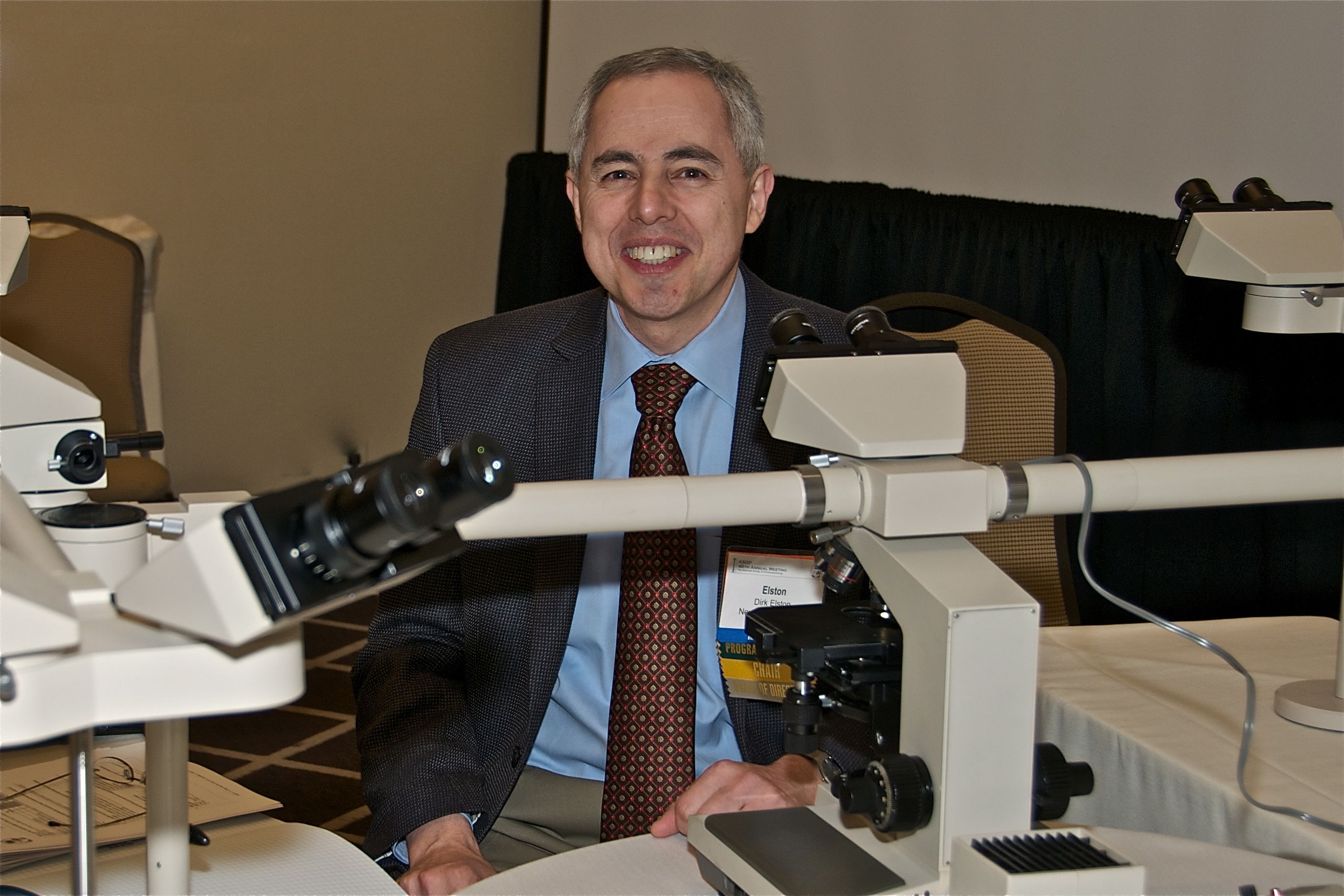
Dr. Elston is one of five authors of “Andrews’ Diseases of the Skin),” coauthor with Tammie Ferringer, MD, of the “Dermatopathology” textbook, and editor in chief of the Requisites in Dermatology series of textbooks. In 2018, he succeeded Bruce H. Thiers, MD, as editor of the Journal of the American Academy of Dermatology and in 2021, received the AAD’s Gold Medal Award, which is the academy’s highest honor.
In an interview, Dr. Elston reflected on his mentors, shared how he manages his many responsibilities as a clinician, teacher, and editor, and talked about the promising future of dermatology.
Who inspired you most to pursue a career in medicine? My grandmother, Annie Elston, was a physician and dedicated her life to helping others. She was a front-line medic during World War I, helped to run a neonatal syphilis ward after the war, and practiced pediatrics in New York City until her death. She was a great role model.
Did you enter medical school knowing that you wanted to become a dermatologist? If not, what was the turning point for you? I didn’t really know much about dermatology when I entered medical school. I fell in love with the specialty during a rotation.
What was the most memorable experience from your dermatology residency at Walter Reed Army Medical Center? There were so many interesting patients, including many tropical diseases.
Why did you choose to pursue a fellowship in dermatopathology? What was it about this subspeciality that piqued your interest? Great teachers, including Tim Berger, MD, George Lupton, MD, and Dean Pearson, MD. They inspired me to seek a dermpath fellowship and I was lucky enough to train with Wilma Bergfeld, MD.
In your opinion, what’s been the most important advance in dermatopathology to date?
Immunohistochemistry changed the specialty. Now molecular diagnostics is a second wave of major advancement.
How do you stay passionate about both dermatology and dermatopathology? The patients, residents, and fellows keep it interesting. It’s a two-way street. I learn as much as I teach.
You’ve had a remarkable run at the Journal of the American Academy of Dermatology, starting as deputy editor in 2008 before becoming editor in 2018. What’s been most rewarding about this role for you? It is a labor of love and such a privilege to see everyone’s best work.
During the peak of the COVID-19 pandemic, what were your most significant challenges from both a clinical and a personal standpoint? Fear of the unknown is always a challenge with a new epidemic and worse with a pandemic. The patients still needed to be seen but it was a challenge with some buildings closed and some personnel afraid to come to work.
Is there anything you would tell your younger self in terms of career advice? Enjoy every step of the journey.
Considering your various work responsibilities as a clinician, teacher, and editor, what’s your strategy for achieving a work-life balance? A good friend of mine is fond of saying that balance is an illusion. There is only resilience. I believe the truth lies somewhere in between. Make time for family, and decide what has to get done today and what can wait until tomorrow.
What development in dermatology are you most excited about in the next 5 years? We are in a golden age of therapeutic innovations that are life changing and lifesaving for our patients. I never would have believed I would see complete cures of patients with widely metastatic melanoma. From psoriasis to eczema to malignancy, our therapeutic armamentarium is dramatically better each year. It makes the practice of medicine exciting.
who has authored more than 600 peer-reviewed publications and 92 textbook chapters.
After earning his undergraduate degree from Pennsylvania State University and his medical degree from Jefferson Medical College in Philadelphia, Dr. Elston completed an internship and a dermatology residency at Walter Reed Army Medical Center in Washington, as well as a dermatopathology fellowship at the Cleveland Clinic. He currently is professor and chair of the department of dermatology and dermatologic surgery at the Medical University of South Carolina in Charleston.
Dr. Elston is one of five authors of “Andrews’ Diseases of the Skin),” coauthor with Tammie Ferringer, MD, of the “Dermatopathology” textbook, and editor in chief of the Requisites in Dermatology series of textbooks. In 2018, he succeeded Bruce H. Thiers, MD, as editor of the Journal of the American Academy of Dermatology and in 2021, received the AAD’s Gold Medal Award, which is the academy’s highest honor.
In an interview, Dr. Elston reflected on his mentors, shared how he manages his many responsibilities as a clinician, teacher, and editor, and talked about the promising future of dermatology.
Who inspired you most to pursue a career in medicine? My grandmother, Annie Elston, was a physician and dedicated her life to helping others. She was a front-line medic during World War I, helped to run a neonatal syphilis ward after the war, and practiced pediatrics in New York City until her death. She was a great role model.
Did you enter medical school knowing that you wanted to become a dermatologist? If not, what was the turning point for you? I didn’t really know much about dermatology when I entered medical school. I fell in love with the specialty during a rotation.
What was the most memorable experience from your dermatology residency at Walter Reed Army Medical Center? There were so many interesting patients, including many tropical diseases.
Why did you choose to pursue a fellowship in dermatopathology? What was it about this subspeciality that piqued your interest? Great teachers, including Tim Berger, MD, George Lupton, MD, and Dean Pearson, MD. They inspired me to seek a dermpath fellowship and I was lucky enough to train with Wilma Bergfeld, MD.
In your opinion, what’s been the most important advance in dermatopathology to date?
Immunohistochemistry changed the specialty. Now molecular diagnostics is a second wave of major advancement.
How do you stay passionate about both dermatology and dermatopathology? The patients, residents, and fellows keep it interesting. It’s a two-way street. I learn as much as I teach.
You’ve had a remarkable run at the Journal of the American Academy of Dermatology, starting as deputy editor in 2008 before becoming editor in 2018. What’s been most rewarding about this role for you? It is a labor of love and such a privilege to see everyone’s best work.
During the peak of the COVID-19 pandemic, what were your most significant challenges from both a clinical and a personal standpoint? Fear of the unknown is always a challenge with a new epidemic and worse with a pandemic. The patients still needed to be seen but it was a challenge with some buildings closed and some personnel afraid to come to work.
Is there anything you would tell your younger self in terms of career advice? Enjoy every step of the journey.
Considering your various work responsibilities as a clinician, teacher, and editor, what’s your strategy for achieving a work-life balance? A good friend of mine is fond of saying that balance is an illusion. There is only resilience. I believe the truth lies somewhere in between. Make time for family, and decide what has to get done today and what can wait until tomorrow.
What development in dermatology are you most excited about in the next 5 years? We are in a golden age of therapeutic innovations that are life changing and lifesaving for our patients. I never would have believed I would see complete cures of patients with widely metastatic melanoma. From psoriasis to eczema to malignancy, our therapeutic armamentarium is dramatically better each year. It makes the practice of medicine exciting.
Why is there an increased risk of cancer in depressed patients?
LAS VEGAS – Is the relationship between major depressive disorder and the development of cancer, cardiovascular disease, and other medical conditions a coincidence, or is there more at play?
According to Charles B. Nemeroff, MD, PhD, a host of circumstances potentially underlies this association, including treatment of the medical disorder itself.

“The best example of that is probably the use of interferon-alpha for the treatment of malignant melanoma,” Dr. Nemeroff, professor and chair of the department of psychiatry and behavioral sciences at the University of Texas at Austin, said during an annual psychopharmacology update held by the Nevada Psychiatric Association. “Many patients treated with interferon-alpha ended up with very severe depression, including several documented suicides. Another possibility of the relationship between depression and medical disorders is that treating a patient for depression could result in a medical disorder. The best example of this is the use of 20 mg of olanzapine to augment the effects of an antidepressant, resulting in a 50-pound weight gain and the development of type 2 diabetes and metabolic syndrome. Both of those scenarios are well understood.”
Then there’s the behavioral aspects of the relationship, he continued, in which patients adopt the mindset that “I’m depressed. I don’t want to exercise. I’m a couch potato. I have been gaining a lot of weight. It’s bad for my heart.”
Converging biology is another possibility. “Is it possible that the biology of depression is linked to the biology of other disorders?” asked Dr. Nemeroff, who directs the university’s Institute for Early Life Adversity Research. “We can talk about this in relation to thyroid disease, a well known cause of depression, but we can also talk about the relationship to other disorders. There’s amazing epidemiologic evidence that patients with PTSD are much more likely to develop Alzheimer’s disease than patients without PTSD.”
Psychosocial issues also play a role. He recalled seeing patient in a clinic for the underserved who had underlying severe ulcerative colitis and anemia and couldn’t afford medical treatment. “The patient had a low hemoglobin, so it was impossible to distinguish between that and whether they had a primary depressive disorder or not,” he said.
In a study that explored the relationship between major depression and cancer, Dr. Nemeroff and colleagues found that the prevalence was highest in those with pancreatic cancer (50%), followed by oropharyngeal (40%), colon (13-25%), breast (18-25%), and gynecologic (23%), and Hodgkin’s lymphoma (17%) (Arch Gen Psychiatry 1995;52[2]:89-99). “Not all cancers have the same rate of depression,” he said. “One of the central questions is, not so much is the cancer patient depressed, but is depression a risk factor for developing cancer? The answer is a resounding yes. But what we don’t know is if you treat the depression aggressively, can you reduce that risk of either developing cancer or the progression of cancer?”
Dr. Nemeroff spotlighted several studies largely from the oncology literature, including a prospective survival analysis of 578 women with early-stage breast cancer (Lancet 1999;354:1331-6). After 5 years, 395 were alive and without relapse, 50 were alive with relapse, and 133 had died. The researchers found a significantly increased risk of death from all causes by 5 years in women with a high depression score (HR 3.59). There was a significantly increased risk of relapse or death at 5 years in women with high scores on helplessness and hopelessness measures.
In an analysis of the association between breast cancer and traumatic events, women who had severe stress or a traumatic event had lower rates of disease-free intervals (J Psychosomatic Res 2007;63:233-9). Another study by the same investigators found that a decrease in depression symptoms is associated with longer survival in patients with metastatic breast cancer (J Clin Oncol 2010;29:413-20). The median survival was 53.6 months for women with decreasing depression scores over 1 year and 25.1 months for women with increasing depression scores.
A more recent study of cervical cancer patients found that those exposed to psychological stress had an increased risk of cancer-specific mortality (HR 1.33) (Cancer Res 2019;79:3965-72). The association was mainly driven by distress experienced within 1 year before or after diagnosis (HR 1.30) but not afterward (HR 1.12). In addition, data from the large longitudinal Nurses’ Health Study II found that women with high PTSD symptoms had a twofold greater risk of ovarian cancer compared with women who had no trauma exposure (Cancer Res 2019;79:5113-20).
Authors of a separate study analyzed data from the Women’s Health Initiative to examine if depression precedes the development of a cancer diagnosis. They found that depression 3 years before a diagnosis of breast cancer was associated with all-cause mortality (HR 1.35) (Cancer 2017;123[16]:3107-15). Meanwhile, among women with late-stage breast cancer, newly developed depression at year 3 was significantly associated with all-cause mortality (HR 2.0) and breast cancer-specific mortality (HR 2.42). “That’s a pretty amazing finding,” Dr. Nemeroff said. “We have to think about depression as a systemic illness. What is depression doing that’s creating a fertile environment for cancer or worsening of cancer?”
He then discussed the risk of suicide in patients who are newly diagnosed with cancer. “No one ever talks about this, and I can’t get anybody to support research in this area,” he said. In one of the first studies on the topic, researchers conducted a case-control study of Medicare patients and determined risk of suicide among those with cancer was 2.3-fold higher compared with controls, even after adjustment for psychiatric illness and the risk of dying within a year (J Clin Oncol 2008;26[29]:4720-4). More recently, authors of a large population-based study in England found that the overall standardized mortality ratio for suicide was 1.20 (JAMA Psychiatry 2019;76[1]51-60). The risk was highest among patients with mesothelioma, with a 4.51-fold risk, followed by pancreatic (3.89-fold), esophageal (2.65-fold), lung (2.57-fold), and stomach cancer (2.20-fold). “They reported that the first 6 months after the diagnosis is associated with an increased risk of suicide – unrelated to prognosis,” Dr. Nemeroff said.
A separate analysis of SEER data from 1973-2014 and comprising more than 8.6 million cancer patients found that newly diagnosed cancer patients are 4.4 times more likely to die from suicide than patients in the same age group without cancer (Nat Commun 2019;10[1]:207). The highest risk was in lung cancer, followed by head and neck, testes, bladder, and Hodgkin’s lymphoma.
According to Dr. Nemeroff, For example, he said, if the depressed environment is associated with a marked increase in tumor necrosis factor, interleukin 6, and other inflammatory markers, “that probably contributes to the body’s ability to fight disease. Ironically, depression is associated with an increase in inflammation but a decreased in T cell function. Remember, there are two fundamental types of immunity: the antibody response and the cellular response. What’s odd about depression is that there’s an increase in inflammatory markers but a decrease in the ability of T cells to function in terms of cellular immunity.”
Dr. Nemeroff disclosed that he has served as a consultant and/or scientific adviser for numerous pharmaceutical companies. He has received research and grant support from the National Institutes of Health.
LAS VEGAS – Is the relationship between major depressive disorder and the development of cancer, cardiovascular disease, and other medical conditions a coincidence, or is there more at play?
According to Charles B. Nemeroff, MD, PhD, a host of circumstances potentially underlies this association, including treatment of the medical disorder itself.
“The best example of that is probably the use of interferon-alpha for the treatment of malignant melanoma,” Dr. Nemeroff, professor and chair of the department of psychiatry and behavioral sciences at the University of Texas at Austin, said during an annual psychopharmacology update held by the Nevada Psychiatric Association. “Many patients treated with interferon-alpha ended up with very severe depression, including several documented suicides. Another possibility of the relationship between depression and medical disorders is that treating a patient for depression could result in a medical disorder. The best example of this is the use of 20 mg of olanzapine to augment the effects of an antidepressant, resulting in a 50-pound weight gain and the development of type 2 diabetes and metabolic syndrome. Both of those scenarios are well understood.”
Then there’s the behavioral aspects of the relationship, he continued, in which patients adopt the mindset that “I’m depressed. I don’t want to exercise. I’m a couch potato. I have been gaining a lot of weight. It’s bad for my heart.”
Converging biology is another possibility. “Is it possible that the biology of depression is linked to the biology of other disorders?” asked Dr. Nemeroff, who directs the university’s Institute for Early Life Adversity Research. “We can talk about this in relation to thyroid disease, a well known cause of depression, but we can also talk about the relationship to other disorders. There’s amazing epidemiologic evidence that patients with PTSD are much more likely to develop Alzheimer’s disease than patients without PTSD.”
Psychosocial issues also play a role. He recalled seeing patient in a clinic for the underserved who had underlying severe ulcerative colitis and anemia and couldn’t afford medical treatment. “The patient had a low hemoglobin, so it was impossible to distinguish between that and whether they had a primary depressive disorder or not,” he said.
In a study that explored the relationship between major depression and cancer, Dr. Nemeroff and colleagues found that the prevalence was highest in those with pancreatic cancer (50%), followed by oropharyngeal (40%), colon (13-25%), breast (18-25%), and gynecologic (23%), and Hodgkin’s lymphoma (17%) (Arch Gen Psychiatry 1995;52[2]:89-99). “Not all cancers have the same rate of depression,” he said. “One of the central questions is, not so much is the cancer patient depressed, but is depression a risk factor for developing cancer? The answer is a resounding yes. But what we don’t know is if you treat the depression aggressively, can you reduce that risk of either developing cancer or the progression of cancer?”
Dr. Nemeroff spotlighted several studies largely from the oncology literature, including a prospective survival analysis of 578 women with early-stage breast cancer (Lancet 1999;354:1331-6). After 5 years, 395 were alive and without relapse, 50 were alive with relapse, and 133 had died. The researchers found a significantly increased risk of death from all causes by 5 years in women with a high depression score (HR 3.59). There was a significantly increased risk of relapse or death at 5 years in women with high scores on helplessness and hopelessness measures.
In an analysis of the association between breast cancer and traumatic events, women who had severe stress or a traumatic event had lower rates of disease-free intervals (J Psychosomatic Res 2007;63:233-9). Another study by the same investigators found that a decrease in depression symptoms is associated with longer survival in patients with metastatic breast cancer (J Clin Oncol 2010;29:413-20). The median survival was 53.6 months for women with decreasing depression scores over 1 year and 25.1 months for women with increasing depression scores.
A more recent study of cervical cancer patients found that those exposed to psychological stress had an increased risk of cancer-specific mortality (HR 1.33) (Cancer Res 2019;79:3965-72). The association was mainly driven by distress experienced within 1 year before or after diagnosis (HR 1.30) but not afterward (HR 1.12). In addition, data from the large longitudinal Nurses’ Health Study II found that women with high PTSD symptoms had a twofold greater risk of ovarian cancer compared with women who had no trauma exposure (Cancer Res 2019;79:5113-20).
Authors of a separate study analyzed data from the Women’s Health Initiative to examine if depression precedes the development of a cancer diagnosis. They found that depression 3 years before a diagnosis of breast cancer was associated with all-cause mortality (HR 1.35) (Cancer 2017;123[16]:3107-15). Meanwhile, among women with late-stage breast cancer, newly developed depression at year 3 was significantly associated with all-cause mortality (HR 2.0) and breast cancer-specific mortality (HR 2.42). “That’s a pretty amazing finding,” Dr. Nemeroff said. “We have to think about depression as a systemic illness. What is depression doing that’s creating a fertile environment for cancer or worsening of cancer?”
He then discussed the risk of suicide in patients who are newly diagnosed with cancer. “No one ever talks about this, and I can’t get anybody to support research in this area,” he said. In one of the first studies on the topic, researchers conducted a case-control study of Medicare patients and determined risk of suicide among those with cancer was 2.3-fold higher compared with controls, even after adjustment for psychiatric illness and the risk of dying within a year (J Clin Oncol 2008;26[29]:4720-4). More recently, authors of a large population-based study in England found that the overall standardized mortality ratio for suicide was 1.20 (JAMA Psychiatry 2019;76[1]51-60). The risk was highest among patients with mesothelioma, with a 4.51-fold risk, followed by pancreatic (3.89-fold), esophageal (2.65-fold), lung (2.57-fold), and stomach cancer (2.20-fold). “They reported that the first 6 months after the diagnosis is associated with an increased risk of suicide – unrelated to prognosis,” Dr. Nemeroff said.
A separate analysis of SEER data from 1973-2014 and comprising more than 8.6 million cancer patients found that newly diagnosed cancer patients are 4.4 times more likely to die from suicide than patients in the same age group without cancer (Nat Commun 2019;10[1]:207). The highest risk was in lung cancer, followed by head and neck, testes, bladder, and Hodgkin’s lymphoma.
According to Dr. Nemeroff, For example, he said, if the depressed environment is associated with a marked increase in tumor necrosis factor, interleukin 6, and other inflammatory markers, “that probably contributes to the body’s ability to fight disease. Ironically, depression is associated with an increase in inflammation but a decreased in T cell function. Remember, there are two fundamental types of immunity: the antibody response and the cellular response. What’s odd about depression is that there’s an increase in inflammatory markers but a decrease in the ability of T cells to function in terms of cellular immunity.”
Dr. Nemeroff disclosed that he has served as a consultant and/or scientific adviser for numerous pharmaceutical companies. He has received research and grant support from the National Institutes of Health.
FROM NPA 2022
Routine pharmacogenetic testing in psychiatry not indicated
LAS VEGAS –
“It’s misleading to rely on results of genetic tests to drive clinical treatment,” Dr. Nurmi, a child and adolescent psychiatrist in the department of psychiatry and biobehavioral sciences at the University of California, Los Angeles, said during an annual psychopharmacology update held by the Nevada Psychiatric Association. “There’s a lot of hope and promise there. But currently, we only know the tip of the iceberg about how drugs work and the genetics influencing these effects. Current testing is probably a very poor reflection of the complexity of drug effects.”

According to Dr. Nurmi, there are at least 165 Food and Drug Administration–approved drugs with pharmacogenetic information on 64 different biomarkers – 37% with CYP p450 notations. Of these, 32 psychiatric drugs have pharmacogenetic information, and most of them are dosing recommendations based on whether a patient has the variant. However, there is wide public acceptance of genetic testing in preventing the wrong drug from being used, in selecting the best drug dose, and avoiding side effects (Pharmacogenomics 2012;12[3]:197-204). “Most people have a lot of hope [for genetic testing in psychiatry],” Dr. Nurmi said. “But is the science really there? It doesn’t matter, because these companies are doing it, and you are being shown these reports from patients. Whether or not the science supports it, we’re going to have to interpret these reports and explain them to our patients – even if we don’t order them.”
Currently, she continued, clinicians practice trial and error prescribing where they might try one treatment in a class that they think that will work based on previous literature. If nothing works, they try another one. If that’s intolerable, they try a third treatment, and so on. “When we finally find the right treatment, it can take some time to get the dosing right,” Dr. Nurmi said. “So, it can take many months to get a child on the right medication. Precision treatment, on the other hand, would start off by taking a saliva or blood sample to get a printout that lets physicians know which drugs might be used with caution because they might lack efficacy at standard doses, which ones would likely have adverse effects at standard doses, and which are the best choices and what are the dosing recommendations for those choices. If we could get all the information to guide us, that would be a useful product, but right now, we don’t know enough to be able to make these determinations.”
Current evidence-based genetic testing supports a limited role for CYP2D6 and CYP2C19 genotyping because most psychiatric drugs are metabolized by those two enzymes. Poor metabolizers have two dysfunctional copies of the enzyme-encoding gene. This results in increased drug plasma levels with a potentially increased rate of adverse effects.
“Intermediate and extensive metabolizers usually have a normal phenotype, but you can also have ultrarapid metabolizers who have duplications or other enhancing mutations of the CYP gene,” Dr. Nurmi said. “This can result in lower bioavailability and possibly efficacy. Psychiatrists treat poor metabolizers and ultrarapid metabolizers all the time, because the variants are very common.” An estimated 10% of White people are poor metabolizers at the CYP2D6 gene while about 7% are ultrarapid metabolizers. At the same time, an estimated 20% of Asians, Africans, and Whites are poor metabolizers at the CYP2C19 gene. “So, you’re seeing a lot of this in your practice, and you’re probably changing dosing based on genetic differences in metabolism,” she said.
The only FDA pharmacodynamic treatment guideline is for the risk of Stevens-Johnson syndrome (SJS) with the use of carbamazepine. In a study of 44 patients with SJS, all were positive for the HLA-B*1502 variant, compared with 3% of carbamazepine-tolerant patients (Nature 2004;428[6982]:486). The frequency of carrying this variant is an estimated 1:10,000 among Whites and 1:1,000 among Asians. In 2007, the FDA recommended that patients of Asian ancestry should be screened for HLA-B*1502 prior to starting carbamazepine.
Genetic variation also predicts clinical outcome with atomoxetine use. “Most child psychiatrists I know think atomoxetine doesn’t work as a second-line nonstimulant medication for ADHD,” Dr. Nurmi said. “I’d like to convince you that why you think it doesn’t work is because of the genetics.” In a study published in 2019, Dr. Nurmi and colleagues reviewed medical literature and provided therapeutic recommendations for atomoxetine therapy based on CYP2D6 genotype (Clin Pharmacol Ther 2019 Jul;106[1]:94-102). They observed 10- to 30-fold plasma differences in drug exposure between normal metabolizers and poor metabolizers.
“Poor metabolizers therefore get more benefit, but they are also going to get more side effects,” she said. “FDA recommended doses are inadequate for normal metabolizers, so they had to make guidelines based on poor metabolizers because there would be too much risk for them at higher doses. One-third of individuals require doses above the FDA limit to achieve a therapeutic drug level.”
Dr. Nurmi warned that the existing evidence base for using these genetic tests in children “is really poor. There is no data in adults with any diagnosis other than depression, and even those studies are plagued by concerns. When you’re implementing decision support tools in your practice, the key factors are patient presentation, history and symptoms, your clinical skills, the evidence base, FDA recommendations, and patient autonomy. Appropriate incorporation of genetic data should include avoiding a medication with high toxicity (like SJS), titration planning (dose and titration speed adjustments), and choosing between medications in the same class with an indication or evidence base for the target disorder.” She added that while the benefit of current genetic testing is limited, it may help some patients feel more comfortable tolerating a medication. “For example, being able to tell someone with anxiety that their genetics suggests that they will not have side effects could be very powerful,” she said.
In a 2018 safety communication, the FDA warned the public about its concerns with companies making claims about how to use genetic test results to manage medication treatments that are not supported by recommendations in the FDA-approved drug labeling or other scientific evidence. The American Academy of Child and Adolescent Psychiatry also published a guide for patients and families.
Dr. Nurmi disclosed that she is an unpaid advisory board member for Myriad Genetics and the Tourette Association of America, a paid adviser for Teva Pharmaceuticals, and a recipient of research support from Emalex Pharmaceuticals. She has received research funding from the National Institutes Health, the International OCD Foundation, the Tourette Association of America, and the Brain & Behavior Research Foundation.
LAS VEGAS –
“It’s misleading to rely on results of genetic tests to drive clinical treatment,” Dr. Nurmi, a child and adolescent psychiatrist in the department of psychiatry and biobehavioral sciences at the University of California, Los Angeles, said during an annual psychopharmacology update held by the Nevada Psychiatric Association. “There’s a lot of hope and promise there. But currently, we only know the tip of the iceberg about how drugs work and the genetics influencing these effects. Current testing is probably a very poor reflection of the complexity of drug effects.”
According to Dr. Nurmi, there are at least 165 Food and Drug Administration–approved drugs with pharmacogenetic information on 64 different biomarkers – 37% with CYP p450 notations. Of these, 32 psychiatric drugs have pharmacogenetic information, and most of them are dosing recommendations based on whether a patient has the variant. However, there is wide public acceptance of genetic testing in preventing the wrong drug from being used, in selecting the best drug dose, and avoiding side effects (Pharmacogenomics 2012;12[3]:197-204). “Most people have a lot of hope [for genetic testing in psychiatry],” Dr. Nurmi said. “But is the science really there? It doesn’t matter, because these companies are doing it, and you are being shown these reports from patients. Whether or not the science supports it, we’re going to have to interpret these reports and explain them to our patients – even if we don’t order them.”
Currently, she continued, clinicians practice trial and error prescribing where they might try one treatment in a class that they think that will work based on previous literature. If nothing works, they try another one. If that’s intolerable, they try a third treatment, and so on. “When we finally find the right treatment, it can take some time to get the dosing right,” Dr. Nurmi said. “So, it can take many months to get a child on the right medication. Precision treatment, on the other hand, would start off by taking a saliva or blood sample to get a printout that lets physicians know which drugs might be used with caution because they might lack efficacy at standard doses, which ones would likely have adverse effects at standard doses, and which are the best choices and what are the dosing recommendations for those choices. If we could get all the information to guide us, that would be a useful product, but right now, we don’t know enough to be able to make these determinations.”
Current evidence-based genetic testing supports a limited role for CYP2D6 and CYP2C19 genotyping because most psychiatric drugs are metabolized by those two enzymes. Poor metabolizers have two dysfunctional copies of the enzyme-encoding gene. This results in increased drug plasma levels with a potentially increased rate of adverse effects.
“Intermediate and extensive metabolizers usually have a normal phenotype, but you can also have ultrarapid metabolizers who have duplications or other enhancing mutations of the CYP gene,” Dr. Nurmi said. “This can result in lower bioavailability and possibly efficacy. Psychiatrists treat poor metabolizers and ultrarapid metabolizers all the time, because the variants are very common.” An estimated 10% of White people are poor metabolizers at the CYP2D6 gene while about 7% are ultrarapid metabolizers. At the same time, an estimated 20% of Asians, Africans, and Whites are poor metabolizers at the CYP2C19 gene. “So, you’re seeing a lot of this in your practice, and you’re probably changing dosing based on genetic differences in metabolism,” she said.
The only FDA pharmacodynamic treatment guideline is for the risk of Stevens-Johnson syndrome (SJS) with the use of carbamazepine. In a study of 44 patients with SJS, all were positive for the HLA-B*1502 variant, compared with 3% of carbamazepine-tolerant patients (Nature 2004;428[6982]:486). The frequency of carrying this variant is an estimated 1:10,000 among Whites and 1:1,000 among Asians. In 2007, the FDA recommended that patients of Asian ancestry should be screened for HLA-B*1502 prior to starting carbamazepine.
Genetic variation also predicts clinical outcome with atomoxetine use. “Most child psychiatrists I know think atomoxetine doesn’t work as a second-line nonstimulant medication for ADHD,” Dr. Nurmi said. “I’d like to convince you that why you think it doesn’t work is because of the genetics.” In a study published in 2019, Dr. Nurmi and colleagues reviewed medical literature and provided therapeutic recommendations for atomoxetine therapy based on CYP2D6 genotype (Clin Pharmacol Ther 2019 Jul;106[1]:94-102). They observed 10- to 30-fold plasma differences in drug exposure between normal metabolizers and poor metabolizers.
“Poor metabolizers therefore get more benefit, but they are also going to get more side effects,” she said. “FDA recommended doses are inadequate for normal metabolizers, so they had to make guidelines based on poor metabolizers because there would be too much risk for them at higher doses. One-third of individuals require doses above the FDA limit to achieve a therapeutic drug level.”
Dr. Nurmi warned that the existing evidence base for using these genetic tests in children “is really poor. There is no data in adults with any diagnosis other than depression, and even those studies are plagued by concerns. When you’re implementing decision support tools in your practice, the key factors are patient presentation, history and symptoms, your clinical skills, the evidence base, FDA recommendations, and patient autonomy. Appropriate incorporation of genetic data should include avoiding a medication with high toxicity (like SJS), titration planning (dose and titration speed adjustments), and choosing between medications in the same class with an indication or evidence base for the target disorder.” She added that while the benefit of current genetic testing is limited, it may help some patients feel more comfortable tolerating a medication. “For example, being able to tell someone with anxiety that their genetics suggests that they will not have side effects could be very powerful,” she said.
In a 2018 safety communication, the FDA warned the public about its concerns with companies making claims about how to use genetic test results to manage medication treatments that are not supported by recommendations in the FDA-approved drug labeling or other scientific evidence. The American Academy of Child and Adolescent Psychiatry also published a guide for patients and families.
Dr. Nurmi disclosed that she is an unpaid advisory board member for Myriad Genetics and the Tourette Association of America, a paid adviser for Teva Pharmaceuticals, and a recipient of research support from Emalex Pharmaceuticals. She has received research funding from the National Institutes Health, the International OCD Foundation, the Tourette Association of America, and the Brain & Behavior Research Foundation.
REPORTING FROM NPA 2022
Self-care tips for clinicians as COVID-19 lingers
LAS VEGAS – according to Jon A. Levenson, MD.
“There are those who will need mental health treatment, so creating an easy way to reach out for help and facilitate linkage with care is critically important,” Dr. Levenson, associate professor of psychiatry at Columbia University Irving Medical Center, New York, said during an annual psychopharmacology update held by the Nevada Psychiatric Association. “The vast majority of our workforce will thrive with proper support. But what can each of us do to take care of ourselves?”

Step one is to recognize common stress reactions as well as signs of distress. He offered the oxygen mask metaphor, the idea that before we can take care of and support anyone else, we must first take care of ourselves. “When people are stressed, they don’t always think about the oxygen mask metaphor,” Dr. Levenson said. Step two is to practice and model self-care by adopting principles often discussed in acceptance and commitment therapy: to focus on what you can control, not on what you can’t control.
“We can’t control the amount of toilet paper at the grocery store, how long the pandemic will last, or how others have reacted,” Dr. Levenson said. “We also can’t control other people’s motives, predict what will happen, or the actions of others, including whether they will follow social distancing guidelines or not.”
How about what we can control? One is a positive attitude, “which can sustain people during times of intense stress,” he said. “Other things that we can do include turn off the news and find fun and enriching activities to do at home, whether it be playing a game with family or reaching out to friends through an iPad or a smartphone. You can also follow [Centers for Disease Control and Prevention] recommendations, control your own social distancing, and limit social media activity, which can be stressful. We can also control our kindness and grace.” He added that resilience does not mean “snapping back” to how you were before the pandemic, but rather “learning to integrate the adverse experiences into who you are and growing with them, which is sometimes known as posttraumatic growth.”
Dr. Levenson encouraged health care workers to use their coping resources, connect to others, and cultivate their values and purpose in life as they navigate these challenging times. “You also want to promote realistic optimism; find a way to stay positive,” he said. “We emphasize to our staff that while you won’t forget this time, focus on what you can control – your positive relationships – and remind yourself of your values and sources of gratitude. Figure out, and reflect on, what you care about, and then care about it. Remind yourself in a deliberate, purposeful way what anchors you to your job, which in the health care setting tends to be a desire to care for others, to assist those in need, and to work in teams. We also encourage staff to refrain from judgment. Guilt is a normal and near-universal response to this stressor, but there are many ways to contribute without a judgmental or guilty tone.”
Other tips for self-support are to remind yourself that it is not selfish to take breaks. “The needs of your patients are not more important than your own needs,” Dr. Levenson said. “Working nonstop can put you at higher risk for stress, exhaustion, and illness. You may need to give yourself more time to step back and recover from workplace challenges or extended coverage for peers; this is important. We remind our staff that your work may feel more emotionally draining than usual because everything is more intense overall during the COVID-19 pandemic. This reminder helps staff normalize what they already may be experiencing, and in turn, to further support each other.”
Soothing activities to relieve stress include meditation, prayer, deep and slow breathing, relaxation exercises, yoga, mindfulness, stretching, staying hydrated, eating healthfully, exercise, and getting sufficient sleep. Other stress management tips include avoiding excessive alcohol intake, reaching out to others, asking for assistance, and delegating when possible. “We want to promote psychological flexibility: the ability to stay in contact with the present moment,” he said. “We encourage our peers to be aware of unpleasant thoughts and feelings, and to try to redirect negative thought patterns to a proactive problem-solving approach; this includes choosing one’s behaviors based on the situation and personal values.”
Dr. Levenson reported having no disclosures related to his presentation.
LAS VEGAS – according to Jon A. Levenson, MD.
“There are those who will need mental health treatment, so creating an easy way to reach out for help and facilitate linkage with care is critically important,” Dr. Levenson, associate professor of psychiatry at Columbia University Irving Medical Center, New York, said during an annual psychopharmacology update held by the Nevada Psychiatric Association. “The vast majority of our workforce will thrive with proper support. But what can each of us do to take care of ourselves?”
Step one is to recognize common stress reactions as well as signs of distress. He offered the oxygen mask metaphor, the idea that before we can take care of and support anyone else, we must first take care of ourselves. “When people are stressed, they don’t always think about the oxygen mask metaphor,” Dr. Levenson said. Step two is to practice and model self-care by adopting principles often discussed in acceptance and commitment therapy: to focus on what you can control, not on what you can’t control.
“We can’t control the amount of toilet paper at the grocery store, how long the pandemic will last, or how others have reacted,” Dr. Levenson said. “We also can’t control other people’s motives, predict what will happen, or the actions of others, including whether they will follow social distancing guidelines or not.”
How about what we can control? One is a positive attitude, “which can sustain people during times of intense stress,” he said. “Other things that we can do include turn off the news and find fun and enriching activities to do at home, whether it be playing a game with family or reaching out to friends through an iPad or a smartphone. You can also follow [Centers for Disease Control and Prevention] recommendations, control your own social distancing, and limit social media activity, which can be stressful. We can also control our kindness and grace.” He added that resilience does not mean “snapping back” to how you were before the pandemic, but rather “learning to integrate the adverse experiences into who you are and growing with them, which is sometimes known as posttraumatic growth.”
Dr. Levenson encouraged health care workers to use their coping resources, connect to others, and cultivate their values and purpose in life as they navigate these challenging times. “You also want to promote realistic optimism; find a way to stay positive,” he said. “We emphasize to our staff that while you won’t forget this time, focus on what you can control – your positive relationships – and remind yourself of your values and sources of gratitude. Figure out, and reflect on, what you care about, and then care about it. Remind yourself in a deliberate, purposeful way what anchors you to your job, which in the health care setting tends to be a desire to care for others, to assist those in need, and to work in teams. We also encourage staff to refrain from judgment. Guilt is a normal and near-universal response to this stressor, but there are many ways to contribute without a judgmental or guilty tone.”
Other tips for self-support are to remind yourself that it is not selfish to take breaks. “The needs of your patients are not more important than your own needs,” Dr. Levenson said. “Working nonstop can put you at higher risk for stress, exhaustion, and illness. You may need to give yourself more time to step back and recover from workplace challenges or extended coverage for peers; this is important. We remind our staff that your work may feel more emotionally draining than usual because everything is more intense overall during the COVID-19 pandemic. This reminder helps staff normalize what they already may be experiencing, and in turn, to further support each other.”
Soothing activities to relieve stress include meditation, prayer, deep and slow breathing, relaxation exercises, yoga, mindfulness, stretching, staying hydrated, eating healthfully, exercise, and getting sufficient sleep. Other stress management tips include avoiding excessive alcohol intake, reaching out to others, asking for assistance, and delegating when possible. “We want to promote psychological flexibility: the ability to stay in contact with the present moment,” he said. “We encourage our peers to be aware of unpleasant thoughts and feelings, and to try to redirect negative thought patterns to a proactive problem-solving approach; this includes choosing one’s behaviors based on the situation and personal values.”
Dr. Levenson reported having no disclosures related to his presentation.
AT NPA 2022