User login
Doug Brunk is a San Diego-based award-winning reporter who began covering health care in 1991. Before joining the company, he wrote for the health sciences division of Columbia University and was an associate editor at Contemporary Long Term Care magazine when it won a Jesse H. Neal Award. His work has been syndicated by the Los Angeles Times and he is the author of two books related to the University of Kentucky Wildcats men's basketball program. Doug has a master’s degree in magazine journalism from the S.I. Newhouse School of Public Communications at Syracuse University. Follow him on Twitter @dougbrunk.
Collaboration helped reduce heart failure readmissions
Hospitals that participated in the Southeast Michigan “See You in 7 Collaborative” experienced substantial reductions in 30-day readmission rates and Medicare payments related to heart failure (HF) care.
Increases in postdischarge 7-day follow-up were modest “but associated processes aimed at this goal may have improved the overall transition from inpatient to outpatient care,” Dr. Scott L. Hummel reported online Sept. 9 in JACC: Heart Failure. “Our study suggests that regional hospital collaboration to share best practices can be an effective strategy to reduce HF readmissions and associated costs.”

Established in 2011, the Southeast Michigan See You in 7 Collaborative is an effort by the Greater Detroit Area Council, the American College of Cardiology’s Michigan Chapter, the Michigan Peer Review Organization, and 11 previously nonaffiliated hospitals to increase 7-day postdischarge follow-up and reduce all-cause 30-day readmission rates in HF patients. Over a 1-year period, Dr. Hummel, a cardiologist at the University of Michigan, Ann Arbor, and his associates examined the rates of 7-day follow-up and 30-day readmissions in Medicare fee-for-service HF patients among the 10 collaborating hospitals, and compared the findings to trends in the state’s 82 nonparticipating hospitals. The preintervention period studied was May 2011-April 2012; the intervention occurred from May 2012-April 2013.
During the intervention period, the rates of 7-day postdischarge follow-up increased significantly but remained low in both groups (from 31.1% to 34.4% in collaborating hospitals, and from 30.2% to 32.6% in nonparticipating hospitals). At the same time, the rates of unadjusted readmissions significantly decreased in both groups (from 29% to 27.3% in collaborating hospitals and from 26.4% to 25.8% in nonparticipating hospitals). The researchers also found that the mean risk-standardized 30-day, all-cause readmission rates improved significantly among collaborating hospitals (from 31.1% to 28.5%), but significantly less so among nonparticipating hospitals (from 26.7% to 26.1%).
Finally, combined Medicare payments for inpatient and 30 days of postdischarge care decreased by an average of $182 per beneficiary in collaborating hospitals and by $63 in nonparticipating hospitals (JACC Heart Fail. 2015 Sept 9; [doi:10.1016/j.jchf.2015.06.007]).
The researchers acknowledged certain limitations of the study, including its observational design and the fact that administrative data sets were used to determine the outcomes of interest. “Given the observational nature of the study, we cannot confirm that improvements in 7-day follow-up or 30-day readmission rates directly resulted from See You in 7 Collaborative participation,” they noted.
The study was funded in part by the Robert Wood Johnson Foundation. Dr. Hummel disclosed that he is supported by a grant from the National Heart, Lung, and Blood Institute. The remaining study authors reported having no financial disclosures.
The investigators should be congratulated for a well-designed intervention that was multifaceted, allowing hospitals to experiment locally with the innovations that proved most useful to the individual health care environment. Unfortunately, the hospitals in the Collaborative were unable to significantly increase 7-day follow-up over the course of 1 year; 7-day follow-up rose from 31% to 34% only, compared with 32% to 34% in matched hospitals in Michigan. Even 14-day follow-up was less than 50% for various hospital groups in the study. These rates are consistent with other national data and are an important reflection of the current state of health care in the United States, yet it remains unclear why it is so difficult to move the needle.
Despite the lack of improvement in early follow-up, all-cause 30-day, risk-standardized readmission rates decreased more in Collaborative hospitals (31.1% to 28.5%), compared with other hospitals in Michigan (26.7% to 26.1%) during the 1-year study, including a cohort of hospitals matched for size, region, and demographics. Nevertheless, it is unclear which tools in the toolkit led to this improvement. Because of other changes noted in the environment, these results were possibly brought about by overall cultural or environmental attention toward readmission among more motivated hospitals. To address this important source of bias, future studies should consider randomization schemes of different interventions, such as cluster-randomized trials or embedded randomization.
There will undoubtedly be more opportunities to establish a better evidence base leveraging national, reusable research infrastructure such as the National Patient-Centered Clinical Research Network (PCORnet), which can serve as a platform for testing new health system delivery models.
Dr. Adrian F. Hernandez and Dr. Adam D. Devore are with the department of medicine at Duke University, as well as the Duke Clinical Research Institute, both in Durham, N.C. They made these comments in an accompanying editorial (JACC Heart Fail. 2015 Sept 9; [doi:10.1016/j.jchf.2015.07.004]). Dr. Hernandez reported receiving honoraria from Amgen, AstraZeneca, Janssen, Merck and Novartis; and research support from the Agency for Healthcare Research and Quality, American Heart Association, Amgen, AstraZeneca, Novartis, Merck, the National Heart, Lung, and Blood Institute, and the Patient Centered Outcomes Research Institute. Dr. Devore reported research funding from Amgen, AHA, Maquet, Novartis, and Thoratec; and consultant fees from Maquet.
Hospitals that participated in the Southeast Michigan “See You in 7 Collaborative” experienced substantial reductions in 30-day readmission rates and Medicare payments related to heart failure (HF) care.
Increases in postdischarge 7-day follow-up were modest “but associated processes aimed at this goal may have improved the overall transition from inpatient to outpatient care,” Dr. Scott L. Hummel reported online Sept. 9 in JACC: Heart Failure. “Our study suggests that regional hospital collaboration to share best practices can be an effective strategy to reduce HF readmissions and associated costs.”
Established in 2011, the Southeast Michigan See You in 7 Collaborative is an effort by the Greater Detroit Area Council, the American College of Cardiology’s Michigan Chapter, the Michigan Peer Review Organization, and 11 previously nonaffiliated hospitals to increase 7-day postdischarge follow-up and reduce all-cause 30-day readmission rates in HF patients. Over a 1-year period, Dr. Hummel, a cardiologist at the University of Michigan, Ann Arbor, and his associates examined the rates of 7-day follow-up and 30-day readmissions in Medicare fee-for-service HF patients among the 10 collaborating hospitals, and compared the findings to trends in the state’s 82 nonparticipating hospitals. The preintervention period studied was May 2011-April 2012; the intervention occurred from May 2012-April 2013.
During the intervention period, the rates of 7-day postdischarge follow-up increased significantly but remained low in both groups (from 31.1% to 34.4% in collaborating hospitals, and from 30.2% to 32.6% in nonparticipating hospitals). At the same time, the rates of unadjusted readmissions significantly decreased in both groups (from 29% to 27.3% in collaborating hospitals and from 26.4% to 25.8% in nonparticipating hospitals). The researchers also found that the mean risk-standardized 30-day, all-cause readmission rates improved significantly among collaborating hospitals (from 31.1% to 28.5%), but significantly less so among nonparticipating hospitals (from 26.7% to 26.1%).
Finally, combined Medicare payments for inpatient and 30 days of postdischarge care decreased by an average of $182 per beneficiary in collaborating hospitals and by $63 in nonparticipating hospitals (JACC Heart Fail. 2015 Sept 9; [doi:10.1016/j.jchf.2015.06.007]).
The researchers acknowledged certain limitations of the study, including its observational design and the fact that administrative data sets were used to determine the outcomes of interest. “Given the observational nature of the study, we cannot confirm that improvements in 7-day follow-up or 30-day readmission rates directly resulted from See You in 7 Collaborative participation,” they noted.
The study was funded in part by the Robert Wood Johnson Foundation. Dr. Hummel disclosed that he is supported by a grant from the National Heart, Lung, and Blood Institute. The remaining study authors reported having no financial disclosures.
FROM JACC: HEART FAILURE
Key clinical point: An interhospital collaborative approach was associated with substantial reductions in 30-day readmission rates and Medicare payments among heart failure patients.
Major finding: Over the course of 1 year, all-cause 30-day, risk-standardized readmission rates decreased more in collaborating hospitals (31.1% to 28.5%; P less than .001), compared with other hospitals in Michigan (26.7% to 26.1%; P = .02).
Data source: An observational study of Medicare heart failure patients discharged from 10 collaborating hospitals participating in the Southeast Michigan See You in 7 Collaborative.
Disclosures: The study was funded in part by the Robert Wood Johnson Foundation. Dr. Hummel disclosed that he is supported by a grant from the National Heart, Lung, and Blood Institute. The remaining study authors reported having no financial disclosures.

Dermatologists should be central to wound care, expert says
The way Dr. Adam Friedman sees it, dermatologists deserve a prominent place at the table when it comes to the treatment of acute and chronic wounds.
“As masters of the integument, we should be central to wound care, whether it be for research, in terms of developing better technologies, medications, approaches, diagnostics, but also in terms of managing these wounds, given the rich breadth of pathophysiology and biology we learn during our residency and maintain during our continuing education as practicing dermatologists,” said Dr. Friedman of the department of dermatology at George Washington University, Washington.
When the Journal of Drugs in Dermatology invited Dr. Friedman to serve as guest editor for a special feature section on wound care for its July 2015 issue, he jumped at the chance “to give the dermatology community a small taste of what’s going on in the wound healing world.”
Currently, he said, there is wide variability in the types of clinicians leading wound care centers in the United States, with dermatologists often sitting on the sidelines. “At one institution, it may be the vascular surgery service, at others it may be the family medicine service or even the emergency medicine department,” said Dr. Friedman, who is an editorial advisor to Dermatology News.
“That’s a big problem, in that there’s no uniformity from one center to the next in terms of who is expected to and should be taking responsibility for the wound healing service at their institutions. The reality is, it should be an interdisciplinary team, which not only involves dermatology but vascular surgery, nutrition, internal medicine, subspecialties of medicine like rheumatology, and rehab medicine. However, what is happening more often than not is that you’re getting just one or two of these elements, which cannot be as effective because you miss out on a broader, holistic view.”

There are two chief reasons why dermatologists aren’t more involved in wound care management, he continued. One stems from a lack of training on the topic. In one of the abstracts from the special JDD wound care section, researchers led by Dr. Emily Stamell Ruiz conducted an online survey of dermatology residents in the United States, to ask them about their preparedness to care for wounds and to assess the amount and type of training devoted to wound care during residency. Of the 175 respondents, 78% and 85% did not feel prepared to manage acute and chronic wounds, respectively, while 77% felt that more education is needed during their residency (J Drugs Dermatol. 2015;14[7]:716-20). “Residents felt that there was a clinical as well as a didactical gap, so they felt that they needed more training both through lectures as well as in clinics,” said Dr. Ruiz of the department of dermatology at Brigham and Women’s Hospital, Boston. “It’s not just a focal problem, it really is a universal curriculum problem. Future reforms to the current dermatology curriculum to include wound care training could help close the gap in wound care training.”
Another reason why dermatologists aren’t more involved in wound care management is the time commitment, said Dr. Friedman, who is also director of translational research at George Washington. The treatment of chronic wounds is “physically and financially burdensome,” he said. “It takes not only yourself being comfortable with managing the whole patient which includes the wound[s] with a side order of comorbidities, but your support staff as well – having nurses who know how to use the different wound dressings and how to help you with debridement. You need the right infrastructure. It also costs a lot on the provider side to manage wounds. You need a setup where you can get these patients in, have support staff to help with the wound dressings once you’ve identified what’s necessary, and be able to move on to the next patient.”
In another manuscript contained in the JDD special section, Dr. Friedman and his associates retrospectively reviewed the characteristics of 51 patients with burn injuries who were seen by seven different dermatologists at the Einstein-Montefiore division of dermatology from April 2010 to July 2014 (J Drugs Dermatol. 2015;14[7]:721-4). It found that the main mechanism of injury was burn from hot metal (22%), followed by contact with hot liquids (18%). It also found that silver sulfadiazine was the most commonly prescribed treatment, “even though there are considerable data illustrating that its use will delay wound closure and healing (J Invest Dermatol. 2015 May;135[5]:1459-62),” Dr. Friedman said. He went on to note that for patients who suffer an acute burn, “the ability to access a dermatologist is somewhat limited because their schedules are heavily booked well in advance, and the format doesn’t allow for these types of emergencies. More often than not they go to the ED or to primary care. That might not necessarily be the right decision because these are physicians who may not have the necessary training in terms of not only proper burn care, but skin care overall.”
Another manuscript in the special section describes a method in which partial thickness wounds were induced by cryosurgery to create wounds that could facilitate wound healing research and development. For the study, researchers led by Dr. Robert Kirsner, interim chairman of the department of dermatology and cutaneous surgery at the University of Miami, used liquid nitrogen spray to induce freeze injuries on the forearms of eight healthy adult volunteers (J. Drugs Dermatol. 2015;14[7]: 734-8). They delivered the spray onto a target area of a 1-cm circular opening at a distance from the cryodevice to the skin of 0.5-1 cm and implemented several freeze-thaw time cycles by administering pulses that ranged from 3-12 seconds.
After a 24-hour follow-up, Dr. Kirsner and his associates observed that freeze times exceeding 5 seconds caused a majority of study participants to develop blisters, while freeze times exceeding 8 seconds caused uniform blister formation. Time to healing among subjects in the 8-second freeze time group was 12-13 days, while time to healing among those in the 12-second time freeze group was 21 days.
“Cryo-induced wound healing is a little bit slower than you’d expect with a scalpel, but that wasn’t really surprising,” Dr. Kirsner said. “The fact that it healed a little bit slower was a pretty good thing because if everything healed too fast then it couldn’t serve as a model to speed or slow epithelialization. We were quite pleased.” He noted that the model “could be used as a safety test for chronic wound treatment and as an efficacy test for acute wound treatment. It’s relatively inexpensive and a relatively simple technique. If you’re developing a product for widespread use, it’s probably a minor cost in the whole development process.”
Other manuscripts in the JDD special section include a preclinical study using a murine multithermal burn model which found that N-acetylcysteine S-nitrosothiol nanoparticles prevent wound expansion and accelerate burn closure, and a practical, systematic approach to using wound dressings for the wound care novice. Dr. Friedman hopes that the special section not only stimulates further interest in wound care, but that it serves as “a call for action. We really need to be more involved in wound care from the acute and chronic perspective,” he said. “Wound centers around the country should be involving dermatologists. We have so much to offer from bench to bedside because the skin is our thing. I hope this is a reminder that we should be part of this picture.”
Dr. Friedman disclosed that he serves as a consultant for Galderma, Biogen, Aveeno, Intraderm, Puracore, La Roche-Posay, Amgen, Pfizer, PHD Skin Care. He also serves as an advisory board member for Nerium International, Valeant, Nano BioMed, MicroCures, and Novartis, and has received research grants from Valeant. Dr. Ruiz and Dr. Kirsner reported no financial disclosures.
The way Dr. Adam Friedman sees it, dermatologists deserve a prominent place at the table when it comes to the treatment of acute and chronic wounds.
“As masters of the integument, we should be central to wound care, whether it be for research, in terms of developing better technologies, medications, approaches, diagnostics, but also in terms of managing these wounds, given the rich breadth of pathophysiology and biology we learn during our residency and maintain during our continuing education as practicing dermatologists,” said Dr. Friedman of the department of dermatology at George Washington University, Washington.
When the Journal of Drugs in Dermatology invited Dr. Friedman to serve as guest editor for a special feature section on wound care for its July 2015 issue, he jumped at the chance “to give the dermatology community a small taste of what’s going on in the wound healing world.”
Currently, he said, there is wide variability in the types of clinicians leading wound care centers in the United States, with dermatologists often sitting on the sidelines. “At one institution, it may be the vascular surgery service, at others it may be the family medicine service or even the emergency medicine department,” said Dr. Friedman, who is an editorial advisor to Dermatology News.
“That’s a big problem, in that there’s no uniformity from one center to the next in terms of who is expected to and should be taking responsibility for the wound healing service at their institutions. The reality is, it should be an interdisciplinary team, which not only involves dermatology but vascular surgery, nutrition, internal medicine, subspecialties of medicine like rheumatology, and rehab medicine. However, what is happening more often than not is that you’re getting just one or two of these elements, which cannot be as effective because you miss out on a broader, holistic view.”
There are two chief reasons why dermatologists aren’t more involved in wound care management, he continued. One stems from a lack of training on the topic. In one of the abstracts from the special JDD wound care section, researchers led by Dr. Emily Stamell Ruiz conducted an online survey of dermatology residents in the United States, to ask them about their preparedness to care for wounds and to assess the amount and type of training devoted to wound care during residency. Of the 175 respondents, 78% and 85% did not feel prepared to manage acute and chronic wounds, respectively, while 77% felt that more education is needed during their residency (J Drugs Dermatol. 2015;14[7]:716-20). “Residents felt that there was a clinical as well as a didactical gap, so they felt that they needed more training both through lectures as well as in clinics,” said Dr. Ruiz of the department of dermatology at Brigham and Women’s Hospital, Boston. “It’s not just a focal problem, it really is a universal curriculum problem. Future reforms to the current dermatology curriculum to include wound care training could help close the gap in wound care training.”
Another reason why dermatologists aren’t more involved in wound care management is the time commitment, said Dr. Friedman, who is also director of translational research at George Washington. The treatment of chronic wounds is “physically and financially burdensome,” he said. “It takes not only yourself being comfortable with managing the whole patient which includes the wound[s] with a side order of comorbidities, but your support staff as well – having nurses who know how to use the different wound dressings and how to help you with debridement. You need the right infrastructure. It also costs a lot on the provider side to manage wounds. You need a setup where you can get these patients in, have support staff to help with the wound dressings once you’ve identified what’s necessary, and be able to move on to the next patient.”
In another manuscript contained in the JDD special section, Dr. Friedman and his associates retrospectively reviewed the characteristics of 51 patients with burn injuries who were seen by seven different dermatologists at the Einstein-Montefiore division of dermatology from April 2010 to July 2014 (J Drugs Dermatol. 2015;14[7]:721-4). It found that the main mechanism of injury was burn from hot metal (22%), followed by contact with hot liquids (18%). It also found that silver sulfadiazine was the most commonly prescribed treatment, “even though there are considerable data illustrating that its use will delay wound closure and healing (J Invest Dermatol. 2015 May;135[5]:1459-62),” Dr. Friedman said. He went on to note that for patients who suffer an acute burn, “the ability to access a dermatologist is somewhat limited because their schedules are heavily booked well in advance, and the format doesn’t allow for these types of emergencies. More often than not they go to the ED or to primary care. That might not necessarily be the right decision because these are physicians who may not have the necessary training in terms of not only proper burn care, but skin care overall.”
Another manuscript in the special section describes a method in which partial thickness wounds were induced by cryosurgery to create wounds that could facilitate wound healing research and development. For the study, researchers led by Dr. Robert Kirsner, interim chairman of the department of dermatology and cutaneous surgery at the University of Miami, used liquid nitrogen spray to induce freeze injuries on the forearms of eight healthy adult volunteers (J. Drugs Dermatol. 2015;14[7]: 734-8). They delivered the spray onto a target area of a 1-cm circular opening at a distance from the cryodevice to the skin of 0.5-1 cm and implemented several freeze-thaw time cycles by administering pulses that ranged from 3-12 seconds.
After a 24-hour follow-up, Dr. Kirsner and his associates observed that freeze times exceeding 5 seconds caused a majority of study participants to develop blisters, while freeze times exceeding 8 seconds caused uniform blister formation. Time to healing among subjects in the 8-second freeze time group was 12-13 days, while time to healing among those in the 12-second time freeze group was 21 days.
“Cryo-induced wound healing is a little bit slower than you’d expect with a scalpel, but that wasn’t really surprising,” Dr. Kirsner said. “The fact that it healed a little bit slower was a pretty good thing because if everything healed too fast then it couldn’t serve as a model to speed or slow epithelialization. We were quite pleased.” He noted that the model “could be used as a safety test for chronic wound treatment and as an efficacy test for acute wound treatment. It’s relatively inexpensive and a relatively simple technique. If you’re developing a product for widespread use, it’s probably a minor cost in the whole development process.”
Other manuscripts in the JDD special section include a preclinical study using a murine multithermal burn model which found that N-acetylcysteine S-nitrosothiol nanoparticles prevent wound expansion and accelerate burn closure, and a practical, systematic approach to using wound dressings for the wound care novice. Dr. Friedman hopes that the special section not only stimulates further interest in wound care, but that it serves as “a call for action. We really need to be more involved in wound care from the acute and chronic perspective,” he said. “Wound centers around the country should be involving dermatologists. We have so much to offer from bench to bedside because the skin is our thing. I hope this is a reminder that we should be part of this picture.”
Dr. Friedman disclosed that he serves as a consultant for Galderma, Biogen, Aveeno, Intraderm, Puracore, La Roche-Posay, Amgen, Pfizer, PHD Skin Care. He also serves as an advisory board member for Nerium International, Valeant, Nano BioMed, MicroCures, and Novartis, and has received research grants from Valeant. Dr. Ruiz and Dr. Kirsner reported no financial disclosures.
Celiac disease ‘as common as psoriasis’
PARK CITY, UTAH – Dr. John J. Zone first began to study gluten sensitivity in 1977, an interest that left some of his clinician colleagues wondering why.
“Everybody told me I was crazy – that this was extremely rare. So I always say I was gluten when gluten wasn’t cool,” Dr. Zone, professor and chairman of dermatology at the University of Utah, Salt Lake City, told attendees at the annual meeting of the Pacific Dermatologic Association.
These days, it’s hard to shop in a food market without noticing all the gluten-free foods available, from pizza dough to beer. Many restaurants also serve gluten-free dishes. But is it hype, or is gluten sensitivity that common? Five percent of people in the United States “will say they are gluten sensitive,” he said. “In fact, 1% of Caucasians actually have celiac disease and 1% of Caucasians have gluten sensitivity that can be documented by challenge but don’t have celiac disease, while 3% have nothing.”
Gluten is a group of proteins contained in wheat, barley, and rye that is insoluble in water. Dr. Zone described celiac disease as a “spectrum of disease” characterized by inflammation of the small intestinal mucosa that occurs with the ingestion of gluten. The condition improves when gluten is removed from the diet. From a genetic standpoint, having a predisposition to express human leukocyte antigen-DQ2 or HLA-DQ8 is required for a diagnosis of celiac disease (CD). An estimated 20%-25% of whites “have that HLA background, but it is rare in Asians,” he said. “The receptors coded by HLA genes are essential for the processing of the gliadin antigen in CD.”
The hallmark for CD is a blood test for immunoglobulin A (IgA) anti-tissue transglutaminase antibodies, which are detectable in patients with untreated disease. “You should be able to get that test for $50 or $60 in any laboratory in the country,” Dr. Zone said. “It’s about 98% reliable. You also want to do a total serum IgA to rule out IgA-deficiency.”
CD clusters with other autoimmune disorders such as Addison’s disease, autoimmune thyroiditis, atrophic gastritis, systemic lupus erythematosus, rheumatoid arthritis, myasthenia gravis, and vitiligo. It’s also common in Down syndrome. “Many patients with histological inflammation have atypical intestinal symptoms or none at all,” he said. “Clinical studies have shown that only 15%-20% of CD patients identified by serology and confirmed by biopsy have classical symptoms of diarrhea and malabsorption.” The presenting symptom in patients with celiac disease may be limited to only aphthous stomatitis, eczema, alopecia areata, psoriasis, or diabetes, along with fatigue or anemia.
Researchers who analyzed the prevalence of CD in the United States estimated the risk to be 1:133 among individuals deemed not to be at risk, 1:56 in symptomatic patients, 1:39 in second-degree relatives, and 1:22 in first-degree relatives (Arch Intern Med. 2003;163[3]:286-92.). “We tested 2,100 people in Utah and found the prevalence among first-degree relatives was 1:12,” Dr. Zone said. “The point is that CD is common, not rare. It’s as common as psoriasis. It profoundly affects the immune system, which is the modulator of inflammatory skin disease.”
Dr. Zone reported having no financial disclosures.
EXPERT ANALYSIS FROM PDA 2015

Benznidazole: No clinical impact on Chagas cardiomyopathy
Among patients with Chagas cardiomyopathy, trypanocidal therapy with benznidazole significantly reduced serum parasite detection but did not reduce cardiac clinical deterioration through 5 years of follow-up, a randomized trial showed.
Those are key findings from the Benznidazole Evaluation for Interrupting Trypanosomiasis (BENEFIT) trial, which were presented at the annual congress of the European Society of Cardiology and published simultaneously in the New England Journal of Medicine (doi: 10.1056/NEJMoa1507574).
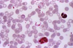
According to lead study authors Dr. Carlos A. Morillo of the Population Health Research Institute at Hamilton Health Sciences and McMaster University, Hamilton, Ont., and Dr. Jose Antonio Marin-Neto of the division of cardiology at the Medical School of Riberao Preto, Brazil, Chagas cardiomyopathy develops in about one-quarter of patients infected with Trypanosoma cruzi. “The identification of T. cruzi antigens in inflamed myocardium with the use of sensitive techniques, such as immunohistochemical analysis and polymerase chain reaction (PCR) assay, suggests that parasite persistence may be an important factor that, in conjunction with individual host factors, triggers the inflammatory process,” they wrote. “In assessing whether trypanocidal therapy prevents or reduces cardiac disease, experimental models of chronic Chagas infection have shown that trypanocidal therapy attenuates the pathologic consequences by reducing the parasite burden. A few small observational and randomized studies involving patients with Chagas disease have shown that benznidazole reduced the circulating parasite load, enhances seroconversion, and may halt the progression of cardiomyopathy.”
In a study of 2,854 patients with chronic Chagas cardiomyopathy conducted at 49 centers in Argentina, Bolivia, Brazil, Columbia, and El Salvador, the researchers set out to evaluate the efficacy and safety of benznidazole, compared with placebo, in reducing the clinical outcomes in patients with chronic Chagas cardiomyopathy. Of the 2,854 patients, slightly more than half (1,431) received benznidazole, while the remaining 1,423 received placebo for up to 80 days and were followed for a mean of 5.4 years. The primary outcome was the first event of any of the components of the composite outcome of death, resuscitated cardiac arrest, sustained ventricular tachycardia, insertion of a pacemaker or implantable cardioverter-defibrillator, cardiac transplantation, new heart failure, stroke, or other thromboembolic event. Secondary outcomes included the response to treatment on the basis of results on PCR assay overall and according to geographic region.
The proportion of study participants who achieved the primary outcome was statistically similar between the two groups: 27.% in the benznidazole group, compared with 29.1% in the placebo group (adjusted hazard ratio of .092; P = .26). The researchers observed no significant between-group differences in any component of the primary outcome.
In terms of secondary outcomes, the rates of conversion to negative PCR results were 66.2% in the benznidazole group, compared with 33.5% in the placebo group at the end of treatment. Between-group differences were sustained at 2 years (55.4% and 35.3%, respectively) and at 5 years or more (46.7% vs. 33.1%; P less than .001 for all comparisons). At the same time, rates of PCR conversion varied according to geographic region. For example, the odds ratio for PCR conversion was highest in Brazil (odds ratio, 3.03 at 2 years and OR, 1.87 at 5 or more years) and lowest in Columbia and El Salvador (OR, 1.33 at 2 years and OR, 0.96 at 5 years). “Since we do not have genotype information, we had to infer that patients in certain geographic regions were likely to have distinct T. cruzi strains,” the authors wrote.
They acknowledged certain limitations of the study, including the fact that “variable responses to benznidazole have been reported previously and may have contributed to our neutral findings. Further analyses of stored blood samples by means of quantitative PCR and genotyping may provide a more precise characterization of T. cruzi that can be used to assess whether genotype influences the clinical response to benznidazole.”
They concluded that the current findings “do not challenge current guidelines that recommend treatment with trypanocidal therapy in the early stages of chronic Chagas infection. ... and should not detract from the pursuit of general goals for exploring more effective or earlier treatments with new drugs or drug combinations.”
The study was funded by several sources, including the Population Health Research Institute, the Canadian Institutes of Health Research, the United Nations Children’s Fund, and the World Bank. Dr. Morillo disclosed that he has received lecture fees from Bayer and Boehringer Ingelheim and grant support from Boston Scientific and Merck & Co. Another study author, Dr. Janis Lazdins, disclosed having received consulting fees from Merck. The remaining study authors reporting having no financial disclosures.
Among patients with Chagas cardiomyopathy, trypanocidal therapy with benznidazole significantly reduced serum parasite detection but did not reduce cardiac clinical deterioration through 5 years of follow-up, a randomized trial showed.
Those are key findings from the Benznidazole Evaluation for Interrupting Trypanosomiasis (BENEFIT) trial, which were presented at the annual congress of the European Society of Cardiology and published simultaneously in the New England Journal of Medicine (doi: 10.1056/NEJMoa1507574).
According to lead study authors Dr. Carlos A. Morillo of the Population Health Research Institute at Hamilton Health Sciences and McMaster University, Hamilton, Ont., and Dr. Jose Antonio Marin-Neto of the division of cardiology at the Medical School of Riberao Preto, Brazil, Chagas cardiomyopathy develops in about one-quarter of patients infected with Trypanosoma cruzi. “The identification of T. cruzi antigens in inflamed myocardium with the use of sensitive techniques, such as immunohistochemical analysis and polymerase chain reaction (PCR) assay, suggests that parasite persistence may be an important factor that, in conjunction with individual host factors, triggers the inflammatory process,” they wrote. “In assessing whether trypanocidal therapy prevents or reduces cardiac disease, experimental models of chronic Chagas infection have shown that trypanocidal therapy attenuates the pathologic consequences by reducing the parasite burden. A few small observational and randomized studies involving patients with Chagas disease have shown that benznidazole reduced the circulating parasite load, enhances seroconversion, and may halt the progression of cardiomyopathy.”
In a study of 2,854 patients with chronic Chagas cardiomyopathy conducted at 49 centers in Argentina, Bolivia, Brazil, Columbia, and El Salvador, the researchers set out to evaluate the efficacy and safety of benznidazole, compared with placebo, in reducing the clinical outcomes in patients with chronic Chagas cardiomyopathy. Of the 2,854 patients, slightly more than half (1,431) received benznidazole, while the remaining 1,423 received placebo for up to 80 days and were followed for a mean of 5.4 years. The primary outcome was the first event of any of the components of the composite outcome of death, resuscitated cardiac arrest, sustained ventricular tachycardia, insertion of a pacemaker or implantable cardioverter-defibrillator, cardiac transplantation, new heart failure, stroke, or other thromboembolic event. Secondary outcomes included the response to treatment on the basis of results on PCR assay overall and according to geographic region.
The proportion of study participants who achieved the primary outcome was statistically similar between the two groups: 27.% in the benznidazole group, compared with 29.1% in the placebo group (adjusted hazard ratio of .092; P = .26). The researchers observed no significant between-group differences in any component of the primary outcome.
In terms of secondary outcomes, the rates of conversion to negative PCR results were 66.2% in the benznidazole group, compared with 33.5% in the placebo group at the end of treatment. Between-group differences were sustained at 2 years (55.4% and 35.3%, respectively) and at 5 years or more (46.7% vs. 33.1%; P less than .001 for all comparisons). At the same time, rates of PCR conversion varied according to geographic region. For example, the odds ratio for PCR conversion was highest in Brazil (odds ratio, 3.03 at 2 years and OR, 1.87 at 5 or more years) and lowest in Columbia and El Salvador (OR, 1.33 at 2 years and OR, 0.96 at 5 years). “Since we do not have genotype information, we had to infer that patients in certain geographic regions were likely to have distinct T. cruzi strains,” the authors wrote.
They acknowledged certain limitations of the study, including the fact that “variable responses to benznidazole have been reported previously and may have contributed to our neutral findings. Further analyses of stored blood samples by means of quantitative PCR and genotyping may provide a more precise characterization of T. cruzi that can be used to assess whether genotype influences the clinical response to benznidazole.”
They concluded that the current findings “do not challenge current guidelines that recommend treatment with trypanocidal therapy in the early stages of chronic Chagas infection. ... and should not detract from the pursuit of general goals for exploring more effective or earlier treatments with new drugs or drug combinations.”
The study was funded by several sources, including the Population Health Research Institute, the Canadian Institutes of Health Research, the United Nations Children’s Fund, and the World Bank. Dr. Morillo disclosed that he has received lecture fees from Bayer and Boehringer Ingelheim and grant support from Boston Scientific and Merck & Co. Another study author, Dr. Janis Lazdins, disclosed having received consulting fees from Merck. The remaining study authors reporting having no financial disclosures.
FROM THE ESC CONGRESS 2015
Key clinical point: Benznidazole for Chagas cardiomyopathy did not reduce long-term cardiac clinical deterioration.
Major finding: The proportion of study participants who achieved the primary outcome was statistically similar between the two groups: 27.% in the benznidazole group, compared with 29.1% in the placebo group (adjusted hazard ratio of .92; P = .26).
Data source: A prospective, randomized trial in which 2,854 patients with Chagas cardiomyopathy received benznidazole or placebo for up to 80 days and were followed for a mean of 5.4 years.
Disclosures: The study was funded by several sources, including the Population Health Research Institute, the Canadian Institutes of Health Research, the United Nations Children’s Fund, and the World Bank. Dr. Morillo disclosed that he has received lecture fees from Bayer and Boehringer Ingelheim and grant support from Boston Scientific and Merck & Co. Another study author, Dr. Janis Lazdins, disclosed having received consulting fees from Merck. The remaining study authors reported having no financial disclosures.

Simple screening tool can help detect psoriatic arthritis
PARK CITY, UTAH – Since most people with psoriatic arthritis (PsA) develop psoriasis before joint symptoms, it’s helpful to have a simple screening test for the condition.
One of Dr. Philip Mease’s favorite PsA screening tools is the Psoriasis Epidemiology Screening Test (PEST), which was first described at the 2009 annual meeting of the Group for Research and Assessment of Psoriasis and Psoriatic Arthritis (GRAPPA) and consists of five simple questions. They are: Have you ever had a swollen joint (or joints)? Has a doctor ever told you that you have arthritis? Do your fingernails or toes have holes or pits? Have you had pain in your heel? Have you had a finger or toe that was completely swollen and painful for no apparent reason? (Clin Exp Rheumatol. 2009;27:469-74).

“Just these five simple questions, or trying to remember a few of them, can help you in your review of systems,” Dr. Mease, director of arthritis research at Swedish Medical Center, Seattle, said at the annual meeting of the Pacific Dermatologic Association. “I think patients appreciate it when you look beyond the skin in your questioning. These can pick up [PsA] with a high sensitivity and specificity” of 0.92 and 0.78, respectively.
He went on to discuss current PsA treatment approaches. According to an evidence review that he and his associates in GRAPPA published in 2009, biologics (anti–tumor necrosis factor inhibitors) as a group were found to be effective in all five domains of the disease: peripheral arthritis, skin and nail disease, axial disease, dactylitis, and enthesitis, while the oral disease-modifying antirheumatic drugs (DMARDs) were effective for peripheral arthritis and skin and nail disease.
Other treatments that were found effective are: psoralen and UVA/UVB for skin and nail disease, physiotherapy for axial disease, intra-articular steroids for peripheral arthritis, and NSAIDs for peripheral arthritis and axial disease (J Rheum. 2009;33:1417-21). “Patients with mild disease can be tried on NSAIDs, especially in a patient with monoarticular disease, but for the most part we need to move on to using systemic medication,” he said. Updated recommendations from GRAPPA include new data regarding ustekinumab, apremilast, and secukinumab, as well as data on comorbidities (J Rheumatol. 2015;42[6]:1052-5).
According to Dr. Mease, controlled trials of DMARDs in PsA patients have yielded treatment effects that range from marginal in the joints to marginal or none at all in the skin. Data from the Methotrexate in Psoriatic Arthritis trial conducted in the United Kingdom and published in 2012 showed no evidence that methotrexate improves inflammatory synovitis in active PsA (Rheumatol. 2012;51:1368-77).
“There were issues with this trial, including the fact that it took 5 years to enroll patients, and many dropped out, so I don’t think it’s a very reliable study,” said Dr. Mease, who is also a professor of medicine in the division of rheumatology at the University of Washington, Seattle. “Currently, Amgen is in the process of starting a trial in which the goal is to enroll 840 subjects with early PsA who are being randomized to methotrexate alone, Enbrel alone, or Enbrel plus methotrexate. This, I think, is going to give us a better answer about the effectiveness of methotrexate. It will also teach us about whether there’s a value in combining an anti-TNF inhibitor with methotrexate. We still don’t know the answer to that question.”
The most recent data on methotrexate come from an open-label trial known as TICOPA, which used a tight control treatment paradigm through 48 weeks of treatment. A subanalysis of 188 patients treated with methotrexate through 12 weeks was presented at the 2015 meeting of the European League Against Rheumatism. It revealed that 41% of patients achieved an ACR 20, 22% achieved minimal disease activity, 62% experienced an improvement in dactylitis, and 25% experienced an improvement in enthesitis.
“So we have a few data suggesting that methotrexate may be modestly effective in treating PsA,” Dr. Mease said. “We often will start with methotrexate unless the patient has really aggressive disease activity. If they get some effect from the drug but not enough, we’ll often add a biologic agent but often keep some methotrexate in the background, even at 10 mg per week, in order to reduce immunogenicity from a biologic.”
Dr. Mease disclosed that he has received research grants, consultation fees, and/or speaker honoraria from AbbVie, Amgen, Bristol-Myers Squibb, Celgene, Crescendo Bioscience, Genentech, GlaxoSmithKline, Janssen, Eli Lilly, Merck, Novartis, Pfizer, and UCB.
PARK CITY, UTAH – Since most people with psoriatic arthritis (PsA) develop psoriasis before joint symptoms, it’s helpful to have a simple screening test for the condition.
One of Dr. Philip Mease’s favorite PsA screening tools is the Psoriasis Epidemiology Screening Test (PEST), which was first described at the 2009 annual meeting of the Group for Research and Assessment of Psoriasis and Psoriatic Arthritis (GRAPPA) and consists of five simple questions. They are: Have you ever had a swollen joint (or joints)? Has a doctor ever told you that you have arthritis? Do your fingernails or toes have holes or pits? Have you had pain in your heel? Have you had a finger or toe that was completely swollen and painful for no apparent reason? (Clin Exp Rheumatol. 2009;27:469-74).
“Just these five simple questions, or trying to remember a few of them, can help you in your review of systems,” Dr. Mease, director of arthritis research at Swedish Medical Center, Seattle, said at the annual meeting of the Pacific Dermatologic Association. “I think patients appreciate it when you look beyond the skin in your questioning. These can pick up [PsA] with a high sensitivity and specificity” of 0.92 and 0.78, respectively.
He went on to discuss current PsA treatment approaches. According to an evidence review that he and his associates in GRAPPA published in 2009, biologics (anti–tumor necrosis factor inhibitors) as a group were found to be effective in all five domains of the disease: peripheral arthritis, skin and nail disease, axial disease, dactylitis, and enthesitis, while the oral disease-modifying antirheumatic drugs (DMARDs) were effective for peripheral arthritis and skin and nail disease.
Other treatments that were found effective are: psoralen and UVA/UVB for skin and nail disease, physiotherapy for axial disease, intra-articular steroids for peripheral arthritis, and NSAIDs for peripheral arthritis and axial disease (J Rheum. 2009;33:1417-21). “Patients with mild disease can be tried on NSAIDs, especially in a patient with monoarticular disease, but for the most part we need to move on to using systemic medication,” he said. Updated recommendations from GRAPPA include new data regarding ustekinumab, apremilast, and secukinumab, as well as data on comorbidities (J Rheumatol. 2015;42[6]:1052-5).
According to Dr. Mease, controlled trials of DMARDs in PsA patients have yielded treatment effects that range from marginal in the joints to marginal or none at all in the skin. Data from the Methotrexate in Psoriatic Arthritis trial conducted in the United Kingdom and published in 2012 showed no evidence that methotrexate improves inflammatory synovitis in active PsA (Rheumatol. 2012;51:1368-77).
“There were issues with this trial, including the fact that it took 5 years to enroll patients, and many dropped out, so I don’t think it’s a very reliable study,” said Dr. Mease, who is also a professor of medicine in the division of rheumatology at the University of Washington, Seattle. “Currently, Amgen is in the process of starting a trial in which the goal is to enroll 840 subjects with early PsA who are being randomized to methotrexate alone, Enbrel alone, or Enbrel plus methotrexate. This, I think, is going to give us a better answer about the effectiveness of methotrexate. It will also teach us about whether there’s a value in combining an anti-TNF inhibitor with methotrexate. We still don’t know the answer to that question.”
The most recent data on methotrexate come from an open-label trial known as TICOPA, which used a tight control treatment paradigm through 48 weeks of treatment. A subanalysis of 188 patients treated with methotrexate through 12 weeks was presented at the 2015 meeting of the European League Against Rheumatism. It revealed that 41% of patients achieved an ACR 20, 22% achieved minimal disease activity, 62% experienced an improvement in dactylitis, and 25% experienced an improvement in enthesitis.
“So we have a few data suggesting that methotrexate may be modestly effective in treating PsA,” Dr. Mease said. “We often will start with methotrexate unless the patient has really aggressive disease activity. If they get some effect from the drug but not enough, we’ll often add a biologic agent but often keep some methotrexate in the background, even at 10 mg per week, in order to reduce immunogenicity from a biologic.”
Dr. Mease disclosed that he has received research grants, consultation fees, and/or speaker honoraria from AbbVie, Amgen, Bristol-Myers Squibb, Celgene, Crescendo Bioscience, Genentech, GlaxoSmithKline, Janssen, Eli Lilly, Merck, Novartis, Pfizer, and UCB.
EXPERT ANALYSIS AT PDA 2015

Understanding of Mycoplasma-induced mucositis improving
PARK CITY, UTAH – According to emerging evidence in the medical literature, Mycoplasma pneumoniae–induced rash and mucositis is a distinct clinical entity with a distinct etiology and a good prognosis.
At the annual meeting of the Pacific Dermatologic Association, Dr. Erin Mathes shared tips on how to distinguish potentially life-threatening causes of rash and mucositis in children. “You want to think about their age, demographics, and history of medication exposures,” said Dr. Mathes, a pediatric dermatologist at the University of California, San Francisco. “Kids are exposed to viruses and bacteria all the time when they’re in day care. Also think about fever, whether or not they’re sick or well appearing, and the morphology.”
According to a retrospective study of 55 children with Stevens-Johnson Syndrome/toxic epidermal necrolysis (SJS/TEN) at the Hospital for Sick Children, Toronto, and Boston Children’s Hospital between 2000 and 2007, the top three causes of these rare conditions were antiepileptic drugs (29%), followed by M. pneumoniae infection (25%) and antibiotics (20%) (Pediatrics 2011;128[4]:723-8).
Dr. Mathes said that she learned from her mentor, Dr. Ilona Frieden, director of pediatric dermatology at UCSF Benioff Children’s Hospital, that in cases of M. pneumoniae-induced rash and mucositis (MIRM), 90% of the disease burden is the mucositis, and the remaining 10% is cutaneous findings. “So when you see mucositis far out of proportion to rash, think Mycoplasma,” Dr. Mathes said.
She and her associates at UCSF conducted a systematic review of all published cases on Mycoplasma-induced rash and mucositis and included 202 cases from 95 reports in the medical literature (J Am Acad Dermatol. 2015;72:239-45). The mean age of patients was 12 years, 66% were male, and most had a mucosal morphology (oral in 94% of cases, ocular in 82%, and genitourinary findings in 63%). “About one-third of patients had mucosal involvement alone, which is important to remember,” she said.
The patients’ skin involvement was generally mild; 46% was considered to be sparse or scattered, and 19% had moderate involvement. Extensive skin involvement “is rare, but it can happen,” Dr. Mathes said. Outcomes for the 202 patients were “generally good,” with 81% having no sequelae. The rates of recurrence and mucosal complications such as scarring around the mouth were both 8%, the rate of pigmentary alterations was 6%, and the rate of mortality was 3%, “but those cases were prior to the introduction of antibiotics in the 1940s,” she emphasized. “Children, in general, are not dying of Mycoplasma-associated rash and mucositis.”
A study published on July 25, 2015 in Pediatrics (doi:10.1542/peds.2015-0278) described an outbreak of eight Mycoplasma-associated SJS cases at Children’s Hospital Colorado, Aurora, that occurred over a 2-month period. In a case-control analysis comparing hospitalized SJS cases with and without evidence of M. pneumoniae infection, the researchers found that cases of Mycoplasma-associated SJS were significantly more likely to have pneumonia (odds ratio, 10), preceding respiratory symptoms (OR, 30), an erythrocyte sedimentation rate of greater than 35 mg/dL (OR, 22.8), and fewer than three affected skin sites (OR, 4.5).
Dr. Mathes was part of a research team that published diagnostic criteria for MIRM. These include less than 10% body surface area affected, involvement of two or more mucosal sites, the presence of few vesiculobullous lesions/scattered atypical targets with or without targetoid lesions, and clinical and laboratory evidence of atypical pneumonia (J Am Acad Dermatol. 2015;72:239-45).
“Rare cases can have more body surface area detachment, or no rash at all,” she said.
Existing tests for Mycoplasma infection are “not that great,” she continued. “It depends on the manufacturer and what is used as the gold standard.” IgM and IgG have specificity ranges from 25% to 100% and sensitivity ranges from 52% to 100%, she said, while polymerase chain reaction is rapidly becoming the gold standard. “That is usually from a nasal wash or bronchoalveolar lavage. It is very sensitive, but is almost too sensitive, because you can actually shed bacteria in your nasal pharynx for 4 months after an infection. That can lead to false positives.”
At UCSF, Dr. Mathes and her colleagues tend to treat MIRM cases with azithromycin followed by supportive care. “We occasionally use steroids but rarely use IVIG [intravenous immunoglobulin],” she said.
Dr. Mathes reported having no financial disclosures.
EXPERT ANALYSIS AT PDA 2015

ESC: Cyclosporine intervention failed to benefit STEMI patients
Intravenous administration of cyclosporine just before percutaneous coronary intervention was not associated with a reduced risk of adverse outcomes at 1 year in a study of patients who underwent the procedure after experiencing an acute anterior ST-segment elevation myocardial infarction.
Findings from the multicenter, randomized study, known as the Does Cyclosporine Improve Clinical Outcome in ST Elevation Myocardial Infarction Patients (CIRCUS) trial, were presented at the annual congress of the European Society of Cardiology and published simultaneously in the New England Journal of Medicine (doi: 10.1056/NEJMoa1505489).
For the phase 3 trial, Dr. Michel Ovize and associates investigated whether a single intravenous dose of cyclosporine, administered just before PCI, would improve clinical outcomes and prevent left adverse left ventricular modeling at 1 year in patients with anterior STEMI.
“Growing evidence from experimental studies and small-size proof-of-concept clinical trials shows that reperfusion injury contributes greatly to the final infarct size,” the researchers wrote. “Preclinical studies indicate that the opening of the mitochondrial permeability transition pore (PTP) in the inner mitochondrial membrane plays a major role in reperfusion injury. Either genetic or pharmacologic inhibition of cyclophilin D, a major component of the PTP, reduces the severity of myocardial reperfusion injury.”
Dr. Ovize of the Clinical Investigation Center of Lyon, France, and his associates chose to test cyclosporine, a pharmacologic inhibitor of cyclophilin D, after a phase 2 trial conducted by the same researchers found that administration of cyclosporine immediately before PCI reduced the myocardial infarct size in patients with STEMI. In CIRCUS, which was conducted from April 2011 through February 2014 at 42 hospitals in three countries, 475 patients with an acute anterior STEMI who were undergoing PCI within 12 hours of symptom onset received IV cyclosporine at a dose of 2.5 mg/kg of body weight, while 495 patients received placebo. The primary outcome was a composite of death from any cause, worsening of heart failure during initial hospitalization, rehospitalization for heart failure, or adverse left ventricular remodeling at 1 year.
At baseline, the mean age of patients was 60 years, 82% were men, and their mean body mass index was 27 kg/m2. One year of follow-up data was available for 395 patients in the cyclosporine group and 396 in the placebo group. The researchers found no significant differences between the two groups in the proportion who experienced a primary outcome event (59% in the cyclosporine group vs. 58% in the control group) or in the rate of all-cause mortality (7.1% vs. 6.6%).
There were also no differences between groups in the rate of initial worsening of heart failure or rehospitalization for heart failure at 1 year (22.8% vs. 22.7%). In addition, a similar proportion of patients in both groups experienced adverse left ventricular remodeling (42.8% vs. 40.7%).
“One important limitation of this trial is that we included in the primary outcome the nonclinical outcome of adverse left ventricular remodeling, which accounted for a substantial proportion of the primary outcome rate,” the researchers wrote. “Left ventricular remodeling is a surrogate outcome – one that was not successfully measured in 17.4% of the trial participants. However, in secondary analyses, no beneficial effect of cyclosporine was detected on any of the hard clinical outcomes.”
They went on to note that nearly one of four patients in CIRCUS died or was hospitalized for heart failure, “a reminder of the substantial residual risk in this population and the persistent room for improvement in the medical treatment of high-risk patients with STEMI. The results of this trial do not challenge the concept that reperfusion injury is clinically important. Recent phase 2 trials that have used either ischemic or pharmacologic postconditioning suggest that interventions applied at the time of reperfusion can limit infarct size and improve functional recovery. Their effect on clinical outcome remains to be determined in phase 3 studies.”
The study was supported by grants from the French Ministry of Health and Research National Program and NeuroVive Pharmaceutical. Dr. Ovize disclosed that he is a consultant for NueroVive Pharmaceutical.
Why, despite the promising experimental and clinical data, did cyclosporine have no effect on clinical outcomes? Several factors may be considered. First, although the experimental data supporting the cardioprotective effect of cyclosporine are extensive, not all the studies have been positive. The clinical evidence in favor of a cardioprotective effect is limited to one study in this patient group. Another study involving patients with STEMI who were treated with thrombolysis showed no effect with cyclosporine.
Second, the use of an increase in left ventricular end-diastolic volume as one of the primary outcomes has to be queried. This outcome, for which data were missing in 17% of patients, is not a clinical one but a surrogate marker of adverse left ventricular modeling.
Finally, the use of CicloMulsion (NeuroVive Pharmaceutical), a new formulation of cyclosporine that was used in this study, instead of Sandimmune (Novartis), which was used in the original study by Piot et al., may have contributed to the neutral results.
CicloMulsion contains a lipid emulsion carrier vehicle, thereby avoiding the risk of anaphylaxis that has been associated with Sandimmune, which uses an ethanol and polyoxyethylated castor oil (Cremophor EL) carrier vehicle.
Although the results of the CIRCUS study are disappointing, they do not disprove the existence or clinical significance of myocardial reperfusion injury, because it appears that the formulation of cyclosporine used in the study might not have been effective at preventing myocardial reperfusion injury. If the benefits of PTP inhibition are to be harnessed, more specific inhibitors will need to be discovered. Therefore, the search to find an effective therapy for preventing myocardial reperfusion injury and improving clinical outcomes in patients with reperfused STEMI should continue.
Dr. Derek J. Hausenloy and Derek M. Yellon, D.Sc., are with the Hatter Cardiovascular Institute, University College London, and the National Institute of Health Research University College London Hospitals Biomedical Research Centre. Both reported having no financial disclosures. This text was extracted from an editorial that appeared online Aug. 30, 2015 in the New England Journal of Medicine (doi: 10.1056/NEJMe1509718).
Intravenous administration of cyclosporine just before percutaneous coronary intervention was not associated with a reduced risk of adverse outcomes at 1 year in a study of patients who underwent the procedure after experiencing an acute anterior ST-segment elevation myocardial infarction.
Findings from the multicenter, randomized study, known as the Does Cyclosporine Improve Clinical Outcome in ST Elevation Myocardial Infarction Patients (CIRCUS) trial, were presented at the annual congress of the European Society of Cardiology and published simultaneously in the New England Journal of Medicine (doi: 10.1056/NEJMoa1505489).
For the phase 3 trial, Dr. Michel Ovize and associates investigated whether a single intravenous dose of cyclosporine, administered just before PCI, would improve clinical outcomes and prevent left adverse left ventricular modeling at 1 year in patients with anterior STEMI.
“Growing evidence from experimental studies and small-size proof-of-concept clinical trials shows that reperfusion injury contributes greatly to the final infarct size,” the researchers wrote. “Preclinical studies indicate that the opening of the mitochondrial permeability transition pore (PTP) in the inner mitochondrial membrane plays a major role in reperfusion injury. Either genetic or pharmacologic inhibition of cyclophilin D, a major component of the PTP, reduces the severity of myocardial reperfusion injury.”
Dr. Ovize of the Clinical Investigation Center of Lyon, France, and his associates chose to test cyclosporine, a pharmacologic inhibitor of cyclophilin D, after a phase 2 trial conducted by the same researchers found that administration of cyclosporine immediately before PCI reduced the myocardial infarct size in patients with STEMI. In CIRCUS, which was conducted from April 2011 through February 2014 at 42 hospitals in three countries, 475 patients with an acute anterior STEMI who were undergoing PCI within 12 hours of symptom onset received IV cyclosporine at a dose of 2.5 mg/kg of body weight, while 495 patients received placebo. The primary outcome was a composite of death from any cause, worsening of heart failure during initial hospitalization, rehospitalization for heart failure, or adverse left ventricular remodeling at 1 year.
At baseline, the mean age of patients was 60 years, 82% were men, and their mean body mass index was 27 kg/m2. One year of follow-up data was available for 395 patients in the cyclosporine group and 396 in the placebo group. The researchers found no significant differences between the two groups in the proportion who experienced a primary outcome event (59% in the cyclosporine group vs. 58% in the control group) or in the rate of all-cause mortality (7.1% vs. 6.6%).
There were also no differences between groups in the rate of initial worsening of heart failure or rehospitalization for heart failure at 1 year (22.8% vs. 22.7%). In addition, a similar proportion of patients in both groups experienced adverse left ventricular remodeling (42.8% vs. 40.7%).
“One important limitation of this trial is that we included in the primary outcome the nonclinical outcome of adverse left ventricular remodeling, which accounted for a substantial proportion of the primary outcome rate,” the researchers wrote. “Left ventricular remodeling is a surrogate outcome – one that was not successfully measured in 17.4% of the trial participants. However, in secondary analyses, no beneficial effect of cyclosporine was detected on any of the hard clinical outcomes.”
They went on to note that nearly one of four patients in CIRCUS died or was hospitalized for heart failure, “a reminder of the substantial residual risk in this population and the persistent room for improvement in the medical treatment of high-risk patients with STEMI. The results of this trial do not challenge the concept that reperfusion injury is clinically important. Recent phase 2 trials that have used either ischemic or pharmacologic postconditioning suggest that interventions applied at the time of reperfusion can limit infarct size and improve functional recovery. Their effect on clinical outcome remains to be determined in phase 3 studies.”
The study was supported by grants from the French Ministry of Health and Research National Program and NeuroVive Pharmaceutical. Dr. Ovize disclosed that he is a consultant for NueroVive Pharmaceutical.
FROM THE ESC CONGRESS 2015
Key clinical point: Cyclosporine administration prior to PCI did not benefit STEMI patients.
Major finding: There were no significant differences between intervention and control groups in the proportion who experienced a primary outcome event (59% in the cyclosporine group vs. 58% in the control group).
Data source: A randomized, placebo-controlled trial of 970 patients with an acute anterior ST-segment elevation myocardial infarction who were undergoing percutaneous coronary intervention within 12 hours after symptom onset.
Disclosures: The study was supported by grants from the French Ministry of Health and Research National Program and NeuroVive Pharmaceutical. Dr. Ovize disclosed that he is a consultant for NueroVive Pharmaceutical.
Pruritus in the elderly often linked to atopic-like dermatosis
PARK CITY, UTAH – When an otherwise healthy 75-year-old patient presents with persistent pruritus as the chief complaint, the first thing to do is rule out specific dermatologic disorders, according to Dr. Kevin C. Wang.
“Thankfully, most of the patients complaining of pruritus have visible dermatoses,” Dr. Wang said at the annual meeting of the Pacific Dermatologic Association. “According to a review of more than 150 elderly patients in the outpatient setting who presented with a chief complaint of persistent pruritus, the five most common diagnoses were atopic-like dermatosis, lichen simplex chronicus/prurigo nodularis, subacute prurigo, transient acantholytic dermatosis, and neuropathic disease.”

Treatment directed at the primary element triggering the pruritus is most effective. “The best treatment for these patients will likely involve multiple modalities/combination therapy, as there is no one major pathway pathophysiologically,” said Dr. Wang of the department of dermatology at Stanford (Calif.) University and the Palo Alto VA Hospital.
“Pruritus can be quite debilitating,” said Dr. Wang, who also is the principal investigator of a research lab at Stanford. “I have not met an itchy patient who has said that it has not ruined their lives somehow: whether it’s their work, social life, things that they like to do. Also, many elderly veterans are already quite debilitated functionally in the first place, so it is a huge problem.”
An estimated 2% of all dermatology visits are for pruritus, “but it’s probably more than that, because a lot of those complaints don’t come up until you ask the patient in person,” he said. “This issue is also important because as physicians we really don’t have any specific ‘itch blockers.’ We just use drugs developed for other conditions that happen to work, but not often enough.”
Dr. Wang then went on to share some of the concepts raised in a 2011 article by Dr. Timothy G. Berger and Dr. Martin Steinhoff of the University of California, San Francisco. They suggested that pruritic conditions afflicting the elderly are the results of a variety of age-related changes they termed “eruptions of senescence” (Semin Cutan Med Surg. 2011;30[2]:113-7).
As people age, Dr. Wang said, the immune system “becomes much more proinflammatory, including significant aberration of T- and B-cell populations. More importantly, the immune system develops an allergic Th2 phenotype, where you have loss of naive T cells as the immune repertoire becomes populated with ‘committed’ T and B cells, and a preponderance of Th2 cells. This means you have an impaired ability to respond to new antigens, with a greater propensity for autoimmune responses, and lingering, low-grade inflammation.”
Aging also brings structural changes to the epidermal barrier, he continued. Specifically, the surface pH becomes less acidic. This is problematic because enzymes that are required to process lipids function best at acidic pH. “You also have a reduction in the rate of barrier repair, and decreased production of filaggrin and aquaporin-3,” he said. “In combination, this impaired barrier has two direct consequences: Barrier failure may lead to increased development of contact dermatitis, because the impaired barrier may not prevent penetration of potential antigens into the epidermis, and when the barrier fails, the cytokines released to induce barrier repair are proinflammatory, resulting in dermatitis.”
He reported having no financial disclosures.
PARK CITY, UTAH – When an otherwise healthy 75-year-old patient presents with persistent pruritus as the chief complaint, the first thing to do is rule out specific dermatologic disorders, according to Dr. Kevin C. Wang.
“Thankfully, most of the patients complaining of pruritus have visible dermatoses,” Dr. Wang said at the annual meeting of the Pacific Dermatologic Association. “According to a review of more than 150 elderly patients in the outpatient setting who presented with a chief complaint of persistent pruritus, the five most common diagnoses were atopic-like dermatosis, lichen simplex chronicus/prurigo nodularis, subacute prurigo, transient acantholytic dermatosis, and neuropathic disease.”
Treatment directed at the primary element triggering the pruritus is most effective. “The best treatment for these patients will likely involve multiple modalities/combination therapy, as there is no one major pathway pathophysiologically,” said Dr. Wang of the department of dermatology at Stanford (Calif.) University and the Palo Alto VA Hospital.
“Pruritus can be quite debilitating,” said Dr. Wang, who also is the principal investigator of a research lab at Stanford. “I have not met an itchy patient who has said that it has not ruined their lives somehow: whether it’s their work, social life, things that they like to do. Also, many elderly veterans are already quite debilitated functionally in the first place, so it is a huge problem.”
An estimated 2% of all dermatology visits are for pruritus, “but it’s probably more than that, because a lot of those complaints don’t come up until you ask the patient in person,” he said. “This issue is also important because as physicians we really don’t have any specific ‘itch blockers.’ We just use drugs developed for other conditions that happen to work, but not often enough.”
Dr. Wang then went on to share some of the concepts raised in a 2011 article by Dr. Timothy G. Berger and Dr. Martin Steinhoff of the University of California, San Francisco. They suggested that pruritic conditions afflicting the elderly are the results of a variety of age-related changes they termed “eruptions of senescence” (Semin Cutan Med Surg. 2011;30[2]:113-7).
As people age, Dr. Wang said, the immune system “becomes much more proinflammatory, including significant aberration of T- and B-cell populations. More importantly, the immune system develops an allergic Th2 phenotype, where you have loss of naive T cells as the immune repertoire becomes populated with ‘committed’ T and B cells, and a preponderance of Th2 cells. This means you have an impaired ability to respond to new antigens, with a greater propensity for autoimmune responses, and lingering, low-grade inflammation.”
Aging also brings structural changes to the epidermal barrier, he continued. Specifically, the surface pH becomes less acidic. This is problematic because enzymes that are required to process lipids function best at acidic pH. “You also have a reduction in the rate of barrier repair, and decreased production of filaggrin and aquaporin-3,” he said. “In combination, this impaired barrier has two direct consequences: Barrier failure may lead to increased development of contact dermatitis, because the impaired barrier may not prevent penetration of potential antigens into the epidermis, and when the barrier fails, the cytokines released to induce barrier repair are proinflammatory, resulting in dermatitis.”
He reported having no financial disclosures.
EXPERT ANALYSIS FROM PDA 2015

Expert Shares Treatment Tips for Molluscum Contagiosum and Warts
PARK CITY, UTAH – In her practice as a pediatric dermatologist, Dr. Sheryll L. Vanderhooft sees her share of children who present with molluscum contagiosum and warts. At the annual meeting of the Pacific Dermatologic Association, she shared her approach to caring for patients who present with these two common conditions.
The root cause of molluscum contagiosum is a poxvirus infection that spreads by skin-to-skin contact. It takes an average of 6 months to 2 years for the host immune response to occur. “During that time parents panic and some need a lot of hand-holding,” said Dr. Vanderhooft, professor of pediatric dermatology at the University of Utah School of Medicine, Salt Lake City. “They seek treatment primarily due to associated dermatitis triggered or exacerbated by the virus. There’s also the risk of spreading the condition to other children and the stigma of visible lesions.”

No FDA-approved therapies for molluscum contagiosum currently exist. “There are many options, but none of them are guaranteed to hasten resolution,” she said. “This is a very important point to deliver to parents before you start trying to do a lot of therapy.” Common treatment options are cryotherapy and cantharadin destruction, curettage, treatment with topical imiquimod or retinoids, oral cimetidine, and injections of Candida antigen.
According to two retrospective studies of Candida antigen injection, complete clearance occurred in 56% of patients after an average of 3 treatments, while partial clearance occurred in 28-38% of patients after an average of 4 treatments (Pediatr. Dermatol. 2008; 25:189-92 and Pediatr. Dermatol 2011; 28:254-8). The main side effect was local pain from the injections.

Antiviral and antitumor effects are seen with imiquimod cream through activation of the innate immunity and upregulation of cytokines, such as interferon-alpha, Dr. Vanderhooft said. It’s FDA approved for genital and perianal warts in patients older than 12 years of age, as well as for actinic keratosis, superficial basal cell carcinoma, and antiviral-resistant HSV in patients older than 12 years of age. In a prospective study, researchers compared imiquimod cream 5 times per week up to 16 weeks, with cryotherapy for the treatment of molluscum contagiosum in children (Pediatr. Dermatol. 2010; 27[4]:388-94). More than half of patients (60%) achieved clearance by week 6 and 92% achieved clearance by week 12. There were no relapses observed at six months. “Liquid nitrogen cleared the lesions faster, but caused more post-inflammatory dyspigmentation, and there were a few relapses in this group,” Dr. Vanderhooft said.
The downside to imiquimod treatment is that it requires prolonged use, it’s expensive, “and a lot of insurance companies will not pay for it because it’s not FDA approved for young children,” she said. “It can also cause irritant dermatitis, which is a difficult problem when you already have a kid who may be atopic and you’re trying to treat molluscum.” Also, according to the package insert for Aldara cream, imiquimod administered three times per week for 16 weeks was not shown to be superior to vehicle alone in two randomized, controlled trials that were completed in 2006 but never published. There were 323 children in one study, with complete clearance at 18 weeks in 24% of children treated with imiquimod, compared with 26% of children treated with vehicle alone. There were 379 in the second study with complete clearance in 24% and 28% of children, respectively.
Children who were treated with imiquimod in these two studies were more likely to experience application site reactions, otitis media, and conjunctivitis. In addition, a pharmacokinetic study of 22 children with molluscum involving at least 10% of body surface area showed systemically detectable drug levels after single and multiple doses 3 times a week for 4 weeks. The findings from these three studies led to changes in the FDA approved package insert in 2008. Section 1.4 of the “indications and usage” section of the package insert now reads: “Aldara cream has been evaluated in children ages 2012 years with molluscum contagiosum and these studies failed to demonstrate efficacy.” With that in mind, Dr. Vanderhooft’s recommendation “is to either stop prescribing it in children or prescribe it once daily or twice daily as tolerated, not every other day.”
While molluscum contagiosum is caused by a poxvirus, warts are growths on the skin caused by infection with human papillomavirus (HPV). On average, 75% of children will develop immunity to HPV within 3 years, “whether you do nothing, or whether you try to treat it,” she said. “Parents seek treatment when they are large, spreading, or causing social stigma. There are many treatment options, but none are guaranteed to hasten resolution. This needs to be driven home to patients when you’re counseling them.” Numerous treatment options exist, ranging from destruction with cryotherapy and pulsed-dye laser to salicylic acid and various topical or injectable agents. The only FDA-approved option is imiquimod cream, which is approved for genital and perianal warts only in patients 12 years of age and older.
In Dr. Vanderhooft’s experience, Candida antigen injections benefit some patients. After injection of 0.3 mL of Candida antigen into 1 or 2 warts at monthly intervals, researchers in one study observed complete clearance in 87% of patients after an average of 3.5 treatments, while 7% demonstrated no improvement after an average of 3.75 treatments (Pediatr. Dermatol. 2008; 25: 189-92).
Zinc supplementation is another option, she said. In a randomized trial, researchers who evaluated oral zinc supplementation versus placebo for two months found complete clearance in 20 out of 23 patients in the treatment group (87%), compared with none of the 20 patients in the placebo group (Br. J. Dermatol; 2002; 146[3]:423-31). All patients in the zinc group reported nausea. “That’s what has limited zinc therapy in my patient population,” Dr. Vanderhooft said.
In a more recent study, researchers conducted a placebo-controlled study of zinc sulfate 10 mg/kg per day up to 600 mg per day for up to two months (J. Am. Acad. Dermatol. 2009; 60[4]:706-8). Complete clearance of all warts was achieved in 78% of patients in the treatment group, compared with 13% in the placebo group. No recurrence of warts was observed at the six-month follow-up.
Topical 5-FU for warts has also been evaluated. One study of once or twice daily application of 5-FU under occlusion for 6 weeks demonstrated improvement in 88% of patients, including 13% with complete clearance (Br. J. Dermatol; 2011; 165[2]:233-46). No blood levels of the drug were detected. “It’s thought to be safe and well-tolerated, but over-the-counter salicylic acid has better efficacy and is quite a bit cheaper,” Dr. Vanderhooft said.
Another strategy for recalcitrant warts involves administration of squaric acid dibutyl ester. In a recent retrospective chart review conducted over a 10.5-year period, researchers evaluated 72 children with recalcitrant warts who had failed therapy with multiple agents and were followed for a period of 6 months to 11 years (Pediatr. Dermatol. 2015; 32:85-90). The protocol involved sensitizing the children to 2% squaric acid dibutyl ester (SADBE). The treatment then started two weeks later, with 0.4% SADBE applied 3 times per week initially, with an extra day added per week as tolerated to reach daily use if possible. The researchers found that 40 of the 48 (83%) patients in whom treatment outcomes could be obtained reported complete resolution of their warts. Seventy percent of patients used a maximum concentration of 0.4% SADBE and 60% of patients reported no adverse effects. The average time to reduction in size of warts was 2.6 months after sensitization, and treatment continued for a mean of 8 months.
Dr. Vanderhooft reported having no financial disclosures.
PARK CITY, UTAH – In her practice as a pediatric dermatologist, Dr. Sheryll L. Vanderhooft sees her share of children who present with molluscum contagiosum and warts. At the annual meeting of the Pacific Dermatologic Association, she shared her approach to caring for patients who present with these two common conditions.
The root cause of molluscum contagiosum is a poxvirus infection that spreads by skin-to-skin contact. It takes an average of 6 months to 2 years for the host immune response to occur. “During that time parents panic and some need a lot of hand-holding,” said Dr. Vanderhooft, professor of pediatric dermatology at the University of Utah School of Medicine, Salt Lake City. “They seek treatment primarily due to associated dermatitis triggered or exacerbated by the virus. There’s also the risk of spreading the condition to other children and the stigma of visible lesions.”
No FDA-approved therapies for molluscum contagiosum currently exist. “There are many options, but none of them are guaranteed to hasten resolution,” she said. “This is a very important point to deliver to parents before you start trying to do a lot of therapy.” Common treatment options are cryotherapy and cantharadin destruction, curettage, treatment with topical imiquimod or retinoids, oral cimetidine, and injections of Candida antigen.
According to two retrospective studies of Candida antigen injection, complete clearance occurred in 56% of patients after an average of 3 treatments, while partial clearance occurred in 28-38% of patients after an average of 4 treatments (Pediatr. Dermatol. 2008; 25:189-92 and Pediatr. Dermatol 2011; 28:254-8). The main side effect was local pain from the injections.
Antiviral and antitumor effects are seen with imiquimod cream through activation of the innate immunity and upregulation of cytokines, such as interferon-alpha, Dr. Vanderhooft said. It’s FDA approved for genital and perianal warts in patients older than 12 years of age, as well as for actinic keratosis, superficial basal cell carcinoma, and antiviral-resistant HSV in patients older than 12 years of age. In a prospective study, researchers compared imiquimod cream 5 times per week up to 16 weeks, with cryotherapy for the treatment of molluscum contagiosum in children (Pediatr. Dermatol. 2010; 27[4]:388-94). More than half of patients (60%) achieved clearance by week 6 and 92% achieved clearance by week 12. There were no relapses observed at six months. “Liquid nitrogen cleared the lesions faster, but caused more post-inflammatory dyspigmentation, and there were a few relapses in this group,” Dr. Vanderhooft said.
The downside to imiquimod treatment is that it requires prolonged use, it’s expensive, “and a lot of insurance companies will not pay for it because it’s not FDA approved for young children,” she said. “It can also cause irritant dermatitis, which is a difficult problem when you already have a kid who may be atopic and you’re trying to treat molluscum.” Also, according to the package insert for Aldara cream, imiquimod administered three times per week for 16 weeks was not shown to be superior to vehicle alone in two randomized, controlled trials that were completed in 2006 but never published. There were 323 children in one study, with complete clearance at 18 weeks in 24% of children treated with imiquimod, compared with 26% of children treated with vehicle alone. There were 379 in the second study with complete clearance in 24% and 28% of children, respectively.
Children who were treated with imiquimod in these two studies were more likely to experience application site reactions, otitis media, and conjunctivitis. In addition, a pharmacokinetic study of 22 children with molluscum involving at least 10% of body surface area showed systemically detectable drug levels after single and multiple doses 3 times a week for 4 weeks. The findings from these three studies led to changes in the FDA approved package insert in 2008. Section 1.4 of the “indications and usage” section of the package insert now reads: “Aldara cream has been evaluated in children ages 2012 years with molluscum contagiosum and these studies failed to demonstrate efficacy.” With that in mind, Dr. Vanderhooft’s recommendation “is to either stop prescribing it in children or prescribe it once daily or twice daily as tolerated, not every other day.”
While molluscum contagiosum is caused by a poxvirus, warts are growths on the skin caused by infection with human papillomavirus (HPV). On average, 75% of children will develop immunity to HPV within 3 years, “whether you do nothing, or whether you try to treat it,” she said. “Parents seek treatment when they are large, spreading, or causing social stigma. There are many treatment options, but none are guaranteed to hasten resolution. This needs to be driven home to patients when you’re counseling them.” Numerous treatment options exist, ranging from destruction with cryotherapy and pulsed-dye laser to salicylic acid and various topical or injectable agents. The only FDA-approved option is imiquimod cream, which is approved for genital and perianal warts only in patients 12 years of age and older.
In Dr. Vanderhooft’s experience, Candida antigen injections benefit some patients. After injection of 0.3 mL of Candida antigen into 1 or 2 warts at monthly intervals, researchers in one study observed complete clearance in 87% of patients after an average of 3.5 treatments, while 7% demonstrated no improvement after an average of 3.75 treatments (Pediatr. Dermatol. 2008; 25: 189-92).
Zinc supplementation is another option, she said. In a randomized trial, researchers who evaluated oral zinc supplementation versus placebo for two months found complete clearance in 20 out of 23 patients in the treatment group (87%), compared with none of the 20 patients in the placebo group (Br. J. Dermatol; 2002; 146[3]:423-31). All patients in the zinc group reported nausea. “That’s what has limited zinc therapy in my patient population,” Dr. Vanderhooft said.
In a more recent study, researchers conducted a placebo-controlled study of zinc sulfate 10 mg/kg per day up to 600 mg per day for up to two months (J. Am. Acad. Dermatol. 2009; 60[4]:706-8). Complete clearance of all warts was achieved in 78% of patients in the treatment group, compared with 13% in the placebo group. No recurrence of warts was observed at the six-month follow-up.
Topical 5-FU for warts has also been evaluated. One study of once or twice daily application of 5-FU under occlusion for 6 weeks demonstrated improvement in 88% of patients, including 13% with complete clearance (Br. J. Dermatol; 2011; 165[2]:233-46). No blood levels of the drug were detected. “It’s thought to be safe and well-tolerated, but over-the-counter salicylic acid has better efficacy and is quite a bit cheaper,” Dr. Vanderhooft said.
Another strategy for recalcitrant warts involves administration of squaric acid dibutyl ester. In a recent retrospective chart review conducted over a 10.5-year period, researchers evaluated 72 children with recalcitrant warts who had failed therapy with multiple agents and were followed for a period of 6 months to 11 years (Pediatr. Dermatol. 2015; 32:85-90). The protocol involved sensitizing the children to 2% squaric acid dibutyl ester (SADBE). The treatment then started two weeks later, with 0.4% SADBE applied 3 times per week initially, with an extra day added per week as tolerated to reach daily use if possible. The researchers found that 40 of the 48 (83%) patients in whom treatment outcomes could be obtained reported complete resolution of their warts. Seventy percent of patients used a maximum concentration of 0.4% SADBE and 60% of patients reported no adverse effects. The average time to reduction in size of warts was 2.6 months after sensitization, and treatment continued for a mean of 8 months.
Dr. Vanderhooft reported having no financial disclosures.
EXPERT ANALYSIS AT PDA 2015

Expert shares treatment tips for molluscum contagiosum and warts
PARK CITY, UTAH – In her practice as a pediatric dermatologist, Dr. Sheryll L. Vanderhooft sees her share of children who present with molluscum contagiosum and warts. At the annual meeting of the Pacific Dermatologic Association, she shared her approach to caring for patients who present with these two common conditions.
The root cause of molluscum contagiosum is a poxvirus infection that spreads by skin-to-skin contact. It takes an average of 6 months to 2 years for the host immune response to occur. “During that time parents panic and some need a lot of hand-holding,” said Dr. Vanderhooft, professor of pediatric dermatology at the University of Utah School of Medicine, Salt Lake City. “They seek treatment primarily due to associated dermatitis triggered or exacerbated by the virus. There’s also the risk of spreading the condition to other children and the stigma of visible lesions.”

No FDA-approved therapies for molluscum contagiosum currently exist. “There are many options, but none of them are guaranteed to hasten resolution,” she said. “This is a very important point to deliver to parents before you start trying to do a lot of therapy.” Common treatment options are cryotherapy and cantharadin destruction, curettage, treatment with topical imiquimod or retinoids, oral cimetidine, and injections of Candida antigen.
According to two retrospective studies of Candida antigen injection, complete clearance occurred in 56% of patients after an average of 3 treatments, while partial clearance occurred in 28-38% of patients after an average of 4 treatments (Pediatr. Dermatol. 2008; 25:189-92 and Pediatr. Dermatol 2011; 28:254-8). The main side effect was local pain from the injections.

Antiviral and antitumor effects are seen with imiquimod cream through activation of the innate immunity and upregulation of cytokines, such as interferon-alpha, Dr. Vanderhooft said. It’s FDA approved for genital and perianal warts in patients older than 12 years of age, as well as for actinic keratosis, superficial basal cell carcinoma, and antiviral-resistant HSV in patients older than 12 years of age. In a prospective study, researchers compared imiquimod cream 5 times per week up to 16 weeks, with cryotherapy for the treatment of molluscum contagiosum in children (Pediatr. Dermatol. 2010; 27[4]:388-94). More than half of patients (60%) achieved clearance by week 6 and 92% achieved clearance by week 12. There were no relapses observed at six months. “Liquid nitrogen cleared the lesions faster, but caused more post-inflammatory dyspigmentation, and there were a few relapses in this group,” Dr. Vanderhooft said.
The downside to imiquimod treatment is that it requires prolonged use, it’s expensive, “and a lot of insurance companies will not pay for it because it’s not FDA approved for young children,” she said. “It can also cause irritant dermatitis, which is a difficult problem when you already have a kid who may be atopic and you’re trying to treat molluscum.” Also, according to the package insert for Aldara cream, imiquimod administered three times per week for 16 weeks was not shown to be superior to vehicle alone in two randomized, controlled trials that were completed in 2006 but never published. There were 323 children in one study, with complete clearance at 18 weeks in 24% of children treated with imiquimod, compared with 26% of children treated with vehicle alone. There were 379 in the second study with complete clearance in 24% and 28% of children, respectively.
Children who were treated with imiquimod in these two studies were more likely to experience application site reactions, otitis media, and conjunctivitis. In addition, a pharmacokinetic study of 22 children with molluscum involving at least 10% of body surface area showed systemically detectable drug levels after single and multiple doses 3 times a week for 4 weeks. The findings from these three studies led to changes in the FDA approved package insert in 2008. Section 1.4 of the “indications and usage” section of the package insert now reads: “Aldara cream has been evaluated in children ages 2012 years with molluscum contagiosum and these studies failed to demonstrate efficacy.” With that in mind, Dr. Vanderhooft’s recommendation “is to either stop prescribing it in children or prescribe it once daily or twice daily as tolerated, not every other day.”
While molluscum contagiosum is caused by a poxvirus, warts are growths on the skin caused by infection with human papillomavirus (HPV). On average, 75% of children will develop immunity to HPV within 3 years, “whether you do nothing, or whether you try to treat it,” she said. “Parents seek treatment when they are large, spreading, or causing social stigma. There are many treatment options, but none are guaranteed to hasten resolution. This needs to be driven home to patients when you’re counseling them.” Numerous treatment options exist, ranging from destruction with cryotherapy and pulsed-dye laser to salicylic acid and various topical or injectable agents. The only FDA-approved option is imiquimod cream, which is approved for genital and perianal warts only in patients 12 years of age and older.
In Dr. Vanderhooft’s experience, Candida antigen injections benefit some patients. After injection of 0.3 mL of Candida antigen into 1 or 2 warts at monthly intervals, researchers in one study observed complete clearance in 87% of patients after an average of 3.5 treatments, while 7% demonstrated no improvement after an average of 3.75 treatments (Pediatr. Dermatol. 2008; 25: 189-92).
Zinc supplementation is another option, she said. In a randomized trial, researchers who evaluated oral zinc supplementation versus placebo for two months found complete clearance in 20 out of 23 patients in the treatment group (87%), compared with none of the 20 patients in the placebo group (Br. J. Dermatol; 2002; 146[3]:423-31). All patients in the zinc group reported nausea. “That’s what has limited zinc therapy in my patient population,” Dr. Vanderhooft said.
In a more recent study, researchers conducted a placebo-controlled study of zinc sulfate 10 mg/kg per day up to 600 mg per day for up to two months (J. Am. Acad. Dermatol. 2009; 60[4]:706-8). Complete clearance of all warts was achieved in 78% of patients in the treatment group, compared with 13% in the placebo group. No recurrence of warts was observed at the six-month follow-up.
Topical 5-FU for warts has also been evaluated. One study of once or twice daily application of 5-FU under occlusion for 6 weeks demonstrated improvement in 88% of patients, including 13% with complete clearance (Br. J. Dermatol; 2011; 165[2]:233-46). No blood levels of the drug were detected. “It’s thought to be safe and well-tolerated, but over-the-counter salicylic acid has better efficacy and is quite a bit cheaper,” Dr. Vanderhooft said.
Another strategy for recalcitrant warts involves administration of squaric acid dibutyl ester. In a recent retrospective chart review conducted over a 10.5-year period, researchers evaluated 72 children with recalcitrant warts who had failed therapy with multiple agents and were followed for a period of 6 months to 11 years (Pediatr. Dermatol. 2015; 32:85-90). The protocol involved sensitizing the children to 2% squaric acid dibutyl ester (SADBE). The treatment then started two weeks later, with 0.4% SADBE applied 3 times per week initially, with an extra day added per week as tolerated to reach daily use if possible. The researchers found that 40 of the 48 (83%) patients in whom treatment outcomes could be obtained reported complete resolution of their warts. Seventy percent of patients used a maximum concentration of 0.4% SADBE and 60% of patients reported no adverse effects. The average time to reduction in size of warts was 2.6 months after sensitization, and treatment continued for a mean of 8 months.
Dr. Vanderhooft reported having no financial disclosures.
PARK CITY, UTAH – In her practice as a pediatric dermatologist, Dr. Sheryll L. Vanderhooft sees her share of children who present with molluscum contagiosum and warts. At the annual meeting of the Pacific Dermatologic Association, she shared her approach to caring for patients who present with these two common conditions.
The root cause of molluscum contagiosum is a poxvirus infection that spreads by skin-to-skin contact. It takes an average of 6 months to 2 years for the host immune response to occur. “During that time parents panic and some need a lot of hand-holding,” said Dr. Vanderhooft, professor of pediatric dermatology at the University of Utah School of Medicine, Salt Lake City. “They seek treatment primarily due to associated dermatitis triggered or exacerbated by the virus. There’s also the risk of spreading the condition to other children and the stigma of visible lesions.”
No FDA-approved therapies for molluscum contagiosum currently exist. “There are many options, but none of them are guaranteed to hasten resolution,” she said. “This is a very important point to deliver to parents before you start trying to do a lot of therapy.” Common treatment options are cryotherapy and cantharadin destruction, curettage, treatment with topical imiquimod or retinoids, oral cimetidine, and injections of Candida antigen.
According to two retrospective studies of Candida antigen injection, complete clearance occurred in 56% of patients after an average of 3 treatments, while partial clearance occurred in 28-38% of patients after an average of 4 treatments (Pediatr. Dermatol. 2008; 25:189-92 and Pediatr. Dermatol 2011; 28:254-8). The main side effect was local pain from the injections.
Antiviral and antitumor effects are seen with imiquimod cream through activation of the innate immunity and upregulation of cytokines, such as interferon-alpha, Dr. Vanderhooft said. It’s FDA approved for genital and perianal warts in patients older than 12 years of age, as well as for actinic keratosis, superficial basal cell carcinoma, and antiviral-resistant HSV in patients older than 12 years of age. In a prospective study, researchers compared imiquimod cream 5 times per week up to 16 weeks, with cryotherapy for the treatment of molluscum contagiosum in children (Pediatr. Dermatol. 2010; 27[4]:388-94). More than half of patients (60%) achieved clearance by week 6 and 92% achieved clearance by week 12. There were no relapses observed at six months. “Liquid nitrogen cleared the lesions faster, but caused more post-inflammatory dyspigmentation, and there were a few relapses in this group,” Dr. Vanderhooft said.
The downside to imiquimod treatment is that it requires prolonged use, it’s expensive, “and a lot of insurance companies will not pay for it because it’s not FDA approved for young children,” she said. “It can also cause irritant dermatitis, which is a difficult problem when you already have a kid who may be atopic and you’re trying to treat molluscum.” Also, according to the package insert for Aldara cream, imiquimod administered three times per week for 16 weeks was not shown to be superior to vehicle alone in two randomized, controlled trials that were completed in 2006 but never published. There were 323 children in one study, with complete clearance at 18 weeks in 24% of children treated with imiquimod, compared with 26% of children treated with vehicle alone. There were 379 in the second study with complete clearance in 24% and 28% of children, respectively.
Children who were treated with imiquimod in these two studies were more likely to experience application site reactions, otitis media, and conjunctivitis. In addition, a pharmacokinetic study of 22 children with molluscum involving at least 10% of body surface area showed systemically detectable drug levels after single and multiple doses 3 times a week for 4 weeks. The findings from these three studies led to changes in the FDA approved package insert in 2008. Section 1.4 of the “indications and usage” section of the package insert now reads: “Aldara cream has been evaluated in children ages 2012 years with molluscum contagiosum and these studies failed to demonstrate efficacy.” With that in mind, Dr. Vanderhooft’s recommendation “is to either stop prescribing it in children or prescribe it once daily or twice daily as tolerated, not every other day.”
While molluscum contagiosum is caused by a poxvirus, warts are growths on the skin caused by infection with human papillomavirus (HPV). On average, 75% of children will develop immunity to HPV within 3 years, “whether you do nothing, or whether you try to treat it,” she said. “Parents seek treatment when they are large, spreading, or causing social stigma. There are many treatment options, but none are guaranteed to hasten resolution. This needs to be driven home to patients when you’re counseling them.” Numerous treatment options exist, ranging from destruction with cryotherapy and pulsed-dye laser to salicylic acid and various topical or injectable agents. The only FDA-approved option is imiquimod cream, which is approved for genital and perianal warts only in patients 12 years of age and older.
In Dr. Vanderhooft’s experience, Candida antigen injections benefit some patients. After injection of 0.3 mL of Candida antigen into 1 or 2 warts at monthly intervals, researchers in one study observed complete clearance in 87% of patients after an average of 3.5 treatments, while 7% demonstrated no improvement after an average of 3.75 treatments (Pediatr. Dermatol. 2008; 25: 189-92).
Zinc supplementation is another option, she said. In a randomized trial, researchers who evaluated oral zinc supplementation versus placebo for two months found complete clearance in 20 out of 23 patients in the treatment group (87%), compared with none of the 20 patients in the placebo group (Br. J. Dermatol; 2002; 146[3]:423-31). All patients in the zinc group reported nausea. “That’s what has limited zinc therapy in my patient population,” Dr. Vanderhooft said.
In a more recent study, researchers conducted a placebo-controlled study of zinc sulfate 10 mg/kg per day up to 600 mg per day for up to two months (J. Am. Acad. Dermatol. 2009; 60[4]:706-8). Complete clearance of all warts was achieved in 78% of patients in the treatment group, compared with 13% in the placebo group. No recurrence of warts was observed at the six-month follow-up.
Topical 5-FU for warts has also been evaluated. One study of once or twice daily application of 5-FU under occlusion for 6 weeks demonstrated improvement in 88% of patients, including 13% with complete clearance (Br. J. Dermatol; 2011; 165[2]:233-46). No blood levels of the drug were detected. “It’s thought to be safe and well-tolerated, but over-the-counter salicylic acid has better efficacy and is quite a bit cheaper,” Dr. Vanderhooft said.
Another strategy for recalcitrant warts involves administration of squaric acid dibutyl ester. In a recent retrospective chart review conducted over a 10.5-year period, researchers evaluated 72 children with recalcitrant warts who had failed therapy with multiple agents and were followed for a period of 6 months to 11 years (Pediatr. Dermatol. 2015; 32:85-90). The protocol involved sensitizing the children to 2% squaric acid dibutyl ester (SADBE). The treatment then started two weeks later, with 0.4% SADBE applied 3 times per week initially, with an extra day added per week as tolerated to reach daily use if possible. The researchers found that 40 of the 48 (83%) patients in whom treatment outcomes could be obtained reported complete resolution of their warts. Seventy percent of patients used a maximum concentration of 0.4% SADBE and 60% of patients reported no adverse effects. The average time to reduction in size of warts was 2.6 months after sensitization, and treatment continued for a mean of 8 months.
Dr. Vanderhooft reported having no financial disclosures.
EXPERT ANALYSIS AT PDA 2015
