User login
Damian McNamara is a journalist for Medscape Medical News and MDedge. He worked full-time for MDedge as the Miami Bureau covering a dozen medical specialties during 2001-2012, then as a freelancer for Medscape and MDedge, before being hired on staff by Medscape in 2018. Now the two companies are one. He uses what he learned in school – Damian has a BS in chemistry and an MS in science, health and environmental reporting/journalism. He works out of a home office in Miami, with a 100-pound chocolate lab known to snore under his desk during work hours.
Attack Acne Early in Skin of Color Patients
MIAMI BEACH – Early and aggressive anti-inflammatory therapy – preferably combination – is the key to treating acne and postinflammatory hyperpigmentation in patients with skin of color.
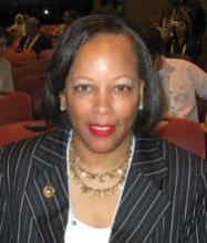
Acne prevalence is about the same in black and white patients, said Dr. Valerie D. Callender. The same mechanisms cause acne and the same treatments, in general, are used regardless of skin type, she said. "What is important is ... there are sequelae of acne that make it a little different, and there are certain, special considerations we have to keep in mind when treating patients with darker skin types."
Prevention of keloids, hypertrophic scars, and postinflammatory hyperpigmentation are among the special considerations in this patient population, Dr. Callender said at the South Beach Symposium.
Keloids and hypertrophic scars usually result from inflammatory acne papules, nodules, and cysts, and can be challenging to treat. The keloids and scarring commonly arise along the jawline and on the chest, shoulder, and back. "It’s important to be very aggressive to resolve the inflammation, to treat them effectively. A lot of these patients do very well with isotretinoin and oral antibiotics," said Dr. Callender of the dermatology department at Howard University in Washington, D.C.
In patients with keloids, consider injection of 20 mg/cc intralesional triamcinolone every 4 weeks, sometimes every 2 weeks, to get these lesions to go down, she said. "Remember that is part of their acne regimen."
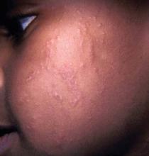
Postinflammatory hyperpigmentation (PIH) is a common presenting complaint among skin of color patients with acne or another inflammatory skin condition.
PIH is "psychologically devastating for these patients. We have to treat the PIH just as aggressively as we treat the acne," Dr. Callender said. In some cases, the disfigurement is severe and the hyperpigmented patches and macules can persist for months or even years.
In a study of 2,895 females aged 10-70, prevalence of PIH varied by ethnicity (J. Eur. Acad. Dermatol. Venereol. 2011;25:1054-60). The researchers at Massachusetts General Hospital in Boston found that PIH affected 65% of 384 black study patients and 48% of 258 Hispanic patients. "The other racial groups were less than 20% for PIH; this goes along with what we do in our practices," Dr. Callender said.
There are multiple options for prevention and treatment of PIH. Sunscreen, sun avoidance, and early diagnosis can prevent or minimize adverse effects. Hydroquinone, retinoids, azelaic acid, and/or kojic acid are recommended treatments.
"I love my hydroquinone, I use it a lot," Dr. Callender said. Hydroquinone lightens areas of hyperpigmentation through inhibition of tyrosine conversion to melanin, reduces the number of melanosomes, and inhibits DNA and RNA synthesis of melanocytes.
Topical retinoid agents are useful because they not only treat acne, but also address the hyperpigmentation, she said. Also, once the hyperpigmentation is under control, the topical retinoids help to exfoliate the skin and keep PIH from recurring. "We love to keep these patients on long-term topical retinoid therapy."
Tolerability is very important when prescribing topical retinoids and other agents. Carefully consider each patient’s potential risk for cutaneous irritation, including erythema, peeling, burning, and dryness. Be sure to inform nurses and office staff that when a patient calls about tolerability, "you have to inquire about any changes in pigmentation [as well], especially in skin of color patients," Dr. Callender said. Moisturizers, cleansers, and less irritating vehicles can improve tolerability.
"We also use adjunctive therapies and sunscreen protection [for PIH]. Remember combination therapy is the way to go," she said.
She and her associates conducted a meta-analysis looking at the tolerability of a fixed combination adapalene 0.1% and benzoyl peroxide 2.5% gel product (Epiduo) for acne in patients by Fitzpatrick skin type (J. Clin. Aesthet. Dermatol. 2010;3:15-9). They found erythema, scaling, and dryness scores higher for white patients in all three studies. Burning and stinging scores were not significantly different. "Tolerability is good for your skin of color patients. You don’t need to be overly concerned about a lot of irritation just because their skin is dark."
Chemical peels, lasers, and light-based therapies are additional treatment options for acne. Peels made with glycolic acid, salicylic acid, Jessner’s solution, or a combination is acceptable in skin of color patients. However, "be very, very careful with peels in skin of color patients. Make sure [to] use superficial peeling agents," Dr. Callender said.
More clinical studies of lasers and light-based therapies to treat acne are including the darker skin types, Dr. Callender said. Blue light, diode laser, intense pulse light, and photodynamic therapy are examples. "As we learn how to adjust the settings, they will be safer for skin of color patients," she said.
Dr. Callender disclosed that she is a consultant for Allergan and Galderma, which markets Epiduo; a researcher for Allergan, Galderma, and Intendis; and a member of the speakers’ bureau for Galderma.
EXPERT ANALYSIS FROM THE SOUTH BEACH SYMPOSIUM
Physician Joins Military at Age 59
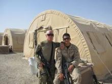
Physicians can learn a great deal after they join the military, such as how to approach an army general and suggest potentially life-saving medical changes on a base, said Dr. Dore J. Gilbert, a dermatologist on the faculty at the University of California Irvine.
A week into Lt. Col. Gilbert's deployment as a brigade surgeon on a base in Kabul, Afghanistan, he identified areas where necessary changes needed to be made and scheduled an appointment with the General.
Dr. Gilbert determined there was insufficient blood stored on the base in the event of an emergency. He suggested a walk-in blood bank with the General as the first volunteer. The General, concerned with the potential consequences, strongly supported the idea of the blood bank.
Next, Dr. Gilbert requested training for the 16 medics on the base and for 14 more at outlying bases once a month for 2 hours. "They were not getting any training," he said. The General again agreed.
"We have these unbelievable mannequins that actually bleed," Dr. Gilbert said. "They are a great way to train people. Sometimes we blow smoke so it's difficult to see the mannequins or do [medic training] at night so they have to put on their head lamps," Dr. Gilbert said during the South Beach Symposium in Miami.
When he raised a third issue, the General said, "Just do it." Dr. Gilbert increased the screening of local Afghan cafeteria employees for endemic intestinal parasites. Their contract only stipulated testing once a year on a base that serves 3,000 soldiers.
And Dr. Gilbert, as a dermatologist, also ran a skin cancer screening clinic. He treated other patients as well, which was part of the multitasking everyone did on the base. "On the second day, I was peeling old shrapnel out of a guy's face."
"I got up every day at 5 am to go to they gym and then work until 9 or 10 at night. I worked 12 to 14 hours a day, 7 days a week," he rcalled.
All his initiative impressed the General, and he invited Dr. Gilbert to become part of the everyday army operations on the base. "I went from being a simple doc to being a part of every general staff meeting," he said. Dr. Gilbert also was called in to the command center every time there was a terrorist-related attack in downtown Kabul. He went out on about 20 missions, most of which were to support medical personnel at other bases.
"I got to do things that 99.9% of doctors do not get to do when they are overseas or here in the United States because of a happenstance meeting I had with the General," Dr. Gilbert said.
Perhaps the most impressive part of the tale is Dr. Gilbert joined and started basic training at Fort Sam at the age of 59 years. "I was not going to allow people to say I was too old. Because there is such a dire need for physicians in the army they are willing to look at people who are slightly more mature."
One day he was doing a 5 mile ruck march with 35 pounds on his back. As he passed one of the younger soldiers, Dr. Gilbert said, "Pick it up. I'm older than your father." The soldier later approached him and said, "Sir, with all due respect, you are not older than my father, you are way older than my father."
Dr. Gilbert always wanted to serve and was inspired by his son Kevin, a corporal in the Marine Corps. "I was happy to see him home with his arms and legs. His battalion had about 25% casualties."
When he finished reflecting on his personal experiences as a dermatologist serving overseas, Dr. Gilbert said: "It was a great experience. I was honored to be able to serve my country."
--Damian McNamara @MedReporter on Twitter
Blog: Dermatologist Joins Military at Age 59
Physicians can learn a great deal after they join the military, such as how to approach an army general and suggest potentially life-saving medical changes on a base, said Dr. Dore J. Gilbert, a dermatologist on the faculty at the University of California Irvine.
A week into Lt. Col. Gilbert's deployment as a brigade surgeon on a base in Kabul, Afghanistan, he identified areas where necessary changes needed to be made and scheduled an appointment with the General.
Dr. Gilbert determined there was insufficient blood stored on the base in the event of an emergency. He suggested a walk-in blood bank with the General as the first volunteer. The General, concerned with the potential consequences, strongly supported the idea of the blood bank.
Next, Dr. Gilbert requested training for the 16 medics on the base and for 14 more at outlying bases once a month for 2 hours. "They were not getting any training," he said. The General again agreed.
"We have these unbelievable mannequins that actually bleed," Dr. Gilbert said. "They are a great way to train people. Sometimes we blow smoke so it's difficult to see the mannequins or do [medic training] at night so they have to put on their head lamps," Dr. Gilbert said during the South Beach Symposium in Miami.
When he raised a third issue, the General said, "Just do it." Dr. Gilbert increased the screening of local Afghan cafeteria employees for endemic intestinal parasites. Their contract only stipulated testing once a year on a base that serves 3,000 soldiers.
And Dr. Gilbert, as a dermatologist, also ran a skin cancer screening clinic. He treated other patients as well, which was part of the multitasking everyone did on the base. "On the second day, I was peeling old shrapnel out of a guy's face."
"I got up every day at 5 am to go to they gym and then work until 9 or 10 at night. I worked 12 to 14 hours a day, 7 days a week," he rcalled.
All his initiative impressed the General, and he invited Dr. Gilbert to become part of the everyday army operations on the base. "I went from being a simple doc to being a part of every general staff meeting," he said. Dr. Gilbert also was called in to the command center every time there was a terrorist-related attack in downtown Kabul. He went out on about 20 missions, most of which were to support medical personnel at other bases.
"I got to do things that 99.9% of doctors do not get to do when they are overseas or here in the United States because of a happenstance meeting I had with the General," Dr. Gilbert said.
Perhaps the most impressive part of the tale is Dr. Gilbert joined and started basic training at Fort Sam Houston at the age of 59 years. "I was not going to allow people to say I was too old. Because there is such a dire need for physicians in the army they are willing to look at people who are slightly more mature."
One day he was doing a 5 mile ruck march with 35 pounds on his back. As he passed one of the younger soldiers, Dr. Gilbert said, "Pick it up. I'm older than your father." The soldier later approached him and said, "Sir, with all due respect, you are not older than my father, you are way older than my father."
Dr. Gilbert always wanted to serve and was inspired by his son Kevin, a corporal in the Marine Corps. "I was happy to see him home with his arms and legs. His battalion had about 25% casualties."
When he finished reflecting on his personal experiences as a dermatologist serving overseas, Dr. Gilbert said: "It was a great experience. I was honored to be able to serve my country."
--Damian McNamara @MedReporter on Twitter
Expert Calls Isotretinoin an Option for Scarring Infantile Acne
MIAMI BEACH – Acne presentation, treatment, and counseling will vary according to whether your patient is a neonate, infant, child, preadolescent, or teenager, according to Dr. Jonette E. Keri.
• Neonatal acne. These small, erythematous papules that arise before 6 weeks of life probably represent a heterogenous set of conditions. Ketoconazole cream 2% twice per day is a treatment option.
However, "a lot of doctors choose not to treat because it’s not a scarring process," Dr. Keri said. You can reassure parents that most neonatal acne improves relatively quickly, usually within a few months. If true comedones are present, consider treatment with the same acne mediations indicated for infantile acne.
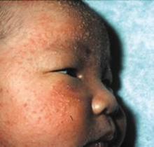
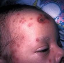
• Infantile acne. Infantile acne appears in children up to 1 year, usually at 3-6 months of age. Male infants are more prone to acne than female infants, and lesions tend to appear on the cheeks and chin and have the appearance of classic adolescent acne. Increased sebum production and some comedones are often present.
"You should treat because it can cause scarring," Dr. Keri said. Also, "this acne may predispose [children] to worse acne in teenage years – that is shown to be true for any form of infantile acne. It doesn’t have to be severe," said Dr. Keri of the University of Miami and chief of dermatology services at the Miami VA Hospital.
She offered the following clinical tips for treating and managing acne based on developmental age:
Combine treatments and use products appropriate for a baby, Dr. Keri advised at the South Beach Symposium. Although some experts recommend benzoyl peroxide, proceed with caution. The concern is getting any benzoyl peroxide near a baby’s eyes, "so you probably want to stay away from the washes."
Treatment options include topical antibiotics, adapalene, or a retinoid like tretinoin. Oral erythromycin is another acceptable option, she said.
"Isotretinoin is actually indicated if a severe, scarring process is going on," said Dr. Keri. She said that she searched the literature and found that some clinicians prescribe isotretinoin in children as young as 5 months.
• Midchildhood acne. "It is a newer concept, but a very important concept," Dr. Keri said. Acne is relatively rare between the ages of 1 and 8 years. During this time androgens in the body should be low and stable.
If acne does arise, it could point to an underlying hormonal abnormality. Evaluate three things: bone age, the growth chart, and hormone levels. "That may be a bit much for a dermatologist, but a pediatrician does these evaluations often," she said.
Accelerated bone age on a wrist radiograph can point to androgen excess, whereas delayed bone age suggests Cushing’s disease. A growth chart that shows a child’s height crossing percentiles upward or increasing faster than would be expected can also suggest androgen excess.
"Hormone levels can be tricky," Dr. Keri said. High levels of androgens, free testosterone, or dehydroepiandrosterone also can occur with tumors or polycystic ovarian syndrome (PCOS). "Another reason they can be tricky is because the child is developing into an adult, so you may need a pediatric endocrinologist to tease this out."
• Preadolescent acne. Treatments for children aged 9-12 years are essentially the same as for infants and midchildhood patients. However, patient counseling plays a bigger role. "Adherence is a big issue with these kids, so try to give them a once-a-day regimen," Dr. Keri said. Isotretinoin is rarely prescribed in this age group, but if severity dictates the need, it is likely the child will need retreatment (two or three courses) over time.
Comedones, seborrhea, and even PCOS are associated with preteen acne. Rule out precocious puberty and distinguish abnormal hormonal changes from normal signs of puberty, Dr. Keri recommended.
"PCOS is very complicated; you are going to need some help, a multispecialty approach," Dr. Keri said. Early diagnosis is worthwhile, she added. "If you identify PCOS when these young ladies are younger, you can prevent infertility, diabetes, [and] coronary artery disease, because they get these things more often than a woman who doesn’t have PCOS."
• Adolescent acne. Speaking directly and appropriately with teenagers about their acne can facilitate better outcomes, Dr. Keri said. For example, instead of asking, "How many days a week do you use your medicines?" ask, "How do you use your acne medicines?" Also determine their expectations before prescribing, and find out if they have a prom or other major social event coming up.
It is also important to evaluate their previous acne-fighting strategies. "It can be painfully tiring to go through all they have done and used, but if you don’t do that, you’re not going to have a good starting point," she said. Find out what they like and dislike, and really listen to their answers. "They will be honest if you listen to them."
Also, adolescents like to use pads to apply their acne medicine, so keep those formulations in mind for this age group, she said.
Dr. Keri said she had no relevant financial disclosures.
EXPERT ANALYSIS FROM THE SOUTH BEACH SYMPOSIUM
Otolaryngologists Right Where You Need Them
MIAMI BEACH – A novel hospitalist program has optimized inpatient otolaryngology care at the University of California, San Francisco, a retrospective study reveals.
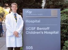
Based on its success, the program is poised to expand further this year and could become a model for other health systems, said Dr. Matthew S. Russell, an otolaryngology hospitalist at UCSF Medical Center and clinical instructor in the department of otolaryngology–head and neck surgery. "Feedback from this service has been overwhelmingly positive. I would encourage others who are interested to undertake a pilot project to see if this model is generalizable to other institutions," he said.
A significant number of requests for otolaryngology care spurred the birth and growth of the program, which may be only one of its kind in the United States. After a pilot feasibility phase, clinicians officially launched the service in July 2009.
Study Data: Volumes of Consults, Interventions Speaks, Well, Volumes
"As an otolaryngologist, I am providing mostly consultative services," Dr. Russell said at the meeting. "The types of problems I see are most commonly acute airway issues and complications of infectious processes in the head and neck. We perform a high volume of bedside procedures, and contrary to a popular assumption, the surgical volume is favorable."
Otolaryngology hospitalists provided a total 384 procedural or surgical interventions during the study period. Endoscopic sinonasal interventions were the most common procedure, followed by transnasal flexible laryngoscopy and operative endoscopy.
The UCSF otolaryngology hospitalists, therefore, work more like specialist consultants and less like traditional primary care hospitalists overseeing a cohort of inpatients, said Dr. Russell.
The study findings could be used to convince hospital administration of the need for this service. Improved time to consultation and surgery for many patients, for example, decreases resource utilization and shortens length of hospital stay, Dr. Russell said. "Prior to our hospitalist model, most of this care was clinically and financially invisible to the institution. We have been able to demonstrate qualitatively the benefit of our unique expertise in solving complex problems as well as mitigating risk from airway catastrophe."
The otolaryngology hospitalists work closely with a number of other clinicians at UCSF. "The program has had a significant clinical impact in both the operating rooms and ICU," Dr. Matt Aldrich, faculty member in the department of anesthesia and perioperative care at UCSF, said in an interview.
"On numerous occasions, Dr. Russell has been very helpful with emergency airway management," Dr. Aldrich said. "He is also an excellent general resource for questions regarding tracheostomy management. This includes being on ‘standby’ for possible emergency tracheostomy during difficult airway management – both intubations and extubations – as well as [providing] assistance in understanding airway anatomic abnormalities during fiber-optic intubation procedures."
"I’ve been very impressed by the speed with which he responds to our requests for help, as well as his clinical skills, knowledge, and professionalism," Dr. Aldrich said. With only a few minutes’ notice, Dr. Russell recently came to the operating room to evaluate a patient with a pharyngeal mucosal cyst prior to placement of a transesophageal echocardiography probe, for example.
UCSF is a 550-bed tertiary care and referral center. Hospitals smaller than approximately 500 beds might not be able to support a 100% full-time equivalent position for this model, he added.
But at UCSF, he is optimistic, predicting a measurably strong impact by year’s end: "This year we are on track to see over 700 unique patients with a significant bedside procedural and surgical volume," Dr. Russell said at the meeting, which was jointly sponsored by the Triological Society and the American College of Surgeons.
Dr. Russell and Dr. Aldrich reported having no financial disclosures.
UCSF pediatric hospitalist Dr. Bradley Monash offers several examples of situations where having an otolaryngologist at hand was a game changer:
• A young boy with NEMO syndrome (a rare immunodeficiency) developed pyoderma gangrenosum of his esophageal inlet and was refusing to eat because of the pain associated with oral intake. This created quite a quandary, as malnutrition was further compromising his overall condition. This patient had already developed a host of issues related to prolonged nasogastric tube feeds, and his subspecialty providers were reluctant to recommend a surgical feeding tube for a variety of reasons. [otolaryngology hospitalist Dr. Matthew] Russell served a pivotal role in this patient’s care, participating in family meetings, providing patient counseling, and offering different options to the patient and primary providers. He was ultimately able to convince the patient to accept topical anesthetic application, which allowed the patient to resume oral intake.
• As the "transfer attending," I was called about several outside hospital requests to transfer patients with invasive fungal sinusitis. Many of these patients were quite systemically ill and required the care of the hospital medicine service. The accessibility of an otolaryngology hospitalist afforded the opportunity to appropriately triage and expeditiously attend to these patients.
• A patient was admitted to the otolaryngology service with presumed head and neck soft tissue cellulitis due to intravenous heroin injection. This patient ended up developing respiratory failure from botulism. The outstanding, collaborative working relationship between the medicine and otolaryngology services contributed to expeditious interdisciplinary diagnostics and management.
— Bradley Monash, M.D.
MIAMI BEACH – A novel hospitalist program has optimized inpatient otolaryngology care at the University of California, San Francisco, a retrospective study reveals.
Based on its success, the program is poised to expand further this year and could become a model for other health systems, said Dr. Matthew S. Russell, an otolaryngology hospitalist at UCSF Medical Center and clinical instructor in the department of otolaryngology–head and neck surgery. "Feedback from this service has been overwhelmingly positive. I would encourage others who are interested to undertake a pilot project to see if this model is generalizable to other institutions," he said.
A significant number of requests for otolaryngology care spurred the birth and growth of the program, which may be only one of its kind in the United States. After a pilot feasibility phase, clinicians officially launched the service in July 2009.
Study Data: Volumes of Consults, Interventions Speaks, Well, Volumes
"As an otolaryngologist, I am providing mostly consultative services," Dr. Russell said at the meeting. "The types of problems I see are most commonly acute airway issues and complications of infectious processes in the head and neck. We perform a high volume of bedside procedures, and contrary to a popular assumption, the surgical volume is favorable."
Otolaryngology hospitalists provided a total 384 procedural or surgical interventions during the study period. Endoscopic sinonasal interventions were the most common procedure, followed by transnasal flexible laryngoscopy and operative endoscopy.
The UCSF otolaryngology hospitalists, therefore, work more like specialist consultants and less like traditional primary care hospitalists overseeing a cohort of inpatients, said Dr. Russell.
The study findings could be used to convince hospital administration of the need for this service. Improved time to consultation and surgery for many patients, for example, decreases resource utilization and shortens length of hospital stay, Dr. Russell said. "Prior to our hospitalist model, most of this care was clinically and financially invisible to the institution. We have been able to demonstrate qualitatively the benefit of our unique expertise in solving complex problems as well as mitigating risk from airway catastrophe."
The otolaryngology hospitalists work closely with a number of other clinicians at UCSF. "The program has had a significant clinical impact in both the operating rooms and ICU," Dr. Matt Aldrich, faculty member in the department of anesthesia and perioperative care at UCSF, said in an interview.
"On numerous occasions, Dr. Russell has been very helpful with emergency airway management," Dr. Aldrich said. "He is also an excellent general resource for questions regarding tracheostomy management. This includes being on ‘standby’ for possible emergency tracheostomy during difficult airway management – both intubations and extubations – as well as [providing] assistance in understanding airway anatomic abnormalities during fiber-optic intubation procedures."
"I’ve been very impressed by the speed with which he responds to our requests for help, as well as his clinical skills, knowledge, and professionalism," Dr. Aldrich said. With only a few minutes’ notice, Dr. Russell recently came to the operating room to evaluate a patient with a pharyngeal mucosal cyst prior to placement of a transesophageal echocardiography probe, for example.
UCSF is a 550-bed tertiary care and referral center. Hospitals smaller than approximately 500 beds might not be able to support a 100% full-time equivalent position for this model, he added.
But at UCSF, he is optimistic, predicting a measurably strong impact by year’s end: "This year we are on track to see over 700 unique patients with a significant bedside procedural and surgical volume," Dr. Russell said at the meeting, which was jointly sponsored by the Triological Society and the American College of Surgeons.
Dr. Russell and Dr. Aldrich reported having no financial disclosures.
FROM THE TRIOLOGICAL SOCIETY COMBINED SECTIONS MEETING
Tips for Spotting Dermatoses in Children With Darker Skin
MIAMI – Some hallmark signs of dermatologic problems in children – especially erythema and hyperpigmentation – often are less obvious in children with skin of color and can require more clinical detective work to diagnose.
Dr. Patricia A. Treadwell narrowed down the most likely dermatoses a pediatrician will encounter in this patient population at a pediatric update sponsored by Miami Children’s Hospital. Atopic and contact dermatitis, phytophotodermatitis, transient neonatal pustular melanosis, and neonatal lupus erythematosus are among the noteworthy clinical challenges, she said at the meeting.
"Children with increased pigmentation in their skin may end up testing your knowledge in terms of looking at their dermatitis and being able to diagnose that. Keep in mind it may be a little bit different in terms of the clinical presentation, but it’s important to identify it and start the proper treatment," said Dr. Treadwell, a pediatric dermatologist at Indiana University Health and Riley Hospital for Children in Indianapolis.
Overcoming this "masking" of a condition by skin pigmentation can be important, Dr. Treadwell said. She cited a patient born with a port-wine stain that went undiagnosed. The infant had subtle erythema and some asymmetry related to the overgrowth of the lesion. "This was not diagnosed based on the fact that the erythema was not apparent. The patient later developed a pyogenic granuloma, which is a complication that can be seen in patients with port-wine stains as they get older."
Erythema can be missed in children of color with atopic dermatitis as well. For this reason, atopic dermatitis may be underdiagnosed in this population overall, she said. Another challenge is the common practice of grading the severity of atopic dermatitis in lighter-skinned patients based on the degree of erythema. "In children with a fair amount of pigmentation in their skin, the erythema may not be noted and the severity will not be recognized."
Similarly, you might need a higher index of clinical suspicion to diagnose a child of color with contact dermatitis. Again, the erythema can be subtle. In contrast, "contact dermatitis can be very clear in a Caucasian patient. But the lesions are the same – linear, asymmetrical, and occurring on exposed areas," Dr. Treadwell said. Pruritus is common, and edema and swelling also occur. Watch for development of vesiculobullous lesions.
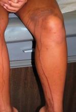
Phytophotodermatitis is another dermatologic condition that may require some additional detective work in children of color. Dr. Treadwell described a girl with a unique hyperpigmentation pattern on her legs and arms. She was referred following a vacation in Cancun, Mexico, with her family, and there was a concern about an autoimmune process. "They asked if she needed blood work. I said no, she was eating a mango and went out in the sun." Some of the mango juice splashed on her legs and arms.
Phytophotodermatitis occurs when furocoumarins from tropical fruit, citrus, celery, fennel, or parsnip come into contact with skin subsequently exposed to the sun.
"This condition can have a fairly bizarre pattern of presentation," Dr. Treadwell said. "Again, if there is more pigment in the skin, hyperpigmentation can present in a less common way than might be expected."
Some dermatoses are noted more commonly in children of color, she said. For example, the hyperpigmented macules or pustules that characterize transient neonatal pustular melanosis are reported in 4.4% of African American infants and 0.2% of Caucasian infants. "The percentages here may be related to the pigmentation in the skin. This does occur in Caucasian infants, but it may not be as noticeable."
The condition can be present at birth. The macules and pustules can appear anywhere on the body, but most often on the chin, neck, upper chest, and/or lower back. The good news is they fade over time and are benign, so no treatment is necessary. The differential diagnosis from such conditions as herpes simplex and erythema toxicum is important, however, said Dr. Treadwell. One tip is to check for lesions on the palms and soles, which can be diagnostic in transient neonatal pustular melanosis, but not for erythema toxicum. A biopsy can confirm your clinical suspicions.
The cutaneous manifestations of neonatal lupus erythematosus can tip you off to this condition, she said. Skin lesions can be annular, discoid, or atrophic. Some children present with "raccoon eyes." Because this condition is related to maternal antibodies passed through the placenta during gestation, lesions generally clear by 6 months to 1 year of age.
The mother may have a diagnosis of an autoimmune disease, or she may be completely asymptomatic. In one instance, a mother brought her newborn to Dr. Treadwell’s clinic. He had annular lesions on his forehead with some erythema. The lesions were more edematous around the edges. He actually got some sun exposure between his first and second visits, and developed more-discoid lesions in his sun-exposed areas. He also presented with lesions in non–sun exposed areas.
"The mother had a positive ANA [antinuclear antibody] test. I told her she should go to her doctor for urine and blood pressure monitoring," Dr. Treadwell said. "She had no symptoms and thought I didn’t know what I was talking about." Two years later, the woman returned with a newborn daughter who also had neonatal lupus with lesions on her face and some patchy alopecia on her scalp.
"The brother came back in, ... and he had telangiectasias already present at age 2 years."
Your evaluation can be more family centered. Consider testing the parent and affected children for anti-Rho, anti-La, and anti-RNP.
"I usually treat them with sun avoidance, sun protection, and possibly hydrocortisone," Dr. Treadwell said. She also recommends one electrocardiogram to rule out any cardiac consequences.
Dr. Treadwell reported that she had no relevant financial disclosures.
EXPERT ANALYSIS FROM A PEDIATRIC UPDATE SPONSORED BY MIAMI CHILDREN'S HOSPITAL
Volume of ENT Hospitalist Consults Speaks, Well, Volumes
To measure the impact of an otolaryngology hospitalist service on patient care and physician collaboration at UCSF Medical Center, Dr. Matthew S. Russell and his colleagues quantified their interventions between mid-2009 and December 2011.
During this period, otolaryngology hospitalists consulted on 375 new inpatients and generated 951 patient encounters. They most often evaluated general/pediatric cases (39%), laryngology concerns (29%), and rhinology issues (19%), according to a subanalysis of the first 18 months.
Respiratory failure was the most common specific diagnosis (12%) associated with a consult. They also consulted on patients with sinusitis (11%), stridor (11%), and dysphonia (8%), according to analysis of a billing database, including ICD-9 and CPT codes.
Otolaryngology hospitalists provided a total of 384 procedural or surgical interventions during the study period. Endoscopic sinonasal interventions were the most common procedure (122 codes), followed by transnasal flexible laryngoscopy (94 codes), and operative endoscopy (71 codes). A tally listing 19 specific codes was included in Dr. Russell’s poster presentation at the meeting.
Use of an administrative database is a potential limitation of the study that likely underestimated the number of otolaryngology hospitalist interventions, Dr. Russell said. "I was a bit surprised by the volume of consultations and procedures in the consortium model data, both of which seemed low compared to my experience this year as a sole service provider. I suspect this is a limitation of how the data was collected for administrative review rather than a true phenomenon."
An online search revealed no comparable otolaryngology service. "To our knowledge, ours is the first full-time otolaryngology hospitalist model in the United States," the investigators noted.
Respiratory failure, sinusitis, stridor, dysphonia,
To measure the impact of an otolaryngology hospitalist service on patient care and physician collaboration at UCSF Medical Center, Dr. Matthew S. Russell and his colleagues quantified their interventions between mid-2009 and December 2011.
During this period, otolaryngology hospitalists consulted on 375 new inpatients and generated 951 patient encounters. They most often evaluated general/pediatric cases (39%), laryngology concerns (29%), and rhinology issues (19%), according to a subanalysis of the first 18 months.
Respiratory failure was the most common specific diagnosis (12%) associated with a consult. They also consulted on patients with sinusitis (11%), stridor (11%), and dysphonia (8%), according to analysis of a billing database, including ICD-9 and CPT codes.
Otolaryngology hospitalists provided a total of 384 procedural or surgical interventions during the study period. Endoscopic sinonasal interventions were the most common procedure (122 codes), followed by transnasal flexible laryngoscopy (94 codes), and operative endoscopy (71 codes). A tally listing 19 specific codes was included in Dr. Russell’s poster presentation at the meeting.
Use of an administrative database is a potential limitation of the study that likely underestimated the number of otolaryngology hospitalist interventions, Dr. Russell said. "I was a bit surprised by the volume of consultations and procedures in the consortium model data, both of which seemed low compared to my experience this year as a sole service provider. I suspect this is a limitation of how the data was collected for administrative review rather than a true phenomenon."
An online search revealed no comparable otolaryngology service. "To our knowledge, ours is the first full-time otolaryngology hospitalist model in the United States," the investigators noted.
Respiratory failure, sinusitis, stridor, dysphonia,
Respiratory failure, sinusitis, stridor, dysphonia,
Palliation Trumps PET in Prolonging Head & Neck Cancer Survival
MIAMI BEACH – Using PET scans to diagnose distant metastasis in patients with advanced head and neck squamous cell carcinoma does not significantly prolong life expectancy, compared with other imaging techniques, according to a retrospective study.
Palliative chemotherapy did make a difference, however, significantly increasing life expectancy by 215 days in patients who received it, Dr. Matthew E. Spector and colleagues from the University of Michigan, Ann Arbor, reported at a meeting of the Triological Society.
"Over 90% of patients at University of Michigan have at least one PET scan at some point in their treatment," Dr. Spector said. Increased sensitivity is one reason for such widespread adoption of the imaging technique. "We were wondering, while it may be more sensitive to identify distant metastatic disease, was it changing what we were doing?"
In a retrospective look at 170 patients with such cancers at their institution, researchers found no significant difference in median survival between patients who had a PET scan (168 days) and those who did not (193 days). Determination of any survival difference was a primary aim of the study.
"A lot of studies have looked at PET scans, and we know in up to one-third of cases it may change our decisions," Dr. Spector said. For example, a negative PET scan might mean definitive treatment, whereas a positive PET finding might lead to palliative therapy. However, "no one has looked at the impact of the PET findings on the life expectancy after diagnosis."
All patients in the study had a distant metastasis diagnosis. "We found PET was more likely to diagnose multiple distant metastasis sites [P = .03]," Dr. Spector said. "But there were no differences in life expectancy when comparing PET to the various other imaging modalities like CT or chest x-ray."
Mean patient age was 59 years, and 135 of the patients were men.
Kaplan-Meier survival curves revealed no difference in survival between patients with a single distant metastatic site vs. multiple distant metastatic sites, said Dr. Spector, a head and neck surgery resident at the University of Michigan Health System in Ann Arbor.
The investigators intentionally controlled for chemotherapy use (110 patients, or 65%) in their survival calculations. "Chemotherapy could alter the course of their distant metastasis. Since [survival] was our main outcome measure, we wanted to control for that."
There were no differences in survival by patient age, sex, or site of primary tumor. Primary head and neck tumor sites included the oropharynx in 75 patients, the oral cavity in 40 patients, and the larynx in 36 others. The hypopharynx, nasopharynx, and some cases with unknown primary sites accounted for the remainder.
Dr. Spector and his associates did find a significant difference between the 86% of patients whose distant metastasis was detected during routine follow-up cancer care and the 14% who presented with symptoms. Median survival was 247 days in the routine surveillance group vs. 73 days for patients who might have come into the clinic complaining of chest pain after which subsequent imaging studies revealed a distant metastasis.
"Patients who were symptomatic, as you would imagine, had a worse life expectancy," Dr. Spector said. For the group detected on routine follow-up, the median time to distant metastasis diagnosis was 324 days.
Identification of any factors that did prolong survival was a second aim of the study. For the 85 patients who received palliative chemotherapy, median survival was significantly longer at 285 days, compared with 70 days for those who did not receive it.
Palliative chemotherapy was an independent factor that increased life expectancy, "and should be promoted for patients with these cancers," Dr. Spector said at the meeting, which was sponsored by the Triological Society and the American College of Surgeons. Previous chemotherapy did not alter patient response to palliative chemotherapy.
"Even for patients who were symptomatic at the time of diagnosis of their distant metastasis, palliative chemotherapy was still found to be effective," he added.
By cancer subtype, there was a nonsignificant trend for palliative chemotherapy to prolong survival among patients with primary oropharyngeal cancers (median, 333 days) compared with patients with primary laryngeal cancers (195 days).
Dr. Spector said that he had no relevant disclosures.
FROM THE TRIOLOGICAL SOCIETY'S COMBINED SECTIONS MEETING
Major Finding: In head and neck cancer patients, median survival was 168 days when distant metastases were detected by PET scan, and 193 days when metastases were detected using other modalities, an insignificant difference.
Data Source: A retrospective study of 170 patients with head and neck cancer diagnosed with distant metastasis at the University of Michigan.
Disclosures: Dr. Spector reported having no relevant financial disclosures.
Many Updates for Hospitalists Promised at Perioperative Medicine Summit
MIAMI – The Perioperative Medicine Summit in Miami Beach promises to deliver several clinical updates for hospitalists presented in innovative and interactive formats, according to summit director Dr. Amir K. Jaffer.
"The meeting in general is about patients having surgery. It’s been documented time and again in surveys that a large number of hospitalists across the country spend part of their time managing the medical aspect of surgical patients," Dr. Jaffer said in an interview.
Therefore, attendees will learn strategies and tools to better manage patients in the perioperative period, a core competency of the Society of Hospital Medicine. Dr. Jaffer is division chief of hospital medicine within the department of medicine at the University of Miami.
The meeting will run from March 15-17. During a plenary session scheduled to kick off the meeting, Dr. Thanjavur S. Ravikumar will outline his models to improve hospitalwide patient outcomes. He will explain how his strategies also decrease health care utilization and costs. Dr. Ravikumar is the chief quality officer, surgery and interventional procedures, and director of the center for surgical innovation at Geisinger Health System in Wilkes Barre, Pa.
A point-counterpoint session on optimization of deep vein thrombosis prophylaxis in major joint replacement surgery is expected to be another highlight. The back and forth between thromboembolism specialist James D. Douketis and orthopedic surgeon Cliff W. Colwell is sure to include relevant clinical guidelines just released in February by the American College of Chest Physicians, Dr. Jaffer said.
Dr. Jaffer, who is on the board of advisers for Hospitalist News, will also present at the meeting. "I’m going to be talking about the perioperative management of new and old antithrombotic therapies."
The second day of the summit is set to start with relevant and practical talk on the role of hospitalist medicine in the era of value-based care. Dr. Joseph Ming Wah Li will share his unique perspective as current president of the Society for Hospital Medicine.
Rapid-fire, 1-minute perioperative consults are a new feature of the summit this year. These will include advice on identification of noncardiopulmonary triggers for transfusion in postoperative anemia, with a discussion of blood transfusion alternatives. Other topics include when noninvasive coronary assessment is indicated for asymptomatic patients undergoing surgery for cancer and whether discontinuation of immunomodulatory medications in the perioperative period is warranted. The idea for these rapid-fire consults came from similar and well-attended sessions at the American College of Physicians meeting in which experts answer specific clinical questions.
The summit has a global reach, Dr. Jaffer said. "We do have people coming from all over the world ... including attendees signed up from New Zealand, Australia, and Singapore." In addition to hospitalists, attendees will include internists, anesthesiologists, physician assistants, and nurse practitioners.
The scope of the summit also goes beyond a single-institution or regional meeting, he added. This is in large part due to the summit codirectors and cosponsors. For example, Dr. Franklin A. Michota Jr. who is on the medicine faculty at the Cleveland Clinic Lerner College of Medicine at Case Western Reserve University brings his expertise as director of academic affairs for the department of hospital medicine at the Cleveland Clinic. The Cleveland Clinic is cosponsoring the meeting along with the University of Miami Miller School of Medicine. Dr. Michota is the medical editor for Hospitalist News. Another codirector, Dr. Darin J. Correll, is director of the acute postoperative pain management service at Brigham and Women’s Hospital in Boston and a member of the governing board of the Society for Perioperative Assessment and Quality Improvement, a third cosponsor of the meeting.
Visit periopmedicine.org for additional information and updates on the seventh Perioperative Medicine Summit.
Posttransplant Head and Neck Tumors Tallied
MIAMI BEACH – Patients who have undergone solid organ transplantation are at greater risk for subsequent tumor development, and head and neck cancers can be particularly aggressive, according to results of a single-institution study.
Dr. Robert H. Deeb and his associates at Henry Ford Hospital in Detroit studied 3,639 patients who underwent solid organ transplantation between January 1990 and December 2011. By retrospectively searching electronic medical records, they identified 95 people who developed cutaneous, salivary gland, or mucosal malignancies.
They found a 2.1% incidence of cutaneous cancers and a 0.5% incidence of noncutaneous head and neck cancers in this population. Despite a relatively low overall 2.6% incidence, there are reasons for concern, Dr. Deeb said at the Triological Society’s Combined Sections Meeting, which was jointly sponsored by the Triological Society and the American College of Surgeons. When cutaneous cancers did occur, they were more aggressive and were associated with shorter 1-year survival rates.
Henry Ford Transplant Institute maintains a noncutaneous tumor registry. This allowed the researchers to compare survival and other factors. Compared with the "nontransplant tumor registry in our institution, we found significantly fewer patients were alive at 1 year," Dr. Deeb said. Specifically, 55% of posttransplant patients with these cancers survived to 1 year, compared with 81% of nontransplant patients. There were no significant differences between groups in terms of age, sex, stage at diagnosis, or 5-year survival.
More than half (52%) of the patients who developed skin cancers had multiple head and neck cutaneous malignancies, pointing to the aggressive nature of these cancers, said Dr. Deeb of the department of otolaryngology–head and neck surgery at Henry Ford Hospital.
"We believe these patients require aggressive screening, treatment, and follow-up," Dr. Deeb said.
Of the 78 cutaneous head and neck cancers, 51% were squamous cell carcinomas, 36% were basal cell carcinomas, and the remaining 13% were other skin cancer types. The cheek and scalp were the most common sites. The patients’ average age was 61 years at the time of diagnosis, and 74% were men. Skin cancer developed a mean 4 years after transplant surgery.
Four patients had a salivary gland cancer and 13 had an upper aerodigestive tract mucosal malignancy. The average patient age in this group was 60 years, and 94% were men. Cancer was diagnosed a mean 66 months post transplantation. All 13 of the mucosal malignancies in the study were squamous cell carcinomas, Dr. Deeb said.
Kidney, liver, and heart were the most commonly transplanted organs among patients who ultimately developed these head and neck malignancies.
Although immunosuppressants are universally prescribed to prevent organ rejection following transplant surgery, the exact mechanism for subsequent cancer development remains unknown, Dr. Deeb said. Loss of immunosurveillance is one possibility; uncontrolled cell proliferation from chronic and low-level antigenic stimulation throughout the body is another. In addition, he said, immunosuppressants might somehow activate oncogenic viruses.
Despite the findings of this study, Dr. Deeb said that the benefits of solid organ transplantation still generally outweigh the risk of subsequent cancer development.
Henry Ford Hospital funded the study. Dr. Deeb said that he had no relevant disclosures.
FROM THE TRIOLOGICAL SOCIETY'S COMBINED SECTIONS MEETING
Major Finding: A total of 95 people who underwent solid organ transplantation over 21 years developed subsequent head and neck cancer, for an incidence of 2.6%.
Data Source: Data are taken from a retrospective study of 3,639 solid organ transplant patients treated at the Henry Ford Hospital since 1990.
Disclosures: Henry Ford Hospital sponsored the study. Dr. Deeb reported having no financial disclosures.
