User login
What’s hot in knee OA rehab research
TORONTO – Emerging evidence indicates that patients with knee osteoarthritis who engage in high-intensity interval training obtain significantly greater improvement in physical function than with conventionally prescribed moderate-intensity exercise, Monica R. Maly, PhD, said at the OARSI 2019 World Congress.
This was one of the key conclusions she and her coworkers drew from their analysis of the past year’s published research on diet and exercise interventions to improve outcomes in patients with OA, where obesity and physical inactivity figure prominently as modifiable lifestyle factors.
Another finding: Exercise interventions are where all the action is at present in the field of lifestyle-modification research aimed at achieving better health-related quality of life and other positive outcomes in OA. Dietary interventions are simply not a hot research topic. Indeed, her review of the past year’s literature included 38 randomized, controlled trials (RCTs) and 15 meta-analyses and systematic reviews – and all 38 RCTs addressed exercise interventions.
“It’s interesting to note that we found no new RCT data on diet to modify obesity in OA in the past year,” Dr. Maly said at the meeting sponsored by the Osteoarthritis Research Society International.
Additionally, 32 of the 38 RCTs devoted to exercise interventions for OA focused specifically on knee OA, noted Dr. Maly of the department of kinesiology at the University of Waterloo (Ont.).
Aerobic exercise dosage and intensity
Australian investigators conducted a pilot randomized trial of high-intensity interval training (HIIT) versus more conventional moderate-intensity exercise to improve health-related quality of life and physical function in 27 patients with knee OA. The exercise programs involved unsupervised home-based cycling, with participants requested to do four roughly 25-minute sessions per week for 8 weeks.
The two exercise intensity groups showed similar gains in health-related quality of life as assessed by the Western Ontario and McMaster Universities Osteoarthritis Index (WOMAC). However, the HIIT group showed significantly greater improvement in physical function as measured on the Timed Up and Go test (PeerJ. 2018 May 9;6:e4738).
Dr. Maly noted that adherence to the home-based exercise programs was a challenge: Only 17 of the 27 patients completed the 8-week Australian study, for a 37% dropout rate. However, most study withdrawals were because of family-related issues, illness, or injuries unrelated to cycling, with no signal that HIIT placed knee OA patients at higher injury risk.
Other investigators performed a systematic review of 45 studies in an effort to generate evidence-based guidance about the optimal exercise dosing in order to improve outcomes in knee OA patients. They concluded that programs comprising 24 therapeutic exercise sessions over the course of 8-12 weeks resulted in the largest improvements in measures of pain and physical function. And, importantly, one exercise session per week conferred no benefits (J Orthop Sports Phys Ther. 2018 Mar;48[3]:146-61).
“Frequency probably matters,” Dr. Maly observed.
Patients and their physicians often wonder if long-term, land-based exercise might have deleterious impacts on joint structure in patients with knee OA. Reassurance on this score was provided by a recent meta-analysis of RCTs that concluded, on the basis of moderate-strength evidence, that exercise therapy of longer than 6 months duration had no adverse effect on tibiofemoral radiographic disease severity, compared with no exercise. Nor was there evidence of a long-term-exercise–associated deterioration of tibiofemoral cartilage morphology or worsening of synovitis or effusion. Plus, the meta-analysis provided limited evidence to suggest long-term exercise had a protective effect on the composition of patellar cartilage (Semin Arthritis Rheum. 2019 Jun;48[6]:941-9).
“While there was a little bit of evidence suggesting that long-term exercise could worsen bone marrow lesions, really there was no other evidence that it could change the structure of a joint,” according to Dr. Maly.
Internet-based exercise training
Using the Internet to deliver an individually tailored exercise-training program for patients with symptomatic knee OA might sound like an efficient strategy, but in fact it proved fruitless in a large randomized trial. The 350 participants were assigned to an 8-visit, 4-month program of physical therapy, a wait-list control group, or an internet-based program that delivered tailored exercises and video demonstrations with no face-to-face contact. The bottom line is that improvement in WOMAC scores didn’t differ significantly between the three groups when evaluated at 4 and 12 months (Osteoarthritis Cartilage. 2018 Mar;26[3]:383-96).
“When we deliver exercise with the use of technology, it may require some support, including face to face,” Dr. Maly concluded from the study results.
Strength training
High-intensity resistance training such as weight lifting aimed at strengthening the quadriceps and other large muscles is often eschewed in patients with knee OA because of concern about possible damage to their already damaged joints. Intriguingly, Brazilian investigators may have found a workaround. They randomized 48 women with knee OA to 12 weeks of either supervised low-intensity resistance training performed with partial blood-flow restriction using an air cuff, to low-intensity resistance training alone, or to high-intensity resistance training. The low-intensity resistance workouts involved exercises such as leg presses and knee extensions performed at 30% of maximum effort.
The low-intensity resistance training performed with blood-flow restriction and the high-intensity strength training programs proved similarly effective in improving quadriceps muscle mass, muscle strength, and physical function to a significantly greater extent than with low-intensity resistance training alone. Moreover, low-intensity resistance training with blood-flow restriction also resulted in a significant improvement in pain scores. That finding, coupled with the fact that 4 of the 16 patients in the high-intensity resistance training group dropped out of the trial because of exercise-induced knee pain, suggests that low-intensity strength training carried out with partial blood-flow restriction may have a bright future (Med Sci Sports Exerc. 2018 May;50[5]:897-905).
Exercise plus diet-induced weight loss
How does the combination of dietary weight loss plus exercise stack up against diet alone in terms of benefits on pain and physical function in obese patients with knee OA? A systematic review and meta-analysis of nine RCTs aimed at answering that question concluded that diet-alone strategies are less effective. Both the diet-plus-exercise and diet-only interventions resulted in comparably moderate improvement in physical function. However, diet-only treatments didn’t reduce pain, whereas diet-plus-exercise interventions achieved moderate pain relief (Semin Arthritis Rheum. 2019 Apr;48[5]:765-77).
Dr. Maly reported having no financial conflicts of interest regarding her presentation.
EXPERT ANALYSIS FROM OARSI 2019
Occupational therapy program helps thumb OA
TORONTO – A multimodal occupational therapy intervention in patients with thumb base osteoarthritis brought clinically meaningful improvements in pain, grip strength, and function, at least short term, in a Norwegian multicenter randomized clinical trial, Anne Therese Tveter reported at the OARSI 2019 World Congress.
OA of the thumb base – that is, the carpometacarpal joint – causes more pain and dysfunction than disease involvement at many other sites because of the evolutionary importance of the opposable thumb. Current guidelines recommend conservative therapies as first line for hand OA; however, there is a dearth of high-quality evidence for multimodal occupational therapy in the special setting of thumb-base OA. This was the impetus for a randomized trial of 170 consecutive patients with thumb OA who presented to three Norwegian rheumatology departments for surgical consultation, explained Ms. Tveter, a physiotherapist at the Norwegian National Advisory Unit on Rehabilitation in Rheumatology at Diakonhjemmet Hospital in Oslo.
Participants were randomized to a 3-month, multimodal self-management intervention. It included education about OA; ergonomic principles; the importance of using separate orthoses as much as possible both day and night to stabilize the joint, improve performance, and relieve pain; and – at the heart of the program – instruction in hand exercises to enhance joint mobility, strength, and stability, as well as hand-stretching exercises. The exercises were to be done at home three times per week. Also, the active intervention group received five common assistive devices to help them in household tasks, such as opening jars. The control group received usual care, which was basically information about hand OA, she said at the meeting sponsored by the Osteoarthritis Research Society International.
Ms. Tveter presented an interim analysis focused on the 3-month outcomes. At 4 months, participants underwent surgical consultation. The study will continue for 2 years, with endpoints including the impact of the occupational therapy intervention on need for joint surgery, as well as long-term pain and function measures.
At baseline, most patients reported mild pain, with a median score of 3 on a 10-point numeric rating scale, and moderate disability. Baseline grip and pinch strength was 60%-65% of normal. The 3-month outcomes included pain at rest and during pinch- and grip-strength testing, range of motion through palmar abduction and abduction in the carpometacarpal joint, and self-reported function as measured using the validated MAP-Hand and QuickDASH physiotherapy measures. Adherence to the program was assessed by review of patient diaries.
At 3 months of follow-up, the active-intervention group showed significant improvements in all measures of pain and function except for the flexion deficit, which was minimal to begin with. In contrast, the control group showed no improvements and a trend towards deterioration in pain and function.
Specifically, the intervention group averaged a 1.4-point reduction in pain at rest on a self-reported 10-point scale, a 1.1-point improvement in pain following a grip strength test, and a 0.8-point improvement in pain following a pinch test. On the MAP-Hand self-reported test of function, the intervention group showed a 0.18-point improvement from a baseline of 2 on the 1-4 scale, coupled with an 8.1-point improvement on the QuickDASH, which is scored 0-100.
Adherence to the program was deemed acceptable: 82% of patients reported doing their hand exercises at least twice per week for at least 8 of the 12 weeks, 61% used their day orthotic devices at least 4 days per week for 8 weeks, 54% used the night orthoses at least 5 nights per week for 8 weeks, and 69% utilized at least three of the five home-assist devices. In total, 64% of patients adhered to at least three of the four program components.
Asked for the rationale in requesting that patients do their home exercises three times per week instead of daily, Ms. Tveter replied that three times per week is more realistic and is consistent with major guidelines.
“It would be nice to exercise every day. I don’t think it would be possible to get adherence to that,” she said.
She reported having no financial conflicts regarding the study, funded by scientific research grants from the Norwegian government.
REPORTING FROM OARSI 2019
Key clinical point: A multimodal occupational therapy intervention brought significant improvements in pain and function in patients with thumb-base OA.
Major finding: The intervention resulted in a mean 1.4-point decrease in self-reported pain at rest from a baseline of 3 on a 10-point scale, while most usual care controls showed a modest trend for worsening.
Study details: This was an interim 3-month report from a 2-year, randomized, multicenter trial including 170 consecutive patients who presented for surgical consultation regarding their thumb base OA.
Disclosures: The presenter reported having no financial conflicts regarding the study, funded by Norwegian governmental scientific research grants.
Liposomal steroid brings durable pain relief in knee OA
TORONTO – A single intra-articular injection of a novel, sustained-release liposomal formulation of dexamethasone in patients with symptomatic knee osteoarthritis brought at least 6 months of pain control in a multicenter, phase 2a trial, David Hunter, MD, reported at the OARSI 2019 World Congress.
This is a product that could fill a significant unmet medical need. Current therapies for knee OA have modest efficacy, and the injectable ones provide only 2-4 weeks of benefit. The ability to obtain significant pain relief with just a couple of intra-articular injections per year would be an important therapeutic advance, observed Dr. Hunter, professor of medicine at the University of Sydney.
He presented a 24-week study of 75 patients with symptomatic knee OA randomized at 13 sites in Australia and Taiwan to a single intra-articular injection of either 12 or 18 mg of the liposomal dexamethasone or to normal saline. One knee per patient was treated.
The primary outcome was the change in the Western Ontario and McMaster Universities Osteoarthritis Index (WOMAC) pain score from baseline to week 12. The 12-mg formulation of steroid significantly outperformed placebo at that time point as well as at all others. From a mean baseline WOMAC pain score of 1.49 on the 0-4 scale, patients in the 12-mg group averaged reductions of 0.83 points at 12 weeks, 0.85 at both weeks 16 and 20, and 0.87 at week 24. A statistically significant between-group difference was seen as early as day 3 after injection.
More than half (52%) of recipients of the 12-mg dose of liposomal dexamethasone, a product known for now simply as TLC599, maintained at least 30% pain relief at all visits through the study close at 24 weeks, as did 22% of controls, the rheumatologist reported at the meeting sponsored by the Osteoarthritis Research Society International.
The 12-mg injection also proved superior to placebo for the secondary endpoint of change in WOMAC function score. From a mean baseline score of 1.53, recipients of the 12-mg dose had improvements ranging from 0.82 points at week 12 to 0.85 points at week 24.
Of note, total acetaminophen intake over the course of the trial in the 12-mg steroid group was less than one-third of that in controls.
The 18-mg dose didn’t result in significantly greater reduction in pain scores than placebo. This is because dexamethasone release in the higher-dose formulation as presently constituted turned out to be less efficient, Dr. Hunter explained.
The safety profile was closely similar in all three study arms.
In phase 3 clinical trials, TLC599 will be compared with standard intra-articular triamcinolone, according to the rheumatologist.
He reported serving as a consultant to the Taiwan Liposome Company, which sponsored the phase 2a study, as well as to a handful of other pharmaceutical companies.
REPORTING FROM OARSI 2019
What’s up in the osteoarthritis drug pipeline
TORONTO – Philip G. Conaghan, MBBS, PhD, observed at the OARSI 2019 World Congress.
“Not only have things not improved during my time in osteoarthritis-land, they’ve gotten worse. We’ve lost therapies,” said Dr. Conaghan, professor of musculoskeletal medicine at the University of Leeds (England) and director of the Leeds Institute of Rheumatic and Musculoskeletal Medicine.
Specifically, opioids are now shunned because of the ongoing epidemic of addiction and a belated recognition that opioids are not a good option for pain relief in OA patients who want to have active lives. And acetaminophen has fallen by the wayside in light of recent evidence of lack of effectiveness: “It’s what our patients have been telling us for a long period of time,” he noted at the meeting sponsored by the Osteoarthritis Research Society International.
But change is in the air.
“I think we’ve got some things coming that look promising. What do I think will be the fastest to get to market? The peripheral nerve modulators look to me like the ones closest to going forward,” according to the rheumatologist, who provided an overview of the OA drug development pipeline, organized by treatment target.
Nerves
“Nerves as a treatment target in OA is a hot area. We’ve seen quite a slew of products recently looking at this. I think it’s a really fascinating area to play in: looking at how we modulate peripheral nociceptive pain,” Dr. Conaghan continued.
Tanezumab, an inhibitor of nerve growth factor, demonstrated very good pain relief and improvement in physical function in a phase 3 trial, although the occurrence of rapidly progressive OA in a subset of patients has bedeviled the drug development program. The hope is that a new subcutaneous drug delivery system coupled with careful patient pretreatment screening will mitigate the problem.
Tanezumab’s efficacy has contributed to a new understanding of the nature of pain in OA.
“I know I’m going to upset some people, but if you think central sensitization is the biggest driver of pain, I’d have to argue that the tanezumab program is the biggest single argument against that, since tanezumab is a large monoclonal antibody that doesn’t cross the blood-brain barrier and yet it has some of the best pain responses that we’ve seen,” Dr. Conaghan said.
Another peripheral nerve modulator, known for now as CNTX-4975, exhibited dose-dependent improvement in knee OA pain in the 175-patient, phase 2b TRIUMPH trial (Arthritis Rheumatol. 2019 Mar 19. doi: 10.1002/art.40894). CNTX-4975, which is delivered by intra-articular injection, is a synthetic form of capsaicin specific to pain nociceptors within the joint. Other sensory fibers remain unaffected.
Cartilage
Sprifermin, a recombinant human fibroblast growth factor 18 given by intra-articular injection, stimulates chondrocyte growth and decreases type 1 collagen expression. At year 2 in the ongoing 549-patient, 5-year, phase 2 FORWARD study, a dose-dependent increase in cartilage thickness at the tibiofemoral joint became apparent in sprifermin-treated patients, compared with those on placebo. This cartilage rebuilding effect was maintained at year 3, Dr. Conaghan said.
Bone
At the OARSI meeting, Dr. Conaghan and coinvestigators presented the results of a 6-month, open-label extension of their previously reported 6-month, placebo-controlled, phase 2 study of MIV-711, a potent selective reversible inhibitor of cathepsin K. The disease-modifying effects of MIV-711 seen in the first 6 months of the study, based on MRI-based measurements of changes in three-dimensional bone shape and cartilage thickness, were maintained in the second 6 months. Notably, MIV-711 slowed the rate of increase in bone area in the medial femur region and reduced loss of cartilage thickness relative to placebo. MIV-711 has also been shown to achieve a rapid and sustained reduction in the bone turnover biomarkers CTX-1 and -2, providing a rational mechanism to explain the drug’s observed structural benefits.
“So we’ve now got two agents – sprifermin for cartilage and MIV-711 for bone – showing that it’s possible to get some structural change, but no symptomatic benefit within the period of those trials,” the rheumatologist noted.
Wnt pathway inhibition
Samumed has launched a phase 3 clinical trials program, known as STRIDES, for lorecivivint, the company’s investigational small molecule inhibitor of the Wnt pathway. In phase 2 studies, including one led by Dr. Conaghan, intra-articular injection of lorecivivint, previously known as SMO4690, improved pain and physical function as well as medial joint space width. The drug’s potential effects on multiple tissues offers the promise of providing both symptomatic improvement and modification of the course of structural disease progression.
Inflammation
Lutikizumab, an anti–interleukin-1 alpha/beta immunoglobulin, proved to be a disappointment in a recent phase 2, placebo-controlled trial carried out in 350 patients with knee OA and synovitis. The IL-1 inhibitor had no benefit on synovitis, joint space narrowing, or cartilage thickness. Nor was it significantly better than placebo for pain reduction (Arthritis Rheumatol. 2019 Jul;71[7]:1056-69).
Anti–tumor necrosis factor agents haven’t exactly set the OA world on fire, either.
“In rheumatoid arthritis we know they’re stupendously effective, but the data from a number of trials in OA show they’re not so effective on symptoms and signs,” Dr. Conaghan said.
Colchicine and hydroxychloroquine are other anti-inflammatory agents which, while in theory might be helpful, have in actuality shown no benefit for OA symptoms in controlled clinical trials and are now considered dead ends.
On the other hand, the sustained delivery of intra-articular corticosteroids through the use of microsphere technology is advancing smartly through the developmental pipeline. Dr. Conaghan was first author of a multicenter, double-blind, phase 3 trial of FX006, a sustained-release formulation of triamcinolone acetonide, which showed that a single intra-articular injection provided at least 3 months of pain relief in knee OA patients, along with reduced systemic drug exposure, compared with standard intra-articular corticosteroids (J Bone Joint Surg Am. 2018 Apr 18;100[8]:666-77).
FX006 also performed well in another phase 3 trial, this one featuring repeated dosing on a flexible schedule based upon patient response (Rheumatol Ther. 2019 Mar;6[1]:109-24).
Reassuringly, this slow-release corticosteroid doesn’t appear to worsen glycemic control in knee OA patients with type 2 diabetes (Rheumatology [Oxford]. 2018 Dec 1;57[12]:2235-41).
“This is the start of a revolution in nanotechnology and the ability to slowly deliver a variety of drugs within the joint,” Dr. Conaghan predicted.
Although he was tasked at OARSI 2019 with providing an overview of the OA pharmacologic pipeline, he stressed that in his clinical practice, as opposed to his work as a clinical trialist, he functions more like a physical therapist.
“I actually spend my whole time in the OA clinic being a physical therapist and trying to get people strong, because that does work and it has no side effects. It’s just that nobody does it. We have a real adherence problem,” he said. “My favorite thought is this: keep people strong. If a patient can’t get out of a chair easily or can’t undo a jar, then they’ve got a problem.”
Dr. Conaghan reported receiving research funding from and serving as a consultant to many of the companies developing novel drug treatments for OA.
EXPERT ANALYSIS FROM OARSI 2019
How common is accelerated knee OA?
TORONTO – Accelerated knee osteoarthritis – a particularly noxious form of the joint disease – occurred in more than one in seven women who developed knee osteoarthritis in the prospective, long-term Chingford Cohort Study, Jeffrey B. Driban, PhD, reported at the OARSI 2019 World Congress.
This finding from a unique prospective study of 1,003 middle-aged U.K. women who were followed for the development of knee osteoarthritis (OA) for 15 years is important because the participants represented a typical community-based population sample. And yet the Chingford results are consistent with and confirmatory of those found earlier in the Osteoarthritis Initiative, a U.S. cohort study of nearly 4,800 individuals, even though the Osteoarthritis Initiative featured a population enriched with established risk factors for knee OA, Dr. Driban explained at the meeting, sponsored by the Osteoarthritis Research Society International.
In Chingford, accelerated knee OA accounted for 15% of all incident cases of knee OA during follow-up, and for 17% of all newly affected knees, whereas 20% of incident knee OA in the Osteoarthritis Initiative was accelerated knee OA, noted Dr. Driban of Tufts University, Boston.
Accelerated knee OA is defined by rapidly progressive structural damage. Affected individuals streak from no radiographic evidence of knee OA to advanced-stage disease marked by a Kellgren-Lawrence score of 3 or more within 4 years, whereas the typical form of knee OA follows a more gradual course. Also, accelerated knee OA features greater pain and disability.
In the Chingford study, the cumulative incidence of accelerated knee OA was 3.9%, while typical knee OA occurred in 21.7% of women. During years 6-15 of follow-up, 21% of women with accelerated knee OA underwent total knee replacement, compared with 2% of those with typical knee OA and 0.9% of women without knee OA.
Dr. Driban reported having no financial conflicts regarding his analysis of the Chingford Cohort Study and the Osteoarthritis Initiative, supported by Arthritis Research UK and the National Institutes of Health, respectively.
SOURCE: Driban JB et al. Osteoarthritis Cartilage. 2019 Apr;27[suppl 1]:S250-S251, Abstract 352.
REPORTING FROM OARSI 2019
Liberalized low–glycemic-index diet effective for seizure reduction
BANGKOK – in a randomized, double-blind, 24-week, noninferiority study.
The low–glycemic-index diet (LGID) was introduced as a kinder, gentler, variant of the classic ketogenic diet for seizure frequency reduction. The ketogenic diet’s efficacy for this purpose is well established, but compliance is a problem and discontinuation rates are high. Yet even though the LGID was designed to be less onerous than the ketogenic diet, many children and parents also find the 7-days-a-week LGID to be excessively burdensome. This was the impetus for pitting the daily LGID against an intermittent version – 5 days on, 2 days off – in a randomized trial, Prateek K. Panda, MD, explained at the International Epilepsy Congress.
The hypothesis of this noninferiority trial was that adherence to the liberalized LGID would be similar to or better than that with the daily LGID regimen, with resultant similar reductions in seizure frequency. And further, that patients on the intermittent LGID would feel better because it would help improve depleted glycogen stores important for daily activity and that the liberalized diet would also be rated more favorably by caregivers, Dr. Panda said at the congress sponsored by the International League Against Epilepsy.
The 24-week, single-center trial included 122 children ages 1-15 years with drug-resistant epilepsy. At baseline they averaged 99 seizures per week by parental diary despite being on a median of four antiepileptic drugs. A total of 88% of participants had some form of structural epilepsy; the rest had a probable or confirmed genetic cause for their seizure disorder, according to Dr. Panda of the All-India Institute of Medical Sciences in New Delhi.
The standard daily LGID was comprised of 10% carbohydrate, 30% protein, and 60% fat, with only low–glycemic-index foods permitted. The cohort randomized to the liberalized diet ate that way on weekdays; however, on Saturdays and Sundays their diet was 20% carbohydrate, 30% protein, and 50% fat, with both medium- and low–glycemic-index foods allowed.
The primary outcome was the mean reduction in seizures per week by caregiver records at 24 weeks. The reduction from baseline was 54% in the strict LGID group and not significantly different at 49% in the intermittent LGID patients. Overall, 54% of patients in the strict LGID arm experienced a greater than 50% reduction in weekly seizure frequency, as did 50% on the liberalized diet, a nonsignificant difference.
There were five study dropouts in the strict LGID group and three in the liberalized LGID cohort. The two groups showed similar improvements over baseline in measures of social function, behavior, and cognition. Parents of children in the liberalized LGID group rated that diet as significantly less difficult to administer than those randomized to the strict LGID therapy.
Mean hemoglobin A1c improved in the strict LGID patients from 5.7% at baseline to 5.1% at both 12 and 24 weeks. The intermittent LGID group went from 5.6% to 5.0% and then to 5.2%. There was no correlation between HbA1c and reduction in seizure frequency. In contrast, serum beta-hydroxybutyrate levels showed a moderate correlation with seizure frequency, a novel finding which if confirmed might render beta-hydroxybutyrate useful as a biomarker, according to Dr. Panda.
Adverse events – mostly dyslipidemia and GI complaints such as vomiting or constipation – occurred in 25% of the strict LGID group and 13% with the intermittent LGID. All adverse events were mild.
Dr. Panda reported having no financial conflicts regarding the study, sponsored by the All-India Institute of Medical Sciences.
SOURCE: Panda PK et al. IEC 2019, Abstract P056.
REPORTING FROM IEC 2019
Statins crush early seizure risk poststroke
BANGKOK – Statin therapy, even when initiated only upon hospitalization for acute ischemic stroke, was associated with a striking reduction in the risk of early poststroke symptomatic seizure in a large observational study.
Using propensity-score matching to control for potential confounders, use of a statin during acute stroke management was associated with a “robust” 77% reduction in the risk of developing a symptomatic seizure within 7 days after hospital admission, Soichiro Matsubara, MD, reported at the International Epilepsy Congress.
This is an important finding because early symptomatic seizure (ESS) occurs in 2%-7% of patients following an acute ischemic stroke. Moreover, an Italian meta-analysis concluded that ESS was associated with a 4.4-fold increased risk of developing poststroke epilepsy (Epilepsia. 2016 Aug;57[8]:1205-14), noted Dr. Matsubara, a neurologist at the National Cerebral and Cardiovascular Center in Suita, Japan, as well as at Kumamoto (Japan) University.
He presented a study of 2,969 consecutive acute ischemic stroke patients with no history of epilepsy who were admitted to the Japanese comprehensive stroke center, of whom 2.2% experienced ESS. At physician discretion, 19% of the ESS cohort were on a statin during their acute stroke management, as were 55% of the no-ESS group. Four-fifths of patients on a statin initiated the drug only upon hospital admission.
Strokes tended to be more severe in the ESS group, with a median initial National Institutes of Health Stroke Scale score of 12.5, compared with 4 in the seizure-free patients. A cortical stroke lesion was evident upon imaging in 89% of the ESS group and 55% of no-ESS patients. Among ESS patients, 46% had a cardiometabolic stroke, compared with 34% of the no-ESS cohort. Mean C-reactive protein levels and white blood cell counts were significantly higher in the ESS cohort as well. Their median hospital length of stay was 25.5 days, versus 18 days in the no-ESS group, Dr. Matsubara said at the congress sponsored by the International League Against Epilepsy.
Of the 76 ESSs that occurred in 66 patients, 37% were focal awareness seizures, 35% were focal to bilateral tonic-clonic seizures, and 28% were focal impaired awareness seizures.
In a multivariate analysis adjusted for age, sex, body mass index, stroke subtype, and other potential confounders, statin therapy during acute management of stroke was independently associated with a 56% reduction in the relative risk of ESS. In contrast, a cortical stroke lesion was associated with a 2.83-fold increased risk.
Since this wasn’t a randomized trial of statin therapy, Dr. Matsubara and his coinvestigators felt the need to go further in analyzing the data. After extensive propensity score matching for atrial fibrillation, current smoking, systolic blood pressure, the presence or absence of a cortical stroke lesion, large vessel stenosis, and other possible confounders, they were left with two closely comparable groups: 886 statin-treated stroke patients and an equal number who were not on statin therapy during their acute stroke management. The key finding: The risk of ESS was reduced by a whopping 77% in the patients on statin therapy.
The neurologist observed that these new findings in acute ischemic stroke patients are consistent with an earlier study in a U.S. Veterans Affairs population, which demonstrated that statin therapy was associated with a significantly lower risk of new-onset geriatric epilepsy (J Am Geriatr Soc. 2009 Feb;57[2]:237-42).
As to the possible mechanism by which statins may protect against ESS, Dr. Matsubara noted that acute ischemic stroke causes toxic neuronal excitation because of blood-brain barrier disruption, ion channel dysfunction, altered gene expression, and increased release of neurotransmitters. In animal models, statins provide a neuroprotective effect by reducing glutamate levels, activating endothelial nitric oxide synthase, and inhibiting production of interleukin-6, tumor necrosis factor-alpha, and other inflammatory cytokines.
Asked about the intensity of the statin therapy, Dr. Matsubara replied that the target was typically an LDL cholesterol below 100 mg/dL.
He reported having no financial conflicts regarding the study, conducted free of commercial support.
SOURCE: Matsubara S et al. IEC 219, Abstract P002.
REPORTING FROM IEC 2019
Noninvasive EEG may speed diagnosis of West syndrome
BANGKOK – , Hiroki Nariai, MD, reported at the International Epilepsy Congress.
Among the most promising of these potential EEG biomarkers for development of epilepsy are ictal or interictal high-frequency oscillations (HFOs) at 80 Hz or more, along with cross-frequency coupling of HFOs and delta wave activity, according to Dr. Nariai, a pediatric neurologist at the University of California, Los Angeles.
West syndrome, the most common epileptic encephalopathy during the first 2 years of life, has diverse etiologies. For example, in 250 infants with West syndrome enrolled in the United Kingdom National Infantile Spasms Consortium, a cause was identified in 64%. The etiology was genetic in 14% of subjects, a structural-congenital anomaly in 11%, tuberous sclerosis – a genetic-structural abnormality – in 10%, stroke in 22%, a metabolic defect in 5%, and infection in 2% (Epilepsia. 2015 Apr;56[4]:617-25).
West syndrome is rare, with an estimated prevalence of roughly 1 per 6,000 live births, but the associated mortality is high: 31% on average. And West syndrome often brings severe neurodevelopmental morbidity, with normal or near-normal intelligence present in only 25% of survivors.
An intensive search is on for an objective, reliable diagnostic biomarker – be it electroencephalographic, biochemical, or perhaps a neuroimaging finding – because the clinical diagnosis of West syndrome is highly subjective. It relies upon the triad of epileptic spasms, developmental regression or psychomotor delay, and hypsarrhythmia, which is a chaotic, disorganized, patternless form of brain electrical activity. And while that description makes hypsarrhythmia sound as if it should be easily recognizable, in fact that’s often not the case: Interrater reliability was poor in a study of six pediatric EEG experts at four centers who viewed 5-minute-long EEG samples obtained from 22 patients with infantile spasms (Epilepsia. 2015 Jan;56[1]:77-81), Dr. Nariai noted at the congress, sponsored by the International League Against Epilepsy.
“The clinical trial is maybe not so useful,” he observed.
The hunt for a reliable biomarker is further fueled by evidence that early diagnosis and treatment of West syndrome and other etiologies of infantile spasms makes a real difference. Indeed, investigators found in the United Kingdom Infantile Spasms Study that increasing lag time from onset of spasms to initiation of treatment was associated in stepwise fashion with significantly lower IQ at 4 years of age. While infants who started treatment within 7 days of onset of the seizure disorder had a mean IQ of 76.2 at age 4 years, those with an 8- to 14-day lag time between symptom onset and treatment averaged an additional 3.9-point decrement in IQ. A 15- to 30-day delay was associated with a 7.8-point reduction in IQ, compared with the reference group, while the decrease in IQ averaged 11.7 points in infants with a 1- to 2-month lag time and 15.6 points in those with a lag time of more than 2 months (Epilepsia. 2011 Jul;52[7]:1359-64).
At the start of the decade, Dr. Nariai and other investigators demonstrated that pathologic HFOs recorded during invasive EEG monitoring in conjunction with epilepsy surgery served as a reliable biomarker of epilepsy. While this was an important observation, a biomarker obtained through invasive monitoring during brain surgery clearly has very limited clinical applicability. But more recently, Dr. Nariai and his coinvestigators in the Tuberous Sclerosis Complex Autism Center of Excellence Network reported that noninvasive detection of interictal HFO fast ripples in the 250-500 Hz range via scalp EEG showed promise as a biomarker of epilepsy. Sensitivity of this far more practical approach to the detection of fast ripples was excellent, whether analyzed visually or by automatic detector (Clin Neurophysiol. 2018 Jul;129[7]:1458-66).
Moreover, in a recent, not-yet published study that Dr. Nariai and coworkers conducted in 24 infants with active epileptic spasms and 6 controls, noninvasive objective measurement of HFO rate using scalp EEG had an 83% sensitivity and 100% specificity for active epileptic spasms, while the modulation index of HFO and delta coupling in the 3-4 Hz range showed 74% sensitivity and 86% specificity.
If future studies validate the utility of detection of HFOs above a defined threshold or another noninvasively obtained EEG biomarker for diagnosis of epilepsy, the same strategy would presumably also be applicable for monitoring response to antiepileptic therapies, thereby eliminating the traditional trial-and-error approach to treatment. This would be a particularly important application in patients with West syndrome, where it’s believed that the electrical activity itself is contributing to the progressive – and often rapid – loss of cognitive function and behavioral disturbances. Thus, unlike in most other forms of epilepsy, the treatment goal isn’t merely to suppress the seizures, but also to achieve disease modification by eliminating the underlying subclinical EEG abnormalities, he explained.
A reliable biomarker would also be a boon in selecting the best participants for clinical trials of new antiseizure therapies.
Dr. Nariai reported having no financial conflicts regarding his presentation. His work is funded by research foundations and the National Institutes of Health.
REPORTING FROM IEC 2019
Cutaneous reaction to AEDs? Think autoimmune epilepsy
BANGKOK – Cutaneous reactions to antiepileptic drugs in patients with chronic epilepsy suggest increased likelihood of an autoimmune element to their seizure disorder, Fernando Cendes, MD, PhD, reported at the International Epilepsy Congress.
“My recommendation based on our findings is that if you have a patient who has a history of skin reactions to AEDs [antiepileptic drugs], or who has psychosis, or who has a very strange response to antiepileptic medication – meaning that at some points they are refractory and at other points they are very well controlled – I think those patients are probably at risk for having an autoantibody,” he said at the congress sponsored by the International League Against Epilepsy.
Screening for autoantibodies in such patients is appropriate. However, there’s a caveat: “The thing is, we don’t have evidence that treating these autoantibodies with immunotherapy will have any benefit on seizure control in these patients. We don’t have that data yet, but we are looking into it,” according to Dr. Cendes, professor of neurology at the State University of Campinas (Brazil).
He presented a study of 221 consecutive adults with severe chronic refractory epilepsy as evidenced by a mean disease duration of nearly 29 years, with an average of 5.93 seizures per month. A total of 77% had a structural etiology for their epilepsy, in most cases hippocampal sclerosis. In 19% of patients, the etiology was unknown. Overall, 95% of subjects had focal epilepsy, and the remainder had generalized epilepsy. All underwent serum testing for a variety of antibodies against neuronal surface antigens that have been implicated in encephalitis, seizures, and/or psychosis. Those who tested positive then underwent confirmatory testing of their cerebrospinal fluid.
The impetus for this study, the neurologist explained, is that although it’s now well established that seizures are a common clinical expression of acute- and subacute-phase autoimmune encephalitis marked by neuronal autoantibodies, little is known about the relationship between chronic epilepsy and such antibodies.
Only five Brazilian patients with chronic epilepsy, or 2.2%, tested positive for autoantibodies, all of whom had mesial temporal lobe epilepsy with hippocampal sclerosis. This suggests a possible autoimmune etiology for hippocampal sclerosis. Three of the five patients had anti-N-methyl-D-aspartate receptor antibodies (anti-NMDA) and two had antiglutamic acid decarboxylate antibodies (anti-GAD). No one was positive for anti–leucine-rich glioma-inactivated 1 antibodies (anti-LGI1), anti–contactin-associated proteinlike 2 (anti-caspr2), anti-glutamate receptor antibodies (anti-AMPAr), or anti–gamma-aminobutyric acid receptor antibodies (anti-GABAr).
The autoantibody-negative and the much smaller autoantibody-positive groups didn’t differ significantly in terms of demographics, seizure frequency, disease duration, drug resistance, cognitive impairment, comorbid autoimmune conditions, or history of status epilepticus. Indeed, only two between-group differences were found: fluctuation in seizure control was an issue in 10.6% of autoantibody-negative and 40% of autoantibody-positive patients, and cutaneous adverse reactions to antiepileptic drugs were noted in 10.6% of antibody-negative and 60% of antibody-positive patients. Psychiatric comorbidities were present in 49.5% of autoantibody-negative patients as compared with 80% – that is, four of five – who were autoantibody-positive, a trend that didn’t achieve statistical significance.
Asked if he thinks the autoantibodies found in a small subset of patients with chronic epilepsy were a cause or an effect of repeated seizures for so long, Dr. Cendes replied, “That’s a very interesting question, and I don’t have an answer, actually. But if seizures trigger development of these antibodies – and remember, this population we’re talking about had many, many seizures over the years – I would expect antibodies to be more frequent than the figure we found.”
He reported having no financial conflicts regarding his study.
SOURCE: Watanabe N et al. IEC 2019, Abstract P004.
REPORTING FROM IEC 2019
What’s new in pediatric sepsis
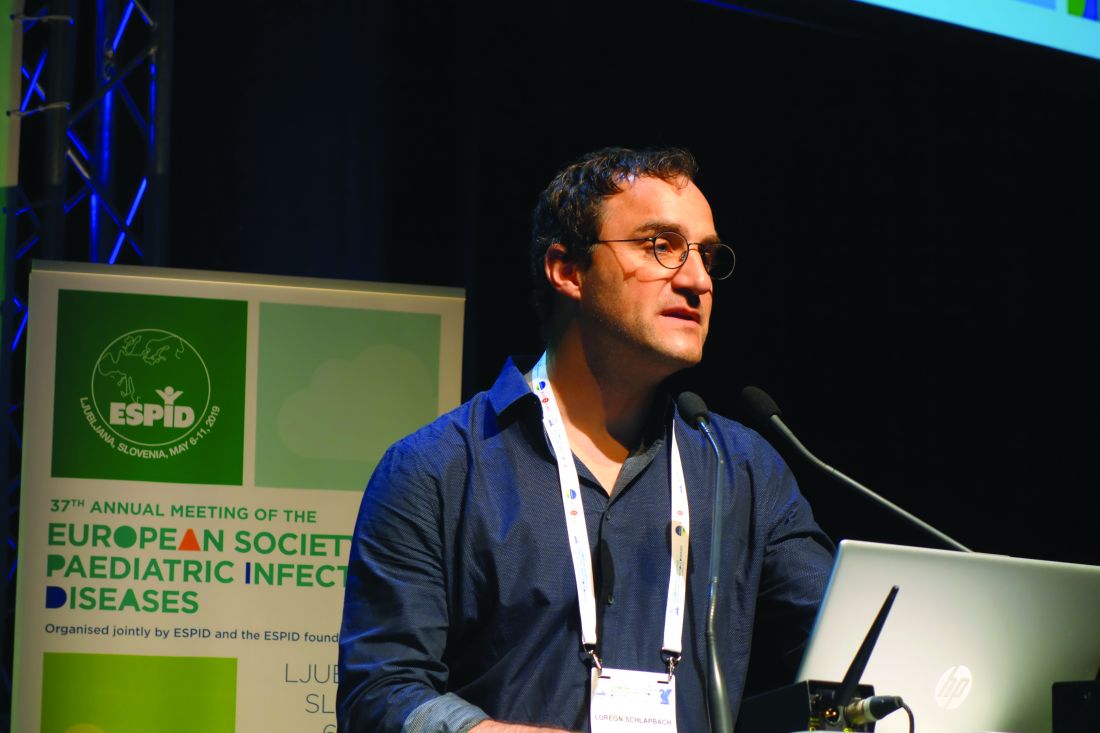
LJUBLJANA, SLOVENIA – The dogma of the “Golden Hour” for the immediate management of pediatric sepsis has been oversold and actually is based upon weak evidence, Luregn J. Schlapbach, MD, asserted at the annual meeting of the European Society for Paediatric Infectious Diseases.
The true Golden Hour – that is, the time frame within which it’s imperative to administer the sepsis bundle comprised of appropriate antibiotics, fluids, and inotropes – is probably more like 3 hours.
“The evidence suggests that up to 3 hours you don’t really have a big difference in outcomes for sepsis. If you recognize shock there’s no question: You should not even wait 1 hour. But if you’re not certain, it may be better to give up to 3 hours to work up the child and get the senior clinician involved before you make decisions about treatment. So I’m not advocating to delay anything, said Dr. Schlapbach, a pediatric intensivist at the Child Health Research Center at the University of Queensland in South Brisbane, Australia.
The problem with a 1-hour mandate for delivery of the sepsis bundle, as recommended in guidelines by the Surviving Sepsis Campaign and the American College of Critical Care Medicine, and endorsed in quality improvement initiatives, is that the time pressure pushes physicians to overprescribe antibiotics to children who don’t actually have a serious bacterial infection. And that, he noted, contributes to the growing problem of antimicrobial resistance.
“You may have a child where you’re not too sure. Usually you would have done a urine culture because UTI [urinary tract infection] is quite a common cause of these infections, and many of these kids aren’t necessarily septic. But if people tell you that within 1 hour you need to treat, are you going to take the time to do the urine culture, or are you just going to decide to treat?” he asked rhetorically.
Dr. Schlapbach is a world-renowned pediatric sepsis researcher. He is far from alone in his reservations about the Golden Hour mandate.
“This is one of the reasons why IDSA [the Infectious Diseases Society of America] has not endorsed the Surviving Sepsis Campaign,” according to the physician, who noted that, in a position statement, IDSA officials have declared that discrimination of sepsis from noninfectious conditions remains a challenge, and that a 60-minute time to antibiotics may jeopardize patient reassessment (Clin Infect Dis. 2018 May 15;66[10]:1631-5).
Dr. Schlapbach highlighted other recent developments in pediatric sepsis.
The definition of adult sepsis has changed, and the pediatric version needs to as well
The revised definition of sepsis, known as Sepsis-3, issued by the International Sepsis Definition Task Force in 2016 notably dropped systemic inflammatory response syndrome (SIRS), as a requirement for sepsis (JAMA. 2016;315[8]:801-10). The revised definition characterizes sepsis as a dysregulated host response to infection resulting in life-threatening organ dysfunction. But Sepsis-3 is based entirely on adult data and is not considered applicable to children.
The current Pediatric Sepsis Consensus Conference definition dates back to 2005. A comprehensive revision is getting underway. It, too, is likely to drop SIRS into the wastebasket, Dr. Schlapbach said.
“It is probably time to abandon the old view of sepsis disease progression, which proposes a progression from infection to SIRS to severe sepsis with organ dysfunction to septic shock, because most children with infection do manifest signs of SIRS, such as tachycardia, tachypnea, and fever, and these probably should be considered as more of an adaptive rather than a maladaptive response,” he explained.
The goal of the pediatric sepsis redefinition project is to come up with something more useful for clinicians than the Sepsis-3 definition. While the Sepsis-3 concept of a dysregulated host response to infection sounds nice, he explained, “we don’t actually know what it is.
“One of the challenges that you all know as pediatricians is that children who develop sepsis get sick very, very quickly. We all have memories of children who we saw and may have discharged, and they were dead 12 hours later,” he noted.
Indeed, he and others have shown in multiple studies that up to 50% of pediatric deaths caused by sepsis happen within 24 hours of presentation.
“So whatever happens, it happens very quickly. The true question for us is actually how and why do children progress from no organ dysfunction, where the mortality is close to zero, to organ dysfunction, where all of a sudden mortality jumps up dramatically. It’s this progression that we don’t understand at all,” according to Dr. Schlapbach.
The genetic contribution to fulminant sepsis in children may be substantial
One-third of pediatric sepsis deaths in high-income countries happen in previously healthy children. In a proof-of-concept study, Dr. Schlapbach and coinvestigators in the Swiss Pediatric Sepsis Study Group conducted exome-sequencing genetic studies in eight previously healthy children with no family history of immunodeficiency who died of severe sepsis because of community-acquired Pseudomonas aeruginosa infection. Two of the eight had rare loss-of-function mutations in genes known to cause primary immunodeficiencies. The investigators proposed that unusually severe sepsis in previously healthy children warrants exome sequencing to look for underlying previously undetected primary immunodeficiencies. That’s important information for survivors and/or affected families to have, they argued (Front Immunol. 2016 Sep 20;7:357. eCollection 2016).
“There are some indications that the genetic contribution in children with sepsis may be larger than previously assumed,” he said.
The longstanding practice of fluid bolus therapy for resuscitation in pediatric sepsis is being reexamined
The FEAST (Fluid Expansion As Supportive Therapy) study, a randomized trial of more than 3,000 children with severe febrile illness and impaired perfusion in sub-Saharan Africa, turned heads with its finding that fluid boluses significantly increased 48-hour mortality (BMC Med. 2013 Mar 14;11:67).
Indeed, the FEAST findings, supported by mechanistic animal studies, were sufficiently compelling that the use of fluid boluses in both pediatric and adult septic shock is now under scrutiny in two major randomized trials: RIFTS (the Restrictive IV Fluid Trial in Severe Sepsis and Septic Shock), and CLOVERS (Crystalloid Liberal or Vasopressors Early Resuscitation in Sepsis). Stay tuned.
Dr. Schlapbach reported having no financial conflicts regarding his presentation.
EXPERT ANALYSIS FROM ESPID 2019
