User login
More Donated Hearts Rejected, Even as Wait List Grows
Only about one in three available hearts was accepted for transplant in the United States in 2010, down from 44% 2 decades ago, researchers reported online Feb. 9 in the American Journal of Transplantation.
The decline stems in part from transplant centers rejecting “marginal” donor hearts, belying a growing need for heart transplants, longer waiting times, and multiple campaigns to expand the use of organs donated for transplantation, said Dr. Kiran Khush of Stanford (Calif.) University and her associates.

The researchers analyzed data on 82,053 potential donor hearts from the Organ Procurement and Transplantation Network. In 1995, 44% of available hearts were accepted for transplant, compared with only 29% in 2006 and 32% in 2010, they found. Meanwhile, rejection rates for donor hearts rose from 37% in 1995 to 52% in 2010, they reported (Am. J. Transplant. 2015 Feb. 10 [doi:10.1111/ajt.13055]).
Several factors might explain the trends, the investigators said. Potential heart donors tended to be older and more often had hypertension and diabetes by the final years of the study period, and transplant centers were less likely to accept hearts from such individuals. Also, mechanical circulatory devices were more commonly used, and centers might hesitate to transplant “marginal” hearts into “stable” recipients of such devices, Dr. Khush and her associates said. Furthermore, government scrutiny of post-transplant outcomes might make centers more conservative when evaluating potential donors, they added.
The study also uncovered regional variations in acceptance rates for donor hearts, with the lowest – about 25%-28% – found primarily in the southeastern United States. “Unfortunately, there are no standard guidelines for donor heart evaluation and acceptance, resulting in considerable inconsistencies in the types of donor hearts that are accepted by different transplant centers, and likely resulting in nonrecovery of potentially useful organs,” the investigators said. The findings “lend support to research and policy efforts aimed at establishing evidence-based criteria for donor heart evaluation and acceptance,” they added.
The work was supported by the National Heart, Lung, and Blood Institute; the National Institute of Diabetes and Digestive and Kidney Diseases; and the Health Resources and Services Administration. The authors reported having no conflicts of interest.
Only about one in three available hearts was accepted for transplant in the United States in 2010, down from 44% 2 decades ago, researchers reported online Feb. 9 in the American Journal of Transplantation.
The decline stems in part from transplant centers rejecting “marginal” donor hearts, belying a growing need for heart transplants, longer waiting times, and multiple campaigns to expand the use of organs donated for transplantation, said Dr. Kiran Khush of Stanford (Calif.) University and her associates.
The researchers analyzed data on 82,053 potential donor hearts from the Organ Procurement and Transplantation Network. In 1995, 44% of available hearts were accepted for transplant, compared with only 29% in 2006 and 32% in 2010, they found. Meanwhile, rejection rates for donor hearts rose from 37% in 1995 to 52% in 2010, they reported (Am. J. Transplant. 2015 Feb. 10 [doi:10.1111/ajt.13055]).
Several factors might explain the trends, the investigators said. Potential heart donors tended to be older and more often had hypertension and diabetes by the final years of the study period, and transplant centers were less likely to accept hearts from such individuals. Also, mechanical circulatory devices were more commonly used, and centers might hesitate to transplant “marginal” hearts into “stable” recipients of such devices, Dr. Khush and her associates said. Furthermore, government scrutiny of post-transplant outcomes might make centers more conservative when evaluating potential donors, they added.
The study also uncovered regional variations in acceptance rates for donor hearts, with the lowest – about 25%-28% – found primarily in the southeastern United States. “Unfortunately, there are no standard guidelines for donor heart evaluation and acceptance, resulting in considerable inconsistencies in the types of donor hearts that are accepted by different transplant centers, and likely resulting in nonrecovery of potentially useful organs,” the investigators said. The findings “lend support to research and policy efforts aimed at establishing evidence-based criteria for donor heart evaluation and acceptance,” they added.
The work was supported by the National Heart, Lung, and Blood Institute; the National Institute of Diabetes and Digestive and Kidney Diseases; and the Health Resources and Services Administration. The authors reported having no conflicts of interest.
FROM THE AMERICAN JOURNAL OF TRANSPLANTATION

More donated hearts rejected, even as wait list grows
Only about one in three available hearts was accepted for transplant in the United States in 2010, down from 44% 2 decades ago, researchers reported online Feb. 9 in the American Journal of Transplantation.
The decline stems in part from transplant centers rejecting “marginal” donor hearts, belying a growing need for heart transplants, longer waiting times, and multiple campaigns to expand the use of organs donated for transplantation, said Dr. Kiran Khush of Stanford (Calif.) University and her associates.

The researchers analyzed data on 82,053 potential donor hearts from the Organ Procurement and Transplantation Network. In 1995, 44% of available hearts were accepted for transplant, compared with only 29% in 2006 and 32% in 2010, they found. Meanwhile, rejection rates for donor hearts rose from 37% in 1995 to 52% in 2010, they reported (Am. J. Transplant. 2015 Feb. 10 [doi:10.1111/ajt.13055]).
Several factors might explain the trends, the investigators said. Potential heart donors tended to be older and more often had hypertension and diabetes by the final years of the study period, and transplant centers were less likely to accept hearts from such individuals. Also, mechanical circulatory devices were more commonly used, and centers might hesitate to transplant “marginal” hearts into “stable” recipients of such devices, Dr. Khush and her associates said. Furthermore, government scrutiny of post-transplant outcomes might make centers more conservative when evaluating potential donors, they added.
The study also uncovered regional variations in acceptance rates for donor hearts, with the lowest – about 25%-28% – found primarily in the southeastern United States. “Unfortunately, there are no standard guidelines for donor heart evaluation and acceptance, resulting in considerable inconsistencies in the types of donor hearts that are accepted by different transplant centers, and likely resulting in nonrecovery of potentially useful organs,” the investigators said. The findings “lend support to research and policy efforts aimed at establishing evidence-based criteria for donor heart evaluation and acceptance,” they added.
The work was supported by the National Heart, Lung, and Blood Institute; the National Institute of Diabetes and Digestive and Kidney Diseases; and the Health Resources and Services Administration. The authors reported having no conflicts of interest.
Only about one in three available hearts was accepted for transplant in the United States in 2010, down from 44% 2 decades ago, researchers reported online Feb. 9 in the American Journal of Transplantation.
The decline stems in part from transplant centers rejecting “marginal” donor hearts, belying a growing need for heart transplants, longer waiting times, and multiple campaigns to expand the use of organs donated for transplantation, said Dr. Kiran Khush of Stanford (Calif.) University and her associates.
The researchers analyzed data on 82,053 potential donor hearts from the Organ Procurement and Transplantation Network. In 1995, 44% of available hearts were accepted for transplant, compared with only 29% in 2006 and 32% in 2010, they found. Meanwhile, rejection rates for donor hearts rose from 37% in 1995 to 52% in 2010, they reported (Am. J. Transplant. 2015 Feb. 10 [doi:10.1111/ajt.13055]).
Several factors might explain the trends, the investigators said. Potential heart donors tended to be older and more often had hypertension and diabetes by the final years of the study period, and transplant centers were less likely to accept hearts from such individuals. Also, mechanical circulatory devices were more commonly used, and centers might hesitate to transplant “marginal” hearts into “stable” recipients of such devices, Dr. Khush and her associates said. Furthermore, government scrutiny of post-transplant outcomes might make centers more conservative when evaluating potential donors, they added.
The study also uncovered regional variations in acceptance rates for donor hearts, with the lowest – about 25%-28% – found primarily in the southeastern United States. “Unfortunately, there are no standard guidelines for donor heart evaluation and acceptance, resulting in considerable inconsistencies in the types of donor hearts that are accepted by different transplant centers, and likely resulting in nonrecovery of potentially useful organs,” the investigators said. The findings “lend support to research and policy efforts aimed at establishing evidence-based criteria for donor heart evaluation and acceptance,” they added.
The work was supported by the National Heart, Lung, and Blood Institute; the National Institute of Diabetes and Digestive and Kidney Diseases; and the Health Resources and Services Administration. The authors reported having no conflicts of interest.
FROM THE AMERICAN JOURNAL OF TRANSPLANTATION
Key clinical point: Acceptance rates of hearts donated for transplantation have declined substantially in the United States.
Major finding: Only 32% of donated hearts were accepted for transplant in 2010, compared with 44% in 1995.
Data source: Analysis of 82,053 potential donor hearts from the Organ Procurement and Transplantation Network.
Disclosures: The study was supported by the National Heart, Lung, and Blood Institute; the National Institute of Diabetes and Digestive and Kidney Diseases; and the Health Resources and Services Administration. The authors reported having no conflicts of interest.

Silver sulfadiazine linked to delayed wound healing in mice
Silver sulfadiazine appeared to disrupt cytokine activity, suppress macrophage recruitment, and inhibit collagen deposition and re-epithelialization in mice with full-thickness burns, the researchers reported.
“Our data suggest a mechanism by which [silver sulfadiazine] impairs wound healing … though further investigations are necessary,” said coauthors Jamie Rosen and Angelo Landriscina and their associates at the Albert Einstein College of Medicine, New York.
Silver sulfadiazine (SSD) is a broad-spectrum antimicrobial used widely for treating second- and third-degree burns. Despite its popularity, some past studies have found it inferior to other wound treatments. To explore why, the investigators created two full-thickness thermal burns each on the backs of 102 mice and treated 46 of the mice with 50 mcL of 1% SSD cream per wound/day. The other mice served as untreated controls (J. Invest. Dermatol. 2015 [doi: 10.1038/jid.2015.21])
Photographs and computerized wound measurements showed that by day 3, the SSD group had greater wound expansion than did the control group (49.7%, compared with 23.8%; P < .0002), said the researchers. Furthermore, the SSD group had only 16.3% wound closure by day 10, compared with 42.1% for controls (P < .005). Cytokine assays also showed that the SSD group also had lower expression of IL-1 and IL-1ra, while histologic studies revealed fewer macrophages and less collagen deposition and re-epithelialization.
“We hypothesize that SSD alters gene expression of IL-1 family genes, resulting in impaired downstream cytokine production and ultimately delayed wound healing,” concluded the authors. But because data in mice do not always translate to humans, more studies would be needed to confirm the mechanism.
The investigators reported no funding sources and declared no relevant financial disclosures.
Key clinical point: Silver sulfadiazine may impair wound healing.
Major finding: By day 10, mice treated with silver sulfadiazine had only 16.2% wound closure, compared with 42.1% for untreated controls (P < .005).
Data source: Histologic, clinical, and cytokine evaluations of 102 mice with full-thickness burns.
Disclosures: The investigators reported no funding sources and declared no relevant financial disclosures.
Contraceptive implant, IUD effective beyond FDA-approved use time
Two long-acting reversible contraceptive devices remained highly effective for about a year beyond their approved durations of use, according to the preliminary results of a study of 500 women.
In the Effectiveness of Prolonged use of IUD/Implant for Contraception (EPIC) study, researchers from Washington University in St. Louis followed women who were continuing their use of two long-acting contraceptive devices beyond the durations approved by the Food and Drug Administration.
Among 237 women who used the etonogestrel implant for a median of 12.5 months beyond the FDA-approved duration of 3 years, there were no unintended pregnancies. There was one unintended pregnancy among the 263 women who used the 52-mg levonorgestrel IUD for a median of 11.7 months beyond the 5-year duration approved by the FDA, the researchers wrote Feb. 5 in Obstetrics & Gynecology (2015;125:599-604).

“The longer a contraceptive method is effective, the bigger the impact it can have,” Dr. Jeffrey F. Peipert, professor of obstetrics and gynecology at Washington University, and the senior researcher on the study, said in a statement. “In the long term, this work has the potential to change how we provide contraceptive methods around the world and can enable women to control their reproductive health and family size.”The study is ongoing, and the researchers are seeking to enroll 400 women in each comparison group, they wrote. The interim analysis included 237 women who used the implant for a total of 229.4 woman-years, and 263 women who used the IUD for 197.7 years.
Etonogestrel levels did not vary with body mass index, and median levels of the hormone consistently exceeded those needed to suppress ovulation. The woman who became pregnant presented with pregnancy symptoms less than 1 month after the FDA-approved duration of use had expired. On examination, she had a partially expelled IUD with the stem visible outside the cervix, the researchers wrote.
The research was supported by the Society of Family Planning, the National Center for Advancing Translational Sciences, and the National Institutes of Health. The study’s senior researcher, Dr. Peipert, reported receiving research support from Bayer, Teva, and Merck, and serving on advisory boards for Teva, Bayer, and Watson/Activitis. The other researchers reporting having no financial disclosures.
Two long-acting reversible contraceptive devices remained highly effective for about a year beyond their approved durations of use, according to the preliminary results of a study of 500 women.
In the Effectiveness of Prolonged use of IUD/Implant for Contraception (EPIC) study, researchers from Washington University in St. Louis followed women who were continuing their use of two long-acting contraceptive devices beyond the durations approved by the Food and Drug Administration.
Among 237 women who used the etonogestrel implant for a median of 12.5 months beyond the FDA-approved duration of 3 years, there were no unintended pregnancies. There was one unintended pregnancy among the 263 women who used the 52-mg levonorgestrel IUD for a median of 11.7 months beyond the 5-year duration approved by the FDA, the researchers wrote Feb. 5 in Obstetrics & Gynecology (2015;125:599-604).
“The longer a contraceptive method is effective, the bigger the impact it can have,” Dr. Jeffrey F. Peipert, professor of obstetrics and gynecology at Washington University, and the senior researcher on the study, said in a statement. “In the long term, this work has the potential to change how we provide contraceptive methods around the world and can enable women to control their reproductive health and family size.”The study is ongoing, and the researchers are seeking to enroll 400 women in each comparison group, they wrote. The interim analysis included 237 women who used the implant for a total of 229.4 woman-years, and 263 women who used the IUD for 197.7 years.
Etonogestrel levels did not vary with body mass index, and median levels of the hormone consistently exceeded those needed to suppress ovulation. The woman who became pregnant presented with pregnancy symptoms less than 1 month after the FDA-approved duration of use had expired. On examination, she had a partially expelled IUD with the stem visible outside the cervix, the researchers wrote.
The research was supported by the Society of Family Planning, the National Center for Advancing Translational Sciences, and the National Institutes of Health. The study’s senior researcher, Dr. Peipert, reported receiving research support from Bayer, Teva, and Merck, and serving on advisory boards for Teva, Bayer, and Watson/Activitis. The other researchers reporting having no financial disclosures.
Key clinical point: The etonogestrel implant and the 52-mg levonorgestrel intrauterine device were safe and effective for a year beyond their FDA-approved durations of use.
Major finding: None of the women who used the implant, and only one who used the IUD, reported unintended pregnancies.
Data source: Prospective, observational study of 237 women who used the implant and 263 who used the IUD beyond the FDA-approved durations of use.
Disclosures: The research was supported by the Society of Family Planning, the National Center for Advancing Translational Sciences, and the National Institutes of Health. The senior researcher, Dr. Peipert, reported receiving research support from Bayer, Teva, and Merck, and serving on advisory boards for Teva, Bayer, and Watson/Activitis. The other researchers reported having no financial disclosures.

ACOG, SMFM propose definitions for maternal care sites
In a first-time consensus document, the American College of Obstetricians and Gynecologists and the Society for Maternal-Fetal Medicine have proposed a new classification system for maternal care facilities that includes minimum standards for each level of care.
Implementing these definitions nationally could help decrease maternal and fetal morbidity and mortality, improve regional availability of care for high-risk pregnant women, and enhance the collection of related data, according to the two organizations. The consensus document appears in the February issue of Obstetrics & Gynecology (Obstet. Gynecol. 2015;125:502-15).

The classification of maternal facilities mirrors the model already in use for neonatal care facilities and comes as U.S. maternal mortality rates have climbed. The United States now ranks 60th in the world for maternal mortality, the document notes.
“It is essential to remember that, when we are addressing obstetrical outcomes, we have two very important patients: mother and child,” Dr. Sarah J. Kilpatrick, chair of the department of obstetrics and gynecology at Cedars-Sinai Medical Center in Los Angeles and lead author of the document, said in a statement. “Our goal for these consensus recommendations is to create a system for maternal care that complements and supplements the current neonatal framework in order to reduce maternal morbidity and mortality across the country.”
The consensus document defines five categories of maternal care centers:
Level 1 (basic care) facilities have the same capacities as birth centers but also can perform common emergency procedures, such as unplanned cesarean deliveries and massive transfusions. They must have the appropriate blood products and analgesia and anesthesia services. Unlike birth centers, level 1 facilities can readily handle term deliveries of twins, preeclampsia at term that is not severe, and uncomplicated cesarean deliveries. Managers of these facilities are able to create formal protocols for transferring high-risk patients, as well as education and quality improvement programs.
Level 2 (specialty care) facilities are able to handle moderately complex cases, such as severe preeclampsia or placental previa without prior uterine surgery. These facilities have an attending ob.gyn. available at all times, plus in-person or remote access to a maternal-fetal medicine subspecialist. They also have basic ultrasound equipment, equipment for obese women such as bariatric beds, and CT scanning equipment. They ideally have MRI scanning and interpretation, according to the consensus document.
Level 3 (subspecialty care) facilities meet level 2 criteria and also have continuous, on-site intensive care, maternal-fetal medicine services that are led by a maternal-fetal subspecialist, and advanced imaging and ventilator equipment. These facilities may act as regional referral centers and are appropriate for women with suspected placenta accreta or placenta previa with prior uterine surgery, suspected placenta percreta, adult respiratory syndrome, or severe eclampsia at less than 34 weeks’ gestation.
Level 4 (regional perinatal health care) facilities offer medical and surgical care for the most critical cases, including severe pulmonary hypertension; liver failure; and conditions that require organ transplantation, neurosurgery, or cardiac surgery. These facilities have continuously available maternal-fetal medicine care teams that are highly experienced in handling pregnant and postpartum women in critical condition. Level 4 facilities also coordinate referral and transport, provide outreach education for local facilities and providers, and help collect and analyze regional data for quality improvement programs.
Leaders at these facilities should understand their sites’ capabilities and limitations and have a “well-defined threshold for transferring women to health care facilities that offer a higher level of care,” ACOG and SMFM wrote. The organizations also called on state and regional authorities to partner with maternal care facilities to coordinate a system of care.
The new consensus statement does not address high-risk neonatal care, but such care must be coordinated with maternal care, ACOG and SMFM officials wrote.
Several groups endorsed the consensus definitions, including the American Association of Birth Centers; the American College of Nurse-Midwives; the Association of Women’s Health, Obstetric and Neonatal Nurses; and the Commission for the Accreditation of Birth Centers. The authors reported having no financial disclosures.
In a first-time consensus document, the American College of Obstetricians and Gynecologists and the Society for Maternal-Fetal Medicine have proposed a new classification system for maternal care facilities that includes minimum standards for each level of care.
Implementing these definitions nationally could help decrease maternal and fetal morbidity and mortality, improve regional availability of care for high-risk pregnant women, and enhance the collection of related data, according to the two organizations. The consensus document appears in the February issue of Obstetrics & Gynecology (Obstet. Gynecol. 2015;125:502-15).
The classification of maternal facilities mirrors the model already in use for neonatal care facilities and comes as U.S. maternal mortality rates have climbed. The United States now ranks 60th in the world for maternal mortality, the document notes.
“It is essential to remember that, when we are addressing obstetrical outcomes, we have two very important patients: mother and child,” Dr. Sarah J. Kilpatrick, chair of the department of obstetrics and gynecology at Cedars-Sinai Medical Center in Los Angeles and lead author of the document, said in a statement. “Our goal for these consensus recommendations is to create a system for maternal care that complements and supplements the current neonatal framework in order to reduce maternal morbidity and mortality across the country.”
The consensus document defines five categories of maternal care centers:
Level 1 (basic care) facilities have the same capacities as birth centers but also can perform common emergency procedures, such as unplanned cesarean deliveries and massive transfusions. They must have the appropriate blood products and analgesia and anesthesia services. Unlike birth centers, level 1 facilities can readily handle term deliveries of twins, preeclampsia at term that is not severe, and uncomplicated cesarean deliveries. Managers of these facilities are able to create formal protocols for transferring high-risk patients, as well as education and quality improvement programs.
Level 2 (specialty care) facilities are able to handle moderately complex cases, such as severe preeclampsia or placental previa without prior uterine surgery. These facilities have an attending ob.gyn. available at all times, plus in-person or remote access to a maternal-fetal medicine subspecialist. They also have basic ultrasound equipment, equipment for obese women such as bariatric beds, and CT scanning equipment. They ideally have MRI scanning and interpretation, according to the consensus document.
Level 3 (subspecialty care) facilities meet level 2 criteria and also have continuous, on-site intensive care, maternal-fetal medicine services that are led by a maternal-fetal subspecialist, and advanced imaging and ventilator equipment. These facilities may act as regional referral centers and are appropriate for women with suspected placenta accreta or placenta previa with prior uterine surgery, suspected placenta percreta, adult respiratory syndrome, or severe eclampsia at less than 34 weeks’ gestation.
Level 4 (regional perinatal health care) facilities offer medical and surgical care for the most critical cases, including severe pulmonary hypertension; liver failure; and conditions that require organ transplantation, neurosurgery, or cardiac surgery. These facilities have continuously available maternal-fetal medicine care teams that are highly experienced in handling pregnant and postpartum women in critical condition. Level 4 facilities also coordinate referral and transport, provide outreach education for local facilities and providers, and help collect and analyze regional data for quality improvement programs.
Leaders at these facilities should understand their sites’ capabilities and limitations and have a “well-defined threshold for transferring women to health care facilities that offer a higher level of care,” ACOG and SMFM wrote. The organizations also called on state and regional authorities to partner with maternal care facilities to coordinate a system of care.
The new consensus statement does not address high-risk neonatal care, but such care must be coordinated with maternal care, ACOG and SMFM officials wrote.
Several groups endorsed the consensus definitions, including the American Association of Birth Centers; the American College of Nurse-Midwives; the Association of Women’s Health, Obstetric and Neonatal Nurses; and the Commission for the Accreditation of Birth Centers. The authors reported having no financial disclosures.

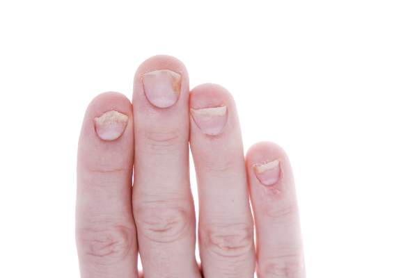
Psoriasis Treatment Recommendations Address Four Clinical Scenarios
New guidelines on nail psoriasis address four clinical manifestations of the disease. The recommendations by the Medical Board of the National Psoriasis Foundation appeared as a consensus statement in the January issue of JAMA Dermatology.
Limitations in clinical trial data make comparing treatments difficult, noted lead author Dr. Jeffrey J. Crowley of Bakersfield (Calif.) Dermatology and his associates. “There are limited data to evaluate or support the use of combination therapy in nail psoriasis. Thus, treatment options recommended in this review are monotherapy,” the guidelines authors added (JAMA Dermatol. 2015;151:87-94).
To develop the guidelines, the research team searched PubMed for articles on nail psoriasis dating from Jan. 1, 1947 through May 11, 2014. They evaluated these studies for level of evidence based on recommendations for writing guidelines from Dr. Paul G. Shekelle of the VA West Los Angeles Medical Center and his associates (BMJ 1999;318:593-6).
They also polled the Medical Board of the National Psoriasis Foundation regarding their treatment approach for four clinical presentations of nail psoriasis:
• For treatment-naive patients with psoriasis of the nails only (affecting at least 3 of 10 fingernails), the board recommended initial treatment with high-potency topical corticosteroids (with or without calcipotriol), with intralesional corticosteroids as a secondary option. Intralesional corticosteroids have been used for decades, but clinical data supporting their use are “extremely limited,” the guidelines state.
• For extensive nail psoriasis (affecting at least five fingernails and causing moderate to severe pain) that has failed topical treatment, the board recommended adalimumab most enthusiastically, followed by etanercept, intralesional corticosteroids, ustekinumab, methotrexate sodium, and acitretin in decreasing order.
• For concurrent skin and nail disease without joint involvement (defined as skin disease on at least 8% of the body surface and moderately to severely painful dystrophy of at least 5 of 10 nails), the board strongly recommended adalimumab, etanercept, and ustekinumab, and also recommended methotrexate, acitretin, infliximab, and apremilast.
• For concurrent nail, skin, and joint involvement (defined as skin disease on 8% of the body surface, a history of dactylitis and morning stiffness (psoriatic arthritis), and severe, painful involvement of at least 5 of 10 nails), the board most strongly recommended adalimumab, followed by etanercept, ustekinumab, infliximab, methotrexate, apremilast, and golimumab.
Continue for study findings >>
Because nails grow slowly, psoriatic joint and skin disease often improve before nail psoriasis does, the authors noted. “Few studies show any significant improvement before 12 weeks, and several studies with etanercept, infliximab, and ustekinumab demonstrate continued improvement beyond 6 months,” they wrote.
About half of patients with psoriasis have some amount of nail involvement, and about 70% of patients with psoriatic arthritis have nail disease, according to the literature review. Dermatophyte infections can further complicate treatment of nail psoriasis, and immunosuppressive therapies can lead to onychomycosis in patients whose psoriasis includes the toenails, the authors added.
Dr. Crowley reported speaker and consulting honoraria from AbbVie, Abbott, and Amgen, and research funding from Abbott, AbbVie, Amgen, AstraZeneca, Celgene, Eli Lilly, Janssen Pharmaceutica, Merck, Pfizer, and Regeneron Pharmaceuticals. Four coauthors reported advisory, consulting, or financial relationships with Amgen, Abbott, Janssen Biotech Inc., Celgene, Novartis International AG, Abbvie, Merck, Celgene, Leo Pharma, Eli Lilly, Pfizer, and the National Psoriasis Foundation.
FROM JAMA DERMATOLOGY
Psoriasis treatment recommendations address four clinical scenarios
New guidelines on nail psoriasis address four clinical manifestations of the disease. The recommendations by the Medical Board of the National Psoriasis Foundation appeared as a consensus statement in the January issue of JAMA Dermatology.
Limitations in clinical trial data make comparing treatments difficult, noted lead author Dr. Jeffrey J. Crowley of Bakersfield (Calif.) Dermatology and his associates. “There are limited data to evaluate or support the use of combination therapy in nail psoriasis. Thus, treatment options recommended in this review are monotherapy,” the guidelines authors added (JAMA Dermatol. 2015;151:87-94).
To develop the guidelines, the research team searched PubMed for articles on nail psoriasis dating from Jan. 1, 1947 through May 11, 2014. They evaluated these studies for level of evidence based on recommendations for writing guidelines from Dr. Paul G. Shekelle of the VA West Los Angeles Medical Center and his associates (BMJ 1999;318:593-6).
They also polled the Medical Board of the National Psoriasis Foundation regarding their treatment approach for four clinical presentations of nail psoriasis:
• For treatment-naive patients with psoriasis of the nails only (affecting at least 3 of 10 fingernails), the board recommended initial treatment with high-potency topical corticosteroids (with or without calcipotriol), with intralesional corticosteroids as a secondary option. Intralesional corticosteroids have been used for decades, but clinical data supporting their use are “extremely limited,” the guidelines state.
• For extensive nail psoriasis (affecting at least five fingernails and causing moderate to severe pain) that has failed topical treatment, the board recommended adalimumab most enthusiastically, followed by etanercept, intralesional corticosteroids, ustekinumab, methotrexate sodium, and acitretin in decreasing order.
• For concurrent skin and nail disease without joint involvement (defined as skin disease on at least 8% of the body surface and moderately to severely painful dystrophy of at least 5 of 10 nails), the board strongly recommended adalimumab, etanercept, and ustekinumab, and also recommended methotrexate, acitretin, infliximab, and apremilast.
• For concurrent nail, skin, and joint involvement (defined as skin disease on 8% of the body surface, a history of dactylitis and morning stiffness (psoriatic arthritis), and severe, painful involvement of at least 5 of 10 nails), the board most strongly recommended adalimumab, followed by etanercept, ustekinumab, infliximab, methotrexate, apremilast, and golimumab.
Because nails grow slowly, psoriatic joint and skin disease often improve before nail psoriasis does, the authors noted. “Few studies show any significant improvement before 12 weeks, and several studies with etanercept, infliximab, and ustekinumab demonstrate continued improvement beyond 6 months,” they wrote.
About half of patients with psoriasis have some amount of nail involvement, and about 70% of patients with psoriatic arthritis have nail disease, according to the literature review. Dermatophyte infections can further complicate treatment of nail psoriasis, and immunosuppressive therapies can lead to onychomycosis in patients whose psoriasis includes the toenails, the authors added.
Dr. Crowley reported speaker and consulting honoraria from AbbVie, Abbott, and Amgen, and research funding from Abbott, AbbVie, Amgen, AstraZeneca, Celgene, Eli Lilly, Janssen Pharmaceutica, Merck, Pfizer, and Regeneron Pharmaceuticals. Four coauthors reported advisory, consulting, or financial relationships with Amgen, Abbott, Janssen Biotech Inc., Celgene, Novartis International AG, Abbvie, Merck, Celgene, Leo Pharma, Eli Lilly, Pfizer, and the National Psoriasis Foundation.
FROM JAMA DERMATOLOGY

CHA2DS2-VASc score of 1 linked to lower stroke risk than previously reported
Patients with atrial fibrillation who had CHA2DS2-VASc scores of 1 were at lower risk of ischemic stroke than previously reported, according to a retrospective analysis of hospital registry data. The research appeared online January 19 in the Journal of American College of Cardiology.
Depending on the definition of stroke used, risk was 0.1% to 0.2% in women and 0.5% to 0.7% in men – so low that oral anticoagulants (OACs) would not be expected to benefit patients of either sex, said Dr. Leif Friberg at the Karolinska Institute in Stockholm and his associates. Past studies had potentially overestimated the risk of stroke in this population, which “may have led to unnecessary, and potentially harmful, OAC treatment of low-risk patients,” they said.

European and U.S. guidelines both recommend using the CHA2DS2-VASc (heart failure, hypertension, age ≥75, diabetes mellitus, prior stroke or transient ischemic attack, vascular disease, age 65-74 years, female) scoring system to assess stroke risk in patients with atrial fibrillation (AF). But past studies have reported a threefold variation (ranging from 0.6% to greater than 2.0%) in stroke risk among AF patients with CHA2DS2-VASc scores of 1 who were not receiving OAC, the researchers noted. Anticoagulation therapy is likely to benefit AF patients whose annual risk of stroke exceeds 1%, but not patients whose risk is only 0.6%, they added.
Their study, which included 140,420 patients in Sweden with nonvalvular AF, assessed the effect of varying definitions of stroke on estimates of stroke risk. Using a broad definition that included ischemic stroke, transient ischemic attack (TIA), and pulmonary embolism led to a 44% greater annual risk of stroke than if only ischemic strokes were considered, the investigators reported. They disagreed with classifying pulmonary embolism events and TIAs as strokes, as some past studies have done. “Primary prevention of pulmonary embolism among patients with AF has, to the best of our knowledge, not been studied and is not an approved indication for OAC treatment,” they said. “We also did not find it relevant to count TIA as an endpoint in studies that describe stroke risk. As a diagnosis, TIA is difficult to validate.”
Several Swedish foundations supported the study. Dr. Friberg reported no relevant financial conflicts of interest.
Given the current state of knowledge, patients with atrial fibrillation who are younger than 65 years but have a CHA2DS2-VASc score of 1 are unlikely to benefit from anticoagulation therapy.
Dr. Friberg and his colleagues make two important observations regarding risk score thresholds for oral anticoagulant therapy. First, they highlight the wide cohort-to-cohort variation in reported CHA2DS2-VASc–stratified rates of stroke for atrial fibrillation patients who are not anticoagulated. Second, they reveal how sensitive estimates of stroke rates are to variations in interrogating administrative databases, which are used repeatedly as sources of “real world” rates of stroke. They conclude that the true stroke rate for patients with a CHA2DS2-VASc score of 1 is less than 0.7% per year, too low for oral anticoagulant therapy to benefit patients with AF.
Going forward, guideline writers should be aware of the drawbacks of the CHA2DS2-VASc score. They should focus on the absolute rates of stroke corresponding to risk prediction point scores and be alert to potential biases in studies reporting these rates. Investigators should work to harmonize methods for analyzing large AF databases. If variation in reported rates cannot be reconciled, then recommendations should reflect this uncertainty.
Dr. Daniel E. Singer is at Harvard Medical School in Boston, and Dr. Michael D. Ezekowitz is at Sidney Kimmel Medical College at Thomas Jefferson University in Philadelphia. Dr. Singer has been a consultant to, advised, and or received research funding from Boehringer Ingelheim, Bristol-Myers Squibb, Johnson & Johnson, Merck, and St. Jude Medical, and Medtronic. Dr. Ezekowitz reported having been a consultant and advisory board member for all those companies and several others. These remarks were taken from their editorial accompanying Dr. Friberg’s report (J. Am. Coll. Cardiol. 2015 Jan. 19 [doi:10.1016/j.jacc.2014.11.013]).
Patients with atrial fibrillation who had CHA2DS2-VASc scores of 1 were at lower risk of ischemic stroke than previously reported, according to a retrospective analysis of hospital registry data. The research appeared online January 19 in the Journal of American College of Cardiology.
Depending on the definition of stroke used, risk was 0.1% to 0.2% in women and 0.5% to 0.7% in men – so low that oral anticoagulants (OACs) would not be expected to benefit patients of either sex, said Dr. Leif Friberg at the Karolinska Institute in Stockholm and his associates. Past studies had potentially overestimated the risk of stroke in this population, which “may have led to unnecessary, and potentially harmful, OAC treatment of low-risk patients,” they said.
European and U.S. guidelines both recommend using the CHA2DS2-VASc (heart failure, hypertension, age ≥75, diabetes mellitus, prior stroke or transient ischemic attack, vascular disease, age 65-74 years, female) scoring system to assess stroke risk in patients with atrial fibrillation (AF). But past studies have reported a threefold variation (ranging from 0.6% to greater than 2.0%) in stroke risk among AF patients with CHA2DS2-VASc scores of 1 who were not receiving OAC, the researchers noted. Anticoagulation therapy is likely to benefit AF patients whose annual risk of stroke exceeds 1%, but not patients whose risk is only 0.6%, they added.
Their study, which included 140,420 patients in Sweden with nonvalvular AF, assessed the effect of varying definitions of stroke on estimates of stroke risk. Using a broad definition that included ischemic stroke, transient ischemic attack (TIA), and pulmonary embolism led to a 44% greater annual risk of stroke than if only ischemic strokes were considered, the investigators reported. They disagreed with classifying pulmonary embolism events and TIAs as strokes, as some past studies have done. “Primary prevention of pulmonary embolism among patients with AF has, to the best of our knowledge, not been studied and is not an approved indication for OAC treatment,” they said. “We also did not find it relevant to count TIA as an endpoint in studies that describe stroke risk. As a diagnosis, TIA is difficult to validate.”
Several Swedish foundations supported the study. Dr. Friberg reported no relevant financial conflicts of interest.
FROM THE JOURNAL OF THE AMERICAN COLLEGE OF CARDIOLOGY
Key clinical point: Stroke risk was low in patients with atrial fibrillation and a CHA2DS2-VASc score of 1.
Major finding: Risk of stroke was 0.1% to 0.2% in women and 0.5% to 0.7% in men.
Data source: Retrospective study of 140,420 patients with nonvalvular AF.
Disclosures: Several Swedish foundations supported the study. Dr. Friberg reported no relevant financial conflicts of interest.

Comorbid substance abuse, psychiatric disorders linked to changes in frontal brain connectivity
Substance abuse by psychiatric inpatients correlated with significantly increased interhemispheric connectivity in two frontal brain areas, the insula and the inferior frontal gyri, according to a case-control study published Jan. 12 in Neuropharmacology.
The results add to evidence linking the insula and the inferior frontal gyri (iFG) with substance abuse disorders (SUD), said Humsini Viswanath at Baylor College of Medicine, Houston, and her coauthors. “We postulate that specific drugs of abuse act upon anatomic, metabolic, or molecular pathways in the insula and iFG, resulting in the observed changes in functional and structural connectivity,” the investigators wrote.

Abuse of tobacco, alcohol, cocaine, sedatives, and hallucinogens was linked with increased resting state functional connectivity (RSFC) of the insula, while use of amphetamines and inhalants correlated with increased RSFC of the iFG, the researchers said. Use of cannabinoids and opiates did not correlate with brain connectivity in either structure, they added (Neuropharmacology 2015 Jan. 12 [doi:10.1016/j.neuropharm.2014.12.030]).
Substance abuse disorders are the leading cause of death among individuals with psychiatric disorders, but studies of mental illness often exclude patients with comorbid SUD, the investigators noted. They therefore used diffusion tensor and RSFC imaging to compare 151 psychiatric inpatients with and without concurrent SUDs, as determined by the World Health Organization Alcohol, Smoking, and Substance Involvement Screening Test (WHOA).
About the same proportion of patients with and without SUDs had depression (38% vs. 40%, respectively), anxiety disorders (50% vs. 53%), bipolar disorders (17% vs. 14%), and personality disorders (41% vs. 37%). But patients with SUDs had increased interhemispheric RSFC for the insula (P < .001) and inferior frontal gyrus (P < .05) and had significantly smaller right inferior frontal gyri, compared with psychiatric inpatients without comorbid SUD, they reported.
Studies of heterogeneous groups of patients “can reveal significant changes in brain structure and function, even in highly comorbid samples,” concluded Ms. Viswanath and her associates. “As additional patients are recruited to this cohort for future studies, we expect to identify even more fine-grained relationships between specific drugs of abuse and brain structure, function, and connectivity in psychiatric conditions.”
The research was supported by the McNair Medical Institute, the Menninger Clinic, the department of psychiatry at Baylor College of Medicine, the National Institutes of Health, and the Brain & Behavior Research Foundation. The researchers declared no financial conflicts.
Substance abuse by psychiatric inpatients correlated with significantly increased interhemispheric connectivity in two frontal brain areas, the insula and the inferior frontal gyri, according to a case-control study published Jan. 12 in Neuropharmacology.
The results add to evidence linking the insula and the inferior frontal gyri (iFG) with substance abuse disorders (SUD), said Humsini Viswanath at Baylor College of Medicine, Houston, and her coauthors. “We postulate that specific drugs of abuse act upon anatomic, metabolic, or molecular pathways in the insula and iFG, resulting in the observed changes in functional and structural connectivity,” the investigators wrote.
Abuse of tobacco, alcohol, cocaine, sedatives, and hallucinogens was linked with increased resting state functional connectivity (RSFC) of the insula, while use of amphetamines and inhalants correlated with increased RSFC of the iFG, the researchers said. Use of cannabinoids and opiates did not correlate with brain connectivity in either structure, they added (Neuropharmacology 2015 Jan. 12 [doi:10.1016/j.neuropharm.2014.12.030]).
Substance abuse disorders are the leading cause of death among individuals with psychiatric disorders, but studies of mental illness often exclude patients with comorbid SUD, the investigators noted. They therefore used diffusion tensor and RSFC imaging to compare 151 psychiatric inpatients with and without concurrent SUDs, as determined by the World Health Organization Alcohol, Smoking, and Substance Involvement Screening Test (WHOA).
About the same proportion of patients with and without SUDs had depression (38% vs. 40%, respectively), anxiety disorders (50% vs. 53%), bipolar disorders (17% vs. 14%), and personality disorders (41% vs. 37%). But patients with SUDs had increased interhemispheric RSFC for the insula (P < .001) and inferior frontal gyrus (P < .05) and had significantly smaller right inferior frontal gyri, compared with psychiatric inpatients without comorbid SUD, they reported.
Studies of heterogeneous groups of patients “can reveal significant changes in brain structure and function, even in highly comorbid samples,” concluded Ms. Viswanath and her associates. “As additional patients are recruited to this cohort for future studies, we expect to identify even more fine-grained relationships between specific drugs of abuse and brain structure, function, and connectivity in psychiatric conditions.”
The research was supported by the McNair Medical Institute, the Menninger Clinic, the department of psychiatry at Baylor College of Medicine, the National Institutes of Health, and the Brain & Behavior Research Foundation. The researchers declared no financial conflicts.
FROM NEUROPHARMACOLOGY
Key clinical point: The results add to evidence linking the insula and the inferior frontal gyri with substance abuse disorders.
Major finding: Substance abuse correlated with increased interhemispheric resting state functional connectivity for the insula (P < .001) and inferior frontal gyrus (P < .05).
Data source: Structured interviews and structural and functional brain imaging of 151 inpatients a single psychiatric clinic.
Disclosures: The research was supported by the McNair Medical Institute, the Menninger Clinic, the department of psychiatry at Baylor College of Medicine, the National Institutes of Health, and the Brain & Behavior Research Foundation. The researchers declared no financial conflicts.

RNA sequencing characterized high-risk squamous cell carcinomas
SAN DIEGO – Cutaneous squamous cell carcinomas from organ transplant recipients had a more aggressive molecular profile than did tumor samples from immunocompetent patients, according to an RNA sequencing study presented at the annual meeting of the American Society for Dermatologic Surgery.
Specimens from organ transplant recipients showed greater induction of biologic pathways related to cancer signaling, fibrosis, and extracellular matrix remodeling, said Dr. Cameron Chesnut, a dermatologist in private practice in Spokane, Wash., who carried out the research while he was a dermatologic surgery resident at the University of California, Los Angeles.
Furthermore, the TP53 tumor suppressor gene was inhibited at least five times more in samples from organ transplant recipients, compared with those from immunocompetent patients, Dr. Chesnut said in an interview.
Squamous cell carcinoma (SCC) is the most common cancer to occur after organ transplantation, Dr. Chesnut and his associates noted. The malignancy is 65-250 times more common, is more than 4 times more likely to metastasize, and has a mortality rate of 5% compared with a rate of less than 1% in immunocompetent patients, based on data published online in the journal F1000 Prime Reports, they said.
To characterize these high-risk SCCs and compare them with lower-risk SCCs, the researchers performed RNA sequencing of three normal skin samples and SCC specimens from 15 patients – 7 organ transplant recipients and 8 otherwise healthy individuals. The researchers used an Illumina GAIIx RNA Seq instrument to generate RNA sequencing libraries of the specimens. They also used the web-based Ingenuity Pathway Analysis technique to identify the major biological pathways regulated within the tumors.
In all, 690 highly expressed genes were induced at least fivefold in SCCs from organ transplant recipients compared with those from otherwise healthy patients. These genes encoded pathways related to fibrosis, extracellular remodeling, the cell cycle, and tumor signaling, the investigators said. The COX-2 pathway for prostaglandin synthesis also was induced fivefold or more in the high-risk SCCs compared with those from immunocompetent patients, Dr. Chesnut added.
The researchers also identified 1,290 highly expressed genes that were inhibited at least fivefold in SCCs from organ transplant recipients compared with specimens from immunocompetent patients. The most strongly inhibited pathways were related to sterol biosynthesis and epithelial differentiation, followed by nucleotide excision repair, interleukin-6 and IL-17, and apoptosis, they said.
Based on these findings, novel therapeutics might someday be able to target specific biologic pathways that are highly induced in SCCs from organ transplant recipients, Dr. Chesnut said. “It’s hard to say what the most likely candidates are,” but based on the study findings, “regulating inflammation may be a target,” he added. Dr. Chesnut and his associates reported no external funding sources or conflicts of interest.
Key clinical point: Squamous cell carcinomas from organ transplant recipients showed a more aggressive molecular profile than did those from immunocompetent individuals.
Major finding: The high-risk tumors showed greater induction of biologic pathways related to cancer signaling, fibrosis, and extracellular matrix remodeling, and inhibition of the tp53 tumor suppressor gene.
Data source: RNA sequencing of 15 squamous cell carcinomas, including seven from organ transplant recipients.
Disclosures: The investigators reported no external funding sources or conflicts of interest.