User login
Long-term safety and tolerability of atogepant in episodic migraine
Key clinical point: The safety and tolerability of once-daily atogepant observed over 40 weeks in this extension trial aligns with profiles from the pivotal phase 3 trials with no new safety signals identified in patients with episodic migraine.
Major finding: Nearly 63% of patients reported treatment-emergent adverse events, most being mild or moderate, with upper respiratory tract infection (5.5%) and urinary tract infection (5.3%) being most frequent. Treatment discontinuation rates due to lack of efficacy (0.6%) or adverse events (3.6%) were low. No deaths were reported.
Study details: Findings are from the 309-OLEX trial, an open-label extension of phase 3 ADVANCE trial, including 685 patients with episodic migraine with or without aura who received 60 mg atogepant once daily for 40 weeks.
Disclosures: This study was supported by AbbVie Inc. (formerly Allergan). Five authors declared being full-time or former employees of or holding stock or stock options in AbbVie. Several authors reported ties with various sources, including AbbVie.
Source: Klein BC et al. Safety and tolerability results of atogepant for the preventive treatment of episodic migraine from a 40-week, open-label multicenter extension of the phase 3 ADVANCE trial. Cephalalgia. 2023 (Jan 9). Doi: 10.1177/03331024221128250
Key clinical point: The safety and tolerability of once-daily atogepant observed over 40 weeks in this extension trial aligns with profiles from the pivotal phase 3 trials with no new safety signals identified in patients with episodic migraine.
Major finding: Nearly 63% of patients reported treatment-emergent adverse events, most being mild or moderate, with upper respiratory tract infection (5.5%) and urinary tract infection (5.3%) being most frequent. Treatment discontinuation rates due to lack of efficacy (0.6%) or adverse events (3.6%) were low. No deaths were reported.
Study details: Findings are from the 309-OLEX trial, an open-label extension of phase 3 ADVANCE trial, including 685 patients with episodic migraine with or without aura who received 60 mg atogepant once daily for 40 weeks.
Disclosures: This study was supported by AbbVie Inc. (formerly Allergan). Five authors declared being full-time or former employees of or holding stock or stock options in AbbVie. Several authors reported ties with various sources, including AbbVie.
Source: Klein BC et al. Safety and tolerability results of atogepant for the preventive treatment of episodic migraine from a 40-week, open-label multicenter extension of the phase 3 ADVANCE trial. Cephalalgia. 2023 (Jan 9). Doi: 10.1177/03331024221128250
Key clinical point: The safety and tolerability of once-daily atogepant observed over 40 weeks in this extension trial aligns with profiles from the pivotal phase 3 trials with no new safety signals identified in patients with episodic migraine.
Major finding: Nearly 63% of patients reported treatment-emergent adverse events, most being mild or moderate, with upper respiratory tract infection (5.5%) and urinary tract infection (5.3%) being most frequent. Treatment discontinuation rates due to lack of efficacy (0.6%) or adverse events (3.6%) were low. No deaths were reported.
Study details: Findings are from the 309-OLEX trial, an open-label extension of phase 3 ADVANCE trial, including 685 patients with episodic migraine with or without aura who received 60 mg atogepant once daily for 40 weeks.
Disclosures: This study was supported by AbbVie Inc. (formerly Allergan). Five authors declared being full-time or former employees of or holding stock or stock options in AbbVie. Several authors reported ties with various sources, including AbbVie.
Source: Klein BC et al. Safety and tolerability results of atogepant for the preventive treatment of episodic migraine from a 40-week, open-label multicenter extension of the phase 3 ADVANCE trial. Cephalalgia. 2023 (Jan 9). Doi: 10.1177/03331024221128250
Pre-pregnancy migraine history not a significant risk factor for spontaneous abortion
Key clinical point: A preconception history of migraine showed no significant association with the risk for spontaneous abortion (SAB); however, routine use of medication, suggesting more severe migraine, may confer a greater SAB risk.
Major finding: Preconception migraine history did not increase the risk for SAB (adjusted hazard ratio [aHR] 1.03; 95% CI 0.91-1.16), but daily migraine medication use (aHR 1.38; 95% CI 0.81-2.35), use of prescription migraine prophylaxis medication (aHR 1.43; 95% CI 0.72-2.84), or analgesic/caffeine medication use (aHR 1.42; 95% CI 0.99-2.04) showed a modest but non-significant association with SAB risk.
Study details: This study evaluated 7890 participants from an ongoing prospective study who conceived during follow-up and had or did not have a preconception diagnosis of migraine or migraine medication use, of which 1537 experienced SAB.
Disclosures: This study was funded by the National Institute of Child Health and Human Development, US National Institutes of Health. The authors declared no conflicts of interest.
Source: Crowe HM et al. Pre‑pregnancy migraine diagnosis, medication use, and spontaneous abortion: A prospective cohort study. J Headache Pain. 2022;23:162 (Dec 20). Doi: 10.1186/s10194-022-01533-6
Key clinical point: A preconception history of migraine showed no significant association with the risk for spontaneous abortion (SAB); however, routine use of medication, suggesting more severe migraine, may confer a greater SAB risk.
Major finding: Preconception migraine history did not increase the risk for SAB (adjusted hazard ratio [aHR] 1.03; 95% CI 0.91-1.16), but daily migraine medication use (aHR 1.38; 95% CI 0.81-2.35), use of prescription migraine prophylaxis medication (aHR 1.43; 95% CI 0.72-2.84), or analgesic/caffeine medication use (aHR 1.42; 95% CI 0.99-2.04) showed a modest but non-significant association with SAB risk.
Study details: This study evaluated 7890 participants from an ongoing prospective study who conceived during follow-up and had or did not have a preconception diagnosis of migraine or migraine medication use, of which 1537 experienced SAB.
Disclosures: This study was funded by the National Institute of Child Health and Human Development, US National Institutes of Health. The authors declared no conflicts of interest.
Source: Crowe HM et al. Pre‑pregnancy migraine diagnosis, medication use, and spontaneous abortion: A prospective cohort study. J Headache Pain. 2022;23:162 (Dec 20). Doi: 10.1186/s10194-022-01533-6
Key clinical point: A preconception history of migraine showed no significant association with the risk for spontaneous abortion (SAB); however, routine use of medication, suggesting more severe migraine, may confer a greater SAB risk.
Major finding: Preconception migraine history did not increase the risk for SAB (adjusted hazard ratio [aHR] 1.03; 95% CI 0.91-1.16), but daily migraine medication use (aHR 1.38; 95% CI 0.81-2.35), use of prescription migraine prophylaxis medication (aHR 1.43; 95% CI 0.72-2.84), or analgesic/caffeine medication use (aHR 1.42; 95% CI 0.99-2.04) showed a modest but non-significant association with SAB risk.
Study details: This study evaluated 7890 participants from an ongoing prospective study who conceived during follow-up and had or did not have a preconception diagnosis of migraine or migraine medication use, of which 1537 experienced SAB.
Disclosures: This study was funded by the National Institute of Child Health and Human Development, US National Institutes of Health. The authors declared no conflicts of interest.
Source: Crowe HM et al. Pre‑pregnancy migraine diagnosis, medication use, and spontaneous abortion: A prospective cohort study. J Headache Pain. 2022;23:162 (Dec 20). Doi: 10.1186/s10194-022-01533-6
No benefits of supportive self-management program in chronic migraine
Key clinical point: A brief group education and supportive self-management program had no beneficial effects on clinically relevant outcomes in patients with chronic migraine or chronic tension type headache and episodic migraine, with or without medication overuse headache.
Major finding: At 12 months, Headache Impact Test scores (adjusted mean difference [AMD] −0.3; P = .56), number of headache days (AMD 0.2; P = .234), duration of headache (estimated difference [ED] 0.4; P = .361), and headache severity (ED 0.2; P = .163) were not significantly different between patients who received self-management intervention vs usual care.
Study details: The data come from CHESS, a randomized controlled trial, including 727 participants with chronic migraine or chronic tension type headache and episodic migraine, with or without medication overuse headache, who received self-management intervention or usual care.
Disclosures: This study was funded by the UK National Institute for Health Research Programme Grants for Applied Research program. Several authors reported receiving grants, personal fees, or honoraria from various sources or owning patent.
Source: Underwood M et al. A supportive self-management program for people with chronic headaches and migraine: A randomized controlled trial and economic evaluation. Neurology. 2022 (Dec 16). Doi: 10.1212/WNL.0000000000201518
Key clinical point: A brief group education and supportive self-management program had no beneficial effects on clinically relevant outcomes in patients with chronic migraine or chronic tension type headache and episodic migraine, with or without medication overuse headache.
Major finding: At 12 months, Headache Impact Test scores (adjusted mean difference [AMD] −0.3; P = .56), number of headache days (AMD 0.2; P = .234), duration of headache (estimated difference [ED] 0.4; P = .361), and headache severity (ED 0.2; P = .163) were not significantly different between patients who received self-management intervention vs usual care.
Study details: The data come from CHESS, a randomized controlled trial, including 727 participants with chronic migraine or chronic tension type headache and episodic migraine, with or without medication overuse headache, who received self-management intervention or usual care.
Disclosures: This study was funded by the UK National Institute for Health Research Programme Grants for Applied Research program. Several authors reported receiving grants, personal fees, or honoraria from various sources or owning patent.
Source: Underwood M et al. A supportive self-management program for people with chronic headaches and migraine: A randomized controlled trial and economic evaluation. Neurology. 2022 (Dec 16). Doi: 10.1212/WNL.0000000000201518
Key clinical point: A brief group education and supportive self-management program had no beneficial effects on clinically relevant outcomes in patients with chronic migraine or chronic tension type headache and episodic migraine, with or without medication overuse headache.
Major finding: At 12 months, Headache Impact Test scores (adjusted mean difference [AMD] −0.3; P = .56), number of headache days (AMD 0.2; P = .234), duration of headache (estimated difference [ED] 0.4; P = .361), and headache severity (ED 0.2; P = .163) were not significantly different between patients who received self-management intervention vs usual care.
Study details: The data come from CHESS, a randomized controlled trial, including 727 participants with chronic migraine or chronic tension type headache and episodic migraine, with or without medication overuse headache, who received self-management intervention or usual care.
Disclosures: This study was funded by the UK National Institute for Health Research Programme Grants for Applied Research program. Several authors reported receiving grants, personal fees, or honoraria from various sources or owning patent.
Source: Underwood M et al. A supportive self-management program for people with chronic headaches and migraine: A randomized controlled trial and economic evaluation. Neurology. 2022 (Dec 16). Doi: 10.1212/WNL.0000000000201518
Chronic migraine: No synergistic effect with erenumab-onabotulinumtoxinA dual therapy
Key clinical point: Erenumab and onabotulinumtoxinA (onabotA) dual therapy appeared less effective than erenumab alone in patients with chronic migraine.
Major finding: After 12 weeks, patients who were taking onabotA while initiating erenumab and maintained it as dual therapy (WBT) vs those who received erenumab alone (NoBT) had a lower reduction in mean monthly headache days (MHD; 4.7 vs 8.21 days; P = .009) and lower mean percentage improvement in MHD (21.7% vs 35.0%; P = .001), with a similar trend being observed among patients who were on onabotA while initiating erenumab but discontinued onabotA (WoBT).
Study details: This retrospective cohort study included 187 patients with chronic migraine who received WBT (n = 73), WoBT (n = 44), or NoBT (n = 70).
Disclosures: This study did not receive any specific funding. A Jaimes and J Rodríguez-Vico declared receiving honoraria or speaking fees from AbbVie and other sources.
Source: Jaimes A et al. Dual therapy with Erenumab and onabotulinumtoxinA: No synergistic effect in chronic migraine: A retrospective cohort study. Pain Pract. 2022 (Dec 12). Doi: 10.1111/papr.13196
Key clinical point: Erenumab and onabotulinumtoxinA (onabotA) dual therapy appeared less effective than erenumab alone in patients with chronic migraine.
Major finding: After 12 weeks, patients who were taking onabotA while initiating erenumab and maintained it as dual therapy (WBT) vs those who received erenumab alone (NoBT) had a lower reduction in mean monthly headache days (MHD; 4.7 vs 8.21 days; P = .009) and lower mean percentage improvement in MHD (21.7% vs 35.0%; P = .001), with a similar trend being observed among patients who were on onabotA while initiating erenumab but discontinued onabotA (WoBT).
Study details: This retrospective cohort study included 187 patients with chronic migraine who received WBT (n = 73), WoBT (n = 44), or NoBT (n = 70).
Disclosures: This study did not receive any specific funding. A Jaimes and J Rodríguez-Vico declared receiving honoraria or speaking fees from AbbVie and other sources.
Source: Jaimes A et al. Dual therapy with Erenumab and onabotulinumtoxinA: No synergistic effect in chronic migraine: A retrospective cohort study. Pain Pract. 2022 (Dec 12). Doi: 10.1111/papr.13196
Key clinical point: Erenumab and onabotulinumtoxinA (onabotA) dual therapy appeared less effective than erenumab alone in patients with chronic migraine.
Major finding: After 12 weeks, patients who were taking onabotA while initiating erenumab and maintained it as dual therapy (WBT) vs those who received erenumab alone (NoBT) had a lower reduction in mean monthly headache days (MHD; 4.7 vs 8.21 days; P = .009) and lower mean percentage improvement in MHD (21.7% vs 35.0%; P = .001), with a similar trend being observed among patients who were on onabotA while initiating erenumab but discontinued onabotA (WoBT).
Study details: This retrospective cohort study included 187 patients with chronic migraine who received WBT (n = 73), WoBT (n = 44), or NoBT (n = 70).
Disclosures: This study did not receive any specific funding. A Jaimes and J Rodríguez-Vico declared receiving honoraria or speaking fees from AbbVie and other sources.
Source: Jaimes A et al. Dual therapy with Erenumab and onabotulinumtoxinA: No synergistic effect in chronic migraine: A retrospective cohort study. Pain Pract. 2022 (Dec 12). Doi: 10.1111/papr.13196
Chronic migraine: No synergistic effect with erenumab-onabotulinumtoxinA dual therapy
Key clinical point: Erenumab and onabotulinumtoxinA (onabotA) dual therapy appeared less effective than erenumab alone in patients with chronic migraine.
Major finding: After 12 weeks, patients who were taking onabotA while initiating erenumab and maintained it as dual therapy (WBT) vs those who received erenumab alone (NoBT) had a lower reduction in mean monthly headache days (MHD; 4.7 vs 8.21 days; P = .009) and lower mean percentage improvement in MHD (21.7% vs 35.0%; P = .001), with a similar trend being observed among patients who were on onabotA while initiating erenumab but discontinued onabotA (WoBT).
Study details: This retrospective cohort study included 187 patients with chronic migraine who received WBT (n = 73), WoBT (n = 44), or NoBT (n = 70).
Disclosures: This study did not receive any specific funding. A Jaimes and J Rodríguez-Vico declared receiving honoraria or speaking fees from AbbVie and other sources.
Source: Jaimes A et al. Dual therapy with Erenumab and onabotulinumtoxinA: No synergistic effect in chronic migraine: A retrospective cohort study. Pain Pract. 2022 (Dec 12). Doi: 10.1111/papr.13196
Key clinical point: Erenumab and onabotulinumtoxinA (onabotA) dual therapy appeared less effective than erenumab alone in patients with chronic migraine.
Major finding: After 12 weeks, patients who were taking onabotA while initiating erenumab and maintained it as dual therapy (WBT) vs those who received erenumab alone (NoBT) had a lower reduction in mean monthly headache days (MHD; 4.7 vs 8.21 days; P = .009) and lower mean percentage improvement in MHD (21.7% vs 35.0%; P = .001), with a similar trend being observed among patients who were on onabotA while initiating erenumab but discontinued onabotA (WoBT).
Study details: This retrospective cohort study included 187 patients with chronic migraine who received WBT (n = 73), WoBT (n = 44), or NoBT (n = 70).
Disclosures: This study did not receive any specific funding. A Jaimes and J Rodríguez-Vico declared receiving honoraria or speaking fees from AbbVie and other sources.
Source: Jaimes A et al. Dual therapy with Erenumab and onabotulinumtoxinA: No synergistic effect in chronic migraine: A retrospective cohort study. Pain Pract. 2022 (Dec 12). Doi: 10.1111/papr.13196
Key clinical point: Erenumab and onabotulinumtoxinA (onabotA) dual therapy appeared less effective than erenumab alone in patients with chronic migraine.
Major finding: After 12 weeks, patients who were taking onabotA while initiating erenumab and maintained it as dual therapy (WBT) vs those who received erenumab alone (NoBT) had a lower reduction in mean monthly headache days (MHD; 4.7 vs 8.21 days; P = .009) and lower mean percentage improvement in MHD (21.7% vs 35.0%; P = .001), with a similar trend being observed among patients who were on onabotA while initiating erenumab but discontinued onabotA (WoBT).
Study details: This retrospective cohort study included 187 patients with chronic migraine who received WBT (n = 73), WoBT (n = 44), or NoBT (n = 70).
Disclosures: This study did not receive any specific funding. A Jaimes and J Rodríguez-Vico declared receiving honoraria or speaking fees from AbbVie and other sources.
Source: Jaimes A et al. Dual therapy with Erenumab and onabotulinumtoxinA: No synergistic effect in chronic migraine: A retrospective cohort study. Pain Pract. 2022 (Dec 12). Doi: 10.1111/papr.13196
Persistent post-traumatic and new daily persistent headache may indicate abrupt migraine onset
Key clinical point: Youth with continuous headache from migraine, persistent post-traumatic headache (PPTH), and new daily persistent headache (NDPH) presented remarkably similar headache features, with most PPTH and NDPH cases resembling abrupt onset of migraine.
Major finding: Youths with migraine, PPTH, and NDPH had similar median usual headache severity score (P = .55), similar frequency of bad headaches (P = .63), and similar raw Pediatric Migraine Disability Assessment score (P = .28). Overall, 72% youths with PPTH and 64% of youths with NDPH met all four diagnostic criteria for migraine.
Study details: This cross-sectional study included 150 age- and sex-matched youths with continuous headache from migraine, PPTH, or NDPH who had prior exposure to ≤2 preventive medications.
Disclosures: This study was supported by the National Institute of Neurological Disorders and Stroke of the US National Institutes of Health and other sources. All authors declared receiving research grants, salary support, or compensation for serving as consultants for or owning stock options in various sources.
Source: Gentile CP et al. Comparison of continuous headache features in youth with migraine, new daily persistent headache, and persistent post-traumatic headache. Cephalalgia. 2023 (Jan 1). Doi: 10.1177/03331024221131331
Key clinical point: Youth with continuous headache from migraine, persistent post-traumatic headache (PPTH), and new daily persistent headache (NDPH) presented remarkably similar headache features, with most PPTH and NDPH cases resembling abrupt onset of migraine.
Major finding: Youths with migraine, PPTH, and NDPH had similar median usual headache severity score (P = .55), similar frequency of bad headaches (P = .63), and similar raw Pediatric Migraine Disability Assessment score (P = .28). Overall, 72% youths with PPTH and 64% of youths with NDPH met all four diagnostic criteria for migraine.
Study details: This cross-sectional study included 150 age- and sex-matched youths with continuous headache from migraine, PPTH, or NDPH who had prior exposure to ≤2 preventive medications.
Disclosures: This study was supported by the National Institute of Neurological Disorders and Stroke of the US National Institutes of Health and other sources. All authors declared receiving research grants, salary support, or compensation for serving as consultants for or owning stock options in various sources.
Source: Gentile CP et al. Comparison of continuous headache features in youth with migraine, new daily persistent headache, and persistent post-traumatic headache. Cephalalgia. 2023 (Jan 1). Doi: 10.1177/03331024221131331
Key clinical point: Youth with continuous headache from migraine, persistent post-traumatic headache (PPTH), and new daily persistent headache (NDPH) presented remarkably similar headache features, with most PPTH and NDPH cases resembling abrupt onset of migraine.
Major finding: Youths with migraine, PPTH, and NDPH had similar median usual headache severity score (P = .55), similar frequency of bad headaches (P = .63), and similar raw Pediatric Migraine Disability Assessment score (P = .28). Overall, 72% youths with PPTH and 64% of youths with NDPH met all four diagnostic criteria for migraine.
Study details: This cross-sectional study included 150 age- and sex-matched youths with continuous headache from migraine, PPTH, or NDPH who had prior exposure to ≤2 preventive medications.
Disclosures: This study was supported by the National Institute of Neurological Disorders and Stroke of the US National Institutes of Health and other sources. All authors declared receiving research grants, salary support, or compensation for serving as consultants for or owning stock options in various sources.
Source: Gentile CP et al. Comparison of continuous headache features in youth with migraine, new daily persistent headache, and persistent post-traumatic headache. Cephalalgia. 2023 (Jan 1). Doi: 10.1177/03331024221131331
Renewed Concern for Navy Conditions Following Suicides
Eight Navy sailors have died by suicide in less than a year. The most recent death was on January 23. Three sailors who died in the past 2 months have more than suicide in common: They were all stationed aboard Navy aircraft carriers undergoing refits: the USS George Washington and the USS Theodore Roosevelt.
These deaths come only a month after the Navy released a report on 3 deaths by suicide on the George Washington, all of which happened in a single week last April. Military.com reported that the ship’s commander, Capt. Brent Gaut, had said 10 sailors had died by suicide in under a year.
In November and December 2022, at least 4 sailors assigned to the Mid-Atlantic Regional Maintenance Center (MARMC) in Virginia died by suicide, multiplying concerns about a fleetwide mental health crisis. “I was inundated with the amount of hopelessness at that command,” Kayla Arestivo, a counselor brought in to help, told nbcnews.com. Sailors spoke of being overworked, undervalued, and not getting the mental health help they needed. “Part of it is toxic leadership. The sailors immediately pointed that out,” Arestivo said.
She noted that many of the people assigned to MARMC are on limited duty due to mental or physical disabilities or have personal stressors that prevent them from full unrestricted duty. Electronics technician Kody Lee Decker, for instance, was on limited duty due to mental health issues when he died by suicide on October 29, 2022, according to a friend. Those people, Arestivo suggested, should have been provided help earlier.
Disabilities are not the only potential risk factors, though. Sailors living aboard the George Washington from April 2021 until April 2022 reported difficult and noisy living conditions with shortages of power, running water, and heat, and poor ventilation. Sailors would sleep in their cars or rent rooms in town rather than stay on board.
The George Washington has been docked at Newport News [Virginia] Shipbuilding for a major overhaul and repairs since 2017 (expected to extend into 2023, nearly 2 years later than the original deadline). The Navy investigation acknowledged “overwhelming” stress and noted that the living conditions created by an “intense and complex” maintenance process were posing hardships for the sailors, including sleep deprivation. (The Theodore Roosevelt has been at the Puget Sound shipyard since August 2021, although none of the sailors live onboard.)
However, the Navy investigation concluded that the 3 April suicide deaths were not directly connected to living conditions. According to the US Fleet Forces Command, “each Sailor was experiencing unique and individualized life stressors, which were contributing factors leading to their deaths.” The 3 suicide deaths were deemed independent events, with no direct correlation among them.
But the report also charged that leaders were oblivious to the problems, and the mental health care the Navy offered was insufficient: “Multiple command members knew or should have known that MASR Mitchell-Sandor [who died by suicide] was experiencing displeasure with Navy life and could have intervened to help him better cope or seek out available support services.”
In the official response to the Navy report, Rear Adm. John Meier, Commander, Naval Air Force Atlantic, noted that he had convened a “second and broader investigation” to assess quality of life issues and other systemic issues for aircraft carriers undergoing extensive maintenance or construction in the Newport News shipyard. “It is safe to say,” he wrote, that “generations of Navy leaders had become accustomed to the reduced quality of life in the shipyard, and accepted the status quo as par for the course…”
He agreed that the general stress of the environment was not the root cause of the deaths but was “certainly a contributing factor” in at least one case. The report, he said, placed too much emphasis on the sailor’s personal decisions to not improve his own living conditions (he was offered the opportunity to change berthing) and thus placed “too much burden on him for his situation.” Senior enlisted leadership knew that the sailor was sleeping in his car and counseled him, but Meier found no evidence of follow-through. More senior sailors or an assigned mentor should have been there to support the sailor, Meier said, and help him make decisions that were in his best interests. “This was a time for intrusive leadership.”
Adm. Daryl Caudle, Commander, US Fleet Forces Command, advised revising the wording in the report to say “No one at the command knew, or had a reason to know, of MASR [Xavier] Mitchell-Sandor’s previous suicidal ideations.” He also advised modifying the wording with: “Had the Navy been aware of MASR Mitchell-Sandor’s previous suicidal ideations, existing programs and procedures were in place that make it likely that he would have been placed in a ‘do not arm’ status and received necessary care.”
Vice Admiral Kenneth Whitesell, Commander, Naval Air Force, US Pacific Fleet, also endorsed the report findings with some revisions, saying, “We cannot assume these issues are isolated to a single ship, or to shipyards alone. Rather, these 3 tragic losses brought to light the ultimate need to remain laser-focused on providing care and guidance to our sailors.”
The Navy is providing mental health support to sailors, including an embedded mental health team and 2 civilian resiliency counselors who work on the George Washington. According to an action update in the report, Commander, Naval Air Force directed all CVNs and Naval Aviation Units to have a minimum of 1 safeTALK (Suicide Alertness For Everyone; Tell, Ask, Listen and KeepSafe) trained member onboard, and 2 to 3 safeTALK trained personnel in each division no later than December 31, 2022.
At MARMC, Arestivo was brought in for several mandatory suicide prevention sessions but without systemic changes, she said, “We’re putting Band-Aids on bullet holes.”
She said she told MARMC’s commanding officer, “You will have another one.” The fourth sailor died by suicide 10 days later.
If you or someone you know is having thoughts of suicide, call or text 988 to reach the National Suicide Prevention Lifeline or contact the Veterans Crisis Line.
Eight Navy sailors have died by suicide in less than a year. The most recent death was on January 23. Three sailors who died in the past 2 months have more than suicide in common: They were all stationed aboard Navy aircraft carriers undergoing refits: the USS George Washington and the USS Theodore Roosevelt.
These deaths come only a month after the Navy released a report on 3 deaths by suicide on the George Washington, all of which happened in a single week last April. Military.com reported that the ship’s commander, Capt. Brent Gaut, had said 10 sailors had died by suicide in under a year.
In November and December 2022, at least 4 sailors assigned to the Mid-Atlantic Regional Maintenance Center (MARMC) in Virginia died by suicide, multiplying concerns about a fleetwide mental health crisis. “I was inundated with the amount of hopelessness at that command,” Kayla Arestivo, a counselor brought in to help, told nbcnews.com. Sailors spoke of being overworked, undervalued, and not getting the mental health help they needed. “Part of it is toxic leadership. The sailors immediately pointed that out,” Arestivo said.
She noted that many of the people assigned to MARMC are on limited duty due to mental or physical disabilities or have personal stressors that prevent them from full unrestricted duty. Electronics technician Kody Lee Decker, for instance, was on limited duty due to mental health issues when he died by suicide on October 29, 2022, according to a friend. Those people, Arestivo suggested, should have been provided help earlier.
Disabilities are not the only potential risk factors, though. Sailors living aboard the George Washington from April 2021 until April 2022 reported difficult and noisy living conditions with shortages of power, running water, and heat, and poor ventilation. Sailors would sleep in their cars or rent rooms in town rather than stay on board.
The George Washington has been docked at Newport News [Virginia] Shipbuilding for a major overhaul and repairs since 2017 (expected to extend into 2023, nearly 2 years later than the original deadline). The Navy investigation acknowledged “overwhelming” stress and noted that the living conditions created by an “intense and complex” maintenance process were posing hardships for the sailors, including sleep deprivation. (The Theodore Roosevelt has been at the Puget Sound shipyard since August 2021, although none of the sailors live onboard.)
However, the Navy investigation concluded that the 3 April suicide deaths were not directly connected to living conditions. According to the US Fleet Forces Command, “each Sailor was experiencing unique and individualized life stressors, which were contributing factors leading to their deaths.” The 3 suicide deaths were deemed independent events, with no direct correlation among them.
But the report also charged that leaders were oblivious to the problems, and the mental health care the Navy offered was insufficient: “Multiple command members knew or should have known that MASR Mitchell-Sandor [who died by suicide] was experiencing displeasure with Navy life and could have intervened to help him better cope or seek out available support services.”
In the official response to the Navy report, Rear Adm. John Meier, Commander, Naval Air Force Atlantic, noted that he had convened a “second and broader investigation” to assess quality of life issues and other systemic issues for aircraft carriers undergoing extensive maintenance or construction in the Newport News shipyard. “It is safe to say,” he wrote, that “generations of Navy leaders had become accustomed to the reduced quality of life in the shipyard, and accepted the status quo as par for the course…”
He agreed that the general stress of the environment was not the root cause of the deaths but was “certainly a contributing factor” in at least one case. The report, he said, placed too much emphasis on the sailor’s personal decisions to not improve his own living conditions (he was offered the opportunity to change berthing) and thus placed “too much burden on him for his situation.” Senior enlisted leadership knew that the sailor was sleeping in his car and counseled him, but Meier found no evidence of follow-through. More senior sailors or an assigned mentor should have been there to support the sailor, Meier said, and help him make decisions that were in his best interests. “This was a time for intrusive leadership.”
Adm. Daryl Caudle, Commander, US Fleet Forces Command, advised revising the wording in the report to say “No one at the command knew, or had a reason to know, of MASR [Xavier] Mitchell-Sandor’s previous suicidal ideations.” He also advised modifying the wording with: “Had the Navy been aware of MASR Mitchell-Sandor’s previous suicidal ideations, existing programs and procedures were in place that make it likely that he would have been placed in a ‘do not arm’ status and received necessary care.”
Vice Admiral Kenneth Whitesell, Commander, Naval Air Force, US Pacific Fleet, also endorsed the report findings with some revisions, saying, “We cannot assume these issues are isolated to a single ship, or to shipyards alone. Rather, these 3 tragic losses brought to light the ultimate need to remain laser-focused on providing care and guidance to our sailors.”
The Navy is providing mental health support to sailors, including an embedded mental health team and 2 civilian resiliency counselors who work on the George Washington. According to an action update in the report, Commander, Naval Air Force directed all CVNs and Naval Aviation Units to have a minimum of 1 safeTALK (Suicide Alertness For Everyone; Tell, Ask, Listen and KeepSafe) trained member onboard, and 2 to 3 safeTALK trained personnel in each division no later than December 31, 2022.
At MARMC, Arestivo was brought in for several mandatory suicide prevention sessions but without systemic changes, she said, “We’re putting Band-Aids on bullet holes.”
She said she told MARMC’s commanding officer, “You will have another one.” The fourth sailor died by suicide 10 days later.
If you or someone you know is having thoughts of suicide, call or text 988 to reach the National Suicide Prevention Lifeline or contact the Veterans Crisis Line.
Eight Navy sailors have died by suicide in less than a year. The most recent death was on January 23. Three sailors who died in the past 2 months have more than suicide in common: They were all stationed aboard Navy aircraft carriers undergoing refits: the USS George Washington and the USS Theodore Roosevelt.
These deaths come only a month after the Navy released a report on 3 deaths by suicide on the George Washington, all of which happened in a single week last April. Military.com reported that the ship’s commander, Capt. Brent Gaut, had said 10 sailors had died by suicide in under a year.
In November and December 2022, at least 4 sailors assigned to the Mid-Atlantic Regional Maintenance Center (MARMC) in Virginia died by suicide, multiplying concerns about a fleetwide mental health crisis. “I was inundated with the amount of hopelessness at that command,” Kayla Arestivo, a counselor brought in to help, told nbcnews.com. Sailors spoke of being overworked, undervalued, and not getting the mental health help they needed. “Part of it is toxic leadership. The sailors immediately pointed that out,” Arestivo said.
She noted that many of the people assigned to MARMC are on limited duty due to mental or physical disabilities or have personal stressors that prevent them from full unrestricted duty. Electronics technician Kody Lee Decker, for instance, was on limited duty due to mental health issues when he died by suicide on October 29, 2022, according to a friend. Those people, Arestivo suggested, should have been provided help earlier.
Disabilities are not the only potential risk factors, though. Sailors living aboard the George Washington from April 2021 until April 2022 reported difficult and noisy living conditions with shortages of power, running water, and heat, and poor ventilation. Sailors would sleep in their cars or rent rooms in town rather than stay on board.
The George Washington has been docked at Newport News [Virginia] Shipbuilding for a major overhaul and repairs since 2017 (expected to extend into 2023, nearly 2 years later than the original deadline). The Navy investigation acknowledged “overwhelming” stress and noted that the living conditions created by an “intense and complex” maintenance process were posing hardships for the sailors, including sleep deprivation. (The Theodore Roosevelt has been at the Puget Sound shipyard since August 2021, although none of the sailors live onboard.)
However, the Navy investigation concluded that the 3 April suicide deaths were not directly connected to living conditions. According to the US Fleet Forces Command, “each Sailor was experiencing unique and individualized life stressors, which were contributing factors leading to their deaths.” The 3 suicide deaths were deemed independent events, with no direct correlation among them.
But the report also charged that leaders were oblivious to the problems, and the mental health care the Navy offered was insufficient: “Multiple command members knew or should have known that MASR Mitchell-Sandor [who died by suicide] was experiencing displeasure with Navy life and could have intervened to help him better cope or seek out available support services.”
In the official response to the Navy report, Rear Adm. John Meier, Commander, Naval Air Force Atlantic, noted that he had convened a “second and broader investigation” to assess quality of life issues and other systemic issues for aircraft carriers undergoing extensive maintenance or construction in the Newport News shipyard. “It is safe to say,” he wrote, that “generations of Navy leaders had become accustomed to the reduced quality of life in the shipyard, and accepted the status quo as par for the course…”
He agreed that the general stress of the environment was not the root cause of the deaths but was “certainly a contributing factor” in at least one case. The report, he said, placed too much emphasis on the sailor’s personal decisions to not improve his own living conditions (he was offered the opportunity to change berthing) and thus placed “too much burden on him for his situation.” Senior enlisted leadership knew that the sailor was sleeping in his car and counseled him, but Meier found no evidence of follow-through. More senior sailors or an assigned mentor should have been there to support the sailor, Meier said, and help him make decisions that were in his best interests. “This was a time for intrusive leadership.”
Adm. Daryl Caudle, Commander, US Fleet Forces Command, advised revising the wording in the report to say “No one at the command knew, or had a reason to know, of MASR [Xavier] Mitchell-Sandor’s previous suicidal ideations.” He also advised modifying the wording with: “Had the Navy been aware of MASR Mitchell-Sandor’s previous suicidal ideations, existing programs and procedures were in place that make it likely that he would have been placed in a ‘do not arm’ status and received necessary care.”
Vice Admiral Kenneth Whitesell, Commander, Naval Air Force, US Pacific Fleet, also endorsed the report findings with some revisions, saying, “We cannot assume these issues are isolated to a single ship, or to shipyards alone. Rather, these 3 tragic losses brought to light the ultimate need to remain laser-focused on providing care and guidance to our sailors.”
The Navy is providing mental health support to sailors, including an embedded mental health team and 2 civilian resiliency counselors who work on the George Washington. According to an action update in the report, Commander, Naval Air Force directed all CVNs and Naval Aviation Units to have a minimum of 1 safeTALK (Suicide Alertness For Everyone; Tell, Ask, Listen and KeepSafe) trained member onboard, and 2 to 3 safeTALK trained personnel in each division no later than December 31, 2022.
At MARMC, Arestivo was brought in for several mandatory suicide prevention sessions but without systemic changes, she said, “We’re putting Band-Aids on bullet holes.”
She said she told MARMC’s commanding officer, “You will have another one.” The fourth sailor died by suicide 10 days later.
If you or someone you know is having thoughts of suicide, call or text 988 to reach the National Suicide Prevention Lifeline or contact the Veterans Crisis Line.
Why Did Nonventilator-Associated HAP Peak During the Pandemic?
Cases of nonventilator-associated hospital-acquired pneumonia (NV-HAP) declined by 32% between 2015 and 2020. Then, of course, COVID-19 changed the trajectory and rates began to rise. After February 2020, the incidence rate rose by 25% among veterans without COVID-19—but by 108% among those who had COVID-19.
Those are findings from a study by researchers at Rocky Mountain Regional VA Medical Center, Aurora, Colorado. They studied data on 1,567,275 veterans admitted to 135 VA facilities in acute care settings between October 2015 and March 2021, with a stay of at least 48 hours.
They say, to their knowledge, this is the first published report of changes in NV-HAP risk associated with the onset of COVID-19 among all hospitalized veterans in a national health care system.
The questions for the researchers were: What drove the increase in NV-HAP rates? Was it the elevated risk among veterans with COVID-19, reduced NV-HAP prevention measures during the extreme pandemic-related stress on the system, and/or increased patient acuity among hospitalized veterans?
They concluded that the observed increase in NV-HAP risk among all patients during the COVID-19 pandemic is “likely multifactorial.” The stresses on clinical workload may have hampered fundamental preventive nursing care, such as early mobility programs, consistent oral care, and aspiration precautions. The researchers also cite barriers including wearing personal protective equipment, which affected communication and the ability to get needed supplies to the bedside without cross-contamination.
Among patients with COVID-19 infections, the greater NV-HAP risk could be due to changes in the lower respiratory tract microbiome, disruption of the immune response, and synergism seen with COVID-19 infection. Moreover, they note, placing patients in a prone position to improve oxygenation might have raised the risk of NV-HAP.
The hospitalized veterans in the study also had a high burden of clinical comorbidities. Those with COVID-19 were more likely to have documented diagnosis of dementia in the previous year, compared with COVID-19-negative veterans or those hospitalized before the pandemic began. The researchers point out that dementia increased the risk of microaspiration, which can lead to secondary bacterial pneumonia.
In addition to reinforcing prevention efforts, the researchers suggest that NV-HAP monitoring via automated electronic surveillance could “serve as a cornerstone of a strong infection prevention program.” A system like that, installed before the pandemic, they say, might have identified the NV-HAP risk sooner.
Most importantly, they add, strategies to reduce NV-HAP risk “should be designed with resilience to significant system stress such as the COVID-19 pandemic.”
Cases of nonventilator-associated hospital-acquired pneumonia (NV-HAP) declined by 32% between 2015 and 2020. Then, of course, COVID-19 changed the trajectory and rates began to rise. After February 2020, the incidence rate rose by 25% among veterans without COVID-19—but by 108% among those who had COVID-19.
Those are findings from a study by researchers at Rocky Mountain Regional VA Medical Center, Aurora, Colorado. They studied data on 1,567,275 veterans admitted to 135 VA facilities in acute care settings between October 2015 and March 2021, with a stay of at least 48 hours.
They say, to their knowledge, this is the first published report of changes in NV-HAP risk associated with the onset of COVID-19 among all hospitalized veterans in a national health care system.
The questions for the researchers were: What drove the increase in NV-HAP rates? Was it the elevated risk among veterans with COVID-19, reduced NV-HAP prevention measures during the extreme pandemic-related stress on the system, and/or increased patient acuity among hospitalized veterans?
They concluded that the observed increase in NV-HAP risk among all patients during the COVID-19 pandemic is “likely multifactorial.” The stresses on clinical workload may have hampered fundamental preventive nursing care, such as early mobility programs, consistent oral care, and aspiration precautions. The researchers also cite barriers including wearing personal protective equipment, which affected communication and the ability to get needed supplies to the bedside without cross-contamination.
Among patients with COVID-19 infections, the greater NV-HAP risk could be due to changes in the lower respiratory tract microbiome, disruption of the immune response, and synergism seen with COVID-19 infection. Moreover, they note, placing patients in a prone position to improve oxygenation might have raised the risk of NV-HAP.
The hospitalized veterans in the study also had a high burden of clinical comorbidities. Those with COVID-19 were more likely to have documented diagnosis of dementia in the previous year, compared with COVID-19-negative veterans or those hospitalized before the pandemic began. The researchers point out that dementia increased the risk of microaspiration, which can lead to secondary bacterial pneumonia.
In addition to reinforcing prevention efforts, the researchers suggest that NV-HAP monitoring via automated electronic surveillance could “serve as a cornerstone of a strong infection prevention program.” A system like that, installed before the pandemic, they say, might have identified the NV-HAP risk sooner.
Most importantly, they add, strategies to reduce NV-HAP risk “should be designed with resilience to significant system stress such as the COVID-19 pandemic.”
Cases of nonventilator-associated hospital-acquired pneumonia (NV-HAP) declined by 32% between 2015 and 2020. Then, of course, COVID-19 changed the trajectory and rates began to rise. After February 2020, the incidence rate rose by 25% among veterans without COVID-19—but by 108% among those who had COVID-19.
Those are findings from a study by researchers at Rocky Mountain Regional VA Medical Center, Aurora, Colorado. They studied data on 1,567,275 veterans admitted to 135 VA facilities in acute care settings between October 2015 and March 2021, with a stay of at least 48 hours.
They say, to their knowledge, this is the first published report of changes in NV-HAP risk associated with the onset of COVID-19 among all hospitalized veterans in a national health care system.
The questions for the researchers were: What drove the increase in NV-HAP rates? Was it the elevated risk among veterans with COVID-19, reduced NV-HAP prevention measures during the extreme pandemic-related stress on the system, and/or increased patient acuity among hospitalized veterans?
They concluded that the observed increase in NV-HAP risk among all patients during the COVID-19 pandemic is “likely multifactorial.” The stresses on clinical workload may have hampered fundamental preventive nursing care, such as early mobility programs, consistent oral care, and aspiration precautions. The researchers also cite barriers including wearing personal protective equipment, which affected communication and the ability to get needed supplies to the bedside without cross-contamination.
Among patients with COVID-19 infections, the greater NV-HAP risk could be due to changes in the lower respiratory tract microbiome, disruption of the immune response, and synergism seen with COVID-19 infection. Moreover, they note, placing patients in a prone position to improve oxygenation might have raised the risk of NV-HAP.
The hospitalized veterans in the study also had a high burden of clinical comorbidities. Those with COVID-19 were more likely to have documented diagnosis of dementia in the previous year, compared with COVID-19-negative veterans or those hospitalized before the pandemic began. The researchers point out that dementia increased the risk of microaspiration, which can lead to secondary bacterial pneumonia.
In addition to reinforcing prevention efforts, the researchers suggest that NV-HAP monitoring via automated electronic surveillance could “serve as a cornerstone of a strong infection prevention program.” A system like that, installed before the pandemic, they say, might have identified the NV-HAP risk sooner.
Most importantly, they add, strategies to reduce NV-HAP risk “should be designed with resilience to significant system stress such as the COVID-19 pandemic.”
Periorbital Orange Spots
The Diagnosis: Orange Palpebral Spots
The clinical presentation of our patient was consistent with a diagnosis of orange palpebral spots (OPSs), an uncommon discoloration that most often appears in White patients in the fifth or sixth decades of life. Orange palpebral spots were first described in 2008 by Assouly et al1 in 27 patients (23 females and 4 males). In 2015, Belliveau et al2 expanded the designation to yellow-orange palpebral spots because they felt the term more fully expressed the color variations depicted in their patients; however, this term more frequently is used in ophthalmology.
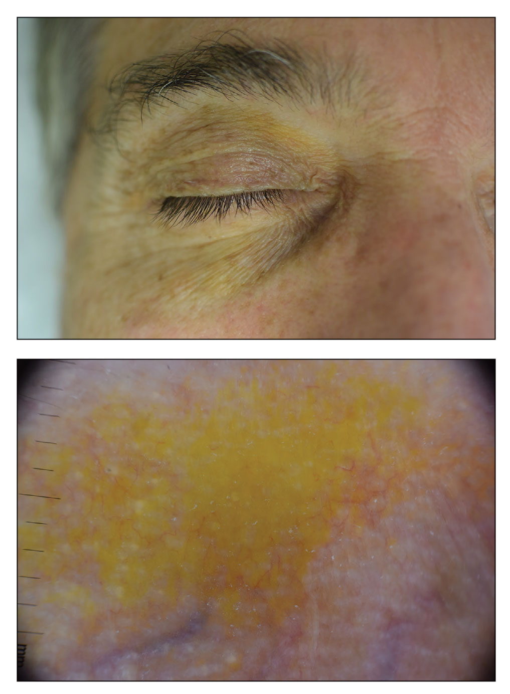
Orange palpebral spots commonly appear as asymptomatic, yellow-orange, symmetric lesions with a predilection for the recessed areas of the superior eyelids but also can present on the canthi and inferior eyelids. The discolorations are more easily visible on fair skin and have been reported to measure from 10 to 15 mm in the long axis.3 Assouly et al1 described the orange spots as having indistinct margins, with borders similar to “sand on a sea shore.” Orange palpebral spots can be a persistent discoloration, and there are no reports of spontaneous regression. No known association with malignancy or systemic illness has been reported.
Case reports of OPSs describe histologic similarities between specimens, including increased adipose tissue and pigment-laden macrophages in the superficial dermis.2 The pigmented deposits sometimes may be found in the basal keratinocytes of the epidermis and turn black with Fontana-Masson stain.1 No inflammatory infiltrates, necrosis, or xanthomization are characteristically found. Stains for iron, mucin, and amyloid also have been negative.2
The cause of pigmentation in OPSs is unknown; however, lipofuscin deposits and high-situated adipocytes in the reticular dermis colored by carotenoids have been proposed as possible mechanisms.1 No unifying cause for pigmentation in the serum (eg, cholesterol, triglycerides, thyroid-stimulating hormone, free retinol, vitamin E, carotenoids) was found in 11 of 27 patients with OPSs assessed by Assouly et al.1 In one case, lipofuscin, a degradation product of lysosomes, was detected by microscopic autofluorescence in the superficial dermis. However, lipofuscin typically is a breakdown product associated with aging, and OPSs have been present in patients as young as 28 years.1 Local trauma related to eye rubbing is another theory that has been proposed due to the finding of melanin in the superficial dermis. However, the absence of hemosiderin deposits as well as the extensive duration of the discolorations makes local trauma a less likely explanation for the etiology of OPSs.2
The clinical differential diagnosis for OPSs includes xanthelasma, jaundice, and carotenoderma. Xanthelasma presents as elevated yellow plaques usually found over the medial aspect of the eyes. In contrast, OPSs are nonelevated with both orange and yellow hues typically present. Histologic samples of xanthelasma are characterized by lipid-laden macrophages (foam cells) in the dermis in contrast to the adipose tissue seen in OPSs that has not been phagocytized.1,2 The lack of scleral icterus made jaundice an unlikely diagnosis in our patient. Bilirubin elevations substantial enough to cause skin discoloration also would be expected to discolor the conjunctiva. In carotenoderma, carotenoids are deposited in the sweat and sebum of the stratum corneum with the orange pigmentation most prominent in regions of increased sweating such as the palms, soles, and nasolabial folds.4 Our patient’s lack of discoloration in places other than the periorbital region made carotenoderma less likely.
In the study by Assouly et al,1 10 of 11 patients who underwent laboratory analysis self-reported eating a diet rich in fruit and vegetables, though no standardized questionnaire was given. One patient was found to have an elevated vitamin E level, and in 5 cases there was an elevated level of β-cryptoxanthin. The significance of these elevations in such a small minority is unknown, and increased β-cryptoxanthin has been attributed to increased consumption of citrus fruits during the winter season. Our patient reported ingesting a daily oral supplement rich in carotenoids that constituted 60% of the daily value of vitamin E including mixed tocopherols as well as 90% of the daily value of vitamin A with many sources of carotenoids including beta-carotenes, lutein/zeaxanthin, lycopene, and astaxanthin. An invasive biopsy was not taken in this case, as OPSs largely are diagnosed clinically. Greater awareness and recognition of OPSs may help to identify common underlying causes for this unique diagnosis.
- Assouly P, Cavelier-Balloy B, Dupré T. Orange palpebral spots. Dermatology. 2008;216:166-170.
- Belliveau MJ, Odashiro AN, Harvey JT. Yellow-orange palpebral spots. Ophthalmology. 2015;122:2139-2140.
- Kluger N, Guillot B. Bilateral orange discoloration of the upper eyelids: a quiz. Acta Derm Venereol. 2011;91:211-212.
- Maharshak N, Shapiro J, Trau H. Carotenoderma—a review of the current literature. Int J Dermatol. 2003;42:178-181.
The Diagnosis: Orange Palpebral Spots
The clinical presentation of our patient was consistent with a diagnosis of orange palpebral spots (OPSs), an uncommon discoloration that most often appears in White patients in the fifth or sixth decades of life. Orange palpebral spots were first described in 2008 by Assouly et al1 in 27 patients (23 females and 4 males). In 2015, Belliveau et al2 expanded the designation to yellow-orange palpebral spots because they felt the term more fully expressed the color variations depicted in their patients; however, this term more frequently is used in ophthalmology.
Orange palpebral spots commonly appear as asymptomatic, yellow-orange, symmetric lesions with a predilection for the recessed areas of the superior eyelids but also can present on the canthi and inferior eyelids. The discolorations are more easily visible on fair skin and have been reported to measure from 10 to 15 mm in the long axis.3 Assouly et al1 described the orange spots as having indistinct margins, with borders similar to “sand on a sea shore.” Orange palpebral spots can be a persistent discoloration, and there are no reports of spontaneous regression. No known association with malignancy or systemic illness has been reported.
Case reports of OPSs describe histologic similarities between specimens, including increased adipose tissue and pigment-laden macrophages in the superficial dermis.2 The pigmented deposits sometimes may be found in the basal keratinocytes of the epidermis and turn black with Fontana-Masson stain.1 No inflammatory infiltrates, necrosis, or xanthomization are characteristically found. Stains for iron, mucin, and amyloid also have been negative.2
The cause of pigmentation in OPSs is unknown; however, lipofuscin deposits and high-situated adipocytes in the reticular dermis colored by carotenoids have been proposed as possible mechanisms.1 No unifying cause for pigmentation in the serum (eg, cholesterol, triglycerides, thyroid-stimulating hormone, free retinol, vitamin E, carotenoids) was found in 11 of 27 patients with OPSs assessed by Assouly et al.1 In one case, lipofuscin, a degradation product of lysosomes, was detected by microscopic autofluorescence in the superficial dermis. However, lipofuscin typically is a breakdown product associated with aging, and OPSs have been present in patients as young as 28 years.1 Local trauma related to eye rubbing is another theory that has been proposed due to the finding of melanin in the superficial dermis. However, the absence of hemosiderin deposits as well as the extensive duration of the discolorations makes local trauma a less likely explanation for the etiology of OPSs.2
The clinical differential diagnosis for OPSs includes xanthelasma, jaundice, and carotenoderma. Xanthelasma presents as elevated yellow plaques usually found over the medial aspect of the eyes. In contrast, OPSs are nonelevated with both orange and yellow hues typically present. Histologic samples of xanthelasma are characterized by lipid-laden macrophages (foam cells) in the dermis in contrast to the adipose tissue seen in OPSs that has not been phagocytized.1,2 The lack of scleral icterus made jaundice an unlikely diagnosis in our patient. Bilirubin elevations substantial enough to cause skin discoloration also would be expected to discolor the conjunctiva. In carotenoderma, carotenoids are deposited in the sweat and sebum of the stratum corneum with the orange pigmentation most prominent in regions of increased sweating such as the palms, soles, and nasolabial folds.4 Our patient’s lack of discoloration in places other than the periorbital region made carotenoderma less likely.
In the study by Assouly et al,1 10 of 11 patients who underwent laboratory analysis self-reported eating a diet rich in fruit and vegetables, though no standardized questionnaire was given. One patient was found to have an elevated vitamin E level, and in 5 cases there was an elevated level of β-cryptoxanthin. The significance of these elevations in such a small minority is unknown, and increased β-cryptoxanthin has been attributed to increased consumption of citrus fruits during the winter season. Our patient reported ingesting a daily oral supplement rich in carotenoids that constituted 60% of the daily value of vitamin E including mixed tocopherols as well as 90% of the daily value of vitamin A with many sources of carotenoids including beta-carotenes, lutein/zeaxanthin, lycopene, and astaxanthin. An invasive biopsy was not taken in this case, as OPSs largely are diagnosed clinically. Greater awareness and recognition of OPSs may help to identify common underlying causes for this unique diagnosis.
The Diagnosis: Orange Palpebral Spots
The clinical presentation of our patient was consistent with a diagnosis of orange palpebral spots (OPSs), an uncommon discoloration that most often appears in White patients in the fifth or sixth decades of life. Orange palpebral spots were first described in 2008 by Assouly et al1 in 27 patients (23 females and 4 males). In 2015, Belliveau et al2 expanded the designation to yellow-orange palpebral spots because they felt the term more fully expressed the color variations depicted in their patients; however, this term more frequently is used in ophthalmology.
Orange palpebral spots commonly appear as asymptomatic, yellow-orange, symmetric lesions with a predilection for the recessed areas of the superior eyelids but also can present on the canthi and inferior eyelids. The discolorations are more easily visible on fair skin and have been reported to measure from 10 to 15 mm in the long axis.3 Assouly et al1 described the orange spots as having indistinct margins, with borders similar to “sand on a sea shore.” Orange palpebral spots can be a persistent discoloration, and there are no reports of spontaneous regression. No known association with malignancy or systemic illness has been reported.
Case reports of OPSs describe histologic similarities between specimens, including increased adipose tissue and pigment-laden macrophages in the superficial dermis.2 The pigmented deposits sometimes may be found in the basal keratinocytes of the epidermis and turn black with Fontana-Masson stain.1 No inflammatory infiltrates, necrosis, or xanthomization are characteristically found. Stains for iron, mucin, and amyloid also have been negative.2
The cause of pigmentation in OPSs is unknown; however, lipofuscin deposits and high-situated adipocytes in the reticular dermis colored by carotenoids have been proposed as possible mechanisms.1 No unifying cause for pigmentation in the serum (eg, cholesterol, triglycerides, thyroid-stimulating hormone, free retinol, vitamin E, carotenoids) was found in 11 of 27 patients with OPSs assessed by Assouly et al.1 In one case, lipofuscin, a degradation product of lysosomes, was detected by microscopic autofluorescence in the superficial dermis. However, lipofuscin typically is a breakdown product associated with aging, and OPSs have been present in patients as young as 28 years.1 Local trauma related to eye rubbing is another theory that has been proposed due to the finding of melanin in the superficial dermis. However, the absence of hemosiderin deposits as well as the extensive duration of the discolorations makes local trauma a less likely explanation for the etiology of OPSs.2
The clinical differential diagnosis for OPSs includes xanthelasma, jaundice, and carotenoderma. Xanthelasma presents as elevated yellow plaques usually found over the medial aspect of the eyes. In contrast, OPSs are nonelevated with both orange and yellow hues typically present. Histologic samples of xanthelasma are characterized by lipid-laden macrophages (foam cells) in the dermis in contrast to the adipose tissue seen in OPSs that has not been phagocytized.1,2 The lack of scleral icterus made jaundice an unlikely diagnosis in our patient. Bilirubin elevations substantial enough to cause skin discoloration also would be expected to discolor the conjunctiva. In carotenoderma, carotenoids are deposited in the sweat and sebum of the stratum corneum with the orange pigmentation most prominent in regions of increased sweating such as the palms, soles, and nasolabial folds.4 Our patient’s lack of discoloration in places other than the periorbital region made carotenoderma less likely.
In the study by Assouly et al,1 10 of 11 patients who underwent laboratory analysis self-reported eating a diet rich in fruit and vegetables, though no standardized questionnaire was given. One patient was found to have an elevated vitamin E level, and in 5 cases there was an elevated level of β-cryptoxanthin. The significance of these elevations in such a small minority is unknown, and increased β-cryptoxanthin has been attributed to increased consumption of citrus fruits during the winter season. Our patient reported ingesting a daily oral supplement rich in carotenoids that constituted 60% of the daily value of vitamin E including mixed tocopherols as well as 90% of the daily value of vitamin A with many sources of carotenoids including beta-carotenes, lutein/zeaxanthin, lycopene, and astaxanthin. An invasive biopsy was not taken in this case, as OPSs largely are diagnosed clinically. Greater awareness and recognition of OPSs may help to identify common underlying causes for this unique diagnosis.
- Assouly P, Cavelier-Balloy B, Dupré T. Orange palpebral spots. Dermatology. 2008;216:166-170.
- Belliveau MJ, Odashiro AN, Harvey JT. Yellow-orange palpebral spots. Ophthalmology. 2015;122:2139-2140.
- Kluger N, Guillot B. Bilateral orange discoloration of the upper eyelids: a quiz. Acta Derm Venereol. 2011;91:211-212.
- Maharshak N, Shapiro J, Trau H. Carotenoderma—a review of the current literature. Int J Dermatol. 2003;42:178-181.
- Assouly P, Cavelier-Balloy B, Dupré T. Orange palpebral spots. Dermatology. 2008;216:166-170.
- Belliveau MJ, Odashiro AN, Harvey JT. Yellow-orange palpebral spots. Ophthalmology. 2015;122:2139-2140.
- Kluger N, Guillot B. Bilateral orange discoloration of the upper eyelids: a quiz. Acta Derm Venereol. 2011;91:211-212.
- Maharshak N, Shapiro J, Trau H. Carotenoderma—a review of the current literature. Int J Dermatol. 2003;42:178-181.
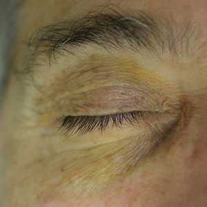
A 63-year-old White man with a history of melanoma presented to our dermatology clinic for evaluation of gradually worsening yellow discoloration around the eyes of 2 years’ duration. Physical examination revealed periorbital yellow-orange patches (top). The discolorations were nonelevated and nonpalpable. Dermoscopy revealed yellow blotches with sparing of the hair follicles (bottom). The remainder of the skin examination was unremarkable.
Pruritic rash on arms and legs
Atopic dermatitis (AD) is one of the most common chronic, inflammatory skin diseases encountered by dermatologists. AD is characterized by pruritus and a chronic course of exacerbations and remissions. AD is thought to involve the interplay of genetic predisposition, immune dysregulation, and environmental factors. It is also associated with other allergic conditions, including asthma.
Although AD typically presents with pruritus as the hallmark symptom in all patients, the appearance of skin lesions may vary among different skin types. In individuals with light-colored skin, AD often appears as erythematous patches and plaques. It also more commonly affects the flexor surfaces of the skin. In individuals with darker skin tones, AD may more often result in follicularly centered papules, lichenification, and pigmentary changes. Lesions may also present on extensor surfaces rather than the typical flexure surfaces. Erythema in darker skin types may appear reddish-brown, have a violaceous hue, or be an ashen gray or darker brown color rather than bright red. Because erythema is more difficult to detect in darker skin types, clinicians may mistakenly minimize the severity of AD.
Clinical severity may also differ between ethnicities. Black patients have an increased tendency toward hyperlinearity of the palms, periorbital dark circles, Dennie-Morgan lines, and diffuse xerosis. Compared with White patients, Black patients with AD are also more likely to develop prurigo nodularis and lichenification. In contrast, Asian patients with AD often experience psoriasiform features, with lesions having more well-defined borders and increased scaling and lichenification.
Beyond differences in clinical appearance, AD may appear molecularly and histologically distinct in ethnic skin. One study suggests that Black patients with AD may have decreased Th1 and Th17 but share similar upregulation of Th2 and Th22 as seen in White patients. Another study showed that Asian patients may have higher Th17 and Th22 and lower Th1/interferon compared with White patients.
Regardless of skin type, treatment goals remain the same. Treatment goals aim to repair and improve the function of the skin barrier while preventing and managing flares. Clinical studies have shown that skincare regimens incorporating ceramide-containing moisturizers may improve AD by increasing the lipid content in the skin. This may offer clinical benefit in patients with skin of color. However, some treatments often used for AD may lead to other skin issues in skin in color. For example, long-term use of topical steroids may worsen hypopigmentation in darker skin types. Management strategies should take into account the unique clinical and genetic features of AD among different patient demographic groups.
William D. James, MD, Professor, Department of Dermatology, University of Pennsylvania, Philadelphia.
Disclosure: William D. James, MD, has disclosed the following relevant financial relationships:
Received income in an amount equal to or greater than $250 from: Elsevier.
Image Quizzes are fictional or fictionalized clinical scenarios intended to provide evidence-based educational takeaways.
Atopic dermatitis (AD) is one of the most common chronic, inflammatory skin diseases encountered by dermatologists. AD is characterized by pruritus and a chronic course of exacerbations and remissions. AD is thought to involve the interplay of genetic predisposition, immune dysregulation, and environmental factors. It is also associated with other allergic conditions, including asthma.
Although AD typically presents with pruritus as the hallmark symptom in all patients, the appearance of skin lesions may vary among different skin types. In individuals with light-colored skin, AD often appears as erythematous patches and plaques. It also more commonly affects the flexor surfaces of the skin. In individuals with darker skin tones, AD may more often result in follicularly centered papules, lichenification, and pigmentary changes. Lesions may also present on extensor surfaces rather than the typical flexure surfaces. Erythema in darker skin types may appear reddish-brown, have a violaceous hue, or be an ashen gray or darker brown color rather than bright red. Because erythema is more difficult to detect in darker skin types, clinicians may mistakenly minimize the severity of AD.
Clinical severity may also differ between ethnicities. Black patients have an increased tendency toward hyperlinearity of the palms, periorbital dark circles, Dennie-Morgan lines, and diffuse xerosis. Compared with White patients, Black patients with AD are also more likely to develop prurigo nodularis and lichenification. In contrast, Asian patients with AD often experience psoriasiform features, with lesions having more well-defined borders and increased scaling and lichenification.
Beyond differences in clinical appearance, AD may appear molecularly and histologically distinct in ethnic skin. One study suggests that Black patients with AD may have decreased Th1 and Th17 but share similar upregulation of Th2 and Th22 as seen in White patients. Another study showed that Asian patients may have higher Th17 and Th22 and lower Th1/interferon compared with White patients.
Regardless of skin type, treatment goals remain the same. Treatment goals aim to repair and improve the function of the skin barrier while preventing and managing flares. Clinical studies have shown that skincare regimens incorporating ceramide-containing moisturizers may improve AD by increasing the lipid content in the skin. This may offer clinical benefit in patients with skin of color. However, some treatments often used for AD may lead to other skin issues in skin in color. For example, long-term use of topical steroids may worsen hypopigmentation in darker skin types. Management strategies should take into account the unique clinical and genetic features of AD among different patient demographic groups.
William D. James, MD, Professor, Department of Dermatology, University of Pennsylvania, Philadelphia.
Disclosure: William D. James, MD, has disclosed the following relevant financial relationships:
Received income in an amount equal to or greater than $250 from: Elsevier.
Image Quizzes are fictional or fictionalized clinical scenarios intended to provide evidence-based educational takeaways.
Atopic dermatitis (AD) is one of the most common chronic, inflammatory skin diseases encountered by dermatologists. AD is characterized by pruritus and a chronic course of exacerbations and remissions. AD is thought to involve the interplay of genetic predisposition, immune dysregulation, and environmental factors. It is also associated with other allergic conditions, including asthma.
Although AD typically presents with pruritus as the hallmark symptom in all patients, the appearance of skin lesions may vary among different skin types. In individuals with light-colored skin, AD often appears as erythematous patches and plaques. It also more commonly affects the flexor surfaces of the skin. In individuals with darker skin tones, AD may more often result in follicularly centered papules, lichenification, and pigmentary changes. Lesions may also present on extensor surfaces rather than the typical flexure surfaces. Erythema in darker skin types may appear reddish-brown, have a violaceous hue, or be an ashen gray or darker brown color rather than bright red. Because erythema is more difficult to detect in darker skin types, clinicians may mistakenly minimize the severity of AD.
Clinical severity may also differ between ethnicities. Black patients have an increased tendency toward hyperlinearity of the palms, periorbital dark circles, Dennie-Morgan lines, and diffuse xerosis. Compared with White patients, Black patients with AD are also more likely to develop prurigo nodularis and lichenification. In contrast, Asian patients with AD often experience psoriasiform features, with lesions having more well-defined borders and increased scaling and lichenification.
Beyond differences in clinical appearance, AD may appear molecularly and histologically distinct in ethnic skin. One study suggests that Black patients with AD may have decreased Th1 and Th17 but share similar upregulation of Th2 and Th22 as seen in White patients. Another study showed that Asian patients may have higher Th17 and Th22 and lower Th1/interferon compared with White patients.
Regardless of skin type, treatment goals remain the same. Treatment goals aim to repair and improve the function of the skin barrier while preventing and managing flares. Clinical studies have shown that skincare regimens incorporating ceramide-containing moisturizers may improve AD by increasing the lipid content in the skin. This may offer clinical benefit in patients with skin of color. However, some treatments often used for AD may lead to other skin issues in skin in color. For example, long-term use of topical steroids may worsen hypopigmentation in darker skin types. Management strategies should take into account the unique clinical and genetic features of AD among different patient demographic groups.
William D. James, MD, Professor, Department of Dermatology, University of Pennsylvania, Philadelphia.
Disclosure: William D. James, MD, has disclosed the following relevant financial relationships:
Received income in an amount equal to or greater than $250 from: Elsevier.
Image Quizzes are fictional or fictionalized clinical scenarios intended to provide evidence-based educational takeaways.
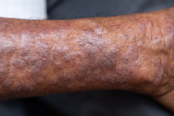
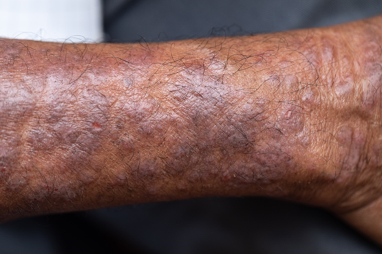
A 27-year-old student presents with a pruritic rash on his hands and in the bends of his arms and legs. He recently started clinical rotations in a nursing facility and has been using hand sanitizer multiple times per day, which has exacerbated the rash on his hands, causing them to ooze and sting. He describes the rash as itchy, especially at night. At times he reports that the itching causes difficulty sleeping. In addition, his skin has little cracks that frequently bleed. He notes that he has experienced similar symptoms in the past, which resolved with moisturizers and topical cream from the drugstore. He has tried over-the-counter hydrocortisone during this episode, with minimal improvement in symptoms. He denies any change in laundry detergents or use of new household products.
Physical examination reveals large erythematous plaques on the hands and flexure surfaces of his neck, antecubital fossa, and behind the knees with scattered excoriations. Erythematous, slightly lichenified coalescing papules are noted on the proximal arms. His face is clear. General skin pigmentation is brown and free of masses and lumps.