User login
Front-line BTK inhibitor+anti-CD20 antibody tops chemoimmunotherapy in CLL, reveals meta-analysis
Key clinical point: Compared with chemoimmunotherapy (CIT), Bruton tyrosine kinase inhibitor (BTKi) therapy combined with anti-CD20 antibody therapy improves clinical outcomes in patients with treatment-naive chronic lymphocytic leukemia (CLL) without causing increased toxicity.
Major finding: Patients receiving BTKi+anti-CD20 antibody vs CIT had significantly prolonged progression-free survival (hazard ratio 0.25; 95% CI 0.15-0.42) and higher objective response rates (risk ratio [RR] 1.16; 95% CI 1.13-1.20) and a comparable risk for grade ≥3 adverse events (RR 1.04; 95% CI 0.92-1.17).
Study details: The data come from a meta-analysis of four randomized controlled trials involving 1479 patients with treatment-naive CLL who had been randomized to receive CIT or BTKi+anti-CD20 antibody therapy.
Disclosures: This study was partly supported by the Ministry of Science and Technology, Taiwan, and Taipei Medical University, Taiwan. The authors declared no conflicts of interest.
Source: Nguyen TT et al. Efficacy and safety of Bruton tyrosine kinase inhibitor plus anti-CD20 antibody therapy compared with chemoimmunotherapy as front-line treatment for chronic lymphocytic leukemia: A systematic review and meta-analysis of randomized controlled trials. J Immunother. 2023 (May 23). doi: 10.1097/CJI.0000000000000471
Key clinical point: Compared with chemoimmunotherapy (CIT), Bruton tyrosine kinase inhibitor (BTKi) therapy combined with anti-CD20 antibody therapy improves clinical outcomes in patients with treatment-naive chronic lymphocytic leukemia (CLL) without causing increased toxicity.
Major finding: Patients receiving BTKi+anti-CD20 antibody vs CIT had significantly prolonged progression-free survival (hazard ratio 0.25; 95% CI 0.15-0.42) and higher objective response rates (risk ratio [RR] 1.16; 95% CI 1.13-1.20) and a comparable risk for grade ≥3 adverse events (RR 1.04; 95% CI 0.92-1.17).
Study details: The data come from a meta-analysis of four randomized controlled trials involving 1479 patients with treatment-naive CLL who had been randomized to receive CIT or BTKi+anti-CD20 antibody therapy.
Disclosures: This study was partly supported by the Ministry of Science and Technology, Taiwan, and Taipei Medical University, Taiwan. The authors declared no conflicts of interest.
Source: Nguyen TT et al. Efficacy and safety of Bruton tyrosine kinase inhibitor plus anti-CD20 antibody therapy compared with chemoimmunotherapy as front-line treatment for chronic lymphocytic leukemia: A systematic review and meta-analysis of randomized controlled trials. J Immunother. 2023 (May 23). doi: 10.1097/CJI.0000000000000471
Key clinical point: Compared with chemoimmunotherapy (CIT), Bruton tyrosine kinase inhibitor (BTKi) therapy combined with anti-CD20 antibody therapy improves clinical outcomes in patients with treatment-naive chronic lymphocytic leukemia (CLL) without causing increased toxicity.
Major finding: Patients receiving BTKi+anti-CD20 antibody vs CIT had significantly prolonged progression-free survival (hazard ratio 0.25; 95% CI 0.15-0.42) and higher objective response rates (risk ratio [RR] 1.16; 95% CI 1.13-1.20) and a comparable risk for grade ≥3 adverse events (RR 1.04; 95% CI 0.92-1.17).
Study details: The data come from a meta-analysis of four randomized controlled trials involving 1479 patients with treatment-naive CLL who had been randomized to receive CIT or BTKi+anti-CD20 antibody therapy.
Disclosures: This study was partly supported by the Ministry of Science and Technology, Taiwan, and Taipei Medical University, Taiwan. The authors declared no conflicts of interest.
Source: Nguyen TT et al. Efficacy and safety of Bruton tyrosine kinase inhibitor plus anti-CD20 antibody therapy compared with chemoimmunotherapy as front-line treatment for chronic lymphocytic leukemia: A systematic review and meta-analysis of randomized controlled trials. J Immunother. 2023 (May 23). doi: 10.1097/CJI.0000000000000471
R-High-CHOP/CHASER/LEED with auto-PBSCT provides favorable long-term survival outcomes in untreated MCL
Key clinical point: High-dose rituximab plus cyclophosphamide, doxorubicin, vincristine, and prednisone (R-High-CHOP)/cyclophosphamide, high-dose cytarabine, dexamethasone, etoposide, and rituximab (CHASER) followed by high-dose chemotherapy (HDC) with melphalan, cyclophosphamide, etoposide, and dexamethasone (LEED), and autologous peripheral blood stem cell transplantation (auto-PBSCT) provided favorable long-term survival outcomes in younger patients with untreated advanced mantle cell lymphoma (MCL).
Major finding: At 6-year median follow-up, the 5- and 8-year progression-free survival rates were 52.3% (95% CI 36.7%-65.7%) and 17.1% (95% CI 5.2%-34.7%), respectively, and the overall survival rates were 75.0% (95% CI 59.4%-85.3%) and 69.0% (95% CI 52.3%-80.9%), respectively. The incidence of secondary malignancies (11.1%) and continuous relapses was high.
Study details: This final analysis of the JCOG0406 study included 45 patients age ≤ 65 years with untreated advanced MCL who received R-High-CHOP/CHASER followed by HDC with LEED and auto-PBSCT.
Disclosures: This study was supported by Health and Labour Sciences Research Grants (Japan) and others. S Nakamura declared being an Editorial Board Member of Cancer Science. Some authors reported ties with various organizations.
Source: Ogura M et al. Long-term follow-up after R-High CHOP/CHASER/LEED with Auto-PBSCT in untreated mantle cell lymphoma-Final analysis of JCOG0406. Cancer Sci. 2023 (May 26). doi: 10.1111/cas.15849
Key clinical point: High-dose rituximab plus cyclophosphamide, doxorubicin, vincristine, and prednisone (R-High-CHOP)/cyclophosphamide, high-dose cytarabine, dexamethasone, etoposide, and rituximab (CHASER) followed by high-dose chemotherapy (HDC) with melphalan, cyclophosphamide, etoposide, and dexamethasone (LEED), and autologous peripheral blood stem cell transplantation (auto-PBSCT) provided favorable long-term survival outcomes in younger patients with untreated advanced mantle cell lymphoma (MCL).
Major finding: At 6-year median follow-up, the 5- and 8-year progression-free survival rates were 52.3% (95% CI 36.7%-65.7%) and 17.1% (95% CI 5.2%-34.7%), respectively, and the overall survival rates were 75.0% (95% CI 59.4%-85.3%) and 69.0% (95% CI 52.3%-80.9%), respectively. The incidence of secondary malignancies (11.1%) and continuous relapses was high.
Study details: This final analysis of the JCOG0406 study included 45 patients age ≤ 65 years with untreated advanced MCL who received R-High-CHOP/CHASER followed by HDC with LEED and auto-PBSCT.
Disclosures: This study was supported by Health and Labour Sciences Research Grants (Japan) and others. S Nakamura declared being an Editorial Board Member of Cancer Science. Some authors reported ties with various organizations.
Source: Ogura M et al. Long-term follow-up after R-High CHOP/CHASER/LEED with Auto-PBSCT in untreated mantle cell lymphoma-Final analysis of JCOG0406. Cancer Sci. 2023 (May 26). doi: 10.1111/cas.15849
Key clinical point: High-dose rituximab plus cyclophosphamide, doxorubicin, vincristine, and prednisone (R-High-CHOP)/cyclophosphamide, high-dose cytarabine, dexamethasone, etoposide, and rituximab (CHASER) followed by high-dose chemotherapy (HDC) with melphalan, cyclophosphamide, etoposide, and dexamethasone (LEED), and autologous peripheral blood stem cell transplantation (auto-PBSCT) provided favorable long-term survival outcomes in younger patients with untreated advanced mantle cell lymphoma (MCL).
Major finding: At 6-year median follow-up, the 5- and 8-year progression-free survival rates were 52.3% (95% CI 36.7%-65.7%) and 17.1% (95% CI 5.2%-34.7%), respectively, and the overall survival rates were 75.0% (95% CI 59.4%-85.3%) and 69.0% (95% CI 52.3%-80.9%), respectively. The incidence of secondary malignancies (11.1%) and continuous relapses was high.
Study details: This final analysis of the JCOG0406 study included 45 patients age ≤ 65 years with untreated advanced MCL who received R-High-CHOP/CHASER followed by HDC with LEED and auto-PBSCT.
Disclosures: This study was supported by Health and Labour Sciences Research Grants (Japan) and others. S Nakamura declared being an Editorial Board Member of Cancer Science. Some authors reported ties with various organizations.
Source: Ogura M et al. Long-term follow-up after R-High CHOP/CHASER/LEED with Auto-PBSCT in untreated mantle cell lymphoma-Final analysis of JCOG0406. Cancer Sci. 2023 (May 26). doi: 10.1111/cas.15849
Tafasitamab+lenalidomide provides survival benefit over pola-BR and R2 in relapsed or refractory DLBCL
Key clinical point: Compared with polatuzumab vedotin+bendamustine+rituximab (pola-BR) and rituximab+lenalidomide (R2), tafasitamab+lenalidomide provided a significant survival benefit in patients with relapsed or refractory (R/R) diffuse large B-cell lymphoma (DLBCL).
Major finding: A significant overall survival (OS) benefit was observed with tafasitamab+lenalidomide vs pola-BR (hazard ratio [HR] 0.441; P = .034) and R2 (HR 0.435; P = .012). However, the OS was similar between the tafasitamab+lenalidomide and CD19-chimeric antigen receptor T-cell groups (CAR-T; HR 0.953; P = .892).
Study details: This expanded analysis of the RE-MIND2 study of propensity score-matched transplant-ineligible patients with R/R DLBCL treated with ≥2 systemic therapies who received tafasitamab+lenalidomide in the L-MIND trial and those who received pola-BR (24 pairs), R2 (33 pairs), or CAR-T (37 pairs) from an observational cohort.
Disclosures: This study was sponsored by MorphoSys AG. Some authors declared serving as consultants, advisory board members, or speakers and receiving research funding, honoraria, or travel support from MorphoSys and others. Four authors declared being employees of or holding equities in MorphoSys.
Source: Nowakowski GS et al. RE-MIND2: Comparative effectiveness of tafasitamab plus lenalidomide versus polatuzumab vedotin/bendamustine/rituximab (pola‑BR), CAR‑T therapies, and lenalidomide/rituximab (R2) based on real‑world data in patients with relapsed/refractory diffuse large B‑cell lymphoma. Ann Hematol. 2023;102:1773-1787 (May 12). doi: 10.1007/s00277-023-05196-4
Key clinical point: Compared with polatuzumab vedotin+bendamustine+rituximab (pola-BR) and rituximab+lenalidomide (R2), tafasitamab+lenalidomide provided a significant survival benefit in patients with relapsed or refractory (R/R) diffuse large B-cell lymphoma (DLBCL).
Major finding: A significant overall survival (OS) benefit was observed with tafasitamab+lenalidomide vs pola-BR (hazard ratio [HR] 0.441; P = .034) and R2 (HR 0.435; P = .012). However, the OS was similar between the tafasitamab+lenalidomide and CD19-chimeric antigen receptor T-cell groups (CAR-T; HR 0.953; P = .892).
Study details: This expanded analysis of the RE-MIND2 study of propensity score-matched transplant-ineligible patients with R/R DLBCL treated with ≥2 systemic therapies who received tafasitamab+lenalidomide in the L-MIND trial and those who received pola-BR (24 pairs), R2 (33 pairs), or CAR-T (37 pairs) from an observational cohort.
Disclosures: This study was sponsored by MorphoSys AG. Some authors declared serving as consultants, advisory board members, or speakers and receiving research funding, honoraria, or travel support from MorphoSys and others. Four authors declared being employees of or holding equities in MorphoSys.
Source: Nowakowski GS et al. RE-MIND2: Comparative effectiveness of tafasitamab plus lenalidomide versus polatuzumab vedotin/bendamustine/rituximab (pola‑BR), CAR‑T therapies, and lenalidomide/rituximab (R2) based on real‑world data in patients with relapsed/refractory diffuse large B‑cell lymphoma. Ann Hematol. 2023;102:1773-1787 (May 12). doi: 10.1007/s00277-023-05196-4
Key clinical point: Compared with polatuzumab vedotin+bendamustine+rituximab (pola-BR) and rituximab+lenalidomide (R2), tafasitamab+lenalidomide provided a significant survival benefit in patients with relapsed or refractory (R/R) diffuse large B-cell lymphoma (DLBCL).
Major finding: A significant overall survival (OS) benefit was observed with tafasitamab+lenalidomide vs pola-BR (hazard ratio [HR] 0.441; P = .034) and R2 (HR 0.435; P = .012). However, the OS was similar between the tafasitamab+lenalidomide and CD19-chimeric antigen receptor T-cell groups (CAR-T; HR 0.953; P = .892).
Study details: This expanded analysis of the RE-MIND2 study of propensity score-matched transplant-ineligible patients with R/R DLBCL treated with ≥2 systemic therapies who received tafasitamab+lenalidomide in the L-MIND trial and those who received pola-BR (24 pairs), R2 (33 pairs), or CAR-T (37 pairs) from an observational cohort.
Disclosures: This study was sponsored by MorphoSys AG. Some authors declared serving as consultants, advisory board members, or speakers and receiving research funding, honoraria, or travel support from MorphoSys and others. Four authors declared being employees of or holding equities in MorphoSys.
Source: Nowakowski GS et al. RE-MIND2: Comparative effectiveness of tafasitamab plus lenalidomide versus polatuzumab vedotin/bendamustine/rituximab (pola‑BR), CAR‑T therapies, and lenalidomide/rituximab (R2) based on real‑world data in patients with relapsed/refractory diffuse large B‑cell lymphoma. Ann Hematol. 2023;102:1773-1787 (May 12). doi: 10.1007/s00277-023-05196-4
First-line rituximab+bendamustine+cytarabine combination shows long-term efficacy in the elderly with MCL
Key clinical point: The rituximab, bendamustine, and low-dose cytarabine (R-BAC) regimen, not succeeded by maintenance therapy, induces long-term responses in previously untreated elderly patients with mantle cell lymphoma (MCL).
Major finding: After a median follow-up of 86 months, the median overall survival (OS) and progression-free survival (PFS) were not reached; the 7-year PFS and OS rates were 55% (95% CI 41%-67%) and 63% (95% CI 49%-74%), respectively. The 7-year duration of response rate among the 52 responding patients was 59% (95% CI 44%-71%). No signal of late toxicity was reported.
Study details: Findings are from a long-term analysis of the FIL-RBAC500 trial that included 57 previously untreated elderly patients with MCL who received the R-BAC regimen.
Disclosures: This study was supported by Progetto di Ricerca Sanitaria Finalizzata (Italy) grants and other sources. Some authors declared serving as advisory board members or consultants and receiving research funding, speaker honoraria, or travel expenses and accommodations from various sources.
Source: Tisi MC et al. Long term follow-up of rituximab plus bendamustine and cytarabine (R-BAC) in elderly patients with newly diagnosed MCL. Blood Adv. 2023 (May 12). doi: 10.1182/bloodadvances.2023009744
Key clinical point: The rituximab, bendamustine, and low-dose cytarabine (R-BAC) regimen, not succeeded by maintenance therapy, induces long-term responses in previously untreated elderly patients with mantle cell lymphoma (MCL).
Major finding: After a median follow-up of 86 months, the median overall survival (OS) and progression-free survival (PFS) were not reached; the 7-year PFS and OS rates were 55% (95% CI 41%-67%) and 63% (95% CI 49%-74%), respectively. The 7-year duration of response rate among the 52 responding patients was 59% (95% CI 44%-71%). No signal of late toxicity was reported.
Study details: Findings are from a long-term analysis of the FIL-RBAC500 trial that included 57 previously untreated elderly patients with MCL who received the R-BAC regimen.
Disclosures: This study was supported by Progetto di Ricerca Sanitaria Finalizzata (Italy) grants and other sources. Some authors declared serving as advisory board members or consultants and receiving research funding, speaker honoraria, or travel expenses and accommodations from various sources.
Source: Tisi MC et al. Long term follow-up of rituximab plus bendamustine and cytarabine (R-BAC) in elderly patients with newly diagnosed MCL. Blood Adv. 2023 (May 12). doi: 10.1182/bloodadvances.2023009744
Key clinical point: The rituximab, bendamustine, and low-dose cytarabine (R-BAC) regimen, not succeeded by maintenance therapy, induces long-term responses in previously untreated elderly patients with mantle cell lymphoma (MCL).
Major finding: After a median follow-up of 86 months, the median overall survival (OS) and progression-free survival (PFS) were not reached; the 7-year PFS and OS rates were 55% (95% CI 41%-67%) and 63% (95% CI 49%-74%), respectively. The 7-year duration of response rate among the 52 responding patients was 59% (95% CI 44%-71%). No signal of late toxicity was reported.
Study details: Findings are from a long-term analysis of the FIL-RBAC500 trial that included 57 previously untreated elderly patients with MCL who received the R-BAC regimen.
Disclosures: This study was supported by Progetto di Ricerca Sanitaria Finalizzata (Italy) grants and other sources. Some authors declared serving as advisory board members or consultants and receiving research funding, speaker honoraria, or travel expenses and accommodations from various sources.
Source: Tisi MC et al. Long term follow-up of rituximab plus bendamustine and cytarabine (R-BAC) in elderly patients with newly diagnosed MCL. Blood Adv. 2023 (May 12). doi: 10.1182/bloodadvances.2023009744
First-line fixed-duration ibrutinib+venetoclax shows promise against high-risk CLL
Key clinical point: First-line ibrutinib+venetoclax led to high response and survival rates in patients with chronic lymphocytic leukemia (CLL) or small lymphocytic lymphoma (SLL) regardless of high-risk genomic features generally associated with poor outcomes.
Major finding: Overall response and 36-month overall survival rates were >95% regardless of high-risk features. Patients with and without high-risk features had similar complete response (61% [95% CI 53%-70%] and 53% [95% CI 41%-65%], respectively) and 36-month progression-free survival (88% [95% CI 81%-93%] and 92% [95% CI 82%-97%], respectively) rates.
Study details: This post hoc analysis of the CAPTIVATE trial analyzed the pooled data of 195 patients with previously untreated CLL/SLL and known status of high-risk features [del(17p), TP53 mutation, and unmutated immunoglobulin heavy chain] treated with fixed-duration ibrutinib+venetoclax, of which 129 had ≥1 high-risk feature.
Disclosures: This study was funded by Pharmacyclics LLC, an AbbVie Company. Some authors declared receiving grants, personal fees, or other support from Pharmacyclics, AbbVie, or others. Two authors declared being employees of Pharmacyclics or AbbVie or having stock or other ownership interests in AbbVie.
Source: Allan JN et al. Outcomes in patients with high-risk features after fixed-duration ibrutinib plus venetoclax: Phase II CAPTIVATE study in first-line chronic lymphocytic leukemia. Clin Cancer Res. 2023 (Jun 7). doi: 10.1158/1078-0432.CCR-22-2779
Key clinical point: First-line ibrutinib+venetoclax led to high response and survival rates in patients with chronic lymphocytic leukemia (CLL) or small lymphocytic lymphoma (SLL) regardless of high-risk genomic features generally associated with poor outcomes.
Major finding: Overall response and 36-month overall survival rates were >95% regardless of high-risk features. Patients with and without high-risk features had similar complete response (61% [95% CI 53%-70%] and 53% [95% CI 41%-65%], respectively) and 36-month progression-free survival (88% [95% CI 81%-93%] and 92% [95% CI 82%-97%], respectively) rates.
Study details: This post hoc analysis of the CAPTIVATE trial analyzed the pooled data of 195 patients with previously untreated CLL/SLL and known status of high-risk features [del(17p), TP53 mutation, and unmutated immunoglobulin heavy chain] treated with fixed-duration ibrutinib+venetoclax, of which 129 had ≥1 high-risk feature.
Disclosures: This study was funded by Pharmacyclics LLC, an AbbVie Company. Some authors declared receiving grants, personal fees, or other support from Pharmacyclics, AbbVie, or others. Two authors declared being employees of Pharmacyclics or AbbVie or having stock or other ownership interests in AbbVie.
Source: Allan JN et al. Outcomes in patients with high-risk features after fixed-duration ibrutinib plus venetoclax: Phase II CAPTIVATE study in first-line chronic lymphocytic leukemia. Clin Cancer Res. 2023 (Jun 7). doi: 10.1158/1078-0432.CCR-22-2779
Key clinical point: First-line ibrutinib+venetoclax led to high response and survival rates in patients with chronic lymphocytic leukemia (CLL) or small lymphocytic lymphoma (SLL) regardless of high-risk genomic features generally associated with poor outcomes.
Major finding: Overall response and 36-month overall survival rates were >95% regardless of high-risk features. Patients with and without high-risk features had similar complete response (61% [95% CI 53%-70%] and 53% [95% CI 41%-65%], respectively) and 36-month progression-free survival (88% [95% CI 81%-93%] and 92% [95% CI 82%-97%], respectively) rates.
Study details: This post hoc analysis of the CAPTIVATE trial analyzed the pooled data of 195 patients with previously untreated CLL/SLL and known status of high-risk features [del(17p), TP53 mutation, and unmutated immunoglobulin heavy chain] treated with fixed-duration ibrutinib+venetoclax, of which 129 had ≥1 high-risk feature.
Disclosures: This study was funded by Pharmacyclics LLC, an AbbVie Company. Some authors declared receiving grants, personal fees, or other support from Pharmacyclics, AbbVie, or others. Two authors declared being employees of Pharmacyclics or AbbVie or having stock or other ownership interests in AbbVie.
Source: Allan JN et al. Outcomes in patients with high-risk features after fixed-duration ibrutinib plus venetoclax: Phase II CAPTIVATE study in first-line chronic lymphocytic leukemia. Clin Cancer Res. 2023 (Jun 7). doi: 10.1158/1078-0432.CCR-22-2779
Depression or anxiety shortens the survival of DLBCL patients
Key clinical point: The preexistence of depression, anxiety, or both is associated with shorter survival in older patients with diffuse large B-cell lymphoma (DLBCL).
Major finding: At a median follow-up of 2.0 years, patients with depression, anxiety, or both vs without any mental disorder had significantly lower 5-year overall survival rates (27.0% vs 37.4%; hazard ratio [HR] 1.37; 95% CI 1.29-1.44), with those with preexisting depression vs without any mental disorder having the worst rate (23.4% vs 38.0%; HR 1.37; P < .0001).
Study details: This retrospective cohort study analyzed the data of 13,244 patients age ≥ 67 years with DLBCL from the Surveillance, Epidemiology, and End Results-Medicare (SEER) registry, of which 2094 had depression, anxiety, or both at the time of their DLBCL diagnosis.
Disclosures: This study was funded by the American Society of Hematology and others. Some authors declared participating on Data Safety Monitoring or Advisory Boards or receiving financial support, royalties or licenses, or payments or honoraria for lectures, presentations, speakers’ bureaus, manuscript writing, or educational events from various organizations.
Source: Kuczmarski TM et al. Mental health disorders and survival among older patients with diffuse large B-cell lymphoma in the USA: A population-based study. Lancet Haematol. 2023 (Jun 1). doi: 10.1016/S2352-3026(23)00094-7
Key clinical point: The preexistence of depression, anxiety, or both is associated with shorter survival in older patients with diffuse large B-cell lymphoma (DLBCL).
Major finding: At a median follow-up of 2.0 years, patients with depression, anxiety, or both vs without any mental disorder had significantly lower 5-year overall survival rates (27.0% vs 37.4%; hazard ratio [HR] 1.37; 95% CI 1.29-1.44), with those with preexisting depression vs without any mental disorder having the worst rate (23.4% vs 38.0%; HR 1.37; P < .0001).
Study details: This retrospective cohort study analyzed the data of 13,244 patients age ≥ 67 years with DLBCL from the Surveillance, Epidemiology, and End Results-Medicare (SEER) registry, of which 2094 had depression, anxiety, or both at the time of their DLBCL diagnosis.
Disclosures: This study was funded by the American Society of Hematology and others. Some authors declared participating on Data Safety Monitoring or Advisory Boards or receiving financial support, royalties or licenses, or payments or honoraria for lectures, presentations, speakers’ bureaus, manuscript writing, or educational events from various organizations.
Source: Kuczmarski TM et al. Mental health disorders and survival among older patients with diffuse large B-cell lymphoma in the USA: A population-based study. Lancet Haematol. 2023 (Jun 1). doi: 10.1016/S2352-3026(23)00094-7
Key clinical point: The preexistence of depression, anxiety, or both is associated with shorter survival in older patients with diffuse large B-cell lymphoma (DLBCL).
Major finding: At a median follow-up of 2.0 years, patients with depression, anxiety, or both vs without any mental disorder had significantly lower 5-year overall survival rates (27.0% vs 37.4%; hazard ratio [HR] 1.37; 95% CI 1.29-1.44), with those with preexisting depression vs without any mental disorder having the worst rate (23.4% vs 38.0%; HR 1.37; P < .0001).
Study details: This retrospective cohort study analyzed the data of 13,244 patients age ≥ 67 years with DLBCL from the Surveillance, Epidemiology, and End Results-Medicare (SEER) registry, of which 2094 had depression, anxiety, or both at the time of their DLBCL diagnosis.
Disclosures: This study was funded by the American Society of Hematology and others. Some authors declared participating on Data Safety Monitoring or Advisory Boards or receiving financial support, royalties or licenses, or payments or honoraria for lectures, presentations, speakers’ bureaus, manuscript writing, or educational events from various organizations.
Source: Kuczmarski TM et al. Mental health disorders and survival among older patients with diffuse large B-cell lymphoma in the USA: A population-based study. Lancet Haematol. 2023 (Jun 1). doi: 10.1016/S2352-3026(23)00094-7
Phase 3 data favor axi-cel over standard care in high-risk large B-cell lymphoma
Key clinical point: Compared with the current standard-of-care chemoimmunotherapy, second-line axicabtagene ciloleucel (axi-cel) significantly prolongs the overall survival of patients with early relapsed or refractory large B-cell lymphoma (LBCL).
Major finding: At a median follow-up of 47.2 months, patients receiving axi-cel vs standard care had a significantly longer median overall survival (not reached vs 31.1 months; hazard ratio 0.73; P = .03) and an absolute improvement in overall survival (8.6 percentage points at 4 years). No new treatment-related deaths were reported since the primary event-free survival analysis.
Study details: This primary overall survival analysis of the phase 3 ZUMA-7 trial included 359 adults with LBCL (refractory to or relapsed after first-line treatment) who were randomly assigned to receive axi-cel (n = 180) or standard care (n = 179).
Disclosures: This study was funded by Kite Pharma. Some authors, including the lead author, declared serving as advisory board members, consultants, or speakers for; receiving research support, speaker fees, travel expenses, or honoraria from; or owning stock or stock options in various sources, including Kite.
Source: Westin JR et al. Survival with axicabtagene ciloleucel in large B-cell lymphoma. N Engl J Med. 2023 (Jun 5). doi: 10.1056/NEJMoa2301665
Key clinical point: Compared with the current standard-of-care chemoimmunotherapy, second-line axicabtagene ciloleucel (axi-cel) significantly prolongs the overall survival of patients with early relapsed or refractory large B-cell lymphoma (LBCL).
Major finding: At a median follow-up of 47.2 months, patients receiving axi-cel vs standard care had a significantly longer median overall survival (not reached vs 31.1 months; hazard ratio 0.73; P = .03) and an absolute improvement in overall survival (8.6 percentage points at 4 years). No new treatment-related deaths were reported since the primary event-free survival analysis.
Study details: This primary overall survival analysis of the phase 3 ZUMA-7 trial included 359 adults with LBCL (refractory to or relapsed after first-line treatment) who were randomly assigned to receive axi-cel (n = 180) or standard care (n = 179).
Disclosures: This study was funded by Kite Pharma. Some authors, including the lead author, declared serving as advisory board members, consultants, or speakers for; receiving research support, speaker fees, travel expenses, or honoraria from; or owning stock or stock options in various sources, including Kite.
Source: Westin JR et al. Survival with axicabtagene ciloleucel in large B-cell lymphoma. N Engl J Med. 2023 (Jun 5). doi: 10.1056/NEJMoa2301665
Key clinical point: Compared with the current standard-of-care chemoimmunotherapy, second-line axicabtagene ciloleucel (axi-cel) significantly prolongs the overall survival of patients with early relapsed or refractory large B-cell lymphoma (LBCL).
Major finding: At a median follow-up of 47.2 months, patients receiving axi-cel vs standard care had a significantly longer median overall survival (not reached vs 31.1 months; hazard ratio 0.73; P = .03) and an absolute improvement in overall survival (8.6 percentage points at 4 years). No new treatment-related deaths were reported since the primary event-free survival analysis.
Study details: This primary overall survival analysis of the phase 3 ZUMA-7 trial included 359 adults with LBCL (refractory to or relapsed after first-line treatment) who were randomly assigned to receive axi-cel (n = 180) or standard care (n = 179).
Disclosures: This study was funded by Kite Pharma. Some authors, including the lead author, declared serving as advisory board members, consultants, or speakers for; receiving research support, speaker fees, travel expenses, or honoraria from; or owning stock or stock options in various sources, including Kite.
Source: Westin JR et al. Survival with axicabtagene ciloleucel in large B-cell lymphoma. N Engl J Med. 2023 (Jun 5). doi: 10.1056/NEJMoa2301665
Pirtobrutinib offers a promising treatment option for covalent BTK-inhibitor pretreated MCL
Key clinical point: Pirtobrutinib demonstrated durable efficacy and a favorable safety profile in patients with covalent Bruton tyrosine kinase inhibitor (cBTKi) pretreated relapsed or refractory mantle cell lymphoma (MCL).
Major finding: The overall response rate was 57.8% (95% CI 46.9%-68.1%), with the complete response rate being 20.0%. At a median follow-up of 12 months, the median duration of response was 21.6 (95% CI 7.5-not reached) months. Grade ≥3 treatment-related adverse events were not frequent, with neutropenia (8.5%) being the most common.
Study details: This multicenter phase 1/2 BRUIN trial included 90 cBTKi pretreated patients with relapsed or refractory MCL in the primary efficacy cohort who received 25-300 mg and 200 mg pirtobrutinib once daily orally in the phases 1 and 2 of the trial, respectively.
Disclosures: This study was sponsored by Loxo Oncology Inc., a wholly owned subsidiary of Eli Lilly and Company. Some authors reported ties with Eli Lilly and others. Seven authors declared being employees of or stockholders in Eli Lilly.
Source: Wang ML et al. Pirtobrutinib in covalent BTK-inhibitor pre-treated mantle cell lymphoma. J Clin Oncol. 2023 (May 16). doi: 10.1200/JCO.23.00562
Key clinical point: Pirtobrutinib demonstrated durable efficacy and a favorable safety profile in patients with covalent Bruton tyrosine kinase inhibitor (cBTKi) pretreated relapsed or refractory mantle cell lymphoma (MCL).
Major finding: The overall response rate was 57.8% (95% CI 46.9%-68.1%), with the complete response rate being 20.0%. At a median follow-up of 12 months, the median duration of response was 21.6 (95% CI 7.5-not reached) months. Grade ≥3 treatment-related adverse events were not frequent, with neutropenia (8.5%) being the most common.
Study details: This multicenter phase 1/2 BRUIN trial included 90 cBTKi pretreated patients with relapsed or refractory MCL in the primary efficacy cohort who received 25-300 mg and 200 mg pirtobrutinib once daily orally in the phases 1 and 2 of the trial, respectively.
Disclosures: This study was sponsored by Loxo Oncology Inc., a wholly owned subsidiary of Eli Lilly and Company. Some authors reported ties with Eli Lilly and others. Seven authors declared being employees of or stockholders in Eli Lilly.
Source: Wang ML et al. Pirtobrutinib in covalent BTK-inhibitor pre-treated mantle cell lymphoma. J Clin Oncol. 2023 (May 16). doi: 10.1200/JCO.23.00562
Key clinical point: Pirtobrutinib demonstrated durable efficacy and a favorable safety profile in patients with covalent Bruton tyrosine kinase inhibitor (cBTKi) pretreated relapsed or refractory mantle cell lymphoma (MCL).
Major finding: The overall response rate was 57.8% (95% CI 46.9%-68.1%), with the complete response rate being 20.0%. At a median follow-up of 12 months, the median duration of response was 21.6 (95% CI 7.5-not reached) months. Grade ≥3 treatment-related adverse events were not frequent, with neutropenia (8.5%) being the most common.
Study details: This multicenter phase 1/2 BRUIN trial included 90 cBTKi pretreated patients with relapsed or refractory MCL in the primary efficacy cohort who received 25-300 mg and 200 mg pirtobrutinib once daily orally in the phases 1 and 2 of the trial, respectively.
Disclosures: This study was sponsored by Loxo Oncology Inc., a wholly owned subsidiary of Eli Lilly and Company. Some authors reported ties with Eli Lilly and others. Seven authors declared being employees of or stockholders in Eli Lilly.
Source: Wang ML et al. Pirtobrutinib in covalent BTK-inhibitor pre-treated mantle cell lymphoma. J Clin Oncol. 2023 (May 16). doi: 10.1200/JCO.23.00562
Erythematous Dermal Facial Plaques in a Neutropenic Patient
THE DIAGNOSIS: Neutrophilic Eccrine Hidradenitis
A biopsy from the left preauricular cheek demonstrated dermal neutrophilic inflammation around eccrine coils with focal necrosis (Figure). No notable diffuse dermal neutrophilic infiltrate was present, ruling out Sweet syndrome, and no notable interstitial neutrophilic infiltrate was present, making cellulitis and erysipelas less likely; panculture of tissue also was negative.1,2 Atypical cells in the deep dermis were positive for CD163 and negative for CD117, CD34, CD123, and myeloperoxidase, consistent with a diagnosis of neutrophilic eccrine hidradenitis (NEH) and reactive histiocytes.3 Treatment with oral prednisone resulted in rapid improvement of symptoms.
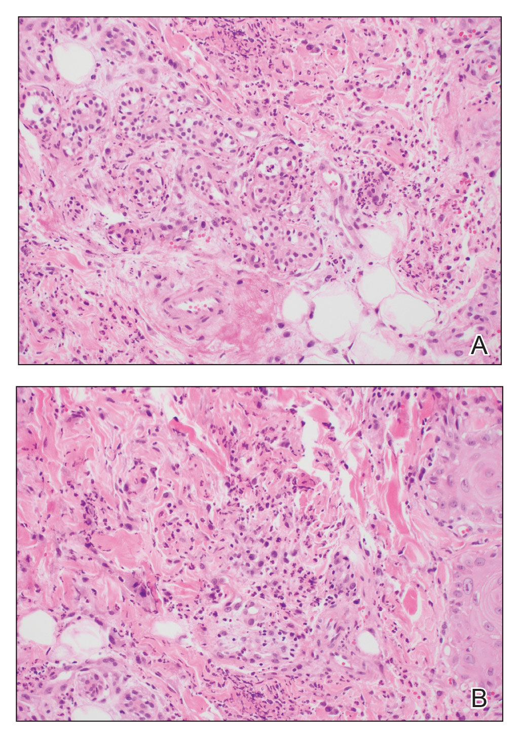
Neutrophilic eccrine hidradenitis is a rare reactive neutrophilic dermatosis characterized by eccrine gland involvement. This benign and self-limited condition presents as asymmetric erythematous papules and plaques.2 Among 8 granulocytopenic patients with neutrophilic dermatoses, 5 were diagnosed with NEH.4 Although first identified in 1982, NEH remains poorly understood.2 Initial theories suggested that NEH developed due to cytotoxic substances secreted in sweat glands causing necrosis and neutrophil chemotaxis; however, chemotherapy exposure cannot be linked to every case of NEH. Neutrophilic eccrine hidradenitis can be extremely difficult to differentiate clinically from conditions such as cellulitis and Sweet syndrome.
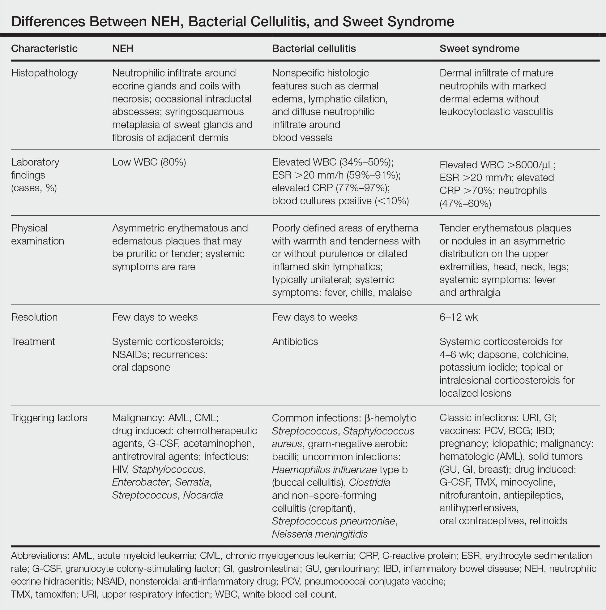
A patient history can be helpful in identifying triggering factors. Neutrophilic eccrine hidradenitis most commonly is associated with malignant, drug-induced, or infectious triggers, while Sweet syndrome has a broad range of associations including infections, vaccines, inflammatory bowel disease, pregnancy, malignancy, and drug-induced etiologies (Table).1 On average, NEH presents 10 days after chemotherapy induction, with 70% of cases presenting after the first chemotherapy cycle.5 Bacterial cellulitis or erysipelas have an infectious etiology, and patients may report symptoms such as fever, chills, or malaise. Immunosuppressed patients are at greater risk for infection; therefore, clinical signs of infection in a granulocytopenic patient should be addressed urgently.
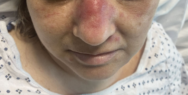
Physical examination may have limited value in differentiating between these diagnoses, as neutrophilic dermatoses notoriously mimic infection. Cutaneous lesions can appear as pruritic or tender erythematous plaques, papules, or nodules in these conditions, though cellulitis and erysipelas tend to be unilateral and may have associated purulence or inflamed skin lymphatics. Given the potential for misdiagnosis, approaching patients with a broad differential can be helpful. In our patient, the differential diagnosis included Sweet syndrome, NEH, bacterial cellulitis, erysipelas, leukemia cutis, sarcoid, and eosinophilic cellulitis.
Leukemia cutis refers to the infiltration of neoplastic leukocytes in the skin and often occurs in patients with peripheral leukemia, most often acute myeloid leukemia or chronic lymphocytic leukemia. Patients with leukemia cutis often have a worse prognosis, as this finding signifies extramedullary spread of disease.6 Clinically, lesions can appear similar to those seen in our patient, though they typically are not symptomatic, can be nodular, tend to exhibit a violaceous hue, and occasionally may be hemorrhagic. Wells syndrome (also known as eosinophilic cellulitis) is an inflammatory dermatosis that presents as painful or pruritic, edematous and erythematous plaques.7,8 A green hue on resolution is present in some cases and may help clinicians differentiate this disease from mimickers.7 Often, eosinophilic cellulitis is misdiagnosed as bacterial cellulitis and treated with antibiotics. The presence of systemic symptoms such as fever or arthralgia is more typical of bacterial cellulitis, erysipelas, eosinophilic cellulitis, or Sweet syndrome than of NEH.1 Additionally, inflammatory markers (ie, C-reactive protein) and white blood cell counts tend to be elevated in bacterial cellulitis and Sweet syndrome, while leukopenia often is seen in NEH.
Histopathology is crucial in distinguishing these disease etiologies. Neutrophilic eccrine hidradenitis is diagnosed by the characteristic neutrophilic infiltrate and necrosis surrounding eccrine glands and coils. There also may be occasional intraductal abscesses and syringosquamous metaplasia of the sweat glands along with fibrosis of the adjacent dermis. In contrast, histologic sections of Sweet syndrome show numerous mature neutrophils infiltrating the dermis with marked papillary dermal edema. The histopathology of bacterial cellulitis and erysipelas is less specific, but common features include dermal edema, lymphatic dilation, and a diffuse neutrophilic infiltrate surrounding blood vessels. Pathogenic organisms may be seen on histopathology but are not required for the diagnosis of bacterial cellulitis or erysipelas.2 Additionally, blood and tissue culture can assist in identification of both the source of infection and the causative organism, but cultures may not always be positive.
Comparatively, the histopathologic features of eosinophilic cellulitis include dermal edema, eosinophilic infiltration, and flame figures that form when eosinophils degranulate and coat collagen fibers with major basic protein. Flame figures are characteristic but not pathognomonic for eosinophilic cellulitis.7 The histopathology of leukemia cutis varies based on the leukemia classification; generally, in acute myeloid leukemia the infiltrate is composed of neoplastic cells in the early stages of development that are positive for myeloid markers such as myeloperoxidase. Atypical and immature granulocytes within the leukocytic infiltrate differentiate this condition from the other diagnoses. Treatment may entail chemotherapy or radiotherapy, and this diagnosis generally carries the worst prognosis of all the conditions in the differential.6
Differentiating between these conditions is important in guiding treatment, especially in patients with febrile neutropenia. Unnecessary steroids in immunosuppressed patients can be dangerous, especially if the patient has an infection such as bacterial cellulitis. Furthermore, unwarranted antibiotic use for noninfectious conditions may expose patients to substantial side effects and not improve the condition. Neutrophilic eccrine hidradenitis typically is self-limited and treated symptomatically with systemic corticosteroids and nonsteroidal anti-inflammatory drugs.3 Sweet syndrome often requires a longer course of oral steroids. Leukemia cutis worsens as the leukemia advances, and treatment of the underlying malignancy is the most effective treatment.9
Early and accurate recognition of the diagnosis can prevent harmful diagnostic delay, unnecessary antibiotic use, or extended steroid taper in neutropenic patients. Appreciating the differences between these diagnoses can assist clinicians in investigating and tailoring a broad differential to specific patient presentations, which is especially critical when considering common mimickers for life-threatening conditions.
- Nelson CA, Stephen S, Ashchyan HJ, et al. Neutrophilic dermatoses. J Am Acad Dermatol. 2018;79:987-1006. doi:10.1016/j.jaad.2017.11.0642
- Srivastava M, Scharf S, Meehan SA, et al. Neutrophilic eccrine hidradenitis masquerading as facial cellulitis. J Am Acad Dermatol. 2007;56:693-696. doi:10.1016/j.jaad.2006.07.032
- Copaescu AM, Castilloux JF, Chababi-Atallah M, et al. A classic clinical case: neutrophilic eccrine hidradenitis. Case Rep Dermatol. 2013; 5:340-346. doi:10.1159/000356229
- Aractingi S, Mallet V, Pinquier L, et al. Neutrophilic dermatoses during granulocytopenia. Arch Dermatol. 1995;131:1141-1145.
- Cohen PR. Neutrophilic dermatoses occurring in oncology patients. Int J Dermatol. 2007;46:106-111. doi:10.1111/j.1365-4632.2006.02605.x
- Wang CX, Pusic I, Anadkat MJ. Association of leukemia cutis with survival in acute myeloid leukemia. JAMA Dermatol. 2019;155:826. doi:10.1001/jamadermatol.2019.0052
- Chung CL, Cusack CA. Wells syndrome: an enigmatic and therapeutically challenging disease. J Drugs Dermatol. 2006;5:908-911.
- Räßler F, Lukács J, Elsner P. Treatment of eosinophilic cellulitis (Wells syndrome): a systematic review. J Eur Acad Dermatol Venereol. 2016;30:1465-1479. doi:10.1111/jdv.13706
- Hobbs LK, Carr PC, Gru AA, et al. Case and review: cutaneous involvement by chronic neutrophilic leukemia vs Sweet syndrome: a diagnostic dilemma. J Cutan Pathol. 2021;48:644-649. doi:10.1111 /cup.13925
THE DIAGNOSIS: Neutrophilic Eccrine Hidradenitis
A biopsy from the left preauricular cheek demonstrated dermal neutrophilic inflammation around eccrine coils with focal necrosis (Figure). No notable diffuse dermal neutrophilic infiltrate was present, ruling out Sweet syndrome, and no notable interstitial neutrophilic infiltrate was present, making cellulitis and erysipelas less likely; panculture of tissue also was negative.1,2 Atypical cells in the deep dermis were positive for CD163 and negative for CD117, CD34, CD123, and myeloperoxidase, consistent with a diagnosis of neutrophilic eccrine hidradenitis (NEH) and reactive histiocytes.3 Treatment with oral prednisone resulted in rapid improvement of symptoms.
Neutrophilic eccrine hidradenitis is a rare reactive neutrophilic dermatosis characterized by eccrine gland involvement. This benign and self-limited condition presents as asymmetric erythematous papules and plaques.2 Among 8 granulocytopenic patients with neutrophilic dermatoses, 5 were diagnosed with NEH.4 Although first identified in 1982, NEH remains poorly understood.2 Initial theories suggested that NEH developed due to cytotoxic substances secreted in sweat glands causing necrosis and neutrophil chemotaxis; however, chemotherapy exposure cannot be linked to every case of NEH. Neutrophilic eccrine hidradenitis can be extremely difficult to differentiate clinically from conditions such as cellulitis and Sweet syndrome.
A patient history can be helpful in identifying triggering factors. Neutrophilic eccrine hidradenitis most commonly is associated with malignant, drug-induced, or infectious triggers, while Sweet syndrome has a broad range of associations including infections, vaccines, inflammatory bowel disease, pregnancy, malignancy, and drug-induced etiologies (Table).1 On average, NEH presents 10 days after chemotherapy induction, with 70% of cases presenting after the first chemotherapy cycle.5 Bacterial cellulitis or erysipelas have an infectious etiology, and patients may report symptoms such as fever, chills, or malaise. Immunosuppressed patients are at greater risk for infection; therefore, clinical signs of infection in a granulocytopenic patient should be addressed urgently.
Physical examination may have limited value in differentiating between these diagnoses, as neutrophilic dermatoses notoriously mimic infection. Cutaneous lesions can appear as pruritic or tender erythematous plaques, papules, or nodules in these conditions, though cellulitis and erysipelas tend to be unilateral and may have associated purulence or inflamed skin lymphatics. Given the potential for misdiagnosis, approaching patients with a broad differential can be helpful. In our patient, the differential diagnosis included Sweet syndrome, NEH, bacterial cellulitis, erysipelas, leukemia cutis, sarcoid, and eosinophilic cellulitis.
Leukemia cutis refers to the infiltration of neoplastic leukocytes in the skin and often occurs in patients with peripheral leukemia, most often acute myeloid leukemia or chronic lymphocytic leukemia. Patients with leukemia cutis often have a worse prognosis, as this finding signifies extramedullary spread of disease.6 Clinically, lesions can appear similar to those seen in our patient, though they typically are not symptomatic, can be nodular, tend to exhibit a violaceous hue, and occasionally may be hemorrhagic. Wells syndrome (also known as eosinophilic cellulitis) is an inflammatory dermatosis that presents as painful or pruritic, edematous and erythematous plaques.7,8 A green hue on resolution is present in some cases and may help clinicians differentiate this disease from mimickers.7 Often, eosinophilic cellulitis is misdiagnosed as bacterial cellulitis and treated with antibiotics. The presence of systemic symptoms such as fever or arthralgia is more typical of bacterial cellulitis, erysipelas, eosinophilic cellulitis, or Sweet syndrome than of NEH.1 Additionally, inflammatory markers (ie, C-reactive protein) and white blood cell counts tend to be elevated in bacterial cellulitis and Sweet syndrome, while leukopenia often is seen in NEH.
Histopathology is crucial in distinguishing these disease etiologies. Neutrophilic eccrine hidradenitis is diagnosed by the characteristic neutrophilic infiltrate and necrosis surrounding eccrine glands and coils. There also may be occasional intraductal abscesses and syringosquamous metaplasia of the sweat glands along with fibrosis of the adjacent dermis. In contrast, histologic sections of Sweet syndrome show numerous mature neutrophils infiltrating the dermis with marked papillary dermal edema. The histopathology of bacterial cellulitis and erysipelas is less specific, but common features include dermal edema, lymphatic dilation, and a diffuse neutrophilic infiltrate surrounding blood vessels. Pathogenic organisms may be seen on histopathology but are not required for the diagnosis of bacterial cellulitis or erysipelas.2 Additionally, blood and tissue culture can assist in identification of both the source of infection and the causative organism, but cultures may not always be positive.
Comparatively, the histopathologic features of eosinophilic cellulitis include dermal edema, eosinophilic infiltration, and flame figures that form when eosinophils degranulate and coat collagen fibers with major basic protein. Flame figures are characteristic but not pathognomonic for eosinophilic cellulitis.7 The histopathology of leukemia cutis varies based on the leukemia classification; generally, in acute myeloid leukemia the infiltrate is composed of neoplastic cells in the early stages of development that are positive for myeloid markers such as myeloperoxidase. Atypical and immature granulocytes within the leukocytic infiltrate differentiate this condition from the other diagnoses. Treatment may entail chemotherapy or radiotherapy, and this diagnosis generally carries the worst prognosis of all the conditions in the differential.6
Differentiating between these conditions is important in guiding treatment, especially in patients with febrile neutropenia. Unnecessary steroids in immunosuppressed patients can be dangerous, especially if the patient has an infection such as bacterial cellulitis. Furthermore, unwarranted antibiotic use for noninfectious conditions may expose patients to substantial side effects and not improve the condition. Neutrophilic eccrine hidradenitis typically is self-limited and treated symptomatically with systemic corticosteroids and nonsteroidal anti-inflammatory drugs.3 Sweet syndrome often requires a longer course of oral steroids. Leukemia cutis worsens as the leukemia advances, and treatment of the underlying malignancy is the most effective treatment.9
Early and accurate recognition of the diagnosis can prevent harmful diagnostic delay, unnecessary antibiotic use, or extended steroid taper in neutropenic patients. Appreciating the differences between these diagnoses can assist clinicians in investigating and tailoring a broad differential to specific patient presentations, which is especially critical when considering common mimickers for life-threatening conditions.
THE DIAGNOSIS: Neutrophilic Eccrine Hidradenitis
A biopsy from the left preauricular cheek demonstrated dermal neutrophilic inflammation around eccrine coils with focal necrosis (Figure). No notable diffuse dermal neutrophilic infiltrate was present, ruling out Sweet syndrome, and no notable interstitial neutrophilic infiltrate was present, making cellulitis and erysipelas less likely; panculture of tissue also was negative.1,2 Atypical cells in the deep dermis were positive for CD163 and negative for CD117, CD34, CD123, and myeloperoxidase, consistent with a diagnosis of neutrophilic eccrine hidradenitis (NEH) and reactive histiocytes.3 Treatment with oral prednisone resulted in rapid improvement of symptoms.
Neutrophilic eccrine hidradenitis is a rare reactive neutrophilic dermatosis characterized by eccrine gland involvement. This benign and self-limited condition presents as asymmetric erythematous papules and plaques.2 Among 8 granulocytopenic patients with neutrophilic dermatoses, 5 were diagnosed with NEH.4 Although first identified in 1982, NEH remains poorly understood.2 Initial theories suggested that NEH developed due to cytotoxic substances secreted in sweat glands causing necrosis and neutrophil chemotaxis; however, chemotherapy exposure cannot be linked to every case of NEH. Neutrophilic eccrine hidradenitis can be extremely difficult to differentiate clinically from conditions such as cellulitis and Sweet syndrome.
A patient history can be helpful in identifying triggering factors. Neutrophilic eccrine hidradenitis most commonly is associated with malignant, drug-induced, or infectious triggers, while Sweet syndrome has a broad range of associations including infections, vaccines, inflammatory bowel disease, pregnancy, malignancy, and drug-induced etiologies (Table).1 On average, NEH presents 10 days after chemotherapy induction, with 70% of cases presenting after the first chemotherapy cycle.5 Bacterial cellulitis or erysipelas have an infectious etiology, and patients may report symptoms such as fever, chills, or malaise. Immunosuppressed patients are at greater risk for infection; therefore, clinical signs of infection in a granulocytopenic patient should be addressed urgently.
Physical examination may have limited value in differentiating between these diagnoses, as neutrophilic dermatoses notoriously mimic infection. Cutaneous lesions can appear as pruritic or tender erythematous plaques, papules, or nodules in these conditions, though cellulitis and erysipelas tend to be unilateral and may have associated purulence or inflamed skin lymphatics. Given the potential for misdiagnosis, approaching patients with a broad differential can be helpful. In our patient, the differential diagnosis included Sweet syndrome, NEH, bacterial cellulitis, erysipelas, leukemia cutis, sarcoid, and eosinophilic cellulitis.
Leukemia cutis refers to the infiltration of neoplastic leukocytes in the skin and often occurs in patients with peripheral leukemia, most often acute myeloid leukemia or chronic lymphocytic leukemia. Patients with leukemia cutis often have a worse prognosis, as this finding signifies extramedullary spread of disease.6 Clinically, lesions can appear similar to those seen in our patient, though they typically are not symptomatic, can be nodular, tend to exhibit a violaceous hue, and occasionally may be hemorrhagic. Wells syndrome (also known as eosinophilic cellulitis) is an inflammatory dermatosis that presents as painful or pruritic, edematous and erythematous plaques.7,8 A green hue on resolution is present in some cases and may help clinicians differentiate this disease from mimickers.7 Often, eosinophilic cellulitis is misdiagnosed as bacterial cellulitis and treated with antibiotics. The presence of systemic symptoms such as fever or arthralgia is more typical of bacterial cellulitis, erysipelas, eosinophilic cellulitis, or Sweet syndrome than of NEH.1 Additionally, inflammatory markers (ie, C-reactive protein) and white blood cell counts tend to be elevated in bacterial cellulitis and Sweet syndrome, while leukopenia often is seen in NEH.
Histopathology is crucial in distinguishing these disease etiologies. Neutrophilic eccrine hidradenitis is diagnosed by the characteristic neutrophilic infiltrate and necrosis surrounding eccrine glands and coils. There also may be occasional intraductal abscesses and syringosquamous metaplasia of the sweat glands along with fibrosis of the adjacent dermis. In contrast, histologic sections of Sweet syndrome show numerous mature neutrophils infiltrating the dermis with marked papillary dermal edema. The histopathology of bacterial cellulitis and erysipelas is less specific, but common features include dermal edema, lymphatic dilation, and a diffuse neutrophilic infiltrate surrounding blood vessels. Pathogenic organisms may be seen on histopathology but are not required for the diagnosis of bacterial cellulitis or erysipelas.2 Additionally, blood and tissue culture can assist in identification of both the source of infection and the causative organism, but cultures may not always be positive.
Comparatively, the histopathologic features of eosinophilic cellulitis include dermal edema, eosinophilic infiltration, and flame figures that form when eosinophils degranulate and coat collagen fibers with major basic protein. Flame figures are characteristic but not pathognomonic for eosinophilic cellulitis.7 The histopathology of leukemia cutis varies based on the leukemia classification; generally, in acute myeloid leukemia the infiltrate is composed of neoplastic cells in the early stages of development that are positive for myeloid markers such as myeloperoxidase. Atypical and immature granulocytes within the leukocytic infiltrate differentiate this condition from the other diagnoses. Treatment may entail chemotherapy or radiotherapy, and this diagnosis generally carries the worst prognosis of all the conditions in the differential.6
Differentiating between these conditions is important in guiding treatment, especially in patients with febrile neutropenia. Unnecessary steroids in immunosuppressed patients can be dangerous, especially if the patient has an infection such as bacterial cellulitis. Furthermore, unwarranted antibiotic use for noninfectious conditions may expose patients to substantial side effects and not improve the condition. Neutrophilic eccrine hidradenitis typically is self-limited and treated symptomatically with systemic corticosteroids and nonsteroidal anti-inflammatory drugs.3 Sweet syndrome often requires a longer course of oral steroids. Leukemia cutis worsens as the leukemia advances, and treatment of the underlying malignancy is the most effective treatment.9
Early and accurate recognition of the diagnosis can prevent harmful diagnostic delay, unnecessary antibiotic use, or extended steroid taper in neutropenic patients. Appreciating the differences between these diagnoses can assist clinicians in investigating and tailoring a broad differential to specific patient presentations, which is especially critical when considering common mimickers for life-threatening conditions.
- Nelson CA, Stephen S, Ashchyan HJ, et al. Neutrophilic dermatoses. J Am Acad Dermatol. 2018;79:987-1006. doi:10.1016/j.jaad.2017.11.0642
- Srivastava M, Scharf S, Meehan SA, et al. Neutrophilic eccrine hidradenitis masquerading as facial cellulitis. J Am Acad Dermatol. 2007;56:693-696. doi:10.1016/j.jaad.2006.07.032
- Copaescu AM, Castilloux JF, Chababi-Atallah M, et al. A classic clinical case: neutrophilic eccrine hidradenitis. Case Rep Dermatol. 2013; 5:340-346. doi:10.1159/000356229
- Aractingi S, Mallet V, Pinquier L, et al. Neutrophilic dermatoses during granulocytopenia. Arch Dermatol. 1995;131:1141-1145.
- Cohen PR. Neutrophilic dermatoses occurring in oncology patients. Int J Dermatol. 2007;46:106-111. doi:10.1111/j.1365-4632.2006.02605.x
- Wang CX, Pusic I, Anadkat MJ. Association of leukemia cutis with survival in acute myeloid leukemia. JAMA Dermatol. 2019;155:826. doi:10.1001/jamadermatol.2019.0052
- Chung CL, Cusack CA. Wells syndrome: an enigmatic and therapeutically challenging disease. J Drugs Dermatol. 2006;5:908-911.
- Räßler F, Lukács J, Elsner P. Treatment of eosinophilic cellulitis (Wells syndrome): a systematic review. J Eur Acad Dermatol Venereol. 2016;30:1465-1479. doi:10.1111/jdv.13706
- Hobbs LK, Carr PC, Gru AA, et al. Case and review: cutaneous involvement by chronic neutrophilic leukemia vs Sweet syndrome: a diagnostic dilemma. J Cutan Pathol. 2021;48:644-649. doi:10.1111 /cup.13925
- Nelson CA, Stephen S, Ashchyan HJ, et al. Neutrophilic dermatoses. J Am Acad Dermatol. 2018;79:987-1006. doi:10.1016/j.jaad.2017.11.0642
- Srivastava M, Scharf S, Meehan SA, et al. Neutrophilic eccrine hidradenitis masquerading as facial cellulitis. J Am Acad Dermatol. 2007;56:693-696. doi:10.1016/j.jaad.2006.07.032
- Copaescu AM, Castilloux JF, Chababi-Atallah M, et al. A classic clinical case: neutrophilic eccrine hidradenitis. Case Rep Dermatol. 2013; 5:340-346. doi:10.1159/000356229
- Aractingi S, Mallet V, Pinquier L, et al. Neutrophilic dermatoses during granulocytopenia. Arch Dermatol. 1995;131:1141-1145.
- Cohen PR. Neutrophilic dermatoses occurring in oncology patients. Int J Dermatol. 2007;46:106-111. doi:10.1111/j.1365-4632.2006.02605.x
- Wang CX, Pusic I, Anadkat MJ. Association of leukemia cutis with survival in acute myeloid leukemia. JAMA Dermatol. 2019;155:826. doi:10.1001/jamadermatol.2019.0052
- Chung CL, Cusack CA. Wells syndrome: an enigmatic and therapeutically challenging disease. J Drugs Dermatol. 2006;5:908-911.
- Räßler F, Lukács J, Elsner P. Treatment of eosinophilic cellulitis (Wells syndrome): a systematic review. J Eur Acad Dermatol Venereol. 2016;30:1465-1479. doi:10.1111/jdv.13706
- Hobbs LK, Carr PC, Gru AA, et al. Case and review: cutaneous involvement by chronic neutrophilic leukemia vs Sweet syndrome: a diagnostic dilemma. J Cutan Pathol. 2021;48:644-649. doi:10.1111 /cup.13925
A 50-year-old woman undergoing cytarabine induction therapy for acute myeloid leukemia developed tender, erythematous, dermal plaques on the nasal dorsum, left medial eyebrow, left preauricular cheek, and right cheek. The rash erupted 7 days after receiving the cytarabine induction regimen. She had a fever (temperature, 39.9 °C [103.8 °F]) and also was neutropenic.
2023 Update on gynecologic cancer
In 2022, the most significant advances in the treatment of gynecologic cancers were achieved for patients with ovarian cancer. While ovarian cancer continues to carry the worst prognosis of all gynecologic cancers, 5-year relative survival has gradually increased, from 34.4% in 1975 to 52.4% in 2014.1
In this Update, we highlight the recent advances in our understanding of targeted therapy in ovarian cancer. We review SORAYA, a trial that demonstrated that mirvetuximab soravtansine, an antibody-drug conjugate, has promising efficacy in platinum-resistant ovarian cancers that overexpress folate receptor α. We also spotlight progress in the treatment of low-grade serous ovarian cancer, another notoriously chemotherapy-resistant disease, in GOG 281/LOGS, a phase 2 study of the MEK inhibitor trametinib. Finally, we discuss emerging long-term follow-up data on poly(ADP-ribose) polymerase (PARP) inhibitors, which are helping to refine the role of these groundbreaking drugs.
New drug approved for platinum-resistant epithelial ovarian cancer—the first since 2014
Matulonis UA, Lorusso D, Oaknin A, et al. Efficacy and safety of mirvetuximab soravtansine in patients with platinum-resistant ovarian cancer with high folate receptor alpha expression: results from the SORAYA study. J Clin Oncol. 2023;41:2436-2445. doi:10.1200/JCO.22.01900.
While most patients diagnosed with advanced ovarian cancer will respond to platinum-based chemotherapy, those whose disease recurs eventually develop resistance to platinum agents. Treatment options for platinum-resistant ovarian cancer are limited and prognosis is poor. Most regimens have a response rate of only 10%. Since the approval of bevacizumab combined with chemotherapy in 2014, no new agents have been approved by the US Food and Drug Administration (FDA) for use in platinum-resistant ovarian cancer.
Efficacy shown with mirvetuximab
Recently, Matulonis and colleagues published results of the SORAYA study, a single-arm,phase 2 trial, that examined the efficacy and safety of mirvetuximab soravtansine-gynx among women with platinum-resistant ovarian cancer.2 Mirvetuximab is an antibody-drug conjugate composed of an antibody directed at the folate receptor α attached to a cytotoxic microtubule inhibitor.
The study included 106 patients with platinum-resistant ovarian cancer whose tumors expressed folate receptor α at a high level—a feature of approximately 50% of patients screened for the study. Twenty-nine patients experienced a partial response and 5 had a complete response, corresponding to a remarkable objective response rate of 32.4%. The median progression-free survival was 4.3 months.
Like other antibody-drug conjugates, ocular toxicities, including blurred vision (41%) and keratopathy (29%), were common. However, toxicity was manageable and rarely led to drug discontinuation.
The FDA has granted accelerated approval to mirvetuximab soravtansine-gynx for women with platinum-resistant ovarian cancer with high folate receptor α expression who have received 1 to 3 prior systemic treatment regimens.
Continue to: A novel agent for recurrent low-grade serous ovarian carcinoma...
A novel agent for recurrent low-grade serous ovarian carcinoma
Low-grade serous carcinoma is a histologic subtype that makes up approximately 5% of all epithelial ovarian cancers.3 Patients with low-grade serous carcinoma are often younger and, because of the indolent nature of the histology, generally have a longer overall survival compared with patients with high-grade serous carcinoma. Unlike high-grade disease, however, low-grade serous carcinoma usually is resistant to chemotherapy, making treatment options limited for patients with advanced and recurrent disease.
Trametinib: A potential option
In an international, randomized, open-label trial (GOG 281/LOGS), Gershenson and colleagues investigated the efficacy of trametinib compared with standard-of-care chemotherapy in patients with recurrent low-grade serous ovarian cancer.4 Trametinib, a mitogen-activated protein kinase MEK inhibitor, is a targeted agent that is FDA approved for treatment in BRAF-mutated melanoma, lung, and thyroid cancers.
Patients with recurrent low-grade serous ovarian cancer were randomly assigned to trametinib (n = 130) or 1 of 5 standard-of-care treatment options (n = 130), including both chemotherapy and hormonal treatments. Those assigned to trametinib were significantly less likely to have disease progression (78% vs 89%), with a median progression-free survival of 13 months, compared with7.2 months in controls (hazard ratio [HR], 0.48; 95% confidence interval [CI], 0.36–0.64). Additionally, patients who had a radiographic response to trametinib experienced a longer duration of response compared with those who responded to standard-of-care treatment (13.6 months vs 5.9 months).
While there was no statistically significant difference in overall survival (HR, 0.76; 95% CI, 0.51–1.12), crossover to trametinib from the standard-of-care group was allowed and occurred among 68% of patients, which limits the study’s ability to measure differences in overall survival.
Trametinib was well tolerated by patients, but skin rash and anemia followed by hypertension were the most common adverse effects. In the standard-of-care group, the most common toxicities were abdominal pain, nausea, and anemia. A slightly higher proportion of patients in the trametinib group discontinued the drug due to toxicity compared with the standard-of-care group (36% vs 30%), but the there was no difference between the 2 groups in scores on quality-of-life assessments.
Although trametinib is not yet FDA approved for the treatment of ovarian cancer, the National Comprehensive Cancer Network has added trametinib as a treatment option for recurrent low-grade serous ovarian carcinoma, given the significant improvement in progression-free survival compared with standard-of-care treatment.
Continue to: PARP inhibitors benefit many women with ovarian cancer, but they may hurt others...
PARP inhibitors benefit many women with ovarian cancer, but they may hurt others
Monk BJ, Parkinson C, Lim MC, et al. A randomized, phase III trial to evaluate rucaparib monotherapy as maintenance treatment in patients with newly diagnosed ovarian cancer (ATHENA-MONO/GOG-3020/ENGOT-ov45). J Clin Oncol. 2022;40:3952-3964. doi:10.1200/JCO.22.01003.
Poly(ADP-ribose) polymerase (PARP) inhibitors are a class of oral anticancer agents that target DNA repair. Since the initial FDA approval in 2014 of olaparib for the treatment of patients with recurrent BRCA-mutated ovarian cancer, PARP inhibitors have been approved for maintenance in both the frontline setting and after platinum-sensitive recurrence, and as single-agenttreatment for ovarian cancer with BRCA mutations or evidence of homologous repair deficiency (HRD), a BRCA-like molecular phenotype.5 The expanding role for PARP inhibitors in ovarian cancer seemed inexorable.
Restricted prescribing advised
In 2022, we learned that in certain settings, PARP inhibitors may be the wrong choice. Several “Dear Health Care Provider” letters were issued by AstraZeneca, Clovis, and GSK to advise physicians to restrict the prescribing of olaparib, rucaparib, and niraparib.6,7
AstraZeneca and Clovis issued letters spurred by the final analysis of ARIEL4 and SOLO3 studies, 2 randomized trials that investigated, respectively, rucaparib and olaparib monotherapy compared with chemotherapy in recurrent ovarian cancer.8,9 In both cases patients randomized to PARP inhibitors may have experienced an overall survival decrement compared with those who received chemotherapy.
At the FDA’s request, Clovis has withdrawn rucaparib as a treatment for patients with recurrent BRCA-mutant ovarian cancer who had received 2 or more lines of chemotherapy, and AstraZeneca withdrew olaparib monotherapy in germline BRCA-mutant patients with recurrent ovarian cancer. Shortly after these withdrawals, GSK also withdrew its indication for niraparib as a treatment for women with HRD, platinum-sensitive ovarian cancer who have received 3 or more prior chemotherapies. Furthermore, based on the final overall survival analysis of the NOVA study, GSK also restricted its indication for niraparib maintenance for recurrent ovarian cancer to patients with germline BRCA mutations, due to evidence of an overall survival detriment in this setting.10
Positive study results
Fortunately, 2022 was not all bad news for PARP inhibitors in ovarian cancer. In June 2022, the ATHENA-MONO trial, a phase 3 double-blind randomized controlled trial, demonstrated that rucaparib maintenance in patients with newly diagnosed epithelial ovarian cancer was associated with a significantly longer progression-free survival compared with placebo.11 The effect was most pronounced in the BRCA-mutant/HRD population, with a median progression-free survival of 28.7 months in the rucaparib group compared with 11.3 months in the placebo group (HR, 0.47; 95% CI, 0.31–0.72). Thus, rucaparib was added to the list of PARP inhibitors approved for upfront maintenance therapy in epithelial ovarian cancer.
Similarly, the long-term overall survival analysis from the upfront trials SOLO-1 and PAOLA-1 showed an overall survival advantage of PARP inhibitor, compared with placebo, maintenance in patients with BRCA mutations or HRD tumors.12,13 ●
PARP inhibitor maintenance therapy after upfront chemotherapy in women with BRCA-mutant and HRD epithelial ovarian cancer has been game changing in ovarian cancer. However, PARP inhibitors have a more limited role than previously thought for patients with recurrent ovarian cancer.
- Cancer stat facts: ovarian cancer. National Cancer Institute Surveillance, Epidemiology, and End Results Program. Accessed March 11, 2023. https://seer.cancer.gov/statfacts /html/ovary.html
- Matulonis UA, Lorusso D, Oaknin A, et al. Efficacy and safety of mirvetuximab soravtansine in patients with platinumresistant ovarian cancer with high folate receptor alpha expression: results from the SORAYA study. J Clin Oncol. 2023;41:2436-2445. doi:10.1200/JCO.22.01900
- Prat J, D’Angelo E, Espinosa I. Ovarian carcinomas: at least five different diseases with distinct histological features and molecular genetics. Hum Pathol. 2018;80:11-27. doi:10.1016 /j.humpath.2018.06.018
- Gershenson DM, Miller A, Brady WE, et al. Trametinib versus standard of care in patients with recurrent low-grade serous ovarian cancer (GOG 281/LOGS): an international, randomised, open-label, multicentre, phase 2/3 trial. Lancet. 2022;399:541-553. doi:10.1016/S0140-6736(21)02175-9
- Tew WP, Lacchetti C, Ellis A, et al. PARP inhibitors in the management of ovarian cancer: ASCO guideline. J Clin Oncol. 2020;38:3468-3493. doi:10.1200/JCO.20.01924
- Rubraca (rucaparib) for treatment of BRCA-mutated ovarian cancer after 2 or more chemotherapies is voluntarily withdrawn in the US. Clovis Oncology. June 2022. Accessed May 11, 2022. chrome-extension://efaidnbmnnnibpcajpcglcle findmkaj/https://clovisoncology.com/pdfs/US_DHCPL _final_signed.pdf
- Lynparza (olaparib) for treatment of adult patients with deleterious or suspected deleterious germline BRCA-mutated (gBRCAm) advanced ovarian cancer who have been treated with three or more prior lines of chemotherapy is voluntarily withdrawn in the US. AstraZeneca. August 26, 2022. Accessed May 11, 2023. https://www.lynparzahcp.com/content/dam /physician-services/us/590-lynparza-hcp-branded/hcp -global/pdf/solo3-dhcp-final-signed.pdf
- Penson RT, Valencia RV, Cibula D, et al. Olaparib versus nonplatinum chemotherapy in patients with platinum-sensitive relapsed ovarian cancer and a germline BRCA1/2 mutation (SOLO3): a randomized phase III trial. J Clin Oncol. 2020;38:1164-1174. doi:10.1200/JCO.19.02745
- Kristeleit R, Lisyanskaya A, Fedenko A, et al. Rucaparib versus standard-of-care chemotherapy in patients with relapsed ovarian cancer and a deleterious BRCA1 or BRCA2 mutation (ARIEL4): an international, open-label, randomised, phase 3 trial. Lancet Oncol. 2022;23:465-478. doi:10.1016 /S1470-2045(22)00122-X
- Dear Health Care Provider Letter (Niraparib). GSK. November 2022. Accessed May 11, 2023. https://www.zejulahcp .com/content/dam/cf-pharma/hcp-zejulahcp-v2/en_US /pdf/ZEJULA%20(niraparib)%20Dear%20HCP%20Letter%20 November%202022.pdf
- Monk BJ, Parkinson C, Lim MC, et al. A randomized, phase III trial to evaluate rucaparib monotherapy as maintenance treatment in patients with newly diagnosed ovarian cancer (ATHENA-MONO/GOG-3020/ENGOT-ov45). J Clin Oncol. 2022;40:3952-3964. doi:10.1200/JCO.22.01003
- Moore K, Colombo N, Scambia G, et al. Maintenance olaparib in patients with newly diagnosed advanced ovarian cancer. N Engl J Med. 2018;379:2495-2505. doi:10.1056 /NEJMoa1810858
- Ray-Coquard I, Pautier P, Pignata S, et al; PAOLA-1 Investigators. Olaparib plus bevacizumab as first-line maintenance in ovarian cancer. N Engl J Med. 2019;381:2416-2428. doi:10.1056/NEJMoa1911361

Alexandra S. Bercow, MD
Dr. Bercow is a Clinical and Research
Fellow in the Meigs Division of
Gynecologic Oncology, Vincent
Department of Obstetrics and Gynecology,
at Massachusetts General Hospital,
a Harvard Medical School
affiliated hospital.

Alexander Melamed, MD, MPH
Dr. Melamed is an Assistant Professor
in the Meigs Division of Gynecologic
Oncology, Vincent Department
of Obstetrics and Gynecology, at
Massachusetts General Hospital, a Harvard
Medical School affiliated hospital.
He is also the Norman F. Gant American
Board of Obstetrics and Gynecology Fellow
at the National Academy of Medicine.
Dr. Melamed serves on the advisory board of AstraZeneca and is a consultant for Kaya17. Dr. Bercow reports no financial relationships relevant to this article.
Alexandra S. Bercow, MD
Dr. Bercow is a Clinical and Research
Fellow in the Meigs Division of
Gynecologic Oncology, Vincent
Department of Obstetrics and Gynecology,
at Massachusetts General Hospital,
a Harvard Medical School
affiliated hospital.
Alexander Melamed, MD, MPH
Dr. Melamed is an Assistant Professor
in the Meigs Division of Gynecologic
Oncology, Vincent Department
of Obstetrics and Gynecology, at
Massachusetts General Hospital, a Harvard
Medical School affiliated hospital.
He is also the Norman F. Gant American
Board of Obstetrics and Gynecology Fellow
at the National Academy of Medicine.
Dr. Melamed serves on the advisory board of AstraZeneca and is a consultant for Kaya17. Dr. Bercow reports no financial relationships relevant to this article.
Alexandra S. Bercow, MD
Dr. Bercow is a Clinical and Research
Fellow in the Meigs Division of
Gynecologic Oncology, Vincent
Department of Obstetrics and Gynecology,
at Massachusetts General Hospital,
a Harvard Medical School
affiliated hospital.
Alexander Melamed, MD, MPH
Dr. Melamed is an Assistant Professor
in the Meigs Division of Gynecologic
Oncology, Vincent Department
of Obstetrics and Gynecology, at
Massachusetts General Hospital, a Harvard
Medical School affiliated hospital.
He is also the Norman F. Gant American
Board of Obstetrics and Gynecology Fellow
at the National Academy of Medicine.
Dr. Melamed serves on the advisory board of AstraZeneca and is a consultant for Kaya17. Dr. Bercow reports no financial relationships relevant to this article.
In 2022, the most significant advances in the treatment of gynecologic cancers were achieved for patients with ovarian cancer. While ovarian cancer continues to carry the worst prognosis of all gynecologic cancers, 5-year relative survival has gradually increased, from 34.4% in 1975 to 52.4% in 2014.1
In this Update, we highlight the recent advances in our understanding of targeted therapy in ovarian cancer. We review SORAYA, a trial that demonstrated that mirvetuximab soravtansine, an antibody-drug conjugate, has promising efficacy in platinum-resistant ovarian cancers that overexpress folate receptor α. We also spotlight progress in the treatment of low-grade serous ovarian cancer, another notoriously chemotherapy-resistant disease, in GOG 281/LOGS, a phase 2 study of the MEK inhibitor trametinib. Finally, we discuss emerging long-term follow-up data on poly(ADP-ribose) polymerase (PARP) inhibitors, which are helping to refine the role of these groundbreaking drugs.
New drug approved for platinum-resistant epithelial ovarian cancer—the first since 2014
Matulonis UA, Lorusso D, Oaknin A, et al. Efficacy and safety of mirvetuximab soravtansine in patients with platinum-resistant ovarian cancer with high folate receptor alpha expression: results from the SORAYA study. J Clin Oncol. 2023;41:2436-2445. doi:10.1200/JCO.22.01900.
While most patients diagnosed with advanced ovarian cancer will respond to platinum-based chemotherapy, those whose disease recurs eventually develop resistance to platinum agents. Treatment options for platinum-resistant ovarian cancer are limited and prognosis is poor. Most regimens have a response rate of only 10%. Since the approval of bevacizumab combined with chemotherapy in 2014, no new agents have been approved by the US Food and Drug Administration (FDA) for use in platinum-resistant ovarian cancer.
Efficacy shown with mirvetuximab
Recently, Matulonis and colleagues published results of the SORAYA study, a single-arm,phase 2 trial, that examined the efficacy and safety of mirvetuximab soravtansine-gynx among women with platinum-resistant ovarian cancer.2 Mirvetuximab is an antibody-drug conjugate composed of an antibody directed at the folate receptor α attached to a cytotoxic microtubule inhibitor.
The study included 106 patients with platinum-resistant ovarian cancer whose tumors expressed folate receptor α at a high level—a feature of approximately 50% of patients screened for the study. Twenty-nine patients experienced a partial response and 5 had a complete response, corresponding to a remarkable objective response rate of 32.4%. The median progression-free survival was 4.3 months.
Like other antibody-drug conjugates, ocular toxicities, including blurred vision (41%) and keratopathy (29%), were common. However, toxicity was manageable and rarely led to drug discontinuation.
The FDA has granted accelerated approval to mirvetuximab soravtansine-gynx for women with platinum-resistant ovarian cancer with high folate receptor α expression who have received 1 to 3 prior systemic treatment regimens.
Continue to: A novel agent for recurrent low-grade serous ovarian carcinoma...
A novel agent for recurrent low-grade serous ovarian carcinoma
Low-grade serous carcinoma is a histologic subtype that makes up approximately 5% of all epithelial ovarian cancers.3 Patients with low-grade serous carcinoma are often younger and, because of the indolent nature of the histology, generally have a longer overall survival compared with patients with high-grade serous carcinoma. Unlike high-grade disease, however, low-grade serous carcinoma usually is resistant to chemotherapy, making treatment options limited for patients with advanced and recurrent disease.
Trametinib: A potential option
In an international, randomized, open-label trial (GOG 281/LOGS), Gershenson and colleagues investigated the efficacy of trametinib compared with standard-of-care chemotherapy in patients with recurrent low-grade serous ovarian cancer.4 Trametinib, a mitogen-activated protein kinase MEK inhibitor, is a targeted agent that is FDA approved for treatment in BRAF-mutated melanoma, lung, and thyroid cancers.
Patients with recurrent low-grade serous ovarian cancer were randomly assigned to trametinib (n = 130) or 1 of 5 standard-of-care treatment options (n = 130), including both chemotherapy and hormonal treatments. Those assigned to trametinib were significantly less likely to have disease progression (78% vs 89%), with a median progression-free survival of 13 months, compared with7.2 months in controls (hazard ratio [HR], 0.48; 95% confidence interval [CI], 0.36–0.64). Additionally, patients who had a radiographic response to trametinib experienced a longer duration of response compared with those who responded to standard-of-care treatment (13.6 months vs 5.9 months).
While there was no statistically significant difference in overall survival (HR, 0.76; 95% CI, 0.51–1.12), crossover to trametinib from the standard-of-care group was allowed and occurred among 68% of patients, which limits the study’s ability to measure differences in overall survival.
Trametinib was well tolerated by patients, but skin rash and anemia followed by hypertension were the most common adverse effects. In the standard-of-care group, the most common toxicities were abdominal pain, nausea, and anemia. A slightly higher proportion of patients in the trametinib group discontinued the drug due to toxicity compared with the standard-of-care group (36% vs 30%), but the there was no difference between the 2 groups in scores on quality-of-life assessments.
Although trametinib is not yet FDA approved for the treatment of ovarian cancer, the National Comprehensive Cancer Network has added trametinib as a treatment option for recurrent low-grade serous ovarian carcinoma, given the significant improvement in progression-free survival compared with standard-of-care treatment.
Continue to: PARP inhibitors benefit many women with ovarian cancer, but they may hurt others...
PARP inhibitors benefit many women with ovarian cancer, but they may hurt others
Monk BJ, Parkinson C, Lim MC, et al. A randomized, phase III trial to evaluate rucaparib monotherapy as maintenance treatment in patients with newly diagnosed ovarian cancer (ATHENA-MONO/GOG-3020/ENGOT-ov45). J Clin Oncol. 2022;40:3952-3964. doi:10.1200/JCO.22.01003.
Poly(ADP-ribose) polymerase (PARP) inhibitors are a class of oral anticancer agents that target DNA repair. Since the initial FDA approval in 2014 of olaparib for the treatment of patients with recurrent BRCA-mutated ovarian cancer, PARP inhibitors have been approved for maintenance in both the frontline setting and after platinum-sensitive recurrence, and as single-agenttreatment for ovarian cancer with BRCA mutations or evidence of homologous repair deficiency (HRD), a BRCA-like molecular phenotype.5 The expanding role for PARP inhibitors in ovarian cancer seemed inexorable.
Restricted prescribing advised
In 2022, we learned that in certain settings, PARP inhibitors may be the wrong choice. Several “Dear Health Care Provider” letters were issued by AstraZeneca, Clovis, and GSK to advise physicians to restrict the prescribing of olaparib, rucaparib, and niraparib.6,7
AstraZeneca and Clovis issued letters spurred by the final analysis of ARIEL4 and SOLO3 studies, 2 randomized trials that investigated, respectively, rucaparib and olaparib monotherapy compared with chemotherapy in recurrent ovarian cancer.8,9 In both cases patients randomized to PARP inhibitors may have experienced an overall survival decrement compared with those who received chemotherapy.
At the FDA’s request, Clovis has withdrawn rucaparib as a treatment for patients with recurrent BRCA-mutant ovarian cancer who had received 2 or more lines of chemotherapy, and AstraZeneca withdrew olaparib monotherapy in germline BRCA-mutant patients with recurrent ovarian cancer. Shortly after these withdrawals, GSK also withdrew its indication for niraparib as a treatment for women with HRD, platinum-sensitive ovarian cancer who have received 3 or more prior chemotherapies. Furthermore, based on the final overall survival analysis of the NOVA study, GSK also restricted its indication for niraparib maintenance for recurrent ovarian cancer to patients with germline BRCA mutations, due to evidence of an overall survival detriment in this setting.10
Positive study results
Fortunately, 2022 was not all bad news for PARP inhibitors in ovarian cancer. In June 2022, the ATHENA-MONO trial, a phase 3 double-blind randomized controlled trial, demonstrated that rucaparib maintenance in patients with newly diagnosed epithelial ovarian cancer was associated with a significantly longer progression-free survival compared with placebo.11 The effect was most pronounced in the BRCA-mutant/HRD population, with a median progression-free survival of 28.7 months in the rucaparib group compared with 11.3 months in the placebo group (HR, 0.47; 95% CI, 0.31–0.72). Thus, rucaparib was added to the list of PARP inhibitors approved for upfront maintenance therapy in epithelial ovarian cancer.
Similarly, the long-term overall survival analysis from the upfront trials SOLO-1 and PAOLA-1 showed an overall survival advantage of PARP inhibitor, compared with placebo, maintenance in patients with BRCA mutations or HRD tumors.12,13 ●
PARP inhibitor maintenance therapy after upfront chemotherapy in women with BRCA-mutant and HRD epithelial ovarian cancer has been game changing in ovarian cancer. However, PARP inhibitors have a more limited role than previously thought for patients with recurrent ovarian cancer.
In 2022, the most significant advances in the treatment of gynecologic cancers were achieved for patients with ovarian cancer. While ovarian cancer continues to carry the worst prognosis of all gynecologic cancers, 5-year relative survival has gradually increased, from 34.4% in 1975 to 52.4% in 2014.1
In this Update, we highlight the recent advances in our understanding of targeted therapy in ovarian cancer. We review SORAYA, a trial that demonstrated that mirvetuximab soravtansine, an antibody-drug conjugate, has promising efficacy in platinum-resistant ovarian cancers that overexpress folate receptor α. We also spotlight progress in the treatment of low-grade serous ovarian cancer, another notoriously chemotherapy-resistant disease, in GOG 281/LOGS, a phase 2 study of the MEK inhibitor trametinib. Finally, we discuss emerging long-term follow-up data on poly(ADP-ribose) polymerase (PARP) inhibitors, which are helping to refine the role of these groundbreaking drugs.
New drug approved for platinum-resistant epithelial ovarian cancer—the first since 2014
Matulonis UA, Lorusso D, Oaknin A, et al. Efficacy and safety of mirvetuximab soravtansine in patients with platinum-resistant ovarian cancer with high folate receptor alpha expression: results from the SORAYA study. J Clin Oncol. 2023;41:2436-2445. doi:10.1200/JCO.22.01900.
While most patients diagnosed with advanced ovarian cancer will respond to platinum-based chemotherapy, those whose disease recurs eventually develop resistance to platinum agents. Treatment options for platinum-resistant ovarian cancer are limited and prognosis is poor. Most regimens have a response rate of only 10%. Since the approval of bevacizumab combined with chemotherapy in 2014, no new agents have been approved by the US Food and Drug Administration (FDA) for use in platinum-resistant ovarian cancer.
Efficacy shown with mirvetuximab
Recently, Matulonis and colleagues published results of the SORAYA study, a single-arm,phase 2 trial, that examined the efficacy and safety of mirvetuximab soravtansine-gynx among women with platinum-resistant ovarian cancer.2 Mirvetuximab is an antibody-drug conjugate composed of an antibody directed at the folate receptor α attached to a cytotoxic microtubule inhibitor.
The study included 106 patients with platinum-resistant ovarian cancer whose tumors expressed folate receptor α at a high level—a feature of approximately 50% of patients screened for the study. Twenty-nine patients experienced a partial response and 5 had a complete response, corresponding to a remarkable objective response rate of 32.4%. The median progression-free survival was 4.3 months.
Like other antibody-drug conjugates, ocular toxicities, including blurred vision (41%) and keratopathy (29%), were common. However, toxicity was manageable and rarely led to drug discontinuation.
The FDA has granted accelerated approval to mirvetuximab soravtansine-gynx for women with platinum-resistant ovarian cancer with high folate receptor α expression who have received 1 to 3 prior systemic treatment regimens.
Continue to: A novel agent for recurrent low-grade serous ovarian carcinoma...
A novel agent for recurrent low-grade serous ovarian carcinoma
Low-grade serous carcinoma is a histologic subtype that makes up approximately 5% of all epithelial ovarian cancers.3 Patients with low-grade serous carcinoma are often younger and, because of the indolent nature of the histology, generally have a longer overall survival compared with patients with high-grade serous carcinoma. Unlike high-grade disease, however, low-grade serous carcinoma usually is resistant to chemotherapy, making treatment options limited for patients with advanced and recurrent disease.
Trametinib: A potential option
In an international, randomized, open-label trial (GOG 281/LOGS), Gershenson and colleagues investigated the efficacy of trametinib compared with standard-of-care chemotherapy in patients with recurrent low-grade serous ovarian cancer.4 Trametinib, a mitogen-activated protein kinase MEK inhibitor, is a targeted agent that is FDA approved for treatment in BRAF-mutated melanoma, lung, and thyroid cancers.
Patients with recurrent low-grade serous ovarian cancer were randomly assigned to trametinib (n = 130) or 1 of 5 standard-of-care treatment options (n = 130), including both chemotherapy and hormonal treatments. Those assigned to trametinib were significantly less likely to have disease progression (78% vs 89%), with a median progression-free survival of 13 months, compared with7.2 months in controls (hazard ratio [HR], 0.48; 95% confidence interval [CI], 0.36–0.64). Additionally, patients who had a radiographic response to trametinib experienced a longer duration of response compared with those who responded to standard-of-care treatment (13.6 months vs 5.9 months).
While there was no statistically significant difference in overall survival (HR, 0.76; 95% CI, 0.51–1.12), crossover to trametinib from the standard-of-care group was allowed and occurred among 68% of patients, which limits the study’s ability to measure differences in overall survival.
Trametinib was well tolerated by patients, but skin rash and anemia followed by hypertension were the most common adverse effects. In the standard-of-care group, the most common toxicities were abdominal pain, nausea, and anemia. A slightly higher proportion of patients in the trametinib group discontinued the drug due to toxicity compared with the standard-of-care group (36% vs 30%), but the there was no difference between the 2 groups in scores on quality-of-life assessments.
Although trametinib is not yet FDA approved for the treatment of ovarian cancer, the National Comprehensive Cancer Network has added trametinib as a treatment option for recurrent low-grade serous ovarian carcinoma, given the significant improvement in progression-free survival compared with standard-of-care treatment.
Continue to: PARP inhibitors benefit many women with ovarian cancer, but they may hurt others...
PARP inhibitors benefit many women with ovarian cancer, but they may hurt others
Monk BJ, Parkinson C, Lim MC, et al. A randomized, phase III trial to evaluate rucaparib monotherapy as maintenance treatment in patients with newly diagnosed ovarian cancer (ATHENA-MONO/GOG-3020/ENGOT-ov45). J Clin Oncol. 2022;40:3952-3964. doi:10.1200/JCO.22.01003.
Poly(ADP-ribose) polymerase (PARP) inhibitors are a class of oral anticancer agents that target DNA repair. Since the initial FDA approval in 2014 of olaparib for the treatment of patients with recurrent BRCA-mutated ovarian cancer, PARP inhibitors have been approved for maintenance in both the frontline setting and after platinum-sensitive recurrence, and as single-agenttreatment for ovarian cancer with BRCA mutations or evidence of homologous repair deficiency (HRD), a BRCA-like molecular phenotype.5 The expanding role for PARP inhibitors in ovarian cancer seemed inexorable.
Restricted prescribing advised
In 2022, we learned that in certain settings, PARP inhibitors may be the wrong choice. Several “Dear Health Care Provider” letters were issued by AstraZeneca, Clovis, and GSK to advise physicians to restrict the prescribing of olaparib, rucaparib, and niraparib.6,7
AstraZeneca and Clovis issued letters spurred by the final analysis of ARIEL4 and SOLO3 studies, 2 randomized trials that investigated, respectively, rucaparib and olaparib monotherapy compared with chemotherapy in recurrent ovarian cancer.8,9 In both cases patients randomized to PARP inhibitors may have experienced an overall survival decrement compared with those who received chemotherapy.
At the FDA’s request, Clovis has withdrawn rucaparib as a treatment for patients with recurrent BRCA-mutant ovarian cancer who had received 2 or more lines of chemotherapy, and AstraZeneca withdrew olaparib monotherapy in germline BRCA-mutant patients with recurrent ovarian cancer. Shortly after these withdrawals, GSK also withdrew its indication for niraparib as a treatment for women with HRD, platinum-sensitive ovarian cancer who have received 3 or more prior chemotherapies. Furthermore, based on the final overall survival analysis of the NOVA study, GSK also restricted its indication for niraparib maintenance for recurrent ovarian cancer to patients with germline BRCA mutations, due to evidence of an overall survival detriment in this setting.10
Positive study results
Fortunately, 2022 was not all bad news for PARP inhibitors in ovarian cancer. In June 2022, the ATHENA-MONO trial, a phase 3 double-blind randomized controlled trial, demonstrated that rucaparib maintenance in patients with newly diagnosed epithelial ovarian cancer was associated with a significantly longer progression-free survival compared with placebo.11 The effect was most pronounced in the BRCA-mutant/HRD population, with a median progression-free survival of 28.7 months in the rucaparib group compared with 11.3 months in the placebo group (HR, 0.47; 95% CI, 0.31–0.72). Thus, rucaparib was added to the list of PARP inhibitors approved for upfront maintenance therapy in epithelial ovarian cancer.
Similarly, the long-term overall survival analysis from the upfront trials SOLO-1 and PAOLA-1 showed an overall survival advantage of PARP inhibitor, compared with placebo, maintenance in patients with BRCA mutations or HRD tumors.12,13 ●
PARP inhibitor maintenance therapy after upfront chemotherapy in women with BRCA-mutant and HRD epithelial ovarian cancer has been game changing in ovarian cancer. However, PARP inhibitors have a more limited role than previously thought for patients with recurrent ovarian cancer.
- Cancer stat facts: ovarian cancer. National Cancer Institute Surveillance, Epidemiology, and End Results Program. Accessed March 11, 2023. https://seer.cancer.gov/statfacts /html/ovary.html
- Matulonis UA, Lorusso D, Oaknin A, et al. Efficacy and safety of mirvetuximab soravtansine in patients with platinumresistant ovarian cancer with high folate receptor alpha expression: results from the SORAYA study. J Clin Oncol. 2023;41:2436-2445. doi:10.1200/JCO.22.01900
- Prat J, D’Angelo E, Espinosa I. Ovarian carcinomas: at least five different diseases with distinct histological features and molecular genetics. Hum Pathol. 2018;80:11-27. doi:10.1016 /j.humpath.2018.06.018
- Gershenson DM, Miller A, Brady WE, et al. Trametinib versus standard of care in patients with recurrent low-grade serous ovarian cancer (GOG 281/LOGS): an international, randomised, open-label, multicentre, phase 2/3 trial. Lancet. 2022;399:541-553. doi:10.1016/S0140-6736(21)02175-9
- Tew WP, Lacchetti C, Ellis A, et al. PARP inhibitors in the management of ovarian cancer: ASCO guideline. J Clin Oncol. 2020;38:3468-3493. doi:10.1200/JCO.20.01924
- Rubraca (rucaparib) for treatment of BRCA-mutated ovarian cancer after 2 or more chemotherapies is voluntarily withdrawn in the US. Clovis Oncology. June 2022. Accessed May 11, 2022. chrome-extension://efaidnbmnnnibpcajpcglcle findmkaj/https://clovisoncology.com/pdfs/US_DHCPL _final_signed.pdf
- Lynparza (olaparib) for treatment of adult patients with deleterious or suspected deleterious germline BRCA-mutated (gBRCAm) advanced ovarian cancer who have been treated with three or more prior lines of chemotherapy is voluntarily withdrawn in the US. AstraZeneca. August 26, 2022. Accessed May 11, 2023. https://www.lynparzahcp.com/content/dam /physician-services/us/590-lynparza-hcp-branded/hcp -global/pdf/solo3-dhcp-final-signed.pdf
- Penson RT, Valencia RV, Cibula D, et al. Olaparib versus nonplatinum chemotherapy in patients with platinum-sensitive relapsed ovarian cancer and a germline BRCA1/2 mutation (SOLO3): a randomized phase III trial. J Clin Oncol. 2020;38:1164-1174. doi:10.1200/JCO.19.02745
- Kristeleit R, Lisyanskaya A, Fedenko A, et al. Rucaparib versus standard-of-care chemotherapy in patients with relapsed ovarian cancer and a deleterious BRCA1 or BRCA2 mutation (ARIEL4): an international, open-label, randomised, phase 3 trial. Lancet Oncol. 2022;23:465-478. doi:10.1016 /S1470-2045(22)00122-X
- Dear Health Care Provider Letter (Niraparib). GSK. November 2022. Accessed May 11, 2023. https://www.zejulahcp .com/content/dam/cf-pharma/hcp-zejulahcp-v2/en_US /pdf/ZEJULA%20(niraparib)%20Dear%20HCP%20Letter%20 November%202022.pdf
- Monk BJ, Parkinson C, Lim MC, et al. A randomized, phase III trial to evaluate rucaparib monotherapy as maintenance treatment in patients with newly diagnosed ovarian cancer (ATHENA-MONO/GOG-3020/ENGOT-ov45). J Clin Oncol. 2022;40:3952-3964. doi:10.1200/JCO.22.01003
- Moore K, Colombo N, Scambia G, et al. Maintenance olaparib in patients with newly diagnosed advanced ovarian cancer. N Engl J Med. 2018;379:2495-2505. doi:10.1056 /NEJMoa1810858
- Ray-Coquard I, Pautier P, Pignata S, et al; PAOLA-1 Investigators. Olaparib plus bevacizumab as first-line maintenance in ovarian cancer. N Engl J Med. 2019;381:2416-2428. doi:10.1056/NEJMoa1911361
- Cancer stat facts: ovarian cancer. National Cancer Institute Surveillance, Epidemiology, and End Results Program. Accessed March 11, 2023. https://seer.cancer.gov/statfacts /html/ovary.html
- Matulonis UA, Lorusso D, Oaknin A, et al. Efficacy and safety of mirvetuximab soravtansine in patients with platinumresistant ovarian cancer with high folate receptor alpha expression: results from the SORAYA study. J Clin Oncol. 2023;41:2436-2445. doi:10.1200/JCO.22.01900
- Prat J, D’Angelo E, Espinosa I. Ovarian carcinomas: at least five different diseases with distinct histological features and molecular genetics. Hum Pathol. 2018;80:11-27. doi:10.1016 /j.humpath.2018.06.018
- Gershenson DM, Miller A, Brady WE, et al. Trametinib versus standard of care in patients with recurrent low-grade serous ovarian cancer (GOG 281/LOGS): an international, randomised, open-label, multicentre, phase 2/3 trial. Lancet. 2022;399:541-553. doi:10.1016/S0140-6736(21)02175-9
- Tew WP, Lacchetti C, Ellis A, et al. PARP inhibitors in the management of ovarian cancer: ASCO guideline. J Clin Oncol. 2020;38:3468-3493. doi:10.1200/JCO.20.01924
- Rubraca (rucaparib) for treatment of BRCA-mutated ovarian cancer after 2 or more chemotherapies is voluntarily withdrawn in the US. Clovis Oncology. June 2022. Accessed May 11, 2022. chrome-extension://efaidnbmnnnibpcajpcglcle findmkaj/https://clovisoncology.com/pdfs/US_DHCPL _final_signed.pdf
- Lynparza (olaparib) for treatment of adult patients with deleterious or suspected deleterious germline BRCA-mutated (gBRCAm) advanced ovarian cancer who have been treated with three or more prior lines of chemotherapy is voluntarily withdrawn in the US. AstraZeneca. August 26, 2022. Accessed May 11, 2023. https://www.lynparzahcp.com/content/dam /physician-services/us/590-lynparza-hcp-branded/hcp -global/pdf/solo3-dhcp-final-signed.pdf
- Penson RT, Valencia RV, Cibula D, et al. Olaparib versus nonplatinum chemotherapy in patients with platinum-sensitive relapsed ovarian cancer and a germline BRCA1/2 mutation (SOLO3): a randomized phase III trial. J Clin Oncol. 2020;38:1164-1174. doi:10.1200/JCO.19.02745
- Kristeleit R, Lisyanskaya A, Fedenko A, et al. Rucaparib versus standard-of-care chemotherapy in patients with relapsed ovarian cancer and a deleterious BRCA1 or BRCA2 mutation (ARIEL4): an international, open-label, randomised, phase 3 trial. Lancet Oncol. 2022;23:465-478. doi:10.1016 /S1470-2045(22)00122-X
- Dear Health Care Provider Letter (Niraparib). GSK. November 2022. Accessed May 11, 2023. https://www.zejulahcp .com/content/dam/cf-pharma/hcp-zejulahcp-v2/en_US /pdf/ZEJULA%20(niraparib)%20Dear%20HCP%20Letter%20 November%202022.pdf
- Monk BJ, Parkinson C, Lim MC, et al. A randomized, phase III trial to evaluate rucaparib monotherapy as maintenance treatment in patients with newly diagnosed ovarian cancer (ATHENA-MONO/GOG-3020/ENGOT-ov45). J Clin Oncol. 2022;40:3952-3964. doi:10.1200/JCO.22.01003
- Moore K, Colombo N, Scambia G, et al. Maintenance olaparib in patients with newly diagnosed advanced ovarian cancer. N Engl J Med. 2018;379:2495-2505. doi:10.1056 /NEJMoa1810858
- Ray-Coquard I, Pautier P, Pignata S, et al; PAOLA-1 Investigators. Olaparib plus bevacizumab as first-line maintenance in ovarian cancer. N Engl J Med. 2019;381:2416-2428. doi:10.1056/NEJMoa1911361