User login
Early endoscopic follow-up nets dysplasia in 9.5% of Barrett’s
CHICAGO – Early endoscopic follow-up within 24 months detected dysplasia in nearly one in 10 patients with nondysplastic or low-grade Barrett’s esophagus in a retrospective study at the Mayo Clinic.
Initial endoscopy missed four cases of high-grade dysplasia or esophageal adenocarcinoma (1.9%) and 16 cases of low-grade dysplasia (7.6%) for an overall miss-rate of 9.5%.
Patients on proton pump inhibitors were less likely to have dysplasia missed than were those off PPIs (20% vs. 52.6%, P = .008).
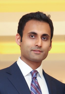
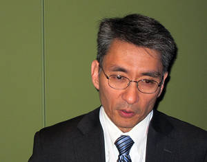
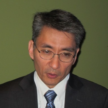
Those with long- versus short-segment Barrett’s esophagus were more likely to have dysplasia overlooked (85% vs. 53.6%; P = .008; mean 6 mm vs. 4 mm; P = .006), Dr. Kavel Visrodia said at the annual Digestive Disease Week.

Current American College of Gastroenterology (ACG) guidelines recommend early repeat esophagogastroduodenoscopy (EGD) to exclude the presence of missed dysplasia in newly diagnosed nondysplastic Barrett’s esophagus (BE), while the ACG and American Society for Gastrointestinal Endoscopy call for repeat EGD within 6 months for those with low-grade dysplasia.
The yield for repeat EGD has not been established, and only one study exists in the literature, said Dr. Visrodia of the department of medicine, Mayo Clinic, Rochester, Minn.
That study (Dis. Esophagus 2012 Sept. 28. [doi:10.1111/j.1442-2050.2012.01431.x]) showed a miss-rate of 8.2% among 146 patients with newly diagnosed nondysplastic BE. Long-segment BE was the only significant predictor of dysplasia on follow-up (odds ratio, 9.18; P = .008).
The cohort was relatively small and had no long-term follow-up, and with an interval to follow-up of 36 months, "it’s possible that some of these were actually incident cases of dysplasia and not prevalent cases," he said.
To address these gaps, Dr. Visrodia and his colleagues identified 488 BE cases from 1977 to 2011 in the Rochester Epidemiology Project in Olmsted County, Minn. A total of 278 patients were excluded because of high-grade dysplasia (HGD) or esophageal cancer on index endoscopy or repeat endoscopy after 24 months, leaving 181 patients with nondysplastic BE and 29 with low-grade dysplasia (LGD).
Repeat endoscopy within 24 months revealed 2 cases of HGD or cancer and 16 cases of LGD in the nondysplastic BE group, and 2 cases of HGD or cancer in the LGD group, Dr. Visrodia said.
Three of the four HGD/cancer cases were in patients with long-segment BE, defined as at least 3 cm of columnar mucosa.
Biopsies were insufficient in 63% of patients with missed dysplasia, compared with 55% in the group without missed dysplasia. Biopsies were considered adequate if the number of biopsies divided by the BE length was at least 2, indicating that samples were taken every 2 cm in accordance with guidelines. This risk factor is noteworthy, although the difference between groups was not statistically significant, possibly because of the small sample size, he said.
Finally, after a median of 6.8 years of follow-up, 30 asymptomatic, prevalent HGDs or cancers were detected within 24 months, compared with 22 incident cases detected after 24 months. This suggests that "a greater number of high-grade dysplasias and cancers were detected up front rather than during long-term careful surveillance," Dr. Visrodia said.
During a discussion of the study, one attendee asked whether the results make a better case for aggressive ablation up front rather than for surveillance, while others expressed surprise at the high miss rate at an institution such as the Mayo Clinic.
Dr. Visrodia replied that the results do give them pause, and suggested that tighter early endoscopic surveillance may be warranted, particularly in those with long-segment BE.
Dr. Visrodia and his coauthors reported no financial disclosures.
CHICAGO – Early endoscopic follow-up within 24 months detected dysplasia in nearly one in 10 patients with nondysplastic or low-grade Barrett’s esophagus in a retrospective study at the Mayo Clinic.
Initial endoscopy missed four cases of high-grade dysplasia or esophageal adenocarcinoma (1.9%) and 16 cases of low-grade dysplasia (7.6%) for an overall miss-rate of 9.5%.
Patients on proton pump inhibitors were less likely to have dysplasia missed than were those off PPIs (20% vs. 52.6%, P = .008).
Those with long- versus short-segment Barrett’s esophagus were more likely to have dysplasia overlooked (85% vs. 53.6%; P = .008; mean 6 mm vs. 4 mm; P = .006), Dr. Kavel Visrodia said at the annual Digestive Disease Week.
Current American College of Gastroenterology (ACG) guidelines recommend early repeat esophagogastroduodenoscopy (EGD) to exclude the presence of missed dysplasia in newly diagnosed nondysplastic Barrett’s esophagus (BE), while the ACG and American Society for Gastrointestinal Endoscopy call for repeat EGD within 6 months for those with low-grade dysplasia.
The yield for repeat EGD has not been established, and only one study exists in the literature, said Dr. Visrodia of the department of medicine, Mayo Clinic, Rochester, Minn.
That study (Dis. Esophagus 2012 Sept. 28. [doi:10.1111/j.1442-2050.2012.01431.x]) showed a miss-rate of 8.2% among 146 patients with newly diagnosed nondysplastic BE. Long-segment BE was the only significant predictor of dysplasia on follow-up (odds ratio, 9.18; P = .008).
The cohort was relatively small and had no long-term follow-up, and with an interval to follow-up of 36 months, "it’s possible that some of these were actually incident cases of dysplasia and not prevalent cases," he said.
To address these gaps, Dr. Visrodia and his colleagues identified 488 BE cases from 1977 to 2011 in the Rochester Epidemiology Project in Olmsted County, Minn. A total of 278 patients were excluded because of high-grade dysplasia (HGD) or esophageal cancer on index endoscopy or repeat endoscopy after 24 months, leaving 181 patients with nondysplastic BE and 29 with low-grade dysplasia (LGD).
Repeat endoscopy within 24 months revealed 2 cases of HGD or cancer and 16 cases of LGD in the nondysplastic BE group, and 2 cases of HGD or cancer in the LGD group, Dr. Visrodia said.
Three of the four HGD/cancer cases were in patients with long-segment BE, defined as at least 3 cm of columnar mucosa.
Biopsies were insufficient in 63% of patients with missed dysplasia, compared with 55% in the group without missed dysplasia. Biopsies were considered adequate if the number of biopsies divided by the BE length was at least 2, indicating that samples were taken every 2 cm in accordance with guidelines. This risk factor is noteworthy, although the difference between groups was not statistically significant, possibly because of the small sample size, he said.
Finally, after a median of 6.8 years of follow-up, 30 asymptomatic, prevalent HGDs or cancers were detected within 24 months, compared with 22 incident cases detected after 24 months. This suggests that "a greater number of high-grade dysplasias and cancers were detected up front rather than during long-term careful surveillance," Dr. Visrodia said.
During a discussion of the study, one attendee asked whether the results make a better case for aggressive ablation up front rather than for surveillance, while others expressed surprise at the high miss rate at an institution such as the Mayo Clinic.
Dr. Visrodia replied that the results do give them pause, and suggested that tighter early endoscopic surveillance may be warranted, particularly in those with long-segment BE.
Dr. Visrodia and his coauthors reported no financial disclosures.
AT DDW 2014
Key clinical point: Early endoscopic follow-up catches 9.5% of dysplasia missed on initial exam, but is not universally recommended by medical societies.
Major finding: Index endoscopy missed 1.9% of high-grade dysplasia or esophageal cancer and 7.6% of low-grade dysplasia.
Data source: A retrospective study in 181 patients with Barrett’s esophagus.
Disclosures: Dr. Visrodia and his coauthors reported no financial disclosures.
GERD may boost risk of MI
CHICAGO – Gastroesophageal reflux disease may constitute a heretofore unrecognized risk factor for coronary heart disease.
In a nationwide case-control study of prodigious proportions, endoscopically confirmed GERD in patients without known coronary or peripheral artery disease at baseline was independently associated with a 57% increased risk of having a first acute MI within the next 5 years, Dr. Ravi K. Prakash reported at the annual Digestive Disease Week.
If this novel finding is confirmed in other databases, the clinical implications would be profound. GERD is after all an extremely common problem, affecting 30%-40% of the U.S. population, noted Dr. Prakash, a gastroenterology fellow at MetroHealth Medical Center and Case Western Reserve University, Cleveland.
The good news: When Dr. Prakash and his coinvestigators divided the 316,390 study participants with GERD into those who were on proton pump inhibitor therapy and those who weren’t, they found that the PPI users weren’t at increased MI risk, compared with the more than 13.6 million similarly aged control subjects who didn’t have GERD, had never undergone an upper endoscopy, and were free of known atherosclerotic disease at baseline.
"Effective treatment of GERD appears to protect against MI," he concluded.
The study population was drawn from Explorys, a private, cloud-based health database containing the electronic health records of 35 million U.S. patients as provided by more than 200,000 physicians in 300-plus health care systems.
A first MI occurred during follow-up in 18,860 GERD patients, or 5.96%, compared with 144,140 controls, or 1.05%. This translates into an unadjusted sixfold increased risk of acute MI in patients with rigorously diagnosed GERD, compared with GERD-free controls.
The investigators then performed a multivariate logistic regression analysis adjusted for six major cardiovascular risk factors: obesity, hypertension, diabetes, tobacco use, hyperlipidemia, and sex. Many of these risk factors were substantially more prevalent in the GERD population. As a result, the unadjusted sixfold increase in MI risk associated with GERD was attenuated in this adjusted risk model; however, the adjusted 57% increased risk remained highly significant.
Moreover, the increased relative risk of GERD seen in the multivariate analysis was comparable with the risks posed by many of the established risk factors. Obesity, for example, showed a 49% increase in MI risk, smoking a 90% increase, and hypertension a 10% increased relative risk, compared with normotension. Only hyperlipidemia and diabetes were in another league, with relative risks of 9.5- and 2.2-fold, respectively, Dr. Prakash continued.
He observed that during the past decade, the research focus has shifted away from GERD as a caustic disease process causing local injury to GERD as a systemic inflammatory disease. Among the proinflammatory cytokines shown to be elevated both in the esophagus and circulation of GERD patients are interleukins-6, -8, and -1B as well as platelet-activating factor. These cytokines are well established as important players in the formation of atherosclerotic plaque in arteries. It was the shared elevations in proinflammatory cytokines that led Dr. Prakash and coworkers to hypothesize that patients with GERD might have an increased incidence of MI.
It’s only relatively recently that several other common chronic diseases have been reconceptualized as systemic inflammatory diseases associated with increased cardiovascular risk. Diabetes, for example, has been repositioned from a straightforward endocrine disease to new status as a coronary heart disease equivalent. And strong bodies of evidence link psoriasis and periodontitis to increased cardiovascular risk, Dr. Prakash noted.
Audience member Dr. Nimish B. Vakil, a Milwaukee gastroenterologist, rose to caution about the possibility of residual confounding by other factors not accounted for in the multivariate analysis, and thus the need for confirmatory studies. That being said, he added that he finds the notion of a GERD/acute MI link mechanistically quite plausible. Acid exposure from GERD might very well trigger exaggerated firing of the esophagocardiac reflex, with resultant episodes of coronary hypoperfusion. Effective PPI therapy for GERD would be expected to reduce such events.
Dr. Prakash said his literature search suggested another possible mechanism for the apparent protective effect of PPIs against MI: effective treatment of GERD, whether using PPIs or via a fundoplication procedure, has been reported to reduce levels of the inflammatory cytokines present in GERD.
Dr. Prakash reported having no financial conflicts with regard to his study, which was supported by institutional funds.
AT DDW 2014
Major finding: GERD was found to be independently associated with a 57% increased risk of acute MI.
Data source: This was a case-control study that harnessed the huge national Explorys database to compare first MI rates in 316,390 subjects with confirmed GERD to more than 13.6 million controls without GERD who’d never had an upper endoscopy.
Disclosures: This study was supported by institutional funds. The presenter reported having no financial conflicts.
Initial dilation no help in eosinophilic esophagitis
CHICAGO – Esophageal dilation combined with standard medical management of eosinophilic esophagitis doesn’t provide added benefit over medication alone in terms of dysphagia relief, according to a randomized, blinded clinical trial.
"In our group of patients with moderate endoscopic findings and without severe stricturing disease, esophageal dilation does not appear to be a necessary additional treatment strategy," Dr. Robert T. Kavitt stated at the annual Digestive Disease Week.
The study involved 31 patients newly diagnosed with eosinophilic esophagitis and baseline moderate to severe difficulty in swallowing. They were randomized to dilation or no dilation at initial endoscopy. Then all patients received standard medical management with 440 mcg of swallowed fluticasone b.i.d. and dexlansoprazole at 60 mg/day for 2 months. Patients were blinded as to their dilation status, as were the physicians who rated their change in dysphagia scores during follow-up.
Both groups experienced robust albeit equal reductions in overall dysphagia scores upon assessment at 30 and 60 days after endoscopy. At baseline, dysphagia scores averaged 6-6.5 on a 0-9 scale, indicative of moderate to severe dysphagia. At follow-up, scores in both groups had dropped to an average of 3 or less, reported Dr. Kavitt of the University of Chicago.
Complete resolution of dysphagia, defined as a dysphagia score of 0, occurred in only 23% of the dilation group and 57% of the no-dilation controls, which was not a statistically significant difference. The looser standard of "significant improvement" – meaning a dysphagia score of 3 or less – was met by 92% of the dilation group and 86% of controls.
Two patients in the dilation group and one control reported odynophagia.
Patients in the dilation group were dilated to the endpoint of mucosal tear. Three-quarters of the patients were dilated to a maximum size of 50 French or larger.
Baseline endoscopic scores assessing strictures, fissures, rings, and other abnormalities were in the moderate range on a 0-13 severity scale, so the study finding of a lack of benefit for dilation as part of an initial treatment strategy in eosinophilic esophagitis may not extend to the minority of patients having truly severe stricturing disease, according to Dr. Kavitt.
He noted that, before this study, the role of dilation in the treatment of eosinophilic esophagitis was a matter of divergent expert opinion unsupported by randomized trial evidence. The 2013 American College of Gastroenterology guidelines state that "the role of dilation as a primary monotherapy of eosinophilic esophagitis is still controversial and should be individualized."
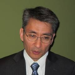
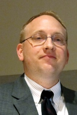
Later during the meeting, in his state-of-the-art lecture on changing therapeutic concepts in eosinophilic esophagitis, Dr. Ikuo Hirano cited Dr. Kavitt’s randomized trial in support of his argument against dilation as primary therapy.
"Dilation does nothing to address the underlying inflammatory response that’s causing strictures to form," noted Dr. Hirano, professor of medicine at Northwestern University, Chicago. "I believe that dilation is inappropriate therapy for children and adults with a predominantly inflammatory phenotype of disease."
Medication and diet therapies not only relieve the symptoms of eosinophilic esophagitis, he continued, they also improve the histopathology.
Dilation entails considerable pain as well as a risk of perforation. Recent reassuring safety data regarding dilation for eosinophilic esophagitis come from specialized esophageal centers with an unusual amount of experience with the procedure, the gastroenterologist said.
Dr. Kavitt reported having no financial conflicts of interest with regard to the randomized trial, which was supported by institutional funds. Dr. Hirano serves as a consultant to Meritage Pharma, Receptos, and Aptalis.
This is the first prospective examination of the role of esophageal dilation in eosinophilic esophagitis (EoE). The symptom-based outcomes suggest that dilation is not necessary for the initial therapy of EoE. The improvement in dysphagia was equivalent in patients treated with dilation combined with medical therapy, compared with those treated with medical therapy alone.
|
| Dr. Ikuo Hirano |
Moreover, while all patients randomized to dilation had dysphagia, not all had endoscopically identified esophageal strictures. One would not expect therapeutic gain for esophageal dilation in the absence of an identifiable stricture. Furthermore, randomized controlled trials of medical therapy for EoE have identified a surprisingly high placebo-response rate for the outcome of dysphagia that may have made demonstration of the benefits of dilation difficult to detect, especially in patients with mild disease severity. Symptom improvement in dysphagia may occur in response to modifications in eating habits such as meticulous mastication or avoidance of highly textured foods.
Finally, the inability to discern a difference in treatment groups may reflect the use of a nonvalidated dysphagia severity assessment instrument.
Despite these shortcomings, the results support current guideline recommendations that dilation should target aspects of esophageal remodeling in EoE that are not amenable to currently available medical or diet therapies.
The data, however, do not exclude an important therapeutic benefit of esophageal dilation in EoE. The study excluded patients with significant strictures that could not be traversed with a standard adult endoscope. The findings, therefore, would not apply to EoE patients with substantial fibrostenosis.
In this study, medical therapy was highly effective at relieving symptoms, but prior studies have shown that symptoms recur in almost all patients following cessation of medications.
Dilation, on the other hand, is generally safe and provides long-lasting relief of dysphagia in adults, even in the absence of medical or diet therapy. Additional prospective studies are needed to define the most appropriate patient subgroups that would benefit from dilation.
Dr. Ikuo Hirano is professor of medicine in the division of gastroenterology at Northwestern University's Feinberg School of Medicine in Chicago. He serves as a consultant to Meritage Pharma, Receptos, and Aptalis.
This is the first prospective examination of the role of esophageal dilation in eosinophilic esophagitis (EoE). The symptom-based outcomes suggest that dilation is not necessary for the initial therapy of EoE. The improvement in dysphagia was equivalent in patients treated with dilation combined with medical therapy, compared with those treated with medical therapy alone.
|
|
| Dr. Ikuo Hirano |
Moreover, while all patients randomized to dilation had dysphagia, not all had endoscopically identified esophageal strictures. One would not expect therapeutic gain for esophageal dilation in the absence of an identifiable stricture. Furthermore, randomized controlled trials of medical therapy for EoE have identified a surprisingly high placebo-response rate for the outcome of dysphagia that may have made demonstration of the benefits of dilation difficult to detect, especially in patients with mild disease severity. Symptom improvement in dysphagia may occur in response to modifications in eating habits such as meticulous mastication or avoidance of highly textured foods.
Finally, the inability to discern a difference in treatment groups may reflect the use of a nonvalidated dysphagia severity assessment instrument.
Despite these shortcomings, the results support current guideline recommendations that dilation should target aspects of esophageal remodeling in EoE that are not amenable to currently available medical or diet therapies.
The data, however, do not exclude an important therapeutic benefit of esophageal dilation in EoE. The study excluded patients with significant strictures that could not be traversed with a standard adult endoscope. The findings, therefore, would not apply to EoE patients with substantial fibrostenosis.
In this study, medical therapy was highly effective at relieving symptoms, but prior studies have shown that symptoms recur in almost all patients following cessation of medications.
Dilation, on the other hand, is generally safe and provides long-lasting relief of dysphagia in adults, even in the absence of medical or diet therapy. Additional prospective studies are needed to define the most appropriate patient subgroups that would benefit from dilation.
Dr. Ikuo Hirano is professor of medicine in the division of gastroenterology at Northwestern University's Feinberg School of Medicine in Chicago. He serves as a consultant to Meritage Pharma, Receptos, and Aptalis.
CHICAGO – Esophageal dilation combined with standard medical management of eosinophilic esophagitis doesn’t provide added benefit over medication alone in terms of dysphagia relief, according to a randomized, blinded clinical trial.
"In our group of patients with moderate endoscopic findings and without severe stricturing disease, esophageal dilation does not appear to be a necessary additional treatment strategy," Dr. Robert T. Kavitt stated at the annual Digestive Disease Week.
The study involved 31 patients newly diagnosed with eosinophilic esophagitis and baseline moderate to severe difficulty in swallowing. They were randomized to dilation or no dilation at initial endoscopy. Then all patients received standard medical management with 440 mcg of swallowed fluticasone b.i.d. and dexlansoprazole at 60 mg/day for 2 months. Patients were blinded as to their dilation status, as were the physicians who rated their change in dysphagia scores during follow-up.
Both groups experienced robust albeit equal reductions in overall dysphagia scores upon assessment at 30 and 60 days after endoscopy. At baseline, dysphagia scores averaged 6-6.5 on a 0-9 scale, indicative of moderate to severe dysphagia. At follow-up, scores in both groups had dropped to an average of 3 or less, reported Dr. Kavitt of the University of Chicago.
Complete resolution of dysphagia, defined as a dysphagia score of 0, occurred in only 23% of the dilation group and 57% of the no-dilation controls, which was not a statistically significant difference. The looser standard of "significant improvement" – meaning a dysphagia score of 3 or less – was met by 92% of the dilation group and 86% of controls.
Two patients in the dilation group and one control reported odynophagia.
Patients in the dilation group were dilated to the endpoint of mucosal tear. Three-quarters of the patients were dilated to a maximum size of 50 French or larger.
Baseline endoscopic scores assessing strictures, fissures, rings, and other abnormalities were in the moderate range on a 0-13 severity scale, so the study finding of a lack of benefit for dilation as part of an initial treatment strategy in eosinophilic esophagitis may not extend to the minority of patients having truly severe stricturing disease, according to Dr. Kavitt.
He noted that, before this study, the role of dilation in the treatment of eosinophilic esophagitis was a matter of divergent expert opinion unsupported by randomized trial evidence. The 2013 American College of Gastroenterology guidelines state that "the role of dilation as a primary monotherapy of eosinophilic esophagitis is still controversial and should be individualized."
Later during the meeting, in his state-of-the-art lecture on changing therapeutic concepts in eosinophilic esophagitis, Dr. Ikuo Hirano cited Dr. Kavitt’s randomized trial in support of his argument against dilation as primary therapy.
"Dilation does nothing to address the underlying inflammatory response that’s causing strictures to form," noted Dr. Hirano, professor of medicine at Northwestern University, Chicago. "I believe that dilation is inappropriate therapy for children and adults with a predominantly inflammatory phenotype of disease."
Medication and diet therapies not only relieve the symptoms of eosinophilic esophagitis, he continued, they also improve the histopathology.
Dilation entails considerable pain as well as a risk of perforation. Recent reassuring safety data regarding dilation for eosinophilic esophagitis come from specialized esophageal centers with an unusual amount of experience with the procedure, the gastroenterologist said.
Dr. Kavitt reported having no financial conflicts of interest with regard to the randomized trial, which was supported by institutional funds. Dr. Hirano serves as a consultant to Meritage Pharma, Receptos, and Aptalis.
AT DDW 2014
Key clinical point: Dilation offers no added benefit over medication alone in relieving dysphagia symptoms in eosinophilic esophagitis.
Major finding: Significant improvement in dysphagia symptoms was documented 30 and 60 days post endoscopy in 92% of eosinophilic esophagitis patients who underwent dilation plus medical therapy and in 86% who got medication alone.
Data source: This was a randomized trial involving 31 patients newly diagnosed with eosinophilic esophagitis who were assigned to dilation or no dilation at initial endoscopy, after which all participants received 2 months of medical therapy with a swallowed corticosteroid and a proton pump inhibitor. Patients as well as the physicians who rated their dysphagia symptoms 30 and 60 days post endoscopy were blinded as to dilation status.
Disclosures: Dr. Kavitt reported having no financial conflicts regarding this study, which was supported by institutional funds. Dr. Hirano serves as a consultant to Meritage Pharma, Receptos, and Aptalis.
Patient selection can keep the lid on esophagectomy costs
PHOENIX, ARIZ. – Cost and quality are not always synonymous, particularly when it comes to complex surgical procedures such as esophagectomy.
A review of records on more than 6,700 patients who underwent esophagectomy during a 4-year period showed that factors such as patient age, severity of illness, and hospital/surgeon volume can have a major effect on resource utilization and costs, said Dr. Daniel E. Abbott, assistant professor of surgery at the University of Cincinnati.
"There are certainly actionable risk factors for poor outcomes, such as mortality, and increased resource utilization, including dollar costs, the opportunity costs of increased length of stay, readmission, and rehabilitation and skilled nursing facilities," he reported at the annual Society of Surgical Oncology Cancer Symposium.
"I would argue that careful patient selection can have profound influences on cost-effectiveness. I think that as our health care is evolving and our outcomes are increasingly scrutinized, there will be increasing pressure to have better outcomes at lower costs," he added.
Dr. Abbott and his colleagues examined clinical variables in the cases of 6,737 esophagectomy patients treated from 2009 through 2012 in the University Healthsystems Consortium (UHC), an organization comprising 120 university hospitals and 299 affiliates.
They evaluated patient characteristics such as age and race, severity of illness index (1-4), esophagectomy type, and center and surgeon volume, and evaluated the effects of these variables on clinical outcomes that contribute to resource utilization, including deaths, readmissions, length of stay (LOS), and discharge disposition.
They found that the median LOS for all patients was 10 days (interquartile range, 8-17 days), but for patients over age 70, the median LOS was 11 days (P less than .01 vs. patients 70 and under).
Older patients did not have significantly higher readmission rates, but of the 4.2% of all patients who died in hospital, those over age 70 had more than twice the death rate of younger patients (7.0% vs. 3.2%, P less than .01).
Older patients were also significantly more likely to be discharged to a skilled nursing or rehabilitation facility than were younger patients (31.9% vs. 10.6%; P less than .01).
Total median cost per patient was $25,952, but again, older patients accounted for more of the expenses, at a median of $27,628 vs. $25,841 for those 70 and under (P less than .01).
In a multivariate analysis, patients over 70 had a more than twofold increase in risk of death (odds ratio, 2.12; P less than .01). Other factors significantly associated with greater risk for death were greater severity of illness (OR, 14.0; P less than .01) and black race vs. other races (OR, 1.88; P less than .01).
Factors associated with more frequent readmissions included greater severity of illness (OR, 1.33; P less than .01), and black race (OR, 1.34; P = .01), while patients of high-volume surgeons were less likely to need readmission (OR, 0.87; P = .04).
Lengths of stay were greater among patients over age 70, with every year over 70 translating into a 6% greater LOS (OR, 1.06; P =.03); older patients had a 16% increase in LOS for every year over 70 (OR, 1.16; P less than .01).
Similarly, each increase in severity of illness index score above 2 was associated with a 31% increase in LOS (OR, 1.31) and a 475% increase in intensive care unit (ICU) days (OR, 4.75; P less than .01 for both LOS and ICU days).
Black patients had a 22% increase in LOS vs. other races (OR, 1.22; P less than .01) and a 31% relative increase in ICU days (OR, 1.31; P = .01).
Because age and severity of illness were both strong predictors for mortality, readmission, and other perioperative outcomes, the authors conducted a further analysis combining the two variables, and found that for every 5 years of age, there were significant increases in the risk for death among patients with a greater severity of illness, compared with low severity.
Dr. Abbott noted that some of the odds ratios for older, sicker patients were "ridiculously high," probably because of the smaller sample sizes.
In a multivariate analysis of cost, factors associated with higher costs were age (OR, 1.14; P less than .01), greater severity of illness (OR, 2.14; P less than .01), and black race (OR, 1.15; P less than .01).
And in an analysis of cumulative resource use, the authors found what Dr. Abbott called a "snowball effect," in that hospitals with the lowest total costs discharged the majority of patients home, and that as costs increased, hospitals were less likely to discharge patients home and more likely to discharge them either to home health services or to extended care. In addition, as costs rose, the percentage of patients who died in hospital also rose.
He acknowledged that because the study used administrative data from university hospitals, it was skewed toward high-volume centers, and that the database did not include survival data, information about longitudinal resource use, or procedure-specific complications.
The authors did not disclose the funding source of the study. Dr. Abbott reported having no financial disclosures.
AT SSO 2014
Major finding: Factors associated with higher costs for esophagectomy were age, greater severity of illness, and black race.
Data source: Retrospective analysis of demographic and clinical factors associated with costs of esophagectomy in 6,737 patients treated in university-based hospitals and affiliates.
Disclosures: The authors did not disclose the funding source of the study. Dr. Abbott reported having no financial disclosures.

Foods can trigger EoE after allergy is outgrown
SAN DIEGO – Seventeen of 425 children who had eosinophilic esophagitis caused by a specific food developed the condition after outgrowing the allergy to that food, a retrospective study found.
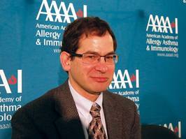
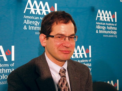
People who outgrow a food allergy may be at risk of developing eosinophilic esophagitis (EoE) to the same food, Dr. Jonathan Spergel said during a press briefing at the annual meeting of the American Academy of Allergy, Asthma, and Immunology.
He and his associates studied data on 1,025 children with EoE seen at the Children’s Hospital of Philadelphia in 2000-2012 to assess the prevalence of food allergy. In 425 children (42%), a specific food was identified as the EoE culprit – reintroducing the food to the diet caused esophageal changes on biopsy or biopsy changes normalized when the food was removed from the diet.
Eighty-four children had a history of IgE-mediated food allergy. Milk, egg, wheat, and soy were the most common food triggers of EoE in the 425 children in the study and in a subset of 17 who had outgrown IgE-mediated allergy to the specific food, reported Dr. Spergel, chief of the allergy section at the Children’s Hospital of Philadelphia. Sixteen of the 17 patients had atopic disease. The most common foods causing IgE-mediated allergy were peanuts, tree nuts, eggs, and milk.
The development of EoE coincided with reintroducing the food triggers. The time between outgrowing an allergy and reintroducing the food, triggering EoE, averaged 2 years but ranged from 6 months to 5 years.
Notably, two of the children who outgrew their food allergy had a normal biopsy of the esophagus when they had the food allergy, he said.
The findings support other recent studies suggesting that the pathophysiologies of EoE and IgE-mediated food allergy are distinct from each other, and that both can occur in the same individual to the same food, Dr. Spergel said. The mechanism by which EoE develops is poorly understood.
"I think these kids probably always had EoE to the food, but they weren’t eating it" because of the allergy, he said. "From 1% to 15% on oral immunotherapy get EoE, depending on which group you look at. I don’t think we caused" EoE by giving oral immunotherapy, he added. "We uncovered it."
Although it is rare for children who outgrow a food allergy to later develop EoE to that food, it’s worth keeping in mind if a child starts vomiting often or complains of stomachaches months or years later, Dr. Spergel said. Keeping the possibility in mind may help clinicians rule out other etiologies and detect EoE faster. "You have to take it seriously and get it checked out," he said.
Of the 84 patients with IgE-mediated food allergy, the 17 who outgrew the allergy and then developed EoE to the same food were significantly older (12 years, on average), compared with 67 patients who developed EoE from a different food from the one that caused their allergy.
The lead author on the study was Dr. Solrun Melkorka Meggadottir, a fellow at the Children’s Hospital of Philadelphia. The findings have been submitted to the Journal of Allergy and Clinical Immunology.
Dr. Spergel and Dr. Meggadottir reported having no disclosures.
[email protected] On Twitter @sherryboschert
Eosinophilic esophagitis (EoE) is a chronic condition that affects children and adults across the world. Children present with feeding problems, abdominal pain, and reflux-like symptoms, whereas adults complain of dysphagia and food impactions. No Food and Drug Administration–approved treatments are available leaving practitioners with off-label use of swallowed steroids administered from multi-dose inhalers or dietary restrictions.

|
|
The elimination of protein antigens has been accomplished through the use of elemental formulas, empiric elimination of the most common allergens, or targeted elimination based on skin or blood testing. A literature review revealed that these approaches are effective in inducing histologic remission in 90.8%, 72.1%, and 45.5% of patients, respectively.
This report by Meggadottir and Spergel provides at least two important findings for patients with EoE. Their data contribute to a growing body of literature and clinical observations indicating that mechanistic pathways other than IgE-mediated food reactions may contribute to the pathogenesis of EoE. Some recent reports and clinical experiences suggest the children with EoE may tolerate some allergenic foods that are cooked in a different way or children may outgrow their allergies. Results from this study suggest that this may not be the case. Clinical implications of their findings suggest that children and adults may need to continue to restrict common foods from their diets; the long-term impact of this on quality of life is not certain.
Dr. Glenn T. Furuta, professor of pediatrics, University of Colorado School of Medicine, Director of the Gastrointestinal Eosinophilic Diseases Program, Children’s Hospital Colorado, Denver. He is the co-founder of EnteroTrack.
Eosinophilic esophagitis (EoE) is a chronic condition that affects children and adults across the world. Children present with feeding problems, abdominal pain, and reflux-like symptoms, whereas adults complain of dysphagia and food impactions. No Food and Drug Administration–approved treatments are available leaving practitioners with off-label use of swallowed steroids administered from multi-dose inhalers or dietary restrictions.
|
|
|
The elimination of protein antigens has been accomplished through the use of elemental formulas, empiric elimination of the most common allergens, or targeted elimination based on skin or blood testing. A literature review revealed that these approaches are effective in inducing histologic remission in 90.8%, 72.1%, and 45.5% of patients, respectively.
This report by Meggadottir and Spergel provides at least two important findings for patients with EoE. Their data contribute to a growing body of literature and clinical observations indicating that mechanistic pathways other than IgE-mediated food reactions may contribute to the pathogenesis of EoE. Some recent reports and clinical experiences suggest the children with EoE may tolerate some allergenic foods that are cooked in a different way or children may outgrow their allergies. Results from this study suggest that this may not be the case. Clinical implications of their findings suggest that children and adults may need to continue to restrict common foods from their diets; the long-term impact of this on quality of life is not certain.
Dr. Glenn T. Furuta, professor of pediatrics, University of Colorado School of Medicine, Director of the Gastrointestinal Eosinophilic Diseases Program, Children’s Hospital Colorado, Denver. He is the co-founder of EnteroTrack.
SAN DIEGO – Seventeen of 425 children who had eosinophilic esophagitis caused by a specific food developed the condition after outgrowing the allergy to that food, a retrospective study found.
People who outgrow a food allergy may be at risk of developing eosinophilic esophagitis (EoE) to the same food, Dr. Jonathan Spergel said during a press briefing at the annual meeting of the American Academy of Allergy, Asthma, and Immunology.
He and his associates studied data on 1,025 children with EoE seen at the Children’s Hospital of Philadelphia in 2000-2012 to assess the prevalence of food allergy. In 425 children (42%), a specific food was identified as the EoE culprit – reintroducing the food to the diet caused esophageal changes on biopsy or biopsy changes normalized when the food was removed from the diet.
Eighty-four children had a history of IgE-mediated food allergy. Milk, egg, wheat, and soy were the most common food triggers of EoE in the 425 children in the study and in a subset of 17 who had outgrown IgE-mediated allergy to the specific food, reported Dr. Spergel, chief of the allergy section at the Children’s Hospital of Philadelphia. Sixteen of the 17 patients had atopic disease. The most common foods causing IgE-mediated allergy were peanuts, tree nuts, eggs, and milk.
The development of EoE coincided with reintroducing the food triggers. The time between outgrowing an allergy and reintroducing the food, triggering EoE, averaged 2 years but ranged from 6 months to 5 years.
Notably, two of the children who outgrew their food allergy had a normal biopsy of the esophagus when they had the food allergy, he said.
The findings support other recent studies suggesting that the pathophysiologies of EoE and IgE-mediated food allergy are distinct from each other, and that both can occur in the same individual to the same food, Dr. Spergel said. The mechanism by which EoE develops is poorly understood.
"I think these kids probably always had EoE to the food, but they weren’t eating it" because of the allergy, he said. "From 1% to 15% on oral immunotherapy get EoE, depending on which group you look at. I don’t think we caused" EoE by giving oral immunotherapy, he added. "We uncovered it."
Although it is rare for children who outgrow a food allergy to later develop EoE to that food, it’s worth keeping in mind if a child starts vomiting often or complains of stomachaches months or years later, Dr. Spergel said. Keeping the possibility in mind may help clinicians rule out other etiologies and detect EoE faster. "You have to take it seriously and get it checked out," he said.
Of the 84 patients with IgE-mediated food allergy, the 17 who outgrew the allergy and then developed EoE to the same food were significantly older (12 years, on average), compared with 67 patients who developed EoE from a different food from the one that caused their allergy.
The lead author on the study was Dr. Solrun Melkorka Meggadottir, a fellow at the Children’s Hospital of Philadelphia. The findings have been submitted to the Journal of Allergy and Clinical Immunology.
Dr. Spergel and Dr. Meggadottir reported having no disclosures.
[email protected] On Twitter @sherryboschert
AT 2014 AAAAI ANNUAL MEETING
Major finding: Seventeen of 425 children with eosinophilic esophagitis caused by a specific food redeveloped the condition after outgrowing an allergy to the same food.
Data source: A retrospective study of data on 1,025 children seen at one institution for eosinophilic esophagitis.
Disclosures: Dr. Spergel and Dr. Meggadottir reported having no relevant financial disclosures.
Fluorescent cholangiography as effective as standard, but cheaper
MIAMI BEACH – Intraoperative fluorescent cholangiography is just as effective as traditional cholangiography but costs hundreds of dollars less and is significantly faster to perform.
It’s also a great teaching tool, Dr. Fernando Dip said at the annual meeting of the Americas Hepato-Pancreato-Biliary Association. In just one session, all of the third- and fourth-year surgical residents were able to correctly identify 100% of the biliary structures.
"It appears to be an additional tool for the laparoscopic surgeon," said Dr. Dip, chief of surgical research at the Cleveland Clinic in Weston, Fla. "It’s quick, inexpensive, real-time, there are no adverse events, and it’s an inciscionless procedure."
Common bile duct injury is the most frequent injury seen in laparoscopic cholecystectomy, he said, and although the overall incidence is low, the number of injuries each year is not inconsiderable, since more than 750,000 laparoscopic cholecystectomies are performed in the United States annually.
"Only 3% of these injuries are due to problems with technical skill," Dr. Dip said. "The other 97% are problems of visual perception – illusions of where the ducts are."
Intraoperative cholangiography helps surgeons visualize this anatomy, but its true usefulness is somewhat controversial. Dr. Dip cited a recent study of almost 93,000 patients – 40% of whom underwent the procedure. It showed that intraoperative cholangiography is not effective as a preventive strategy against common duct injury during cholecystectomy.
Intraoperative fluorescent cholangiography is sometimes used to identify biliary anatomy in extrahepatic surgery. Dr. Dip and his colleagues examined its usefulness in 45 patients undergoing laparoscopic cholecystectomy. Senior residents performed all of the procedures under the supervision of experienced laparoscopic surgeons. All patients underwent the investigational procedure, followed by standard cholangiography.
The patients had a mean age of 49 years and were evenly split between men and women. The mean body mass index was 28 kg/m2. Surgical indications were cholelithiasis (22), acute cholecystitis (17), chronic cholecystitis (5), and polyp (1). About 1 hour before surgery, patients received an infusion of indocyanine green 0.5 mg/kg. During the laparoscopic exploration, surgeons used near-infrared light to make the marker fluoresce as it was excreted by the liver.
In a picture review before surgery, all residents correctly identified 100% of the anatomic structures visualized by the fluorescent procedure. They were able to complete the fluorescent procedure in all of the patients. The completion rate for cholangiography was 93% (42 patients). In three patients, cholangiography failed because the cystic duct could not be cannulated.
The residents identified the cystic duct in 44 of the patients (98%), the common bile duct in 36 (80%), and the common hepatic ducts in 27 (60%). Neither technique identified any aberrant or accessory ducts.
Because fluorescent cholangiography is a real-time surgical procedure, it allowed checking of the transection and resection of the gallbladder pedicle before smooth dissection in all of the patients.
The procedure was significantly quicker than standard cholangiography (0.71 minutes vs. 7.15 minutes). It was also significantly cheaper, costing a mean of $14 vs. $778. There were no adverse surgical events, and no adverse reactions to the dye or at the infusion site, Dr. Dip said. Additionally, he noted, fluorescent cholangiography does not rely on x-rays, and so spared the patients any radiation exposure.
He added that the Cleveland Clinic, in conjunction with the University of Tokyo, will soon launch a randomized clinical trial comparing the two methods. An ongoing trial from another institution is evaluating fluorescent cholangiography compared with critical view technique in visualizing anatomy during laparoscopic cholecystectomy. It was set to wrap up in January, but according to the trial record on clinicaltrials.gov, is still recruiting patients.
Dr. Dip had no financial disclosures.
On Twitter @alz_gal
AT AHPBA 2014
Major finding: Fluorescent cholangiography identified 100% of relevant anatomy during laparoscopic cholecystectomy, while costing less than standard cholangiography ($14 vs. $778).
Data source: Prospective study of 45 patients.
Disclosures: Dr. Dip had no financial disclosures.

Affordable Care Act requires insurers to cover clinical trials
The Affordable Care Act now requires health insurance companies to cover routine costs for clinical trial participants, the American Society of Clinical Oncology reminded clinicians in educational materials about the provision it released Feb. 4.
The group is concerned, it said, because "the federal government has not yet issued regulations to guide implementation of the new law," and, at least so far, has characterized it as a "self-implementing" statute that insurers are "expected to implement ... using a good faith, reasonable interpretation of the law."
"While much of the statutory language is clear, in the absence of federal guidance, payers will likely vary on the legal interpretation of each element of the provision. It is likely that securing compliance with the law may require considerable negotiations with some insurers or health plans. There is no assurance that all parties will agree on the legal interpretation of each element of the provision," the group said.
To help, the American Society of Clinical Oncology (ASCO) issued a detailed explanation of the measure, plus educational materials for patients and a form investigators can fill out to demonstrate that a trial and potential subject meet the law’s requirements.
"ASCO and other groups fought long and hard for this law requiring insurers nationwide to cover the routine costs of care for individuals participating in clinical trials," ASCO president Dr. Clifford Hudis noted in a statement.
The hope is to counter poor study enrollment, the main reason that about 20% of cancer trials are never completed, according to a study reported at the 2014 Genitourinary Cancers Symposium earlier this year. Sometimes patients simply can’t afford to participate, because "some health plans have denied coverage ... of routine costs that are offered as part of the clinical trial." The new law might help patients afford clinical trial participation, the group said.
The law applies to plans newly issued or renewed after Jan. 1, 2014. Routine costs include all items and services that an insurance company would cover for a patient not enrolled in a clinical trial. Plans cannot prohibit participation in clinical trials; deny or limit coverage of routine patient costs for items and services furnished in connection with participation in a trial; or discriminate against an individual because they are enrolled in a trial.
The provision covers studies that are either federally funded, conducted under an Investigational New Drug Application, or exempt from an Investigational New Drug Application.
It does not apply to Medicaid plans, and payers are only required to cover routine costs delivered by out-of-network providers if out-of-network benefits are part of a patient’s insurance plan. "An insurer may attempt to deny coverage on the grounds that the service or item is ‘clearly inconsistent with the established standard of care.’ Providers may consider requesting that the insurers prove that the item or service is inconsistent with the standard of care," ASCO noted.

Sleeve gastrectomy often worsens GERD
Laparoscopic sleeve gastrectomy not only fails to improve gastroesophageal reflux disease in most patients who undergo the weight-loss procedure, it actually worsens GERD symptoms in many of them and induces GERD in 9%, according to a report published online Feb. 5 in JAMA Surgery.
In addition, patients with preexisting GERD who undergo laparoscopic sleeve gastrectomy (LSG) have high rates of surgical complications; revision surgery; failure to achieve weight loss; and failure to resolve weight-related comorbidities such as diabetes, obstructive sleep apnea, and hypertension. In contrast, patients who undergo gastric bypass show significant improvement in all of these outcomes, said Dr. Cecily E. DuPree and her associates in the department of surgery, Madigan Army Medical Center, Fort Lewis, Wash.

Based on the findings from their study of a national database including 4,832 patients who had laparoscopic sleeve gastrectomy (LSG) and 33,867 who had gastric bypass (GB), "we believe that all patients should be evaluated for the presence and severity of GERD and counseled regarding the relative efficacy of LSG vs. GB or other bariatric operations before surgery. Although there is no definitive evidence to support the listing of GERD as an absolute contraindication to LSG, the available data suggest that the presence of preexisting severe GERD or esophageal dysmotility may be considered a relative contraindication," they said.
Dr. DuPree and her colleagues noted that until now, the sleeve procedure’s effect on GERD was unknown. Small, single-center series "have raised significant concerns," but no large study has examined the issue. So she and her associates used data from a large, nationwide database (the Bariatric Outcomes Longitudinal Database) to track the resolution, persistence, or development of GERD in 4,832 patients who underwent laparoscopic sleeve gastrectomy in 2007-2010, comparing their outcomes with those of 33,867 patients who underwent gastric bypass during the same period and served as controls.
The overall prevalence of GERD was 49.7% in the entire study population, and that of severe GERD was 25.7%, confirming that this disorder is very common in candidates for bariatric surgery.
The prevalence of GERD was 44.5% among patients undergoing the sleeve procedure. "This highlights the concern that there is a large population at risk of adverse outcomes after LSG if the procedure is associated with anatomical or physiologic changes that increase the risk of postoperative GERD," the investigators noted.
Most LSG patients (84.1%) had persistent GERD symptoms after the procedure; only 15.9% reported resolution of symptoms. An additional 9.0% of LSG patients reported postoperative worsening of GERD symptoms. And 8.6% of patients who didn’t have GERD before undergoing sleeve gastrectomy developed the disorder afterward.
In contrast, most patients who underwent gastric bypass showed complete resolution (62.8%) or stabilization (17.6%) of GERD symptoms. Only 2.2% reported worsening GERD symptoms, and none developed de novo symptoms.
Among the LSG patients, the complication rate was significantly higher in those with preexisting GERD (15.1%) or preexisting severe GERD (16.3%) than in those without GERD (10.6%). "There was also a small but statistically significant increase in the need for revisional surgery between LSG patients with and without preoperative GERD symptoms (0.6% vs. 0.3%)," the investigators wrote (JAMA Surg. 2014 [doi:10.1001/jamasurg.2013.4323]).
In contrast, the presence of GERD had no effect on complications in the control group.
Similarly, the rate of failure to lose weight was higher in LSG patients with preoperative GERD and in LSG patients with severe preoperative GERD than in those without GERD. Again, the presence of GERD had no such effect on weight loss in the gastric bypass patients.
In addition, the percentage of patients who showed resolution of comorbidities was significantly decreased among patients with preoperative GERD who underwent LSG, compared with all other groups.
"These data raise significant concerns about the effect of LSG on the obesity-related comorbidity of GERD and suggest that most patients with preexisting GERD will have either no improvement or possibly worsening of their symptoms after LSG," Dr. DuPree and her associates said.
The exact reason why the sleeve procedure could contribute to the worsening of reflux or the de novo development of GERD is not known, but there are several anatomical or physiologic factors that may play a role. Laparoscopic sleeve gastrectomy may decrease lower esophageal sphincter resting tone, and it may disrupt the antropyloric pump mechanism or narrow the pylorus.
It is also possible that an excessively large or dilated gastric sleeve may retain the capacity for increased acid production, causing reflux, or that it may decrease esophageal acid clearance. And a hiatal hernia that is unrecognized at the time of surgery or that develops afterward could also produce reflux symptoms.
Modifying surgical technique so that sleeve size and volume are attended to, narrowing of the gastric body or pylorus is avoided, and hiatal hernias are assiduously identified and repaired may reduce the risk of post-LSG GERD, the investigators said.
Laparoscopic sleeve gastrectomy not only fails to improve gastroesophageal reflux disease in most patients who undergo the weight-loss procedure, it actually worsens GERD symptoms in many of them and induces GERD in 9%, according to a report published online Feb. 5 in JAMA Surgery.
In addition, patients with preexisting GERD who undergo laparoscopic sleeve gastrectomy (LSG) have high rates of surgical complications; revision surgery; failure to achieve weight loss; and failure to resolve weight-related comorbidities such as diabetes, obstructive sleep apnea, and hypertension. In contrast, patients who undergo gastric bypass show significant improvement in all of these outcomes, said Dr. Cecily E. DuPree and her associates in the department of surgery, Madigan Army Medical Center, Fort Lewis, Wash.
Based on the findings from their study of a national database including 4,832 patients who had laparoscopic sleeve gastrectomy (LSG) and 33,867 who had gastric bypass (GB), "we believe that all patients should be evaluated for the presence and severity of GERD and counseled regarding the relative efficacy of LSG vs. GB or other bariatric operations before surgery. Although there is no definitive evidence to support the listing of GERD as an absolute contraindication to LSG, the available data suggest that the presence of preexisting severe GERD or esophageal dysmotility may be considered a relative contraindication," they said.
Dr. DuPree and her colleagues noted that until now, the sleeve procedure’s effect on GERD was unknown. Small, single-center series "have raised significant concerns," but no large study has examined the issue. So she and her associates used data from a large, nationwide database (the Bariatric Outcomes Longitudinal Database) to track the resolution, persistence, or development of GERD in 4,832 patients who underwent laparoscopic sleeve gastrectomy in 2007-2010, comparing their outcomes with those of 33,867 patients who underwent gastric bypass during the same period and served as controls.
The overall prevalence of GERD was 49.7% in the entire study population, and that of severe GERD was 25.7%, confirming that this disorder is very common in candidates for bariatric surgery.
The prevalence of GERD was 44.5% among patients undergoing the sleeve procedure. "This highlights the concern that there is a large population at risk of adverse outcomes after LSG if the procedure is associated with anatomical or physiologic changes that increase the risk of postoperative GERD," the investigators noted.
Most LSG patients (84.1%) had persistent GERD symptoms after the procedure; only 15.9% reported resolution of symptoms. An additional 9.0% of LSG patients reported postoperative worsening of GERD symptoms. And 8.6% of patients who didn’t have GERD before undergoing sleeve gastrectomy developed the disorder afterward.
In contrast, most patients who underwent gastric bypass showed complete resolution (62.8%) or stabilization (17.6%) of GERD symptoms. Only 2.2% reported worsening GERD symptoms, and none developed de novo symptoms.
Among the LSG patients, the complication rate was significantly higher in those with preexisting GERD (15.1%) or preexisting severe GERD (16.3%) than in those without GERD (10.6%). "There was also a small but statistically significant increase in the need for revisional surgery between LSG patients with and without preoperative GERD symptoms (0.6% vs. 0.3%)," the investigators wrote (JAMA Surg. 2014 [doi:10.1001/jamasurg.2013.4323]).
In contrast, the presence of GERD had no effect on complications in the control group.
Similarly, the rate of failure to lose weight was higher in LSG patients with preoperative GERD and in LSG patients with severe preoperative GERD than in those without GERD. Again, the presence of GERD had no such effect on weight loss in the gastric bypass patients.
In addition, the percentage of patients who showed resolution of comorbidities was significantly decreased among patients with preoperative GERD who underwent LSG, compared with all other groups.
"These data raise significant concerns about the effect of LSG on the obesity-related comorbidity of GERD and suggest that most patients with preexisting GERD will have either no improvement or possibly worsening of their symptoms after LSG," Dr. DuPree and her associates said.
The exact reason why the sleeve procedure could contribute to the worsening of reflux or the de novo development of GERD is not known, but there are several anatomical or physiologic factors that may play a role. Laparoscopic sleeve gastrectomy may decrease lower esophageal sphincter resting tone, and it may disrupt the antropyloric pump mechanism or narrow the pylorus.
It is also possible that an excessively large or dilated gastric sleeve may retain the capacity for increased acid production, causing reflux, or that it may decrease esophageal acid clearance. And a hiatal hernia that is unrecognized at the time of surgery or that develops afterward could also produce reflux symptoms.
Modifying surgical technique so that sleeve size and volume are attended to, narrowing of the gastric body or pylorus is avoided, and hiatal hernias are assiduously identified and repaired may reduce the risk of post-LSG GERD, the investigators said.
FROM JAMA SURGERY
Major finding: 84.1% of the sleeve gastrectomy patients had persistent GERD symptoms after the procedure, and 9% had worsening of GERD; another 8.6% who didn’t have GERD before undergoing the procedure developed it afterward.
Data source: An analysis of data on 4,832 adults across the United States who underwent laparoscopic sleeve gastrectomy and 33,867 who underwent gastric bypass in 2007-2010, of whom approximately half had preexisting GERD.
Disclosures: No financial conflicts of interest were reported.
LES electrical stimulation shows promise for treating refractory GERD
SAN DIEGO – Electrical stimulation therapy of the lower esophageal sphincter is effective for treating refractory gastroesophageal reflux disease, according to interim results from a trial presented at the annual meeting of the American College of Gastroenterology.
A total of 32 patients underwent implantation of a pair of electrodes in the muscular layer of the gastroesophageal junction. The electrodes were connected to a pulse generator in the abdomen that delivered intermittent stimulation.

Six months later, the patients had a sustained, more than one-half reduction from baseline in exposure of the esophagus to acid and a two-thirds reduction in symptoms, reported first author Dr. Edy E. Soffer at the meeting.
Additionally, whereas most of the patients had been taking proton pump inhibitors (PPIs) at baseline, only about 1 in 10 were doing so at 6 months. None experienced any device-related adverse events.
The findings support the effectiveness and safety of electrical stimulation therapy in this population, according to Dr. Soffer, who is a professor in the gastroenterology division, Keck School of Medicine, University of Southern California, Los Angeles.
"Currently, there are ongoing studies, and there will be future sham-controlled studies that should clarify further the role of this intervention in the treatment of GERD [gastroesophageal reflux disease] and its mechanism of action," he noted.
Session attendee Dr. Marcelo Vela of the Baylor Clinic, Houston, noted that earlier studies have shown that this therapy increases basal pressure of the lower esophageal sphincter (LES). "Because the main mechanism for reflux is transient LES relaxations [TLESRs], particularly with small hernias as in your patients, do you have any data on TLESRs?" he asked.
"No, LES pressure was not an endpoint in these studies," Dr. Soffer replied. "Initially, we thought that this [basal pressure] is the main mechanism; as we are learning more, it is probably not going to be the main mechanism. We are looking at others, including TLESRs. ... We don’t have data yet."
Dr. Joel Richter, of the University of South Florida, Tampa, wondered whether patients experienced any of the oft-feared complications of antireflux surgery, such as bloating, diarrhea, and dysphagia.
"There were only two cases of dysphagia, and they basically resolved on their own," Dr. Soffer replied. "We didn’t see any of the symptoms that you see with postsurgical therapy. ... The intervention really should not result in the symptoms that come after standard surgical care."
Dr. Richter also requested more information on hiatal hernia repairs undertaken in some patients. "If you had a hernia repair, how do you know what part of the success was related to the hernia repair versus the stimulation?" he asked.
Thirteen patients had a hernia repair, with technique left up to the treating physician, Dr. Soffer replied. "We will look separately, when we complete that phase, at those that had the hernia repair versus those that did not."
"Was there a bit of a trend toward any improvement depending on your hernia size?" Dr. Richard McCallam of Texas Tech University in El Paso asked, while also noting that patients with hernias measuring 3 cm or more were excluded.
There were too few patients to assess trends, according to Dr. Soffer. "We tried not to take patients who had particularly severe disease at the beginning," he added. "The 3-cm hernia repair will be the one perhaps that will need to be looked at more carefully. This is a more sustained hernia situation as compared to the 1- and 2-cm [ones]."
The investigators enrolled GERD patients in the study who had a GERD health-related quality of life score of 20 or higher when not taking PPIs, and who had at least a partial response to these agents. They were required to have a basal LES end-expiratory pressure of at least 5 mm Hg, and to have a pH below 4.0 for more than 5% of the time on 24-hour esophageal pH monitoring.
The patients’ mean age was 50 years, and 18 of them were male, according to Dr. Soffer.
The main results showed a sustained reduction in esophageal acid exposure; the percentage of time at pH less than 4.0 was 10.3% at baseline, compared with 3.7% at 3 months (P less than .01), and 4.6% at 6 months (P less than .01).
There was also a sustained improvement in symptoms as assessed from GERD health-related quality of life; the score was 15 at baseline on PPIs and 31 at baseline off PPIs. These figures compared with a score of 4 at 3 months and 5 at 6 months.
Large proportions of patients met criteria for successful treatment at 6 months: 65% had control of esophageal acid exposure (either normalization or a greater than one-half reduction), 86% had control of GERD symptoms compared with level without medical therapy (a greater than 50% improvement versus baseline level not on PPIs), and 76% had control of GERD symptoms compared with level with medical therapy (any improvement of symptoms versus baseline level on PPIs).
Quality of life as assessed with the Short Form-12 showed improvement at 6 months in physical health scores compared with those measured at baseline while off PPIs.
Patients also had significant reductions from the level at baseline off PPIs at both 3 and 6 months in the median percentages of daily diary days with heartburn and with regurgitation symptoms.
Analyses of daytime versus nighttime symptoms are still ongoing, according to Dr. Soffer.
Stimulation therapy was also associated with a dramatic reduction in PPI use. Whereas only 10% of patients did not use any of these agents at baseline, 89% were not using any at 6 months.
There were just two serious adverse events: a procedure-related trocar perforation of the small bowel during laparoscopy (the stimulation device was prophylactically removed and there was resolution after surgical repair) and atrioventricular nodal reentrant tachycardia unrelated to the device or procedure.
There were no device-related serious adverse events. There were 35 nonserious events possibly or probably related to the device or procedure.
Dr. Soffer is a consultant for and shareholder in EndoStim*. The study was supported by EndoStim.
*Correction, 2/4/2014: An earlier version of the story misstated Dr. Soffer's conflicts of interest.
Electrical stimulation of the lower esophageal sphincter (LES) to compensate for sphincter incompetence seems a rather direct approach to treating GERD, and the data presented by Dr. Soffer seem to indicate that this can work. Most convincing is the reduction in esophageal acid exposure from a mean of 10.3% at baseline to a mean of 3.7% after implantation. True, there is the variable of surgical correction of hiatal hernia done in 13 of the 32 implanted patients to deal with, but nonetheless, these are very encouraging proof-of-concept data.

|
| Dr. Peter J. Kahrilas |
So now the more difficult questions: Who needs this therapy, and how should its efficacy be further evaluated? With regard to who needs this therapy, I would argue against it being a mainstream approach. PPIs are simply too good, too inexpensive, and too safe to compete with. However, there are a number of intriguing niche applications for which the EndoStim device might be suitable, for example, patients with poor peristalsis or obesity for whom the conventional surgical approaches are not advised, lung transplant or bariatric surgery patients, or patients with excessive regurgitation after gastrostomy placement. In these situations, conventional approaches of PPIs or Nissen fundoplication are either ineffective or ill-advised. A true unmet need exists.
The other question that arises is how to further test the device. The issue raised by Dr. Richter of discriminating between the benefits from hiatus hernia repair and from the stimulator itself needs to be resolved. Certainly, a subgroup analysis of the stimulator-only population is in order, as is a future study of the device in hernia patients without adding a hernia repair. Another interesting potential with this device is that it can serve as its own control in future trials, assuming that the implanter can resist the temptation to attempt an anatomical correction at the time of implantation and presuming that the stimulator can be switched on and off. Then, as in the studies done with gastric pacing, patients could be studied physiologically or symptomatically with the device on or off in a blinded fashion. I look forward to hearing more on the development of the LES stimulator.
Dr. Peter J. Kahrilas is the Gilbert H. Marquardt Professor of Medicine at the Feinberg School of Medicine, Northwestern University, Chicago. He is a consultant for AstraZeneca, GlaxoSmithKline, Pfizer, Torax, and Reckitt Benckiser.
Electrical stimulation of the lower esophageal sphincter (LES) to compensate for sphincter incompetence seems a rather direct approach to treating GERD, and the data presented by Dr. Soffer seem to indicate that this can work. Most convincing is the reduction in esophageal acid exposure from a mean of 10.3% at baseline to a mean of 3.7% after implantation. True, there is the variable of surgical correction of hiatal hernia done in 13 of the 32 implanted patients to deal with, but nonetheless, these are very encouraging proof-of-concept data.
|
|
| Dr. Peter J. Kahrilas |
So now the more difficult questions: Who needs this therapy, and how should its efficacy be further evaluated? With regard to who needs this therapy, I would argue against it being a mainstream approach. PPIs are simply too good, too inexpensive, and too safe to compete with. However, there are a number of intriguing niche applications for which the EndoStim device might be suitable, for example, patients with poor peristalsis or obesity for whom the conventional surgical approaches are not advised, lung transplant or bariatric surgery patients, or patients with excessive regurgitation after gastrostomy placement. In these situations, conventional approaches of PPIs or Nissen fundoplication are either ineffective or ill-advised. A true unmet need exists.
The other question that arises is how to further test the device. The issue raised by Dr. Richter of discriminating between the benefits from hiatus hernia repair and from the stimulator itself needs to be resolved. Certainly, a subgroup analysis of the stimulator-only population is in order, as is a future study of the device in hernia patients without adding a hernia repair. Another interesting potential with this device is that it can serve as its own control in future trials, assuming that the implanter can resist the temptation to attempt an anatomical correction at the time of implantation and presuming that the stimulator can be switched on and off. Then, as in the studies done with gastric pacing, patients could be studied physiologically or symptomatically with the device on or off in a blinded fashion. I look forward to hearing more on the development of the LES stimulator.
Dr. Peter J. Kahrilas is the Gilbert H. Marquardt Professor of Medicine at the Feinberg School of Medicine, Northwestern University, Chicago. He is a consultant for AstraZeneca, GlaxoSmithKline, Pfizer, Torax, and Reckitt Benckiser.
SAN DIEGO – Electrical stimulation therapy of the lower esophageal sphincter is effective for treating refractory gastroesophageal reflux disease, according to interim results from a trial presented at the annual meeting of the American College of Gastroenterology.
A total of 32 patients underwent implantation of a pair of electrodes in the muscular layer of the gastroesophageal junction. The electrodes were connected to a pulse generator in the abdomen that delivered intermittent stimulation.
Six months later, the patients had a sustained, more than one-half reduction from baseline in exposure of the esophagus to acid and a two-thirds reduction in symptoms, reported first author Dr. Edy E. Soffer at the meeting.
Additionally, whereas most of the patients had been taking proton pump inhibitors (PPIs) at baseline, only about 1 in 10 were doing so at 6 months. None experienced any device-related adverse events.
The findings support the effectiveness and safety of electrical stimulation therapy in this population, according to Dr. Soffer, who is a professor in the gastroenterology division, Keck School of Medicine, University of Southern California, Los Angeles.
"Currently, there are ongoing studies, and there will be future sham-controlled studies that should clarify further the role of this intervention in the treatment of GERD [gastroesophageal reflux disease] and its mechanism of action," he noted.
Session attendee Dr. Marcelo Vela of the Baylor Clinic, Houston, noted that earlier studies have shown that this therapy increases basal pressure of the lower esophageal sphincter (LES). "Because the main mechanism for reflux is transient LES relaxations [TLESRs], particularly with small hernias as in your patients, do you have any data on TLESRs?" he asked.
"No, LES pressure was not an endpoint in these studies," Dr. Soffer replied. "Initially, we thought that this [basal pressure] is the main mechanism; as we are learning more, it is probably not going to be the main mechanism. We are looking at others, including TLESRs. ... We don’t have data yet."
Dr. Joel Richter, of the University of South Florida, Tampa, wondered whether patients experienced any of the oft-feared complications of antireflux surgery, such as bloating, diarrhea, and dysphagia.
"There were only two cases of dysphagia, and they basically resolved on their own," Dr. Soffer replied. "We didn’t see any of the symptoms that you see with postsurgical therapy. ... The intervention really should not result in the symptoms that come after standard surgical care."
Dr. Richter also requested more information on hiatal hernia repairs undertaken in some patients. "If you had a hernia repair, how do you know what part of the success was related to the hernia repair versus the stimulation?" he asked.
Thirteen patients had a hernia repair, with technique left up to the treating physician, Dr. Soffer replied. "We will look separately, when we complete that phase, at those that had the hernia repair versus those that did not."
"Was there a bit of a trend toward any improvement depending on your hernia size?" Dr. Richard McCallam of Texas Tech University in El Paso asked, while also noting that patients with hernias measuring 3 cm or more were excluded.
There were too few patients to assess trends, according to Dr. Soffer. "We tried not to take patients who had particularly severe disease at the beginning," he added. "The 3-cm hernia repair will be the one perhaps that will need to be looked at more carefully. This is a more sustained hernia situation as compared to the 1- and 2-cm [ones]."
The investigators enrolled GERD patients in the study who had a GERD health-related quality of life score of 20 or higher when not taking PPIs, and who had at least a partial response to these agents. They were required to have a basal LES end-expiratory pressure of at least 5 mm Hg, and to have a pH below 4.0 for more than 5% of the time on 24-hour esophageal pH monitoring.
The patients’ mean age was 50 years, and 18 of them were male, according to Dr. Soffer.
The main results showed a sustained reduction in esophageal acid exposure; the percentage of time at pH less than 4.0 was 10.3% at baseline, compared with 3.7% at 3 months (P less than .01), and 4.6% at 6 months (P less than .01).
There was also a sustained improvement in symptoms as assessed from GERD health-related quality of life; the score was 15 at baseline on PPIs and 31 at baseline off PPIs. These figures compared with a score of 4 at 3 months and 5 at 6 months.
Large proportions of patients met criteria for successful treatment at 6 months: 65% had control of esophageal acid exposure (either normalization or a greater than one-half reduction), 86% had control of GERD symptoms compared with level without medical therapy (a greater than 50% improvement versus baseline level not on PPIs), and 76% had control of GERD symptoms compared with level with medical therapy (any improvement of symptoms versus baseline level on PPIs).
Quality of life as assessed with the Short Form-12 showed improvement at 6 months in physical health scores compared with those measured at baseline while off PPIs.
Patients also had significant reductions from the level at baseline off PPIs at both 3 and 6 months in the median percentages of daily diary days with heartburn and with regurgitation symptoms.
Analyses of daytime versus nighttime symptoms are still ongoing, according to Dr. Soffer.
Stimulation therapy was also associated with a dramatic reduction in PPI use. Whereas only 10% of patients did not use any of these agents at baseline, 89% were not using any at 6 months.
There were just two serious adverse events: a procedure-related trocar perforation of the small bowel during laparoscopy (the stimulation device was prophylactically removed and there was resolution after surgical repair) and atrioventricular nodal reentrant tachycardia unrelated to the device or procedure.
There were no device-related serious adverse events. There were 35 nonserious events possibly or probably related to the device or procedure.
Dr. Soffer is a consultant for and shareholder in EndoStim*. The study was supported by EndoStim.
*Correction, 2/4/2014: An earlier version of the story misstated Dr. Soffer's conflicts of interest.
AT THE ACG ANNUAL MEETING
Major finding: At 6 months, there was a sustained, more than one-half reduction from baseline in esophageal acid exposure and a two-thirds reduction in GERD symptoms; 89% of patients were not taking any PPIs, up from 10% at baseline.
Data source: An interim analysis of an open-label trial in 32 patients with refractory GERD.
Disclosures: Dr. Soffer is a consultant for and shareholder in EndoStim*. The study was supported by EndoStim.
Delay in diagnosing eosinophilic esophagitis raises stricture risk
The longer the interval between symptom onset and the diagnosis of eosinophilic esophagitis, the greater the chance that the patient will have developed esophageal strictures, according to a report in the December issue of Gastroenterology (doi:10.1053/j.gastro.2013.08.015).
In a retrospective study of 523 cases of eosinophilic esophagitis (EoE) recorded in a national Swiss database, patients "were likely to present with purely inflammatory endoscopic EoE features early in the disease course and then progress to develop fibrotic endoscopic features, in addition to inflammatory features," said Dr. Alain M. Schoepfer of the division of gastroenerology and hepatology, Centre Hospitalier Universitaire Vaudois, Lausanne, Switzerland, and his associates.
Moreover, an additional analysis of numerous potential disease-, environmental-, and patient-related risk factors demonstrated that the length of diagnostic delay is the single strongest risk factor for stricture formation, the investigators noted.
The natural history of untreated EoE has not been investigated extensively, and data regarding stricture formation are particularly lacking. In addition, eosinophilic esophagitis has long been associated with a substantial delay in diagnosis, with a median of 5 years elapsing between symptom onset and correct identification of the disorder.
Dr. Schoepfer and his colleagues examined the relationship between the duration of untreated disease (before EoE was diagnosed) and the prevalence of esophageal stricture using a database with detailed medical records of 323 patients they personally diagnosed and treated plus 200 others who were diagnosed and treated by other gastroenterologists throughout Switzerland. The database included the results from numerous biopsies of the proximal and distal esophagus for every patient, as well as information on 98 clinical factors that might influence stricture formation.
The median diagnostic delay was 6 years (range, 0 to more than 20 years).
Strictures were present at diagnosis in 75 patients (37.5%).
Features of active inflammation, such as edema, furrows, and whitish exudates, were present in 79.5% of patients while features of fibrotic activity, such as strictures, rings, and crepe-paper esophagus, were seen in 63.0%.
The prevalence of fibrotic features including strictures increased with increasing duration of diagnostic delay. This prevalence was 46.5% among patients who were diagnosed as having EoE within 0-2 years of symptom onset, rising to 87.5% among those diagnosed 20 years or more after symptom onset.
In contrast, the prevalence of inflammatory features alone decreased with increasing duration of diagnostic delay.
The prevalence of esophageal strictures likewise correlated with diagnostic delay. It was 17.2% among patients diagnosed within 0-2 years of symptom onset, compared with 70.8% among those diagnosed 20 years or more after symptom onset, the investigators said.
The prevalence of strictures did not differ by patient age at diagnosis. It was comparable between patients who were diagnosed before they reached 20 years of age and those diagnosed after age 20. The prevalence of strictures also did not differ between the 323 patients diagnosed and treated by Dr. Schoepfer and his associates, who were managed according to a strict standardized protocol, and the 200 other patients who were managed by numerous other gastroenterologists according to their own individual practice preferences.
In an analysis of nearly 100 potential risk factors for stricture formation, only the length of diagnostic delay was found to be significantly associated with the presence of strictures at diagnosis.
These findings demonstrate that a patient’s disease course "is a continuum – a march from a disease predominantly inflammatory in nature to a disease with endoscopic fibrotic features, including strictures, in addition to existing inflammation," the researchers said.
Clinicians should make every effort to reduce the delay in diagnosis of EoE, they added.
This study was supported by the Swiss National Science Foundation. No relevant financial conflicts of interest were reported.
FROM GASTROENTEROLOGY
Major finding: The prevalence of esophageal strictures rose as diagnostic delay increased: 17.2% among patients diagnosed within 0-2 years of symptom onset, compared with 70.8% among those diagnosed 20 years or more after symptom onset.
Data source: A retrospective cohort study involving 523 cases of EoE, including 75 patients (37.5%) who had esophageal strictures at diagnosis, in a national Swiss database; the median delay in diagnosis was 6 years.
Disclosures: This study was supported by the Swiss National Science Foundation. No relevant financial conflicts of interest were reported.