User login
Next stop, Hawai’i: A look into the Scientific Program Committee Meeting
Planning for the CHEST Annual Meeting is no small undertaking and begins shortly after the past year’s conference concludes – if not even earlier. that is both broad in scope, yet also tailored to each specific area of pulmonary, critical care, and sleep medicine.
In mid-February, the CHEST 2023 program began to take shape as the members of the committee met at CHEST Headquarters in Glenview, Illinois, to critically review each and every session. By the end of the 2-day meeting, barring last minute changes, the program was all but completed, and Hawai’i began to feel very close.
Scenic images of the destination were projected onto the walls, and the room was brimming with excitement for a CHEST meeting unlike any other. Chair of the CHEST Annual Meeting 2023, Aneesa Das, MD, FCCP, focused heavily on the educational experience the meeting will offer while also embracing the culture of Hawai’i. With two representatives from the state, CHEST 2023 looks to respectfully incorporate Hawaiian customs at every opportunity to supplement the education.
Chair of the Interstitial Lung Disease and Transplant curriculum, Debbie Levine, MD, MS, FCCP, shared that, at least for her section, it was likely the irresistible destination that contributed to the submissions. “Because this meeting is in Hawai’i, we received the most submissions our group has ever seen,” said Dr. Levine. “And these submissions were top notch – we had really excellent topics to pick from, so this is going to be our best curriculum yet. This is likely true for the other groups, too, so anyone who goes to CHEST 2023 in Hawai’i will get the best of the best in the most beautiful place in the world.”
Breadth of coverage
With something for everyone in chest medicine, the CHEST 2023 meeting will feature hundreds of sessions covering eight curriculum groups:
- Critical Care
- Interdisciplinary/Practice Operations/Education
- Cardiovascular/Pulmonary Vascular Disease
- Interstitial Lung Disease/Transplant
- Lung Cancer/Interventional Pulmonary/Radiology
- Chest Infections/Disaster Medicine/Systemic Disease
- Airways Disease
- Sleep
During the planning meeting, each of the curriculum chairs presented their slate to ensure diversity of content, panelist characteristics – including clinical backgrounds – and something for every level of clinician: from student to accomplished professional.
Chair of the Sleep Medicine curriculum, Carolyn D’Ambrosio, MD, MS, FCCP, said, “Our curriculum covers the typical topics like obstructive sleep apnea, but we also have sessions on difficult titrations in the sleep laboratory and how to work with noninvasive ventilation in the outpatient setting. Anyone who specializes in sleep medicine should come to CHEST 2023 because we have something for every piece of practice.”
The CHEST atmosphere
Chair of the Airway Disorders curriculum, Marcos Restrepo, MD, PhD, FCCP, encouraged attendees who may not be involved with the college saying, “CHEST is very welcoming to everyone, no matter what the level of knowledge or experience is; it is a very collegial group. That’s what first attracted me to CHEST from the beginning – how nice everyone was. I think this is a fantastic opportunity for all of us and particularly for those that are willing to really be part of something. And this is something really special.” In addition to the slate of programming, CHEST will host master courses before and after the annual meeting. Requiring advance registration, these will include a wide variety of problem-based learning scenarios taught by renowned experts in the field.
Returning to Hawai’i for the first time since 2011, this year’s CHEST Annual Meeting is expected to offer an unmatched educational lineup and countless other opportunities for career advancement. When asked why they are looking forward to the meeting, Chair of the Pulmonary Vascular/Cardiovascular Disease curriculum, Jean Elwing, MD, FCCP, said, “There are so many reasons I am looking forward to CHEST 2023 – I want to see my friends. I want to network. And I want to learn together in these interactive, unique ways that only CHEST can offer. From the pro/con debates to the interactive sessions we have planned in our curriculum, anyone who attends will have a great learning experience and have fun doing it. I can’t wait to see everyone there!”
Visit www.chestnet.org/Learning-and-Events/Events/CHEST-Annual-Meeting to sign up for updates about CHEST 2023 and to apply to be a moderator.
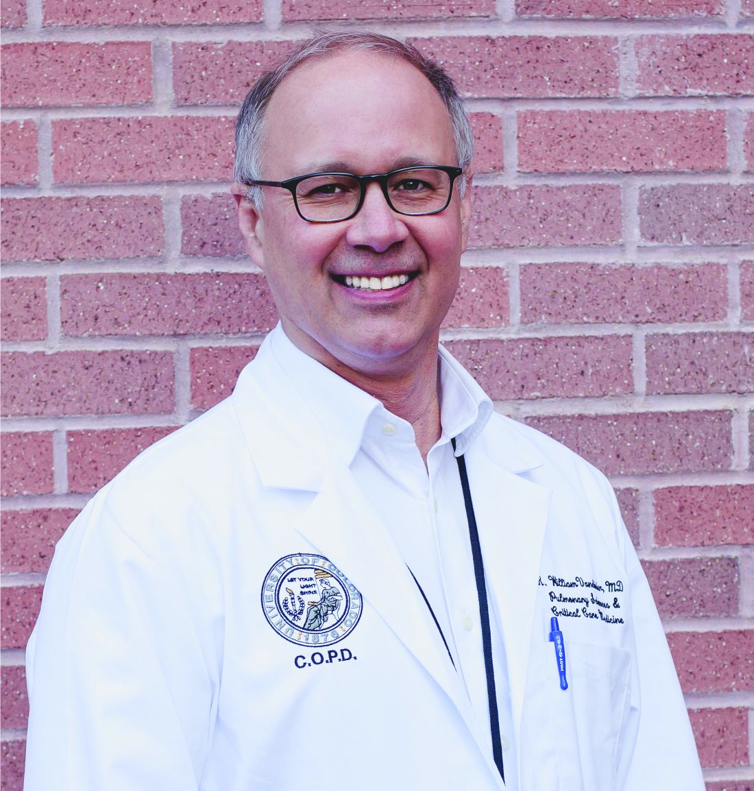
ECMO for refractory asthma exacerbations
The overnight shift in the MCU began as it does for many intensivists, by hearing about ED admissions, transfers from outside hospitals, sick floor patients, and high-risk patients in the MICU. Earlier in the day, the MICU team had admitted a 39-year-old woman with a severe asthma attack that required endotracheal intubation and mechanical ventilation in the ED for hypercarbic respiratory failure. After intubation, she had no audible air movement on chest exam, severe hypercarbic respiratory acidosis determined by an arterial blood gas, a clear chest radiograph, and negative findings on a respiratory viral panel. Her family said that she had run out of her steroid inhaler a month earlier and could not afford a refill. She had been using increasing amounts of albuterol over the past week before developing severe shortness of breath on the day of admission. The ED and MICU teams aggressively treated her with high-dose inhaled albuterol, ipratropium, and IV magnesium sulfate for bronchodilation; methylprednisolone for airway inflammation; and continuous ketamine for sedation, analgesia, and bronchodilation (Rehder KJ, et al. Respir Care. 2017;62[6]:849). Her airway pressures continued to be high despite using lung protective ventilation, so she was shifted to a permissive hypercapnia ventilation strategy using neuromuscular blockade, deep sedation, and low minute-ventilation (Laher AE, et al. J Intensive Care Med. 2018;33[9]:491).
Two hours into the shift, the bedside nurse noted that the patient had become hypotensive. Her ventilator pressures remained stable with peak inspiratory pressures of 38-42 cm H2O, plateau pressures of 28-30 cm H2O, auto-positive end-expiratory pressure (auto-PEEP) of 10-12 cm H2O, and fractional inspiratory oxygen (FiO2) of 40%. A repeat chest radiograph showed no signs of barotrauma, but arterial blood gas values showed severe respiratory acidosis with a pH of 7.05 and a PCO2 > 100 mm Hg. Her condition stabilized when she received a continuous infusion of bicarbonate to control her acidosis and low-dose IV norepinephrine for blood pressure control. It was at that moment that the bedside nurse astutely asked whether we should consider starting ECMO for the patient, as coauthor Dr. Arun Kannappan had done for a similar patient with asthma a month earlier. Dr. Vandivier notes, “My first response was that ECMO was not needed, because our patient had stabilized, and I had taken care of many patients like this in the past. But as I considered the situation more carefully, it was clear that . In short, my ‘traditional’ approach left little room for error in a patient with high ventilator pressures and hemodynamic instability.”
ECMO is a technique used to add oxygen or remove CO2 from the blood of people with different forms of respiratory failure (Fan E, et al. Intensive Care Med. 2016;2:712) that was first used by Hill and colleagues in 1966 for trauma-induced ARDS (Hill JD, et al. N Engl J Med. 1972;286:629). The ECMO circuit pumps blood from the venous system into an oxygenator that adds oxygen and removes CO2 before blood is returned to either the venous or arterial circulation (Intensive Care Med. 2016;42:712). Venovenous ECMO (vvECMO) is used in clinical scenarios where only oxygenation and/or CO2 removal is needed, whereas venoarterial ECMO (vaECMO) is reserved for situations where additional hemodynamic support is necessary. ECMO is traditionally thought of as a means to increase blood oxygenation, but it is less widely appreciated that ECMO is particularly effective at removing blood CO2. In addition to ECMO helping to normalize oxygenation or eliminate CO2, it can also be used to lower tidal volumes, decrease airway pressures, and allow “lungs to rest” with the goal of avoiding ventilator-induced lung injury (VILI).
Standing at the bedside, it seemed to the authors that it was the right time to think about instituting a salvage therapy. But was there evidence that ECMO could improve survival? Were there clear guidelines for when to initiate ECMO, and was ECMO more effective than other salvage therapies such as inhaled volatile anesthetics?
Since McDonnell and colleagues first described the use of ECMO for a severe asthma exacerbation in 1981 (Ann Thoracic Surg. 1981;31[2]:171), about 95 articles have been published. Other than two registry studies and a recent epidemiologic study, all of these publications were case reports, case series, and reviews. Mikkelsen and colleagues (ASAIO J. 2009;55[1]:47) performed a retrospective, cohort study using the International Extracorporeal Life Support (ECLS) Organization Registry to determine whether ECMO use for status asthmaticus was associated with greater survival than the use of ECMO for other causes of respiratory failure. From 1986 through 2006, a total of 2,127 cases of respiratory failure were identified that required ECMO, including 27 for status asthmaticus and 1,233 for other causes. Their analysis showed that 83.3% of asthmatics treated with ECMO survived to hospital discharge, compared with 50.8% of people treated with ECMO for respiratory failure not due to asthma, with an odds ratio (OR) of 4.86 favoring survival of asthmatics (OR = 4.86; 95% CI, 1.65-14.31, P = .004).
Yeo and colleagues (Yeo HJ, et al. Critical Care. 2017;21:297) also used the ECLS Organization Registry to measure survival to hospital discharge, complications, and clinical factors associated with in-hospital mortality for asthmatics treated with ECMO. They included 272 people treated with ECMO for asthma between 1992 and 2016, after excluding people treated with ECMO for cardiopulmonary resuscitation or cardiac dysfunction. ECMO was associated with improvements in ventilator mechanics, including a reduction in respiratory rate, FiO2, peak inspiratory pressure, mean airway pressure, and driving pressure. Use of ECMO for status asthmaticus was also associated with an 83.5% survival to hospital discharge, similar to the study by Mikkelsen and colleagues. Hemorrhage, the most common complication, occurred in roughly a quarter of people treated with ECMO. In the multivariate analysis, age, bleeding, pre-ECMO PEEP, post-ECMO FiO2, and driving pressure were all associated with higher in-hospital mortality.
Although there are no formal criteria to guide use of ECMO for asthma exacerbations with respiratory failure, a number of physicians and a physician organization have recommended that ECMO be considered for persistently high ventilator pressures, uncontrolled respiratory acidosis, or hemodynamic instability. Because our patient qualified for ECMO based on all three suggested criteria, we consulted cardiac surgery who quickly started her on vvECMO. She remained on ECMO for 4 days until she was decannulated, extubated, and discharged home.
Despite this positive outcome, the lack of a high-quality, controlled study to help guide our decision was surprising given the ability of ECMO to efficiently remove CO2 and to decrease ventilator pressures. The lack of guidance prompted us to perform a retrospective, epidemiologic cohort study to determine whether treatment with ECMO for asthma exacerbations with respiratory failure was associated with reduced mortality, compared with people treated without ECMO (Zakrajsek JK, Chest. 2023;163[1]:38). The study included 13,714 people admitted to an ECMO-capable hospital with respiratory failure that required invasive ventilation because of an asthma exacerbation between 2010 and 2020, of which 127 were treated with ECMO and 13,587 were not. During this period, use of ECMO as a salvage therapy for severe asthma exacerbations was a rare event, but it became more common over time. With the limitation that 40% of asthma patients were transferred from an outside hospital, 74% were started on ECMO in the first 2 hospital days, and 94% were started within the first week of hospitalization. Once started, ECMO was continued for a median of 1.0 day and range of 1-49 days. Hospital mortality was 14.6% in the ECMO group versus 26.2% in the no ECMO group, which equated to an 11.6% absolute risk reduction (P = 0.03) and 52% relative risk reduction (P = 0.04) in mortality. ECMO was associated with hospital costs that were $114,000 higher per patient, compared with the no ECMO group, but did not affect intensive care unit length of stay, hospital length of stay, or time on invasive mechanical ventilation.
We were pleased that our patient had a good outcome, and were reassured by our study results. But we were left to wonder whether ECMO really was the best salvage therapy for asthma exacerbations with respiratory failure, and if it was initiated for the right indications at the best time. These are important treatment considerations that take on new urgency given that physicians are increasingly looking to ECMO as a salvage therapy for refractory asthma, and the recent FDA approval of low-flow, extracorporeal CO2 removal systems that could make CO2 removal a more available, and perhaps less expensive, strategy. Despite promising epidemiological data, it will be important that these questions are answered with well-designed clinical trials so that physicians can be armed with the knowledge needed to navigate complex clinical scenarios, and ultimately to prevent unfortunate deaths from a reversible disease.
2023 GOLD update: Changes in COPD nomenclature and initial therapy
Airways Disorders Network
Asthma & COPD Section
The 2023 GOLD committee proposed changes in nomenclature and therapy for various subgroups of patients with COPD.
The mainstay of initial treatment for symptomatic COPD should include combination LABA/LAMA bronchodilators in a single inhaler. For patients with features of concomitant asthma or eosinophils greater than or equal to 300 cells/microliter, an ICS/LABA/LAMA combination inhaler is recommended.
People with “young COPD” develop respiratory symptoms and meet spirometric criteria for COPD between the ages of 25 and 50 years old. Other terminology changes center around those with functional and/or structural changes suggesting COPD, but who do not meet the postbronchodilator spirometric criteria to confirm the COPD diagnosis.
Those with “pre-COPD” have normal spirometry, including the FEV1 and FEV1/FVC ratio, but have functional and/or structural changes concerning for COPD. Functional changes include air trapping and/or hyperinflation on PFTs, low diffusion capacity, and/or decline in FEV1 of > 40 mL per year.
Structural changes include emphysematous changes and/or bronchial wall changes on CT scans. “PRISm” stands for preserved ratio with impaired spirometry, where the postbronchodilator FEV1/FVC is greater than or equal to 0.70, but FEV1 is < 80% predicted with similar functional and/or structural changes to those with “pre-COPD.” People with PRISm have increased all-cause mortality. Not all people with pre-COPD or PRISm progress clinically and spirometrically to COPD; however, they should be treated because they have symptoms as well as functional and/or structural abnormalities. Despite increasing data regarding pre-COPD and PRISm, many gaps remain regarding optimal management.
Maria Ashar, MD, MBBS
AGA guidelines, CPUs lead education at DDW® 2023
Below is a sampling of AGA’s invited-speaker sessions we’re excited about this year for clinical practitioners. To view other AGA program highlights, check out the DDW Preliminary Program.
- Guidelines Highlights 2023
- Clinical Practice Updates: Battle of the Heavyweights
- AGA Clinical Symposium
- Case Studies in Measuring Care and Improving Quality
- Optimizing Your GI Practice: Guidelines, Quality and Delivery
- AGA Postgraduate Course ($)
- Surviving the First Years in Clinical Practice: Roundtable With the Experts
Protect the next generation of GI investigators
Investing in research is the only way we will identify new diagnostics and treatments. However, at this time of unparalleled scientific and clinical opportunity, promising early stage investigators are leaving the field because of the instability of federal research funding.
Fortunately, the AGA Research Foundation has a proven track record of funding young investigators whose work advances the field of gastroenterology and hepatology.
Help the AGA build a community of investigators through the AGA Research Foundation.
Your donation to the AGA Research Foundation can fund future success stories by keeping young scientists working to advance our understanding of digestive diseases.
Donate today to help protect the GI research pipeline. Make a tax-deductible donation at www.foundation.gastro.org.
AGA News – May 2023
Season 2 of Small Talk, Big Topics is here!
AGA’s podcast for trainees and early career GIs, Small Talk, Big Topics, is back for season two. To kick off the new season, hosts Drs. Matthew Whitson, Nina Nandy, and CS Tse sit down with AGA President Dr. John Carethers in a two-part special to chat about his career and how his involvement with AGA has impacted him.
In episode one, Drs. Whitson, Nandy and Tse take a deep dive with Dr. Carethers to reflect on how he first got involved with AGA, his experience with different committees, and how those roles paved the way to leadership positions.
Now, as president, he says, “I am having so much fun. AGA has been with me for my entire GI career. It’s really the voice of the science and practice of gastroenterology.”
In episode two, Dr. Carethers examines the career advice he’s received, how it shaped his leadership style and provides guidance to early career GIs.
“What’s important about some of these higher-level [decisions] is to set a vision. You can’t be a leader if you have no followers, and people have to believe in something, that they’re moving toward something.”
Listen to more of Dr. Carethers’ insight in the first two episodes of Small Talk, Big Topics wherever you listen to podcasts and subscribe to stay up to date on new episodes.
Maximize your first day at DDW® 2023
Held during the first day of Digestive Disease Week®, this year’s AGA Postgraduate Course will be held live on Saturday, May 6, from 8:30 a.m. to 5 p.m. CT. This year’s theme – Advances in Gastroenterology: News You Can Use – will help you cut through the noise surrounding best practices for GI physicians.
Pricing is the same for both in-person and virtual attendees, giving you the flexibility to experience the course in-person or from the comfort of your home. All registrants will have on-demand access to the course for three months and the opportunity to earn up to 17.5 total credits when you complete all on-demand content.
What’s new this year?
General session format
Presentations will be given in an engaging format that will feel less didactic and more akin to a discussion among faculty, or a conversation with the experts! It’s also an exciting opportunity to mix junior and senior lecturers on the same platform.
Recent clinical practices
Session panelists will work together to select the key papers in their topic areas for discussion. Only the newest — within one year — and most important papers, clinical guidelines and pathways in the field will be selected.
Register to attend DDW and the Postgraduate Course today.
And the winner of this year’s Shark Tank is …
The 13th annual AGA Tech Summit took place in San Francisco, Calif., recently, bringing together GI entrepreneurs, clinicians, medical technology companies, venture capitalists, and regulatory agencies working to improve patient care in the field. A highlight of the event is the annual Shark Tank competition, where forward-thinking companies showcase and pitch their innovations to a panel of expert judges.
Congratulations to this year’s winner – Endiatx!
From devices providing rapid cancer detection to technology that makes endoscopy safer, the five companies selected for the 2023 AGA Shark Tank represented a glimpse of the future of GI patient care.
While each team offered a creative solution to modern-day GI challenges, only one could be declared the winner. Congratulations to our 2023 winner, Endiatx! Endiatx will represent AGA in the upcoming Shark Tank competition at DDW®.
Endiatx has developed a vitamin-sized intrabody robot
PillBot is a miniature robotic capsule endoscopy. Shipped to a patient’s home or picked up from a pharmacy, the standard size capsule is swallowed and then controlled by an external joystick-like device or a phone app by a physician in a physically separate location. Using real-time video transmissions visible to both operator and patient, the capsule navigates the entire stomach in a few minutes without anesthesia and ultimately is excreted outside the body without the need for recapture.
Future GI physician innovators
This year the AGA Center for GI Innovation and Technology (CGIT) welcomed 22 first-year to advanced endoscopy fellows to the AGA Innovation Fellows Program. The program provides a unique opportunity for the fellows to learn from GI clinicians, innovators, entrepreneurs, and medical technology executives on how new technologies are developed and brought to market.
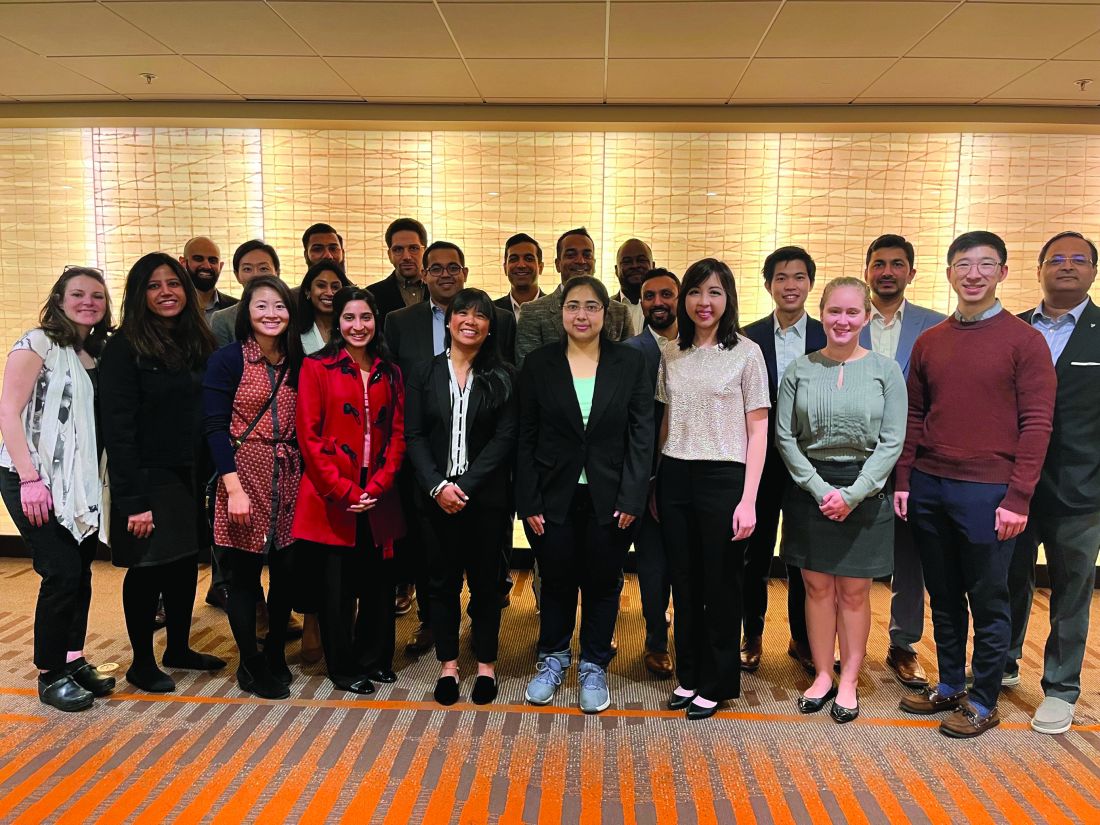
The fellows received an exclusive behind-the-scenes tour of Medtronic’s R&D facility in Santa Clara, Calif., and got to experience hands-on demonstrations of GI GeniusTM, PillCamTM, EndoflipTM, NexpowderTM, BravoTM, BarrxTM and ProdiGITM technologies. The group was also hosted by Boston Scientific Corporation, Castle Biosciences and PENTAX Medical at a dinner that included an innovators panel discussion. The program will continue throughout the year with monthly educational sessions moderated by members of the AGA CGIT committee.
- Mohd Amer Alsamman, MD, Georgetown University
- Mohammad Arfeen, MD, Franciscan Health Olympia Fields
- Alexis Bayudan, MD, University of California, San Francisco
- Aileen Bui, MD, University of Southern California
- Divya Chalikonda, MD, Thomas Jefferson University Hospital
- Alec Faggen, MD, University of California, San Francisco
- Sweta Ghosh, PhD, University of Louisville School of Medicine
- Hemant Goyal, MD, University of Texas Houston
- Averill Guo, MD, Brown University
- Omar Jamil, MD, University of Chicago
- Christina Kratschmer, MD, Washington University in St. Louis
- Thi Khuc, MD, University of Maryland School of Medicine
- Anand Kumar, MD, Northwell Health – Lenox Hill Hospital
- Xing Li, MD, Massachusetts General Hospital
- Alana Persaud, MD, SUNY Downstate Medical Center
- Itegbemie Obaitan, MD, Indiana University School of Medicine
- Chethan Ramprasad, MD, University of Pennsylvania
- Abhishek Satishchandran, MD, University of Michigan
- Kevin Shah, MD, Emory University School of Medicine
- Shifa Umar, MD, University of Chicago
- Kornpong Vantanasiri, MD, Mayo Clinic Rochester
- Shaleen Vasavada, MD, Baylor College of Medicine
Highlights from social media
See what else attendees shared with #AGATech on Twitter.
The 2023 AGA Tech Summit was made possible by support from Castle Biosciences and Medtronic (Diamond Sponsors), AI Medical Services, Boston Scientific, Exact Sciences Corporation, FUJIFILM Medical Systems and Olympus Corporation (Gold Sponsors), Cook Medical Inc., and STERIS Endoscopy (Silver Sponsors), and Apollo Endosurgery and EvoEndo (Bronze Sponsors).
AGA takes CRC month to Capitol Hill
Participating in Colorectal Cancer Awareness Month in Washington, D.C., means one thing – taking the fight to save lives from CRC to Capitol Hill and advocating for increased access to screening and research to improve outcomes.

In March, AGA joined the national advocacy organization Fight Colorectal Cancer (Fight CRC) and partners in the colorectal cancer community for events in our nation’s capital. The goal was to destigmatize talking about gut health and CRC and to collaboratively develop solutions that will improve and increase access to CRC screening.
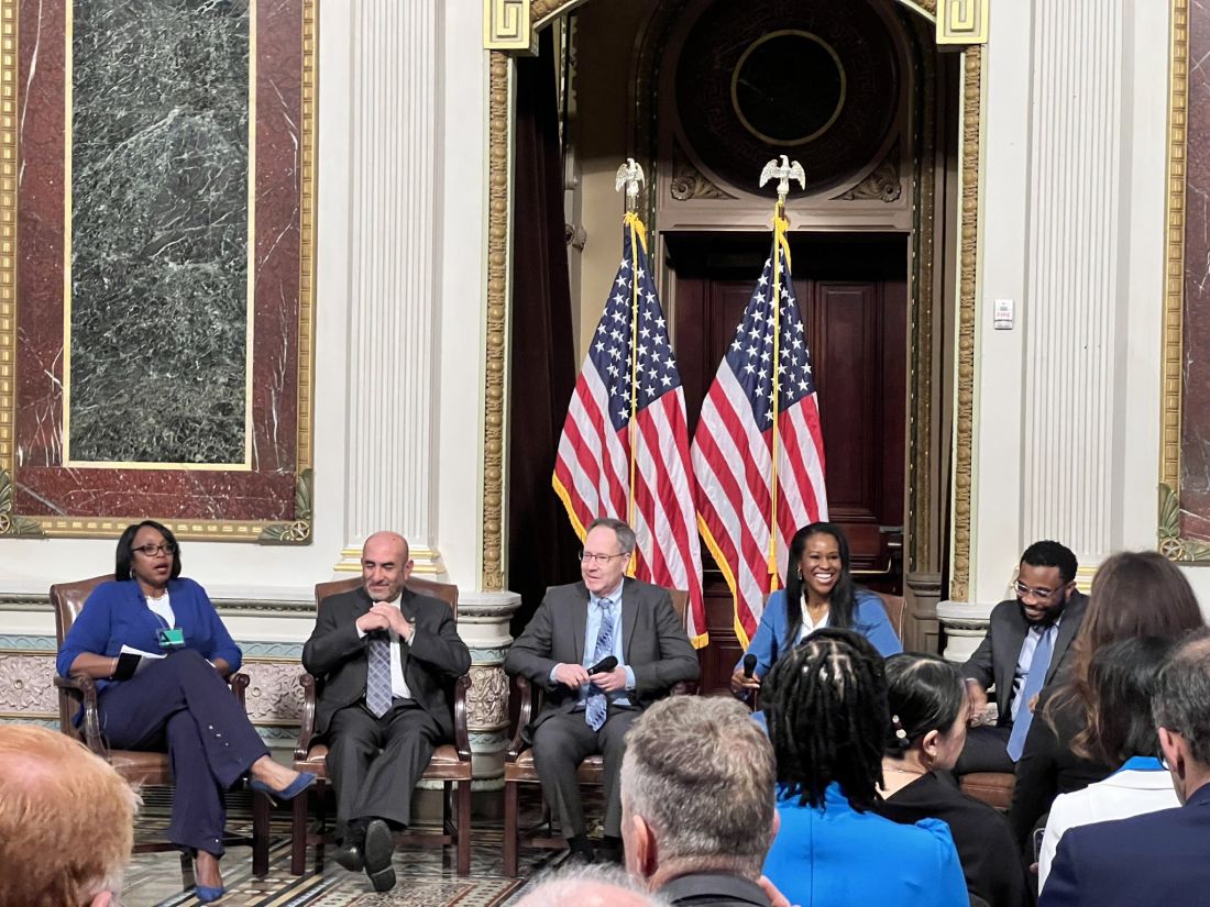
Fight CRC working lunch
Former AGA president Dr. David Lieberman and fellow AGA member and FORWARD graduate Dr. Fola May served as facilitators for the coalition of public and private leaders assembled by Fight CRC. The group is working to develop an action plan to further equitable CRC screening and lower the number of lives impacted by CRC. Among the participants were insurers, industry, federal agencies, healthcare providers, retail businesses, and patients.
White House Cancer Moonshot colorectal cancer forum
In partnership with President Biden’s reignited Cancer Moonshot initiative, we joined Fight CRC and other advocacy and industry leaders in the colorectal cancer community for the Cancer Moonshot Colorectal Cancer Forum, hosted by the White House.
Dr. May participated as a panelist during the forum and discussed how we should address disparities in CRC. “Research dollars are essential in [combating CRC inequity]. We do not know how to effectively deliver care and preventive services to these populations unless we do deep dives into these particular settings to understand how to best deliver that care. This is not a “pick a model and apply broadly” approach. We need to go to the people, and we need to go to the people with the methods that work for that particular setting, and that’s going to be different in every community.”
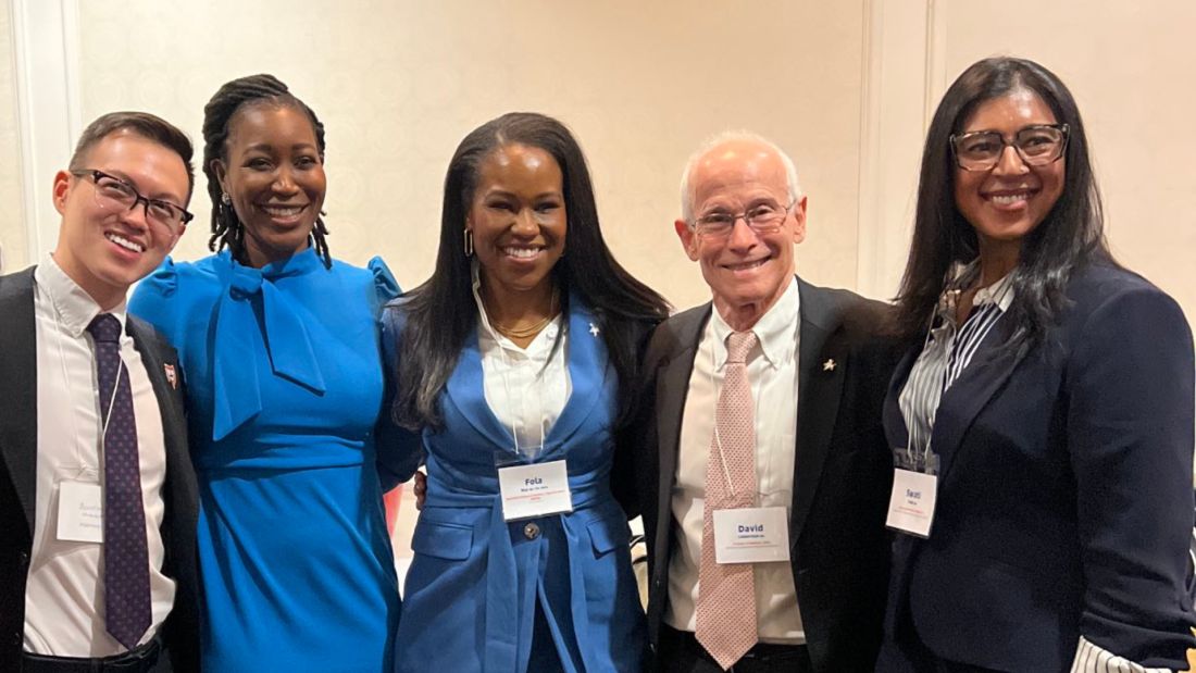
In addition to Dr. Lieberman, who attended on behalf of AGA, fellow AGA members Drs. Austin Chiang, Swati Patel and AGA FORWARD Scholar Rachel Issaka were in attendance. We are appreciative of the opportunity to be included in these important discussions with the Administration and partners in the CRC community as we work together to reduce the burden of CRC and save lives.
Fight CRC United in Blue rally on the National Mall
It’s become an annual tradition for us to join Fight CRC’s United in Blue rally and blue flag installation on the National Mall, and this year was no different. We joined industry and patient advocacy groups in the CRC community to raise our voices about the need for screening, research, and advocacy to improve colon cancer outcomes.
The rally included inspiring calls to action and CRC testimonials from individuals who have been personally impacted by the disease, including Rep. Donald Payne Jr. (D-NJ), who lost his father to CRC and who personally underwent screening, which led to the discovery of 13 polyps.
Dr. Manish Singla from Capital Digestive Care spoke on behalf of AGA and provided encouragement and a reminder for patients and providers.
“What I keep hearing here is patients feel like they’re not being heard – so we’re listening. We’re trying and we’re here to fight the disease with you all. Everyone here knows somebody who is due for a colonoscopy and isn’t getting it, so use your persuasion – talk about it, convince, cajole, shame – use whatever you need so that everyone gets the screenings they need,” Dr. Singla said.
Our work is just beginning: Let’s work together to encourage screenings for colorectal cancer and save lives. Join us as we remind everyone that 45 is the new 50.
Season 2 of Small Talk, Big Topics is here!
AGA’s podcast for trainees and early career GIs, Small Talk, Big Topics, is back for season two. To kick off the new season, hosts Drs. Matthew Whitson, Nina Nandy, and CS Tse sit down with AGA President Dr. John Carethers in a two-part special to chat about his career and how his involvement with AGA has impacted him.
In episode one, Drs. Whitson, Nandy and Tse take a deep dive with Dr. Carethers to reflect on how he first got involved with AGA, his experience with different committees, and how those roles paved the way to leadership positions.
Now, as president, he says, “I am having so much fun. AGA has been with me for my entire GI career. It’s really the voice of the science and practice of gastroenterology.”
In episode two, Dr. Carethers examines the career advice he’s received, how it shaped his leadership style and provides guidance to early career GIs.
“What’s important about some of these higher-level [decisions] is to set a vision. You can’t be a leader if you have no followers, and people have to believe in something, that they’re moving toward something.”
Listen to more of Dr. Carethers’ insight in the first two episodes of Small Talk, Big Topics wherever you listen to podcasts and subscribe to stay up to date on new episodes.
Maximize your first day at DDW® 2023
Held during the first day of Digestive Disease Week®, this year’s AGA Postgraduate Course will be held live on Saturday, May 6, from 8:30 a.m. to 5 p.m. CT. This year’s theme – Advances in Gastroenterology: News You Can Use – will help you cut through the noise surrounding best practices for GI physicians.
Pricing is the same for both in-person and virtual attendees, giving you the flexibility to experience the course in-person or from the comfort of your home. All registrants will have on-demand access to the course for three months and the opportunity to earn up to 17.5 total credits when you complete all on-demand content.
What’s new this year?
General session format
Presentations will be given in an engaging format that will feel less didactic and more akin to a discussion among faculty, or a conversation with the experts! It’s also an exciting opportunity to mix junior and senior lecturers on the same platform.
Recent clinical practices
Session panelists will work together to select the key papers in their topic areas for discussion. Only the newest — within one year — and most important papers, clinical guidelines and pathways in the field will be selected.
Register to attend DDW and the Postgraduate Course today.
And the winner of this year’s Shark Tank is …
The 13th annual AGA Tech Summit took place in San Francisco, Calif., recently, bringing together GI entrepreneurs, clinicians, medical technology companies, venture capitalists, and regulatory agencies working to improve patient care in the field. A highlight of the event is the annual Shark Tank competition, where forward-thinking companies showcase and pitch their innovations to a panel of expert judges.
Congratulations to this year’s winner – Endiatx!
From devices providing rapid cancer detection to technology that makes endoscopy safer, the five companies selected for the 2023 AGA Shark Tank represented a glimpse of the future of GI patient care.
While each team offered a creative solution to modern-day GI challenges, only one could be declared the winner. Congratulations to our 2023 winner, Endiatx! Endiatx will represent AGA in the upcoming Shark Tank competition at DDW®.
Endiatx has developed a vitamin-sized intrabody robot
PillBot is a miniature robotic capsule endoscopy. Shipped to a patient’s home or picked up from a pharmacy, the standard size capsule is swallowed and then controlled by an external joystick-like device or a phone app by a physician in a physically separate location. Using real-time video transmissions visible to both operator and patient, the capsule navigates the entire stomach in a few minutes without anesthesia and ultimately is excreted outside the body without the need for recapture.
Future GI physician innovators
This year the AGA Center for GI Innovation and Technology (CGIT) welcomed 22 first-year to advanced endoscopy fellows to the AGA Innovation Fellows Program. The program provides a unique opportunity for the fellows to learn from GI clinicians, innovators, entrepreneurs, and medical technology executives on how new technologies are developed and brought to market.
The fellows received an exclusive behind-the-scenes tour of Medtronic’s R&D facility in Santa Clara, Calif., and got to experience hands-on demonstrations of GI GeniusTM, PillCamTM, EndoflipTM, NexpowderTM, BravoTM, BarrxTM and ProdiGITM technologies. The group was also hosted by Boston Scientific Corporation, Castle Biosciences and PENTAX Medical at a dinner that included an innovators panel discussion. The program will continue throughout the year with monthly educational sessions moderated by members of the AGA CGIT committee.
- Mohd Amer Alsamman, MD, Georgetown University
- Mohammad Arfeen, MD, Franciscan Health Olympia Fields
- Alexis Bayudan, MD, University of California, San Francisco
- Aileen Bui, MD, University of Southern California
- Divya Chalikonda, MD, Thomas Jefferson University Hospital
- Alec Faggen, MD, University of California, San Francisco
- Sweta Ghosh, PhD, University of Louisville School of Medicine
- Hemant Goyal, MD, University of Texas Houston
- Averill Guo, MD, Brown University
- Omar Jamil, MD, University of Chicago
- Christina Kratschmer, MD, Washington University in St. Louis
- Thi Khuc, MD, University of Maryland School of Medicine
- Anand Kumar, MD, Northwell Health – Lenox Hill Hospital
- Xing Li, MD, Massachusetts General Hospital
- Alana Persaud, MD, SUNY Downstate Medical Center
- Itegbemie Obaitan, MD, Indiana University School of Medicine
- Chethan Ramprasad, MD, University of Pennsylvania
- Abhishek Satishchandran, MD, University of Michigan
- Kevin Shah, MD, Emory University School of Medicine
- Shifa Umar, MD, University of Chicago
- Kornpong Vantanasiri, MD, Mayo Clinic Rochester
- Shaleen Vasavada, MD, Baylor College of Medicine
Highlights from social media
See what else attendees shared with #AGATech on Twitter.
The 2023 AGA Tech Summit was made possible by support from Castle Biosciences and Medtronic (Diamond Sponsors), AI Medical Services, Boston Scientific, Exact Sciences Corporation, FUJIFILM Medical Systems and Olympus Corporation (Gold Sponsors), Cook Medical Inc., and STERIS Endoscopy (Silver Sponsors), and Apollo Endosurgery and EvoEndo (Bronze Sponsors).
AGA takes CRC month to Capitol Hill
Participating in Colorectal Cancer Awareness Month in Washington, D.C., means one thing – taking the fight to save lives from CRC to Capitol Hill and advocating for increased access to screening and research to improve outcomes.
In March, AGA joined the national advocacy organization Fight Colorectal Cancer (Fight CRC) and partners in the colorectal cancer community for events in our nation’s capital. The goal was to destigmatize talking about gut health and CRC and to collaboratively develop solutions that will improve and increase access to CRC screening.
Fight CRC working lunch
Former AGA president Dr. David Lieberman and fellow AGA member and FORWARD graduate Dr. Fola May served as facilitators for the coalition of public and private leaders assembled by Fight CRC. The group is working to develop an action plan to further equitable CRC screening and lower the number of lives impacted by CRC. Among the participants were insurers, industry, federal agencies, healthcare providers, retail businesses, and patients.
White House Cancer Moonshot colorectal cancer forum
In partnership with President Biden’s reignited Cancer Moonshot initiative, we joined Fight CRC and other advocacy and industry leaders in the colorectal cancer community for the Cancer Moonshot Colorectal Cancer Forum, hosted by the White House.
Dr. May participated as a panelist during the forum and discussed how we should address disparities in CRC. “Research dollars are essential in [combating CRC inequity]. We do not know how to effectively deliver care and preventive services to these populations unless we do deep dives into these particular settings to understand how to best deliver that care. This is not a “pick a model and apply broadly” approach. We need to go to the people, and we need to go to the people with the methods that work for that particular setting, and that’s going to be different in every community.”
In addition to Dr. Lieberman, who attended on behalf of AGA, fellow AGA members Drs. Austin Chiang, Swati Patel and AGA FORWARD Scholar Rachel Issaka were in attendance. We are appreciative of the opportunity to be included in these important discussions with the Administration and partners in the CRC community as we work together to reduce the burden of CRC and save lives.
Fight CRC United in Blue rally on the National Mall
It’s become an annual tradition for us to join Fight CRC’s United in Blue rally and blue flag installation on the National Mall, and this year was no different. We joined industry and patient advocacy groups in the CRC community to raise our voices about the need for screening, research, and advocacy to improve colon cancer outcomes.
The rally included inspiring calls to action and CRC testimonials from individuals who have been personally impacted by the disease, including Rep. Donald Payne Jr. (D-NJ), who lost his father to CRC and who personally underwent screening, which led to the discovery of 13 polyps.
Dr. Manish Singla from Capital Digestive Care spoke on behalf of AGA and provided encouragement and a reminder for patients and providers.
“What I keep hearing here is patients feel like they’re not being heard – so we’re listening. We’re trying and we’re here to fight the disease with you all. Everyone here knows somebody who is due for a colonoscopy and isn’t getting it, so use your persuasion – talk about it, convince, cajole, shame – use whatever you need so that everyone gets the screenings they need,” Dr. Singla said.
Our work is just beginning: Let’s work together to encourage screenings for colorectal cancer and save lives. Join us as we remind everyone that 45 is the new 50.
De Marco gift to CHEST makes more than one dream possible
As a member of CHEST leadership for years, Bob De Marco, MD, FCCP, ruminated over new, exciting ways to increase support of the philanthropic efforts of the American College of Chest Physicians.
Dr. De Marco knows all too well that the percentage of CHEST members who donate to support CHEST’s philanthropic initiatives is – in a word - underwhelming. For those who are involved, they do so greatly and with their whole selves, but Dr. De Marco believed more could be done.
In the months leading up to the CHEST Annual Meeting 2022 in Nashville, Dr. De Marco discussed fundraising with CHEST staff and was already thinking ahead to CHEST 2023 in Hawai’i.
“That’s when it hit me – we could leverage Hawai’i to get donations and to expose people to CHEST philanthropy,” said Dr. De Marco. “Hawai’i is a dream destination, and that might be the exact motivation it would take to get that first donation from someone.”
Having a good idea is one thing, but making sure it happens requires individual commitment. Dr. DeMarco personally pledged to cover the cost of first-class airfare for two to Hawai’i, hotel accommodations, and registration to CHEST 2023 in Honolulu. For a minimum donation of $250 to CHEST between September and the end of 2022, each donor would be entered into a drawing for a chance to win this dream trip.
“I thought to myself, who wouldn’t want this prize?” said Dr. De Marco.
“You get to go to paradise for free – with a guest – and attend a top tier educational conference. Knowing your entry supported an organization as deserving as CHEST is the cherry on top,” he added.
In launching the Hawai’i trip fundraiser before and during CHEST 2022, attendees from around the world were introduced to CHEST’s philanthropic efforts and its mission to champion lung health. Over $180,000 was donated during this time period, in no small part because of the Hawai’i travel reward.
“I’m happy to say that the fundraiser did a lot better than I expected, and I was elated to see all of the new donors,” said Dr. De Marco.
“It’s my hope that those first-time donors continue their support for all that we do to provide grants – community, research, and diversity – and support CHEST initiatives that impact patient care and change lives.”
During CHEST 2022, Dr. De Marco and other donors reflected on the organization’s philanthropic accomplishments and impact over the past decades.
Former grant recipients were invited to celebrate with donors and speak to what they were able to accomplish because of the support they received.
The celebration also introduced new CHEST initiatives, the First 5 Minutes® program and Bridging Specialties™: Timely Diagnosis for ILD. The former improves patient care through strengthened patient/clinician relationships, and the latter aims to eliminate gaps in diagnosing complex lung diseases like pulmonary fibrosis.
To all who donated to CHEST in 2022, Dr. De Marco said, “A sincere thank you to each and every one of you for helping us fulfill our mission. To the first-time donors, hopefully this will inspire you and your friends to be an active part of the CHEST family.”
And, to the winner of the trip, Dr. De Marco said, “A sincere congratulations and I hope you enjoy beautiful Hawai’i and your time at the meeting.”
Those who are interested in getting involved and supporting the philanthropic work of CHEST can contact [email protected].
Out of the 150+ donors who gave $250 or more to CHEST between September 2022 and the end of 2022, longtime friend of CHEST, Noah Dorsky, was the recipient of two first-class tickets to Hawai’i, hotel accommodations, and registration to CHEST 2023 in Honolulu.
Noah donated specifically to the Mark J. Rosen, MD, Master FCCP Endowment in honor of his late friend, Dr. Mark J. Rosen, who served as CHEST President from 2006 to 2007 and died in 2019.
"Mark was a remarkable doctor and valued life-long friend,” Noah said. “My continued support for CHEST is my way of honoring his memory and how much he meant to me and others."
Dr. Rosen’s distinguished career in pulmonary and critical care medicine spanned more than 4 decades, marked by his deep commitments to medical education and patient care. Before serving as President, Dr. Rosen served on the CHEST Board of Regents for many years. He held positions as Chair or member on numerous CHEST committees, including Education, Nominations, Membership, Marketing, and Finance.
Following his passing, Dr. Rosen’s wife, Ilene, stayed engaged with CHEST by creating the endowment in his name and attending the CHEST Annual Meeting every year to award the Rosen Cup to the winners of the annual CHEST Challenge.
Congratulations, Noah, and thank you for your faithful giving to support the work of CHEST.
Breathing easier: The growing adoption of indwelling pleural catheters
Thoracic Oncology Network
Interventional Procedures Section
The management of recurrent pleural effusions is challenging. These devices have become an important treatment option in patients with malignant pleural effusions (MPE), particularly those with a nonexpandable lung (Feller-Kopman DJ, et al. Am J Respir Crit Care Med. 2018;198[7]:839) and when talc pleurodesis is unsuccessful in patients with an expandable lung (Dresler CM, et al. Chest. 2005;127[3]:909).
Over the last 5 years, studies evaluating the use of IPCs in treating nonmalignant pleural disease have proliferated. These studies have included and shown the successful treatment of pleural effusions due to end-stage renal disease, advanced heart failure (Walker SP, et al. Eur Respir J. 2022;59[2]:2101362), and cirrhosis, especially when a transjugular intrahepatic portosystemic shunt or liver transplant is not an option (Shojaee S, et al., Chest. 2019;155[3]:546). Compared with MPE, the rate of pleurodesis is generally lower and takes longer when an IPC is used to manage a nonmalignant pleural disease. Infection is the most common complication; most cases can be managed without catheter removal.
With many cited advantages, the IPC is an essential tool in the armamentarium of the chest physician and interventional radiologist. Indwelling pleural catheters have proven applications beyond MPE. When applied in a multidisciplinary fashion involving subspecialists and considering the patient’s goals, using an IPC can help achieve a crucial patient-centric goal in managing a recurrent nonmalignant pleural effusion.
Samiksha Gupta, MD
2nd Year Fellow
Sameer Kaushik Avasarala, MD
Section Member-at-Large
Early mobility in the ICU: Working with the TEAM
Critical Care Network
Nonrespiratory Critical Care Section
This is especially true for critically ill patients, in which weakness is more common and can result in worse outcomes (Kress JP, et al. N Engl J Med. 2014;370:1626). This advocacy is endorsed by major societies and guidelines, like the ABCDEF bundle (Balas MC, et al. Crit Care Med. 2013;41:S116), in which “E” stands for Early mobility and exercise. In fact, the PADIS guidelines, addressing Pain, Agitation, Delirium, Immobility, and Sleep in the ICU, added Immobility and Sleep (the “I” and “S” in PADIS) to the prior PAD guidelines in the latest update in 2018, to stress the importance of early mobility in the ICU (Devlin JW, et al. Crit Care Med. 2018;46[9]:e825). Multiple studies have shown a positive impact of early mobility in the ICU on patients’ outcomes (Tipping CJ, et al. Intensive Care Med. 2017;43:171).
The recent TEAM study examined an early mobility approach in mechanically ventilated patients and found no difference in the primary outcome of alive and out-of-hospital at 180 days (N Engl J Med. 2022;387:1747).
Before concluding, it is worth realizing that the usual care arm included mobilization that was otherwise normally provided. The intervention arm protocolized the early mobility to be done simultaneously with the minimization of sedation. Patients’ assessment occurred in 81% in the usual care arm vs 94% in the intervention arm; both numbers are much higher than reported data in the ICU (Jolley SE, et al. Crit Care Med. 2017;45:205).
Revisiting the question of early mobility in the ICU, more data are needed to clarify the best methodology, sedation, timing, amount, and type of patients who will benefit the most. Until then, it should remain a goal for ICUs and part of the daily discussion when caring for critically ill patients.
Mohammed J. Al-Jaghbeer, MBBS, FCCP
Section Member-at-Large
Salim Surani, MD, MPH, FCCP
New AGA guideline recommends blood and stool tests for monitoring ulcerative colitis
These guidelines were published in Gastroenterology.
The AGA guidelines outline use cases for three biomarkers that provide accurate insights into UC disease activity: serum C-reactive protein (CRP) (blood), fecal calprotectin (stool), and fecal lactoferrin (stool). AGA recommends a monitoring strategy that integrates noninvasive biomarkers for patients with UC in remission (no current symptoms) as well as those with current symptoms.
Patients with UC in symptomatic remission
- Perform interval biomarker monitoring every 6-12 months.
- AGA recommends stool-based biomarkers over blood testing.
- If biomarkers are normal, AGA suggests continuing biomarker monitoring and avoiding routine endoscopic assessment.
- If biomarkers are elevated, AGA suggests endoscopic assessment by a gastroenterologist.
- Listen to your body! Talk to your doctor about any new symptoms.
Patients with symptomatically active UC
- Biomarker testing should be the first step to determine the need for endoscopic assessment.
- For patients with mild symptoms who have normal or elevated biomarkers, AGA suggests endoscopic assessment by a gastroenterologist.
- For patients with moderate to severe symptoms who have normal biomarkers, AGA suggests endoscopic assessment by a gastroenterologist.
- For patients with moderate to severe symptoms and elevated biomarkers, AGA suggests treatment adjustment and avoiding endoscopic assessment.
With AGA guidelines guiding the use of noninvasive biomarkers, physicians can confidently offer a more convenient and closer monitoring option for their patients.
AGA will advocate for all insurers to cover the cost of biomarker testing in UC.