User login
How is oncology adapting to COVID-19?
As the coronavirus pandemic escalates in the United States, Medscape Oncology reached out to a group of our contributors and asked them to provide their perspective on how their oncology departments and centers are preparing. Here are their responses to a number of issues facing oncologists in the US and around the world.
Have you shifted nonurgent follow-up visits to telemedicine, either via video or phone?
Kathy Miller, MD, Associate Director of Indiana University Simon Cancer Center: We are reviewing our clinic schedules and identifying “routine” follow-up patients who can be rescheduled. When patients are contacted to reschedule, they are asked if they have any urgent, immediate concerns that need to be addressed before the new appointment. If yes, they are offered a virtual visit.
Don Dizon, MD, Director of Women’s Cancers, Lifespan Cancer Institute; Director of Medical Oncology, Rhode Island Hospital: We have started to do this in preparation for a surge of people with COVID-19. Patients who are in long-term follow-up (no evidence of disease at 3 years or longer, being seen annually) or those in routine surveillance after curative treatment (that is, seen every 3 months) as well as those being seen for supportive care–type visits, like sexual health or survivorship, are all being contacted and visits are being moved to telehealth.
Jeffrey S. Weber, MD, PhD, Deputy Director of the Laura and Isaac Perlmutter Cancer Center at NYU Langone Medical Center: Yes. Any follow-up, nontreatment visits are done by phone or video if the patient agrees. (They all have).
Have you delayed or canceled cancer surgeries?
Ravi B. Parikh, MD, MPP, Medical oncologist at the University of Pennsylvania and the Philadelphia VA Medical Center: The University of Pennsylvania has taken this seriously. We’ve canceled all elective surgeries, have ramped up our telemedicine (video and phone) capabilities significantly, are limiting our appointments mostly to on-treatment visits, and have been asked to reconsider regular scans and reviews.
Dizon: We have not done this. There are apparently differences in interpretation in what institutions might mean as “elective surgeries.” At our institution, surgery for invasive malignancies is not elective. However, this may (or will) change if resources become an issue.
Lidia Schapira, MD, Associate Professor of Medicine and Director of Cancer Survivorship at the Stanford Comprehensive Cancer Institute: Delaying elective surgery is something that hospitals here have already implemented, and I imagine that this trend will spread. But it may be difficult to decide in situations that are not exactly “life-saving” but where an earlier intervention could preserve function or improve quality of life.
Mark A. Lewis, MD, Director of Gastrointestinal Oncology at Intermountain Healthcare in Utah: Cancer surgeries have not been deemed elective or delayed.
Have you delayed or altered the delivery of potentially immune-comprising treatments?
David Kerr, MD, Professor of Cancer Medicine at the University of Oxford in England: We are considering delaying initiation of our adjuvant colorectal cancer treatments, as we have data from our own QUASAR trials suggesting that patients who commence chemotherapy between 2 and 6 weeks do equally as well as those who begin 6-12 weeks after surgery.
Parikh: I personally haven’t delayed giving chemotherapy to avoid immune compromise, but I believe some others may have. It’s a delicate balance between wanting to ensure cancer control and making sure we are flattening the curve. As an example, though, I delayed three on-treatment visits for my clinic last Monday, and I converted 70% of my visits to telemedicine. However, I’m a genitourinary cancer specialist and the treatments I give are very different from others.
Lewis: The most difficult calculus is around adjuvant therapy. For metastatic patients, I am trying to use the least immunosuppressive regimen possible that will still control their disease. As you can imagine, it’s an assessment of competing risks.
Schapira: Patients who need essential anticancer therapy should still get it, but attempts to deintensify therapy should continue—for example, holding or postponing treatment without harm (based on evidence, not opinion). This may be possible for patients considering hormonal therapies for breast or prostate cancer.
Patients who need radiation should discuss the timing with their radiation oncologist. In some cases, it may be possible to delay treatment without affecting outcomes, but these decisions should be made carefully. Alternatively, shorter courses of radiation may be appropriate.
Have you advised your own patients differently given the high risk to cancer patients?
Kerr: We have factored potential infection with the virus into discussions where the benefits of chemotherapy are very marginal. This could tip the balance toward the patient deciding not to pursue chemotherapy.
Dizon: The data from China are not entirely crystal-clear. While they noted that people with active cancer and those who had a history of cancer are at increased risk for more severe infections and worse outcomes, the Chinese cohort was small, and compared with people without cancer, it tended to be much older and to be smokers (former or current). Having said this, we are counseling everyone about the importance of social distancing, washing hands, and not touching your face.
Lewis: If I have a complete blood count with a differential that includes lymphocytes, I can advise my lymphopenic patients (who are particularly vulnerable to viral infection) to take special precautions regarding social distancing in their own families.
Have any of your hospitalized patients been affected by policy changes to prepare beds/departments for the expected increase in COVID-19–positive patients?
Weber: Not yet.
Dizon: No, not at the moment.
Have you been asked to assist with other services or COVID-19 task forces?
Dizon: I am keenly involved in the preparations and modifications to procedures, including staffing decisions in outpatient, movement to telehealth, and work-from-home policies.
Lewis: I am engaged in system-wide COVID-19 efforts around oncology.
Kerr: Perhaps oddest of all, I am learning with some of our junior doctors to care for ventilated patients. I still consider myself enough of a general physician that I would hope to be able to contribute to the truly sick, but I accept that I do need an appropriate refresher course.
Bishal Gyawali, MD, PhD, medical oncologist at Queen’s University Cancer Research Institute: Queen’s Hospital medical students are now volunteering to help with daycare, groceries, and other tasks for staff who are working in the hospital.
Are you experiencing any shortages in personal protective equipment (PPE) at your center?
Miller: Some supplies are running short, though none are frankly out at this point. However, rationing and controls are in place to stretch the supplies as far as possible, including reusing some PPE.
Dizon: We are rationing face masks and N95 respirators, eye shields, and even surgical scrubs. We are talking about postponing elective surgery to save PPE but are not yet to that point. We’re asking that face masks be reused for at least 2 days, maybe longer. PPEs are one per day. Scrubs are kept secure.
Lewis: We are being very careful not to overuse PPE but currently have an adequate inventory. We have had to move gloves and masks to areas where they are not accessible to the general public, as otherwise they were being stolen (this started weeks ago).
Kerr: Our National Health System has an adequate supply of PPE equipment centrally, but there seems to be a problem with distribution, as some hospitals are reporting shortages.
Weber: Masks are in short supply, so they are being used for several days if not wet. We are short of plastic gowns and are using paper chemo gowns. Similar story at many places.
This article first appeared on Medscape.com.
Disruptions in cancer care in the era of COVID-19
Editor’s note: Find the latest COVID-19 news and guidance in Medscape’s Coronavirus Resource Center.
Even in the midst of the COVID-19 pandemic, cancer care must go on, but changes may need to be made in the way some care is delivered.
“We’re headed for a time when there will be significant disruptions in the care of patients with cancer,” said Len Lichtenfeld, MD, deputy chief medical officer of the American Cancer Society (ACS), in a statement. “For some it may be as straightforward as a delay in having elective surgery. For others it may be delaying preventive care or adjuvant chemotherapy that’s meant to keep cancer from returning or rescheduling appointments.”
Lichtenfeld emphasized that cancer care teams are going to do the best they can to deliver care to those most in need. However, even in those circumstances, it won’t be life as usual. “It will require patience on everyone’s part as we go through this pandemic,” he said.
“The way we treat cancer over the next few months will change enormously,” writes a British oncologist in an article published in the Guardian.
“As oncologists, we will have to find a tenuous balance between undertreating people with cancer, resulting in more deaths from the disease in the medium to long term, and increasing deaths from COVID-19 in a vulnerable patient population. Alongside our patients we will have to make difficult decisions regarding treatments, with only low-quality evidence to guide us,” writes Lucy Gossage, MD, consultant oncologist at Nottingham University Hospital, UK.
The evidence to date (from reports from China in Lancet Oncology) suggests that people with cancer have a significantly higher risk of severe illness resulting in intensive care admissions or death when infected with COVID-19, particularly if they recently had chemotherapy or surgery.
“Many of the oncology treatments we currently use, especially those given after surgery to reduce risk of cancer recurrence, have relatively small benefits,” she writes.
“In the current climate, the balance of offering these treatments may shift; a small reduction in risk of cancer recurrence over the next 5 years may be outweighed by the potential for a short-term increase in risk of death from COVID-19. In the long term, more people’s cancer will return if we aren’t able to offer these treatments,” she adds.
Postpone Routine Screening
One thing that can go on the back burner for now is routine cancer screening, which can be postponed for now in order to conserve health system resources and reduce contact with healthcare facilities, says the ACS.
“Patients seeking routine cancer screenings should delay those until further notice,” said Lichtenfeld. “While timely screening is important, the need to prevent the spread of coronavirus and to reduce the strain on the medical system is more important right now.”
But as soon as restrictions to slow the spread of COVID-19 are lifted and routine visits to health facilities are safe, regular screening tests should be rescheduled.
Guidance From ASCO
The American Society of Clinical Oncology (ASCO) has issued new guidance on caring for patients with cancer during the COVID-19 outbreak.
First and foremost, ASCO encourages providers, facilities, and anyone caring for patients with cancer to follow the existing guidelines from the Center for Disease Control and Prevention when possible.
ASCO highlights the CDC’s general recommendation for healthcare facilities that suggests “elective surgeries” at inpatient facilities be rescheduled if possible, which has also been recommended by the American College of Surgeons.
However, in many cases, cancer surgery is not elective but essential, it points out. So this is largely an individual determination that clinicians and patients will need to make, taking into account the potential harms of delaying needed cancer-related surgery.
Systemic treatments, including chemotherapy and immunotherapy, leave cancer patients vulnerable to infection, but ASCO says there is no direct evidence to support changes in regimens during the pandemic. Therefore, routinely stopping anticancer or immunosuppressive therapy is not recommended, as the balance of potential harms that may result from delaying or interrupting treatment versus the potential benefits of possibly preventing or delaying COVID-19 infection remains very unclear.
Clinical decisions must be individualized, ASCO emphasized, and suggested the following practice points be considered:
- For patients already in deep remission who are receiving maintenance therapy, stopping treatment may be an option.
- Some patients may be able to switch from IV to oral therapies, which would decrease the frequency of clinic visits.
- Decisions on modifying or withholding chemotherapy need to consider both the indication and goals of care, as well as where the patient is in the treatment regimen and tolerance to the therapy. As an example, the risk–benefit assessment for proceeding with chemotherapy in patients with untreated extensive small-cell lung cancer is quite different than proceeding with maintenance pemetrexed for metastatic non–small cell lung cancer.
- If local coronavirus transmission is an issue at a particular cancer center, reasonable options may include taking a 2-week treatment break or arranging treatment at a different facility.
- Evaluate if home infusion is medically and logistically feasible.
- In some settings, delaying or modifying adjuvant treatment presents a higher risk of compromised disease control and long-term survival than in others, but in cases where the absolute benefit of adjuvant chemotherapy may be quite small and other options are available, the risk of COVID-19 may be considered an additional factor when evaluating care.
Delay Stem Cell Transplants
For patients who are candidates for allogeneic stem cell transplantation, a delay may be reasonable if the patient is currently well controlled with conventional treatment, ASCO comments. It also directs clinicians to follow the recommendations provided by the American Society of Transplantation and Cellular Therapy and from the European Society for Blood and Marrow Transplantation regarding this issue.
Finally, there is also the question of prophylactic antiviral therapy: Should it be considered for cancer patients undergoing active therapy?
The answer to that question is currently unknown, says ASCO, but “this is an active area of research and evidence may be available at any time.”
This article first appeared on Medscape.com.
Lymphopenia linked to worse response in RCC, bladder cancer patients on checkpoint inhibitors
ORLANDO – treated in a real-world setting.
In a retrospective study of 20 patients, those with lower pALCs and higher pNLRs were less likely to respond to checkpoint inhibitors.
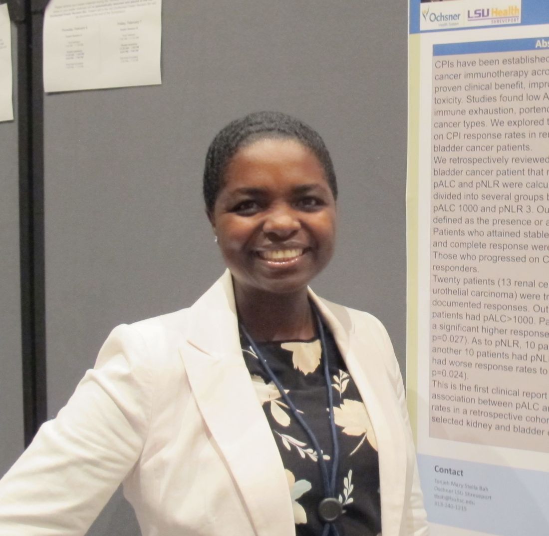
Tonjeh Bah, MD, of Feist-Weiller Cancer Center at LSU Health Shreveport in Louisiana, and colleagues reported these results in a poster at the ASCO-SITC Clinical Immuno-Oncology Symposium.
Response rates were 75% in patients with pALC greater than 1,000 and 25% in patients with pALC less than 1,000. This difference was statistically significant (P = .027), Dr. Bah noted, adding that the groups were comparable with respect to age, sex, race, and type of checkpoint inhibitors used.
Similarly, response rates were 80% in patients with pNLR less than 3 (the established upper limit of normal) and 30% in patients with pNLR greater than 3 (P = .024).
Checkpoint inhibitors targeting cytotoxic T-lymphocyte-associated protein 4 and programmed death-1 and its ligand are essential components of therapy across multiple cancer types, Dr. Bah noted in an interview. She explained that prior studies – mostly in patients with lung cancer and head and neck cancers – have also shown pALC and pNLR to be independently associated with poor checkpoint inhibitor response and worse progression-free survival.
“But this is the first study to look at the connection in kidney and bladder cancer in a real-world setting,” she said.
Dr. Bah and colleagues conducted the study to test the hypothesis that “lymphopenia is a marker of immune exhaustion, which is characterized by dysfunctional T cells that have a limited antitumor effect even in the presence of [checkpoint inhibitors] and by the eventual depletion of antitumor lymphocytes,” they wrote in the poster.
Patients included in the study were all those with renal cell carcinoma (n = 13) or bladder-urothelial cancers (n = 7) who received checkpoint inhibitors at one of two medical centers in Louisiana during 2015-2019 and who had outcomes reported. Patients who attained stable disease or had partial or complete responses were categorized as responders. Patients who progressed on checkpoint inhibitors were considered nonresponders.
“Our findings were not a surprise, but they do document, for the first time and in a real-world setting, that pALC and pNLR may have prognostic utility in patients with kidney and bladder cancers who are treated with [checkpoint inhibitors],” Dr. Bah said.
She added that the findings could help determine which patients are candidates for checkpoint inhibitors, but the results require confirmation in a large, prospective study. Dr. Bah reported having no disclosures, and there was no sponsor for this study.
SOURCE: Bah T et al. ASCO-SITC 2020. Abstract 31.
ORLANDO – treated in a real-world setting.
In a retrospective study of 20 patients, those with lower pALCs and higher pNLRs were less likely to respond to checkpoint inhibitors.
Tonjeh Bah, MD, of Feist-Weiller Cancer Center at LSU Health Shreveport in Louisiana, and colleagues reported these results in a poster at the ASCO-SITC Clinical Immuno-Oncology Symposium.
Response rates were 75% in patients with pALC greater than 1,000 and 25% in patients with pALC less than 1,000. This difference was statistically significant (P = .027), Dr. Bah noted, adding that the groups were comparable with respect to age, sex, race, and type of checkpoint inhibitors used.
Similarly, response rates were 80% in patients with pNLR less than 3 (the established upper limit of normal) and 30% in patients with pNLR greater than 3 (P = .024).
Checkpoint inhibitors targeting cytotoxic T-lymphocyte-associated protein 4 and programmed death-1 and its ligand are essential components of therapy across multiple cancer types, Dr. Bah noted in an interview. She explained that prior studies – mostly in patients with lung cancer and head and neck cancers – have also shown pALC and pNLR to be independently associated with poor checkpoint inhibitor response and worse progression-free survival.
“But this is the first study to look at the connection in kidney and bladder cancer in a real-world setting,” she said.
Dr. Bah and colleagues conducted the study to test the hypothesis that “lymphopenia is a marker of immune exhaustion, which is characterized by dysfunctional T cells that have a limited antitumor effect even in the presence of [checkpoint inhibitors] and by the eventual depletion of antitumor lymphocytes,” they wrote in the poster.
Patients included in the study were all those with renal cell carcinoma (n = 13) or bladder-urothelial cancers (n = 7) who received checkpoint inhibitors at one of two medical centers in Louisiana during 2015-2019 and who had outcomes reported. Patients who attained stable disease or had partial or complete responses were categorized as responders. Patients who progressed on checkpoint inhibitors were considered nonresponders.
“Our findings were not a surprise, but they do document, for the first time and in a real-world setting, that pALC and pNLR may have prognostic utility in patients with kidney and bladder cancers who are treated with [checkpoint inhibitors],” Dr. Bah said.
She added that the findings could help determine which patients are candidates for checkpoint inhibitors, but the results require confirmation in a large, prospective study. Dr. Bah reported having no disclosures, and there was no sponsor for this study.
SOURCE: Bah T et al. ASCO-SITC 2020. Abstract 31.
REPORTING FROM THE CLINICAL IMMUNO-ONCOLOGY SYMPOSIUM
Largest meeting on cancer research canceled: AACR
The biggest cancer research meeting of the year has been canceled as a reaction to the novel coronavirus (COVID-19) outbreak, which has also led to many other medical conferences being canceled or postponed.
The annual meeting of the American Association for Cancer Research (AACR) was due to take place April 24-29 in San Diego, California. More than 24,000 delegates from 80 countries and more than 500 exhibitors were expected to attend.
There are plans to reschedule it for later this year.
This has been a “difficult decision,” said the AACR board of directors, but “we believe that the decision to postpone the meeting is absolutely the correct one to safeguard our meeting participants from further potential exposure to the coronavirus.”
The board goes on to explain that “this evidence-based decision was made after a thorough review and discussion of all factors impacting the annual meeting, including the US government’s enforcement of restrictions on international travelers to enter the US; the imposition of travel restrictions issued by US government agencies, cancer centers, academic institutions, and pharmaceutical and biotech companies; and the counsel of infectious disease experts. It is clear that all of these elements significantly affect the ability of delegates, speakers, presenters of proffered papers, and exhibitors to participate fully in the annual meeting.”
Other cancer conferences that were planned for March and that have been canceled include the following:
- European Breast Cancer Conference (EBCC), Barcelona, Spain, which was to have taken place March 18-20. This conference has been postponed and will now take place September 30 to October 2 at the same venue. Abstracts that have been accepted for the initial conference will remain in the program, and organizers will reopen abstract submissions in May.
- National Comprehensive Cancer Network (NCCN), Orlando, Florida, was scheduled for March 19-22. This conference has been postponed. No new dates have been provided, but the society notes that “NCCN staff is working as quickly as possible to notify all conference registrants about the postponement and further information regarding the refund process.”
- European Association of Urology (EAU), Amsterdam, the Netherlands, at which there is always new research presented on prostate, kidney, and bladder cancer, was due to take place March 20-24. This conference has been postponed to July 2020.
- Society of Gynecologic Oncology (SGO), in Toronto, Canada, which was scheduled for March 28-31. SGO is “exploring alternatives for delivering the science and education.”
Overall, the move to cancel medical conferences over the next few months is a good idea, commented F. Perry Wilson, MD, MSCE, associate professor of medicine and director of Yale’s Program of Applied Translational Research, in a Medscape Medical News commentary.
“There’s a pretty straightforward case here,” he argued. “Medical professionals are at higher risk for exposure to coronavirus because we come into contact with lots and lots of patients. Gathering a large group of medical professionals in a single place increases the risk for exposure further. Factor in airplane flights to and from the conferences, and the chance that infection is spread is significant.”
This article first appeared on Medscape.com.
Bevacizumab/pembrolizumab deemed safe and active in mRCC
The combination of bevacizumab and pembrolizumab demonstrated acceptable safety and activity in patients with metastatic renal cell carcinoma (mRCC) in a phase 1b/2 study, according to researchers.
Grade 3-4 adverse events were seen in 45% of patients, which “compares favorably” with other combinations of immune checkpoint inhibitors and tyrosine kinase inhibitors, according to study author Arkadiusz Z. Dudek, MD, PhD, of HealthPartners Regions Cancer Care Center in St. Paul, Minn. and colleagues. Their report was published in the Journal of Clinical Oncology.
Phase 1b
The phase 1b portion of the study included 13 patients with clear cell mRCC that relapsed after or was refractory to multiple prior lines of therapy. The patients’ median age was 55 years (range, 33-68 years), and most were men (84.6%).
The patients received infusions of pembrolizumab at 200 mg plus bevacizumab at 10 mg/kg or 15 mg/kg every 3 weeks. The primary objective of the phase 1b component was to determine safety and identify the maximum tolerated dose of the combination.
The overall response rate was 41.7%. Five patients had partial responses, six had stable disease, one had progressive disease, and one was not evaluable.
The median progression-free survival was 9.9 months, and the median overall survival was 17.9 months. No dose-limiting toxicities were observed.
Phase 2
The phase 2 component included 48 patients with clear cell mRCC, all of whom were treatment naive. Their median age was 61 years (range, 42-84 years), and most were men (68.8%).
Based on the phase 1b data, the phase 2 dose of bevacizumab was 15 mg/kg every 3 weeks.
After a median time on treatment of 298 days, the overall response rate was 60.9%. One patient achieved a complete response, and two patients had complete responses in target lesions. Of the remaining patients, 25 achieved partial responses, 18 had stable disease, and 2 were unevaluable.
The median progression-free survival was 20.7 months, and the median overall survival was not reached at 28.3 months.
Safety
In the combined safety analysis, the most frequent treatment-related grade 3 adverse events were hypertension (25%), proteinuria (10%), adrenal insufficiency (6.7%), and pain/headaches (5.0%).
The most common grade 3 immune-related adverse events were adrenal insufficiency (6.7%), pneumonitis (3.3%), hepatitis (1.7%), skin rash (1.7%), gastritis (1.7%), hypothyroidism (1.7%), and oral mucositis (1.7%).
Two grade 4 adverse events (hyponatremia and duodenal ulcer) were reported. There were no treatment-related grade 5 events.
“The combination of 200 mg of pembrolizumab and a 15-mg/kg dose of bevacizumab given every 3 weeks is safe and active in metastatic RCC,” the authors wrote. “[The combination] could be further tested in patient populations where TKIs [tyrosine kinase inhibitors] are not well tolerated and can cause early treatment discontinuation.”
This study was funded by Merck. The authors disclosed financial affiliations with Merck and other companies.
SOURCE: Dudek AZ et al. J Clin Oncol. 2020 Feb 25. doi: 10.1200/JCO.19.02394.
FROM THE JOURNAL OF CLINICAL ONCOLOGY
Tumor neoantigenicity metric improves prediction of response to immunotherapy
A new tumor neoantigenicity metric may improve prediction of response to immunotherapy in patients with melanoma, lung cancer, and kidney cancer, a retrospective analysis suggests.
The new metric, known as the Cauchy-Schwarz index of neoantigens (CSiN) score, incorporates both immunogenicity and clonality, according to lead study author Tianshi Lu, a PhD candidate at the University of Texas Southwestern Medical Center in Dallas, and colleagues.
“The major biological insight from this study is that the neoantigen clonal structure in each tumor specimen and the immunogenicity of the neoantigens (represented by the MHC-binding strength in our study) are predictive of response to checkpoint inhibitors and prognosis,” the investigators wrote in Science Immunology.
The study involved 2,479 patients with various cancers, including immunogenic types such as renal cell carcinoma (RCC), and nonimmunogenic types, such as pediatric acute lymphocytic leukemia.
The investigators first evaluated CSiN in relation to clinical outcome among patients with immunogenic cancers who received immunotherapy. Drawing data from multiple cohorts, the investigators found that patients who had better responses to therapy were significantly more likely to have above average CSiN scores than those who had worse responses.
In one cohort of patients with melanoma who received anti–CTLA-4 therapy, those with better responses were more likely to have high CSiN scores (P = .009). In another cohort of melanoma patients who received anti–CTLA-4 therapy, those with higher CSiN scores were more likely to achieve durable clinical benefit (response or stable disease for more than 6 months), compared with patients who had lower CSiN scores (P = .033).
Among patients with clear cell RCC treated with anti-PD-1/PD-L1 therapy, there was a significant positive association between higher CSiN scores and better response (P = .036). Among T effector-high patients with metastatic clear cell RCC, there was a significant association between higher CSiN scores and better response to atezolizumab (P = .028) but not sunitinib (P = .890).
In a cohort of patients with non–small cell lung cancer treated with checkpoint inhibitors, those with sustained responses were more likely to have higher CSiN scores than were patients with short-term progression (P = .015).
The investigators also compared the predictive power of CSiN with existing neoantigenicity metrics, ultimately concluding that CSiN was superior.
“Overall, the neoantigen load and neoantigen fitness models were not as strongly predictive of treatment response as CSiN,” the investigators wrote.
Again using data from patients with immunogenic cancers, the investigators looked for an association between CSiN score and overall survival. Indeed, patients with higher-than-average CSiN scores had significantly better survival than that of those with lower scores (P less than .001). This finding was maintained in a multivariate analysis that accounted for disease type, stage, sex, and age.
In contrast with the above findings, CSiN did not predict survival among patients with nonimmunogenic cancer types.
“Overall, our work offers a rigorous methodology of predicting response to immunotherapy and prognosis from routine patient samples and should be useful for personalizing medicine in the modern era of immunotherapy,” the investigators concluded.
The study was funded by the National Institutes of Health, the Cancer Prevention Research Institute of Texas, and the American Cancer Society. The investigators reported no conflicts of interest.
SOURCE: Lu et al. Sci Immunol. 2020 Feb 21. doi: 10.1126/sciimmunol.aaz3199.
FROM SCIENCE IMMUNOLOGY
Late effects in young cancer survivors underscore importance of high-risk screening
according to data from the Childhood Cancer Survivor Study.
At a median follow-up of 21 years, the SMR for all-cause mortality was 5.9 among survivors aged 15-20 years and 6.2 among diagnosis-matched children under 15 years, compared with expected rates at the same ages in the general population. For health-related causes – excluding primary cancer recurrence or progression but including late effects of cancer therapy – the SMRs were 4.8 in the older group and 6.8 in the younger group.
Eugene Suh, MD, of Loyola University Chicago Medical Center, Maywood, Ill., and colleagues reported these results in Lancet Oncology.
The difference between the older and younger survivors (n = 5,804 in each group) was most evident at least 20 years after cancer diagnosis, the authors noted.
For both groups, but more so for childhood cancer survivors, the risk of developing any chronic health condition and any grade 3-5 health condition was greater than for siblings of the same age who did not have cancer (hazard ratios, 4.2 for adolescents/young adults and 5.6 for childhood survivors). The same was true for grade 3-5 cardiac conditions (HRs, 4.3 and 5.6, respectively), endocrine conditions (HRs, 3.9 and 6.4, respectively), and musculoskeletal conditions (HRs, 6.5 and 8.0, respectively).
These findings, which confirm those of previous studies suggesting that younger children might be more vulnerable to the adverse effects of cancer treatment, “underscore that focused efforts are needed to ensure early-adolescent and young adult cancer survivors are receiving recommended risk-based care, with a focus on high-risk cancer screening, to reduce morbidity and premature mortality,” the researchers concluded, noting that “studies to date indicate that adherence to such high-risk screening is poor.”
In a related editorial, Päivi Lähteenmäki, MD, PhD, of University of Turku (Finland) and Turku University Hospital, wrote that these findings warrant long-term follow-up of adolescent and young adult cancer survivors. She also argued that the results “might not be fully generalizable to patients treated today who might be on different treatment regimens to those treated in previous decades” and that “[m]ore prospectively collected objective data focusing on survivors ... are needed.”
Accurate characterization of patients at high risk who would benefit from a tailored screening program is most important, and identifying underlying genetic or molecular factors that confer higher risk for late sequelae would be useful for “planning approaches to survivorship,” Dr. Lähteenmäki added.
This study was funded by the National Cancer Institute and American Lebanese-Syrian Associated Charities. Dr. Suh and Dr. Lähteenmäki reported having no competing interests.
SOURCES: Suh E et al. Lancet Oncology. 2020 Feb 14. doi: 10.1016/S1470-2045(19)30800-9;Lähteenmäki P. Lancet Oncol. 2020 Feb 14. doi: 10.106/S1470-2045(19)30858-7.
FROM LANCET ONCOLOGY
SBRT may boost efficacy of immunotherapy in renal cell carcinoma
SAN FRANCISCO – (RCC), results of the RADVAX RCC trial suggest.
More than half of patients (56%) who received SBRT plus nivolumab and ipilimumab achieved a response in nonirradiated lesions, but the prespecified threshold for efficacy was not met (70%). Still, the combination was active and well tolerated, and it warrants further investigation, according to Hans J. Hammers, MD, PhD, of the University of Texas, Dallas.
Dr. Hammers presented results from the RADVAX RCC trial at the 2020 Genitourinary Cancers Symposium sponsored by the American Society for Clinical Oncology, ASTRO, and the Society of Urologic Oncology.
Study details
The phase 2 trial enrolled 25 patients with metastatic clear-cell RCC. They had at least two metastatic sites, one of which was measurable for response. Forty percent had received at least one prior line of systemic therapy, and 30% had tumors positive for programmed death–ligand 1 expression. Almost a third had not undergone nephrectomy and therefore still had their primary tumor.
Patients received SBRT (50 Gy in five fractions) at one or two sites while also receiving nivolumab and ipilimumab. SBRT was given right after the first of four nivolumab-ipilimumab treatments and was delivered to lesions in the lung (56% of patients), lymph nodes (20%), bone or soft tissue (12%), and kidney (12%). Patients went on to receive maintenance nivolumab monotherapy.
At a median follow-up of 24 months, 56% of patients achieved a response in nonirradiated lesions. All were partial responses.
The 56% response rate fell short of the trial’s predefined efficacy endpoint of 70%. However, “we felt that an increase to around 60% [with the addition of SBRT] would probably be a reasonable signal for this small signal-finding study,” Dr. Hammers said, noting that this would be in line with the increase seen with intratumoral injections of immune stimulators.
“I would say we are not that far off if the numbers are real,” he added. “That certainly needs to be confirmed and expanded. And I’m sure we can make it more intelligent by choosing which tumors we go after because, right now, we are doing it completely blindly.”
The lack of any complete responses is likely explained, in part, by the large share of patients still having their primary tumor, Dr. Hammers said.
The median duration of response was not reached. The median progression-free survival was 8.21 months, and the median overall survival was not reached.
Thirty-six percent of patients experienced grade 3-4 adverse events, all gastrointestinal. Of the two patients (8%) experiencing grade 2 radiation pneumonitis, one was a case of radiation recall. Some patients needed high-dose corticosteroids (40%) and additional immune suppression (28%) for side effect management.
Should SBRT move forward in RCC?
“We feel that the combination of SBRT with dual immune checkpoint inhibition is feasible and associated with an acceptable safety profile,” Dr. Hammers said. “We do see an encouraging antitumor activity that we believe warrants further investigation.”
A proposed RADVAX II trial would assess programmed death–ligand 1 expression across tumor sites using PET. “We would then direct the radiation to the cold [noninflamed] tumors to derive potentially the maximum benefit in the sense of in situ vaccination,” Dr. Hammers explained.

Invited discussant Thomas Powles, MD, PhD, of the Barts Cancer Institute in London, questioned whether SBRT plus immunotherapy should move forward in RCC patients. He noted that the abscopal effect was first described in the 1950s, but evidence of its clinical efficacy remains limited and inconsistent.
“We are in an era where we are keen to believe that this [phenomenon] exists,” Dr. Powles said. “But just because we can do SBRT in our hospitals doesn’t mean we should be doing it without robust data. The robustness of this data in renal cancer is not there at the moment.”
The 56% response rate seen in the RADVAX RCC trial was somewhat better than the 42% response rate seen in the CheckMate 214 trial with nivolumab and ipilimumab alone among patients with metastatic RCC treated in the first-line setting (Lancet Oncol. 2019;20:1370-85), Dr. Powles noted, while acknowledging the limitations of a cross-trial comparison.
“[However,] there were no complete responses, and this is therefore not a home run, in my opinion,” he said. “Progression-free survival was modest. Clearly, tolerability was okay.”
“In this work, the null hypothesis has not been rejected,” he added. “That’s an English way of saying it didn’t work very well. The question I put to you is, if [SBRT and immunotherapy] were a drug combination, would we be taking it further in randomized trials? And I suspect the answer to that is probably no.”
This trial was funded by KidneyCAN. Dr. Hammers and Dr. Powles disclosed relationships with Bristol-Myers Squibb, which markets nivolumab and ipilimumab, as well as other companies.
SOURCE: Hammers HJ et al. GUCS 2020, Abstract 614.
SAN FRANCISCO – (RCC), results of the RADVAX RCC trial suggest.
More than half of patients (56%) who received SBRT plus nivolumab and ipilimumab achieved a response in nonirradiated lesions, but the prespecified threshold for efficacy was not met (70%). Still, the combination was active and well tolerated, and it warrants further investigation, according to Hans J. Hammers, MD, PhD, of the University of Texas, Dallas.
Dr. Hammers presented results from the RADVAX RCC trial at the 2020 Genitourinary Cancers Symposium sponsored by the American Society for Clinical Oncology, ASTRO, and the Society of Urologic Oncology.
Study details
The phase 2 trial enrolled 25 patients with metastatic clear-cell RCC. They had at least two metastatic sites, one of which was measurable for response. Forty percent had received at least one prior line of systemic therapy, and 30% had tumors positive for programmed death–ligand 1 expression. Almost a third had not undergone nephrectomy and therefore still had their primary tumor.
Patients received SBRT (50 Gy in five fractions) at one or two sites while also receiving nivolumab and ipilimumab. SBRT was given right after the first of four nivolumab-ipilimumab treatments and was delivered to lesions in the lung (56% of patients), lymph nodes (20%), bone or soft tissue (12%), and kidney (12%). Patients went on to receive maintenance nivolumab monotherapy.
At a median follow-up of 24 months, 56% of patients achieved a response in nonirradiated lesions. All were partial responses.
The 56% response rate fell short of the trial’s predefined efficacy endpoint of 70%. However, “we felt that an increase to around 60% [with the addition of SBRT] would probably be a reasonable signal for this small signal-finding study,” Dr. Hammers said, noting that this would be in line with the increase seen with intratumoral injections of immune stimulators.
“I would say we are not that far off if the numbers are real,” he added. “That certainly needs to be confirmed and expanded. And I’m sure we can make it more intelligent by choosing which tumors we go after because, right now, we are doing it completely blindly.”
The lack of any complete responses is likely explained, in part, by the large share of patients still having their primary tumor, Dr. Hammers said.
The median duration of response was not reached. The median progression-free survival was 8.21 months, and the median overall survival was not reached.
Thirty-six percent of patients experienced grade 3-4 adverse events, all gastrointestinal. Of the two patients (8%) experiencing grade 2 radiation pneumonitis, one was a case of radiation recall. Some patients needed high-dose corticosteroids (40%) and additional immune suppression (28%) for side effect management.
Should SBRT move forward in RCC?
“We feel that the combination of SBRT with dual immune checkpoint inhibition is feasible and associated with an acceptable safety profile,” Dr. Hammers said. “We do see an encouraging antitumor activity that we believe warrants further investigation.”
A proposed RADVAX II trial would assess programmed death–ligand 1 expression across tumor sites using PET. “We would then direct the radiation to the cold [noninflamed] tumors to derive potentially the maximum benefit in the sense of in situ vaccination,” Dr. Hammers explained.
Invited discussant Thomas Powles, MD, PhD, of the Barts Cancer Institute in London, questioned whether SBRT plus immunotherapy should move forward in RCC patients. He noted that the abscopal effect was first described in the 1950s, but evidence of its clinical efficacy remains limited and inconsistent.
“We are in an era where we are keen to believe that this [phenomenon] exists,” Dr. Powles said. “But just because we can do SBRT in our hospitals doesn’t mean we should be doing it without robust data. The robustness of this data in renal cancer is not there at the moment.”
The 56% response rate seen in the RADVAX RCC trial was somewhat better than the 42% response rate seen in the CheckMate 214 trial with nivolumab and ipilimumab alone among patients with metastatic RCC treated in the first-line setting (Lancet Oncol. 2019;20:1370-85), Dr. Powles noted, while acknowledging the limitations of a cross-trial comparison.
“[However,] there were no complete responses, and this is therefore not a home run, in my opinion,” he said. “Progression-free survival was modest. Clearly, tolerability was okay.”
“In this work, the null hypothesis has not been rejected,” he added. “That’s an English way of saying it didn’t work very well. The question I put to you is, if [SBRT and immunotherapy] were a drug combination, would we be taking it further in randomized trials? And I suspect the answer to that is probably no.”
This trial was funded by KidneyCAN. Dr. Hammers and Dr. Powles disclosed relationships with Bristol-Myers Squibb, which markets nivolumab and ipilimumab, as well as other companies.
SOURCE: Hammers HJ et al. GUCS 2020, Abstract 614.
REPORTING FROM GUCS 2020
Shorter time to metastases associated with worse RCC outcomes
in an international review of over 7,000 renal cell carcinoma (RCC) patients treated with first-line tyrosine kinase inhibitors.
“Patients with synchronous disease, compared with patients with metachronous disease, have more adverse prognostic features, significantly shorter TTF [time to treatment failure], and poorer survival. This may help in patient counseling and may be taken into consideration in clinical trial designs in the future, in order to avoid an imbalance between treatment arms,” wrote investigators led by Frede Donskov, MD, a clinical professor at Aarhus (Denmark) University Hospital, in European Urology Oncology.
In the largest study to date to address the impact of timing of metastases on outcomes from tyrosine kinase inhibitor (TKI) treatment, something that’s been unclear until now, Dr. Donskov and associates turned to the International Metastatic Renal Cell Carcinoma Database Consortium (IMDC) to compare outcomes of 3,906 patients with synchronous metastases, meaning metastases within 3 months of initial RCC diagnosis, with 3,480 with metachronous disease, meaning metastases after that point.
They found that more patients with synchronous versus metachronous disease had higher T stage (T1-2, 19% vs. 34%), N1 disease (21% vs. 6%), presence of sarcomatoid differentiation (15.8% vs. 7.9%), Karnofsky performance status less than 80 (25.9% vs. 15.1%), anemia (62.5% vs. 42.3%), elevated neutrophils (18.9% vs. 10.9%), elevated platelets (21.6% vs. 11.4%), bone metastases (40.4% vs. 29.8%); and IMDC poor risk (40.6% vs.11.3%).
Synchronous versus metachronous disease by intervals of more than 3-12 months, more than 1-2 years, more than 2-7 years, and more than 7 years correlated with poor TTF (5.6 months vs. 7.3, 8.0, 10.8, and 13.3 months; P less than .0001) and poor overall survival (median, 16.7 months vs. 23.8, 30.2, 34.8, and 41.7 months; P less than .0001).
On multivariable regressions adjusting for baseline variables, metachronous disease was protective versus synchronous RCC on overall survival and TTF, with a greater protective effect the longer it took for the disease to metastasize.
“Synchronous disease may represent a distinct pathologic and molecular phenotype ... a high proportion of patients with synchronous disease have tumors with punctuated evolution, harboring aggressive disease features, consolidating in worse risk factors, requiring systemic therapy earlier, and having almost half the expected survival after the initiation of targeted therapy, compared with the latest metastatic timing, as shown in our study,” the investigators wrote.
The findings “reflect the underlying aggressive tumor biology. Whether [time to metastasis] impacts outcome to checkpoint immunotherapy is yet to be elucidated,” they added.
Patients were a median of 59 years at diagnosis, and 72.9% were men. None of the synchronous patients had a surgical nephrectomy, compared with 95.4% of metachronous patients; 67.2% of patients in both groups were treated with the TKI sunitinib (Sutent).
The work was funded by the IMDC. The lead investigator reported institutional grants from Ipsen and Pfizer, maker of sunitinib.
SOURCE: Donskov F et al. Eur Urol Oncol. 2020 Feb 6. doi: 10.1016/j.euo.2020.01.001.
FROM EUROPEAN UROLOGY ONCOLOGY
FDA: Cell phones still look safe
according to a review by the Food and Drug Administration.

The FDA reviewed the published literature from 2008 to 2018 and concluded that the data don’t support any quantifiable adverse health risks from RFR. However, the evidence is not without limitations.
The FDA’s evaluation included evidence from in vivo animal studies from Jan. 1, 2008, to Aug. 1, 2018, and epidemiologic studies in humans from Jan. 1, 2008, to May 8, 2018. Both kinds of evidence had limitations, but neither produced strong indications of any causal risks from cell phone use.
The FDA noted that in vivo animal studies are limited by variability of methods and RFR exposure, which make comparisons of results difficult. These studies are also impacted by the indirect effects of temperature increases (the only currently established biological effect of RFR) and stress experienced by the animals, which make teasing out the direct effects of RFR difficult.
The FDA noted that strong epidemiologic studies can provide more relevant and accurate information than in vivo studies, but epidemiologic studies are not without limitations. For example, most have participants track and self-report their cell phone use. There’s also no way to directly track certain factors of RFR exposure, such as frequency, duration, or intensity.
Even with those caveats in mind, the FDA wrote that, “based on the studies that are described in detail in this report, there is insufficient evidence to support a causal association between RFR exposure and tumorigenesis. There is a lack of clear dose-response relationship, a lack of consistent findings or specificity, and a lack of biological mechanistic plausibility.”
The full review is available on the FDA website.
according to a review by the Food and Drug Administration.
The FDA reviewed the published literature from 2008 to 2018 and concluded that the data don’t support any quantifiable adverse health risks from RFR. However, the evidence is not without limitations.
The FDA’s evaluation included evidence from in vivo animal studies from Jan. 1, 2008, to Aug. 1, 2018, and epidemiologic studies in humans from Jan. 1, 2008, to May 8, 2018. Both kinds of evidence had limitations, but neither produced strong indications of any causal risks from cell phone use.
The FDA noted that in vivo animal studies are limited by variability of methods and RFR exposure, which make comparisons of results difficult. These studies are also impacted by the indirect effects of temperature increases (the only currently established biological effect of RFR) and stress experienced by the animals, which make teasing out the direct effects of RFR difficult.
The FDA noted that strong epidemiologic studies can provide more relevant and accurate information than in vivo studies, but epidemiologic studies are not without limitations. For example, most have participants track and self-report their cell phone use. There’s also no way to directly track certain factors of RFR exposure, such as frequency, duration, or intensity.
Even with those caveats in mind, the FDA wrote that, “based on the studies that are described in detail in this report, there is insufficient evidence to support a causal association between RFR exposure and tumorigenesis. There is a lack of clear dose-response relationship, a lack of consistent findings or specificity, and a lack of biological mechanistic plausibility.”
The full review is available on the FDA website.