User login
Studies address ibrutinib bleeding risk in patients with CLL receiving Mohs surgery
Patients receiving , new research shows.
“Our cohort of CLL patients on ibrutinib had a two-times greater risk of bleeding complications relative to those on anticoagulants and a nearly 40-times greater risk of bleeding complications relative to those patients on no anticoagulants or CLL therapy,” Kelsey E. Hirotsu, MD, first author of one of two studies on the issue presented at the American College of Mohs Surgery annual meeting, told this news organization.
“It was definitely surprising to see this doubled risk with ibrutinib relative to anticoagulants, and certainly highlights the clinically relevant increased bleeding risk in patients on ibrutinib,” said Dr. Hirotsu, a Mohs micrographic surgery fellow in the department of dermatology, University of California, San Diego (UCSD).
With CLL associated with an increased risk for aggressive skin cancers, particularly squamous cell carcinoma, Mohs surgeons may commonly find themselves treating patients with these unique considerations. Surgical treatment of those cancers can be complicated not only because of potential underlying thrombocytopenia, which occurs in about 5% of untreated CLL patients, but also because of the increased risk for bleeding that is associated with the use of the Bruton tyrosine kinase inhibitor ibrutinib, commonly used for CLL.
While the nature of the increased bleeding-related complications among patients with CLL undergoing Mohs surgery has been documented in some case reports, evidence from larger studies has been lacking.
In one of the studies presented at the ACMS meeting, Dr. Hirotsu and her colleagues evaluated data on patients with CLL who underwent at least one Mohs surgery procedure at UCSD Dermatologic Surgery over 10 years. Of the 362 Mohs cases among 98 patients with CLL, 32 cases had at least one complication. Patients on anticoagulants, including antiplatelet agents, Coumadin, and direct oral anticoagulants (DOACs), not surprisingly, had higher rates of complications, particularly bleeding.
However, those treated with ibrutinib had the highest rates of complications among all of the patients (40.6%), with all of their complications involving bleeding-related events. In comparison, the complication rates, for instance, of patients treated with antiplatelets were 21.9%; Coumadin, 6.2%; and DOACs, 15.6%.
The incidence of bleeding-related complications among the cases in the ibrutinib-treated patients was 30.2% compared with 13.2% among those on blood thinners and no CLL therapy (relative risk [RR], 2.08; 95% confidence interval [CI], 0.85-5.11; P = .11). “Although not statistically significant, these results could trend toward significance with larger sample sizes,” Dr. Hirotsu said.
The risk for bleeding among patients on ibrutinib compared with patients on no medications, however, was significant, with a relative risk of 39.0 (95% CI, 2.35-646; P = .011).
Of note, among 12 patients on ibrutinib who experienced bleeding complications, 7 had previously undergone Mohs surgeries when they were not taking ibrutinib and no bleeding complications had occurred in those procedures. “This may further implicate ibrutinib as a cause of the bleeding-related complications,” Dr. Hirotsu said.
In investigating the role of thrombocytopenia at the time of Mohs surgery, the authors found that, among ibrutinib-treated patients who had no complications, 30% had thrombocytopenia, compared with 70% of those who did have bleeding while on ibrutinib at the time of surgery.
“It was interesting that thrombocytopenia is more common in ibrutinib patients with bleeding-related complications, but further research needs to be done to determine the clinical relevance and possible management implications,” Dr. Hirotsu said.
In a separate study presented at the meeting, 37 patients treated with ibrutinib for CLL while undergoing cutaneous surgery that included Mohs surgery and excisions had a significantly increased bleeding complication rate compared with a control group of 64 age- and sex-matched patients with CLL undergoing cutaneous surgery: 6 of 75 procedures (8%) versus 1 of 115 procedures (0.9%; P = .02).
Those with bleeding complications while on ibrutinib were all male, older (mean age, 82.7 vs. 73.0; P = .01), and had lower mean platelet counts (104 K/mcL vs. 150.5 K/mcL; P = .03).
There were no significant differences between the case and control groups in terms of anatomic site, type of procedure (Mohs versus excision), tumor diagnosis, lesion size, or type of reconstruction, while the control group was more likely to be on aspirin or other anticoagulants (P < .0001).
In an interview, senior author Nahid Y. Vidal, MD, a Mohs surgeon and dermatologic oncologist at the Mayo Clinic, Rochester, Minn., said that “the take-home message is that patients on ibrutinib should be considered higher risk for bleeding events, regardless of whether they are having a simpler surgery [excision] or more involved skin surgery procedure [Mohs with flap].”

Holding treatment
To offset the bleeding risk, Dr. Vidal notes that holding the treatment is considered safe and that the manufacturer recommends holding ibrutinib for at least 3-7 days pre- and post surgery, “depending on type of surgery and risk of bleeding.”
“In our institution, with the hematologist/oncologist’s input, we hold ibrutinib for 5 days preop and 3 days post op, and have not had bleed complications in these patients,” she said, noting that there were no bleeding events in the patients in the study when ibrutinib was held.
Likewise, Dr. Hirotsu noted that at her center at UCSD, patients on ibrutinib are asked during the preop call to hold treatment for 3 days before and after Mohs surgery – but are advised to discuss the decision with their hematologist/oncologist for approval.
The measure isn’t always successful in preventing bleeding, however, as seen in a case study describing two patients who experienced bleeding complications following Mohs surgery despite being taken off ibrutinib 3 days prior to the procedure.
The senior author of that study, Kira Minkis, MD, PhD, department of dermatology, Weill Cornell/New York Presbyterian, New York, told this news organization that her team concluded that in those cases ibrutinib perhaps should have been held longer than 3 days.
“In some cases, especially if the Mohs surgery is a large procedure with a more advanced reconstruction, such as a large flap, it might be more prudent to continue it longer than 3 days,” Dr. Minkis said. She noted that the high bleeding risk observed in the studies at ACMS was notable – but not unexpected.
“I’m not that surprised because if you look at the hematologic literature, the risk is indeed pretty significant, so it makes sense that it would also occur with Mohs surgeries,” she said.
She underscored that a 3-day hold of ibrutinib should be considered the minimum, “and in some cases, it should be held up to 7 days prior to surgery, depending on the specific surgery,” with the important caveat of consulting with the patient’s hematology team.
“Multidisciplinary decision-making is necessary for these cases, and the interruption of therapy should always be discussed with their hematology team,” she added. That said, Dr. Minkis noted that “I’ve never had a hematologist who had any concerns for withholding ibrutinib even for a week around the time of a surgery.”
Dr. Hirotsu, Dr. Vidal, and Dr. Minkis reported no relevant financial relationships.
A version of this article first appeared on Medscape.com.
Patients receiving , new research shows.
“Our cohort of CLL patients on ibrutinib had a two-times greater risk of bleeding complications relative to those on anticoagulants and a nearly 40-times greater risk of bleeding complications relative to those patients on no anticoagulants or CLL therapy,” Kelsey E. Hirotsu, MD, first author of one of two studies on the issue presented at the American College of Mohs Surgery annual meeting, told this news organization.
“It was definitely surprising to see this doubled risk with ibrutinib relative to anticoagulants, and certainly highlights the clinically relevant increased bleeding risk in patients on ibrutinib,” said Dr. Hirotsu, a Mohs micrographic surgery fellow in the department of dermatology, University of California, San Diego (UCSD).
With CLL associated with an increased risk for aggressive skin cancers, particularly squamous cell carcinoma, Mohs surgeons may commonly find themselves treating patients with these unique considerations. Surgical treatment of those cancers can be complicated not only because of potential underlying thrombocytopenia, which occurs in about 5% of untreated CLL patients, but also because of the increased risk for bleeding that is associated with the use of the Bruton tyrosine kinase inhibitor ibrutinib, commonly used for CLL.
While the nature of the increased bleeding-related complications among patients with CLL undergoing Mohs surgery has been documented in some case reports, evidence from larger studies has been lacking.
In one of the studies presented at the ACMS meeting, Dr. Hirotsu and her colleagues evaluated data on patients with CLL who underwent at least one Mohs surgery procedure at UCSD Dermatologic Surgery over 10 years. Of the 362 Mohs cases among 98 patients with CLL, 32 cases had at least one complication. Patients on anticoagulants, including antiplatelet agents, Coumadin, and direct oral anticoagulants (DOACs), not surprisingly, had higher rates of complications, particularly bleeding.
However, those treated with ibrutinib had the highest rates of complications among all of the patients (40.6%), with all of their complications involving bleeding-related events. In comparison, the complication rates, for instance, of patients treated with antiplatelets were 21.9%; Coumadin, 6.2%; and DOACs, 15.6%.
The incidence of bleeding-related complications among the cases in the ibrutinib-treated patients was 30.2% compared with 13.2% among those on blood thinners and no CLL therapy (relative risk [RR], 2.08; 95% confidence interval [CI], 0.85-5.11; P = .11). “Although not statistically significant, these results could trend toward significance with larger sample sizes,” Dr. Hirotsu said.
The risk for bleeding among patients on ibrutinib compared with patients on no medications, however, was significant, with a relative risk of 39.0 (95% CI, 2.35-646; P = .011).
Of note, among 12 patients on ibrutinib who experienced bleeding complications, 7 had previously undergone Mohs surgeries when they were not taking ibrutinib and no bleeding complications had occurred in those procedures. “This may further implicate ibrutinib as a cause of the bleeding-related complications,” Dr. Hirotsu said.
In investigating the role of thrombocytopenia at the time of Mohs surgery, the authors found that, among ibrutinib-treated patients who had no complications, 30% had thrombocytopenia, compared with 70% of those who did have bleeding while on ibrutinib at the time of surgery.
“It was interesting that thrombocytopenia is more common in ibrutinib patients with bleeding-related complications, but further research needs to be done to determine the clinical relevance and possible management implications,” Dr. Hirotsu said.
In a separate study presented at the meeting, 37 patients treated with ibrutinib for CLL while undergoing cutaneous surgery that included Mohs surgery and excisions had a significantly increased bleeding complication rate compared with a control group of 64 age- and sex-matched patients with CLL undergoing cutaneous surgery: 6 of 75 procedures (8%) versus 1 of 115 procedures (0.9%; P = .02).
Those with bleeding complications while on ibrutinib were all male, older (mean age, 82.7 vs. 73.0; P = .01), and had lower mean platelet counts (104 K/mcL vs. 150.5 K/mcL; P = .03).
There were no significant differences between the case and control groups in terms of anatomic site, type of procedure (Mohs versus excision), tumor diagnosis, lesion size, or type of reconstruction, while the control group was more likely to be on aspirin or other anticoagulants (P < .0001).
In an interview, senior author Nahid Y. Vidal, MD, a Mohs surgeon and dermatologic oncologist at the Mayo Clinic, Rochester, Minn., said that “the take-home message is that patients on ibrutinib should be considered higher risk for bleeding events, regardless of whether they are having a simpler surgery [excision] or more involved skin surgery procedure [Mohs with flap].”
Holding treatment
To offset the bleeding risk, Dr. Vidal notes that holding the treatment is considered safe and that the manufacturer recommends holding ibrutinib for at least 3-7 days pre- and post surgery, “depending on type of surgery and risk of bleeding.”
“In our institution, with the hematologist/oncologist’s input, we hold ibrutinib for 5 days preop and 3 days post op, and have not had bleed complications in these patients,” she said, noting that there were no bleeding events in the patients in the study when ibrutinib was held.
Likewise, Dr. Hirotsu noted that at her center at UCSD, patients on ibrutinib are asked during the preop call to hold treatment for 3 days before and after Mohs surgery – but are advised to discuss the decision with their hematologist/oncologist for approval.
The measure isn’t always successful in preventing bleeding, however, as seen in a case study describing two patients who experienced bleeding complications following Mohs surgery despite being taken off ibrutinib 3 days prior to the procedure.
The senior author of that study, Kira Minkis, MD, PhD, department of dermatology, Weill Cornell/New York Presbyterian, New York, told this news organization that her team concluded that in those cases ibrutinib perhaps should have been held longer than 3 days.
“In some cases, especially if the Mohs surgery is a large procedure with a more advanced reconstruction, such as a large flap, it might be more prudent to continue it longer than 3 days,” Dr. Minkis said. She noted that the high bleeding risk observed in the studies at ACMS was notable – but not unexpected.
“I’m not that surprised because if you look at the hematologic literature, the risk is indeed pretty significant, so it makes sense that it would also occur with Mohs surgeries,” she said.
She underscored that a 3-day hold of ibrutinib should be considered the minimum, “and in some cases, it should be held up to 7 days prior to surgery, depending on the specific surgery,” with the important caveat of consulting with the patient’s hematology team.
“Multidisciplinary decision-making is necessary for these cases, and the interruption of therapy should always be discussed with their hematology team,” she added. That said, Dr. Minkis noted that “I’ve never had a hematologist who had any concerns for withholding ibrutinib even for a week around the time of a surgery.”
Dr. Hirotsu, Dr. Vidal, and Dr. Minkis reported no relevant financial relationships.
A version of this article first appeared on Medscape.com.
FROM THE ACMS ANNUAL MEETING
Phase-3 study: Leukemia patients live longer with ibrutinib
“This trial led to the first-line approval of ibrutinib for CLL patients,” lead author Paul M. Barr, MD, of the University of Rochester (N.Y.), said in an interview. “It is important to follow these patients long-term to understand the expected duration of response/disease control and to monitor for late toxicity,” he said “The data are useful in guiding clinicians who treat CLL and patients being treated with single agent BTK inhibitors,” he noted.
In the initial RESONATE-2, a phase 3, open-label study, 269 adults aged 65 years and older who were previously untreated for CLL or small lymphocytic leukemia were randomized to ibrutinib or the standard of care, chlorambucil. Patients received 420 mg of ibrutinib once daily until disease progression or unacceptable toxicity (136 patients) or up to 12 cycles of 0.5-0.8 mg/kg of chlorambucil (133 patients).
The long-term outcome data were published in Blood Advances.
Overall, at a median of 83 months’ follow-up, progression-free survival was significantly higher for ibrutinib patients than for chlorambucil patients (hazard ratio 0.154).
At 7 years, progression-free survival was 59% in the ibrutinib group vs. 9% in the chlorambucil group.
Notably, progression-free survival benefits with ibrutinib also were higher for patients with high-risk genomic features, identified as del(11q) and unmutated immunoglobulin heavy-chain variable region gene (IGHV).
Complete data were available for 54 patients with del(11q) and 118 with unmutated IGHV. In this subset of patients, progression-free survival rates at 7 years were significantly higher for those treated with ibrutinib vs. chlorambucil who had del(11q) or unmutated IGHV (52% vs. 0% and 58% vs. 2%, respectively).
Approximately 42% of patients with chronic lymphocytic leukemia treated with ibrutinib remained on the therapy at up to 8 years, with a median follow-up of 7.4 years. Overall survival at 7 years was 78% for ibrutinib; overall survival data were not collected for chlorambucil for patients with progressive disease after the median of 5 years, as these patients were eligible to switch to ibrutinib in a long-term extension study or exit the study.
Adverse events prompted reduction of ibrutinib in 30 patients and dose holding for at least 7 days in 79 patients. However, dose modification resolved or improved the adverse events in 85% of the patients with held doses and 90% of those with reduced doses.
The overall prevalence of adverse events was similar to previous follow-up data at 5 years. No new safety signals were observed during the longer study period. The rate of treatment discontinuation because of adverse events was highest in the first year.
“We have been surprised at how long the remissions have lasted with ibrutinib,” said Dr. Barr. “Even with up to 8 years of follow-up, we have yet to reach the median progression free-survival,” he noted.
“These data, in combination with other data sets, highlight the impact that ibrutinib and other BTK inhibitors have had in treating CLL,” said Dr. Barr. “Patients are living longer and avoiding the side effects of chemotherapy in the era of novel agent use,” he said.
However, research gaps remain, Dr. Barr noted. “We need to continue following these patients over time given the length of the remissions. Additionally, we need to continue investigating novel combinations,” he said. Such studies will help us understand the benefit of fixed durations regimens compared to single agent BTK inhibitors,” he emphasized.
Safety and efficacy remain promising
“Ibrutinib was approved for the treatment of CLL, but only in the relapsed setting,” Susan M. O’Brien, MD, of the University of California, Irvine, said in an interview. “This trial was important because it led to the approval of ibrutinib in the front-line setting, making it the first, and at the time, only, small molecule that could be used upfront,” said Dr. O’Brien, who was not involved with the study.
“The initial results were certainly not surprising, as given the efficacy of ibrutinib in the relapsed setting, it seemed likely that it would produce a longer PFS than chlorambucil,” said Dr. O’Brien. “What may not have been expected though, is the incredible durability of these responses with ibrutinib,” she noted.
The clinical implications of the long-term data are that ibrutinib is producing “very durable remissions with continuous therapy,” Dr. O’Brien said. “There are no late safety signals and most side effects diminish with time. However, hypertension and atrial fibrillation continue to occur, so continued monitoring of blood pressure in these patients is important,” she emphasized.
Minor, but annoying, side effects are not infrequent early on with ibrutinib and may present a barrier to use for some patients, Dr. O’Brien said. “Some side effects may be overcome with temporary pauses of drug or dose reduction,” she noted. However, “it is important for patients to be aware that most of these side effects will completely abate with time,” she added.
“The main limitation of this trial was that the comparison was to a rather weak chemotherapy agent, albeit it one frequently used in older patients, particularly in Europe,” said Dr. O’Brien. “Nevertheless, two subsequent trials comparing ibrutinib (with or without rituximab) with either BR [bendamustine/rituximab] or FCR [fludarabine/cyclophosphamide/rituximab] showed a longer PFS with ibrutinib, as compared to that seen with either chemoimmunotherapy regimen,” she said.
The study was supported by Pharmacyclics LLC, an AbbVie company. Dr. Barr collaborated with sponsor AbbVie on the study design, and disclosed relationships with companies including AbbVie, AstraZeneca, Bristol Myers Squibb, Celgene, Genentech, Gilead, Janssen, MEI Pharma, Merck, Morphosys, Pharmacyclics LLC (an AbbVie company), Seattle Genetics, and TG Therapeutics. Dr. O’Brien had no relevant financial conflicts to disclose.
FROM BLOOD ADVANCES
Third-generation Black woman physician makes cancer research history
When Jane Cooke Wright, MD, entered the medical profession in 1945, the notion that toxic drugs could target tumors struck many physicians and patients as outlandish. How could one poison be weaponized against another poison – a cancerous tumor – without creating more havoc? Let alone a combination of two or more chemicals?
Dr. Wright’s story would be extraordinary enough if she’d looked like most of her colleagues, but this surgeon and researcher stood apart. An African American woman at a time when medicine and science – like politics and law – were almost entirely the domain of White men, Dr. Wright had determination in her blood. Her father, once honored by a crowd of dignitaries that included a First Lady, persevered despite his horrific encounters with racism. She shared her father’s commitment to progress and added her own personal twists. She balanced elegance and beauty with scientific savvy, fierce ambition, and a refusal to be defined by anything other than her accomplishments.
“She didn’t focus on race, not at all,” her daughter Alison Jones, PhD, a psychologist in East Lansing, Mich., said in an interview. “Wherever she was, she wanted to be the best, not the best Black person. It was not about how she performed in a category, and she would get upset if someone said she was good as a Black physician.”
On the road to being the best, Dr. Jones said, her mother set a goal of curing cancer. National Cancer Research Month is a fitting opportunity to look back on a scientist dedicated to bringing humanity closer to that elusive achievement.
Medical legacy blazed in toil and trauma
A strong case could be made that Dr. Jane C. Wright and her father Louis Tompkins Wright, MD, are the most accomplished father-and-daughter team in all of medicine.
The elder Dr. Wright, son of a formerly enslaved man turned physician and a stepson of the first African American to graduate from Yale University, New Haven, Conn., himself graduated from Harvard Medical School in 1915. He earned a Purple Heart while serving in World War I, then went on to become the first Black surgeon to join the staff at Harlem Hospital.
Dr. Wright, who had witnessed mob violence and the aftermath of a lynching as a young man, became a supporter of the Harlem Renaissance and a prominent advocate for civil rights and integration. He served as chairman of the National Association for the Advancement of Colored People and was only the second Black member of the American College of Surgeons.
According to the 2009 book “Black Genius: Inspirational Portraits of African American Leaders,” he successfully treated the rare but devastating venereal disease lymphogranuloma venereum with a new antibiotic developed by his former colleague Yellapragada SubbaRow, MD. Dr. Wright even tried the drug himself, “as a lot of doctors in the olden days did,” according to another of his daughters, the late Barbara Wright Pierce, MD, who was quoted in “Black Genius.” She, too, was a physician.
In 1948, Dr. Jane C. Wright joined her father at Harlem Hospital’s Cancer Research Foundation. There the duo explored the cancer-fighting possibilities of a nitrogen mustard–like chemical agent that had been known since World War I to kill white blood cells. Ironically, Dr. Louis Wright himself suffered lifelong health problems because of an attack from the poisonous gas phosgene during his wartime service.
“Remissions were observed in patients with sarcoma, Hodgkin disease, and chronic myelogenous leukemia, mycosis fungoides, and lymphoma,” reported a 2013 obituary in the journal Oncology of the younger Dr. Wright. “They also performed early research into the clinical efficacy and toxicity of folic acid antagonists, documenting responses in 93 patients with various forms of incurable blood cancers and solid tumors.”
This research appears in a study that was authored by three Dr. Wrights – Dr. Louis T. Wright and his daughters Jane and Barbara.
“The elder Dr. Wright died in 1952, just months after 1,000 people – including Eleanor Roosevelt – honored him at a dinner to dedicate a Harlem Hospital library named after him. He was 61.
Scientific savvy mixed with modesty and elegance
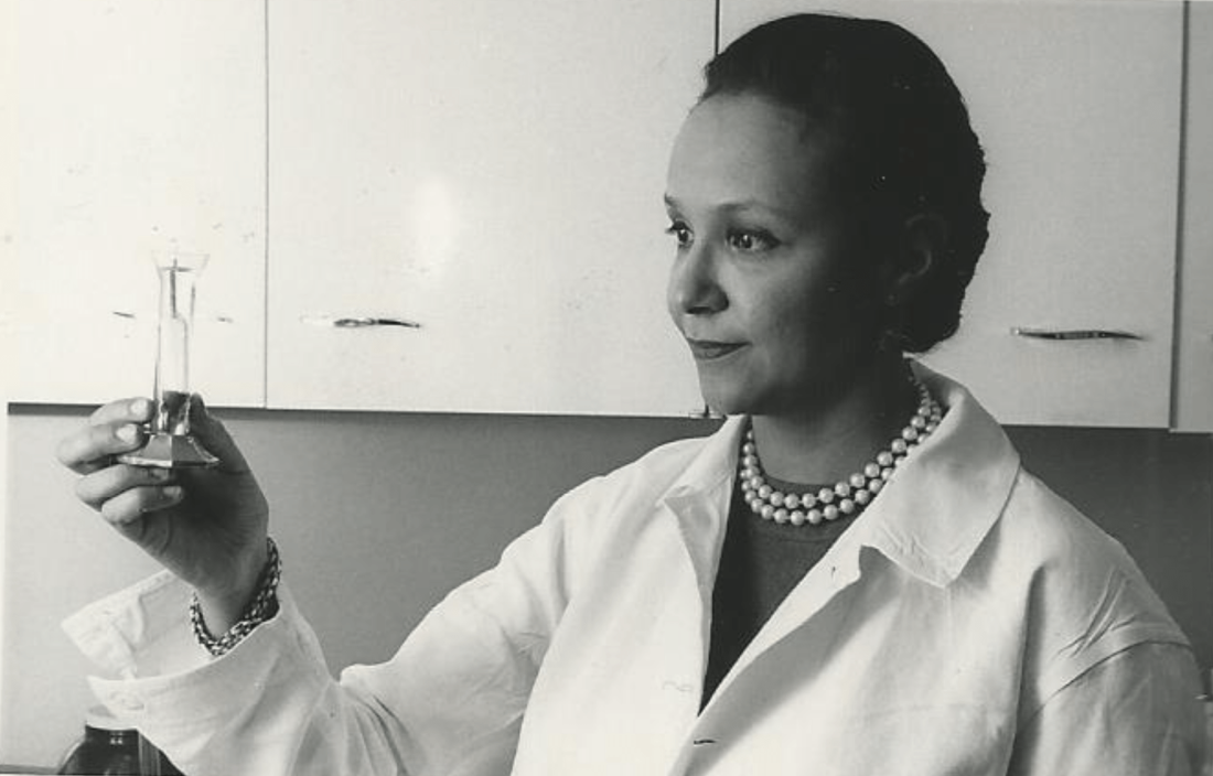
After her father’s death, Dr. Janet C. Wright became director of the hospital’s cancer foundation. From the 1950s to the 1970s, she “worked out ways to use pieces of a patient’s own tumor, removed by surgery and grown in a nutrient culture medium in the laboratory, as a ‘guinea pig for testing drugs,’ ” according to the 1991 book “Black Scientists.” Previously, researchers had focused on mice as test subjects.
This approach also allowed Dr. Wright to determine if specific drugs such as methotrexate, a folic acid antagonist, would help specific patients. “She was looking for predictive activity for chemotherapeutic efficacy in vitro at a time when no one had good predictive tests,” wrote James F. Holland, MD, the late Mount Sinai School of Medicine oncologist, who was quoted in Dr. Wright’s 2013 Oncology obituary.
“Her strict attention to detail and concern for her patients helped determine effective dosing levels and establish treatment guidelines,” the Oncology obituary reported. “She treated patients that other physicians had given up on, and she was among the first small cadre of researchers to carefully test the effects of drugs against cancer in a clinical trial setting.”
Dr. Wright also focused on developing ways to administer chemotherapy, such using a catheter to reach difficult-to-access organs like the spleen without surgery, according to “Black Scientists.”
Along with her work, Dr. Wright’s appearance set her apart. According to “Black Genius,” a newspaper columnist dubbed her one of the 10 most beautiful Back woman in America, and Ebony Magazine in 1966 honored her as one of the best-dressed women in America. It featured a photograph of her in a stunning ivory and yellow brocade gown, noting that she was “in private life Mrs. David J. Jones.” (She’d married the Harvard University Law School graduate in 1946.)
Dr. Wright had a sense of modesty despite her accomplishments, according to her daughter Alison Jones. She even downplayed her own mental powers in a newspaper interview. “I know I’m a member of two minority groups,” she told The New York Post in 1967, “but I don’t think of myself that way. Sure, a woman has to try twice as hard. But – racial prejudice? I’ve met very little of it. It could be I met it – and wasn’t intelligent enough to recognize it.”
Sharp-eyed readers might have glimpsed her modesty nearly 2 decades later. In a 1984 article for the Journal of the National Medical Association, a society of African American physicians, she wrote about the past, present, and future of chemotherapy without noting her own prominent role in its development.
‘Global medical pioneer’ cofounds ASCO – and more
In the 1960s, Dr. Wright joined the influential President’s Commission on Heart Disease, Cancer, and Stroke and was named associate dean at New York Medical College, her alma mater, a first for a black woman at a prominent U.S. medical school. Even more importantly, Dr. Wright was the sole woman among seven physicians who founded the American Society of Clinical Oncology in Chicago in 1964. She served as ASCO’s first Secretary-Treasurer and was honored as its longest surviving founder when she passed away 9 years ago.
“Jane Wright had the vision to see that oncology was an important separate discipline within medicine with far-reaching implications for research and discovery,” Georgetown University Medical Center, Washington, oncologist Sandra M. Swain, MD, a former president of the ASCO and author of the 2013 Oncology obituary of Dr. Wright, said in an interview. “It is truly remarkable that, as a woman and an African American woman, she had a seat at the very small table for the formation of such an important group.”
As her friend and fellow oncologist Edith Mitchell, MD, said in a eulogy, “Dr. Wright led delegations of oncologists to China and the Soviet Union, and countries in Africa and Eastern Europe. She led medical teams providing medical and cancer care and education to other nurses and physicians in Ghana in 1957 and Kenya in 1961. From 1973 to 1984, she served as vice-president of the African Research and Medical foundation.”
Dr. Wright also raised two daughters. A 1968 Ebony article devoted to her career and family declared that neither of her teenagers was interested in medical careers. Their perspectives shifted, however – as had Dr. Wright’s. An undergraduate at Smith College, Dr. Wright majored in art, swam on the varsity team, and had a special affinity for German language studies before she switched to premed.
Like their mother, Dr. Wright’s daughters also changed paths, and they ultimately became the fourth generation of their family to enter the medical field. Dr. Alison Jones, the psychologist, currently works in a prison, while Jane Jones, MD, became a clinical psychiatrist. She’s now retired and lives in Guttenberg, N.J.
Both fondly remember their mother as a supportive force who insisted on excellence. “There couldn’t be any excuses for you not getting where you wanted to go,” Dr. Jane Jones recalled in an interview.
Nevertheless, Dr. Wright was still keenly aware of society’s limits. “She told me I had to be a doctor or lawyer,” Dr. Alison Jones said, “because that’s how you need to survive when you’re Black in America.”
Dr. Wright passed away in 2013 at age 93. “Dr. Jane C. Wright truly has made contributions that have changed the practice of medicine,” noted her friend Dr. Mitchell, an oncologist and a retired brigadier general with the U.S. Air Force who now teaches at Thomas Jefferson University, Philadelphia. “A true pioneer. A concerned mentor. A renowned researcher. A global teacher. A global medical pioneer. A talented researcher, beloved sister, wife, and mother, and a beautiful, kind, and loving human being.”
When Jane Cooke Wright, MD, entered the medical profession in 1945, the notion that toxic drugs could target tumors struck many physicians and patients as outlandish. How could one poison be weaponized against another poison – a cancerous tumor – without creating more havoc? Let alone a combination of two or more chemicals?
Dr. Wright’s story would be extraordinary enough if she’d looked like most of her colleagues, but this surgeon and researcher stood apart. An African American woman at a time when medicine and science – like politics and law – were almost entirely the domain of White men, Dr. Wright had determination in her blood. Her father, once honored by a crowd of dignitaries that included a First Lady, persevered despite his horrific encounters with racism. She shared her father’s commitment to progress and added her own personal twists. She balanced elegance and beauty with scientific savvy, fierce ambition, and a refusal to be defined by anything other than her accomplishments.
“She didn’t focus on race, not at all,” her daughter Alison Jones, PhD, a psychologist in East Lansing, Mich., said in an interview. “Wherever she was, she wanted to be the best, not the best Black person. It was not about how she performed in a category, and she would get upset if someone said she was good as a Black physician.”
On the road to being the best, Dr. Jones said, her mother set a goal of curing cancer. National Cancer Research Month is a fitting opportunity to look back on a scientist dedicated to bringing humanity closer to that elusive achievement.
Medical legacy blazed in toil and trauma
A strong case could be made that Dr. Jane C. Wright and her father Louis Tompkins Wright, MD, are the most accomplished father-and-daughter team in all of medicine.
The elder Dr. Wright, son of a formerly enslaved man turned physician and a stepson of the first African American to graduate from Yale University, New Haven, Conn., himself graduated from Harvard Medical School in 1915. He earned a Purple Heart while serving in World War I, then went on to become the first Black surgeon to join the staff at Harlem Hospital.
Dr. Wright, who had witnessed mob violence and the aftermath of a lynching as a young man, became a supporter of the Harlem Renaissance and a prominent advocate for civil rights and integration. He served as chairman of the National Association for the Advancement of Colored People and was only the second Black member of the American College of Surgeons.
According to the 2009 book “Black Genius: Inspirational Portraits of African American Leaders,” he successfully treated the rare but devastating venereal disease lymphogranuloma venereum with a new antibiotic developed by his former colleague Yellapragada SubbaRow, MD. Dr. Wright even tried the drug himself, “as a lot of doctors in the olden days did,” according to another of his daughters, the late Barbara Wright Pierce, MD, who was quoted in “Black Genius.” She, too, was a physician.
In 1948, Dr. Jane C. Wright joined her father at Harlem Hospital’s Cancer Research Foundation. There the duo explored the cancer-fighting possibilities of a nitrogen mustard–like chemical agent that had been known since World War I to kill white blood cells. Ironically, Dr. Louis Wright himself suffered lifelong health problems because of an attack from the poisonous gas phosgene during his wartime service.
“Remissions were observed in patients with sarcoma, Hodgkin disease, and chronic myelogenous leukemia, mycosis fungoides, and lymphoma,” reported a 2013 obituary in the journal Oncology of the younger Dr. Wright. “They also performed early research into the clinical efficacy and toxicity of folic acid antagonists, documenting responses in 93 patients with various forms of incurable blood cancers and solid tumors.”
This research appears in a study that was authored by three Dr. Wrights – Dr. Louis T. Wright and his daughters Jane and Barbara.
“The elder Dr. Wright died in 1952, just months after 1,000 people – including Eleanor Roosevelt – honored him at a dinner to dedicate a Harlem Hospital library named after him. He was 61.
Scientific savvy mixed with modesty and elegance
After her father’s death, Dr. Janet C. Wright became director of the hospital’s cancer foundation. From the 1950s to the 1970s, she “worked out ways to use pieces of a patient’s own tumor, removed by surgery and grown in a nutrient culture medium in the laboratory, as a ‘guinea pig for testing drugs,’ ” according to the 1991 book “Black Scientists.” Previously, researchers had focused on mice as test subjects.
This approach also allowed Dr. Wright to determine if specific drugs such as methotrexate, a folic acid antagonist, would help specific patients. “She was looking for predictive activity for chemotherapeutic efficacy in vitro at a time when no one had good predictive tests,” wrote James F. Holland, MD, the late Mount Sinai School of Medicine oncologist, who was quoted in Dr. Wright’s 2013 Oncology obituary.
“Her strict attention to detail and concern for her patients helped determine effective dosing levels and establish treatment guidelines,” the Oncology obituary reported. “She treated patients that other physicians had given up on, and she was among the first small cadre of researchers to carefully test the effects of drugs against cancer in a clinical trial setting.”
Dr. Wright also focused on developing ways to administer chemotherapy, such using a catheter to reach difficult-to-access organs like the spleen without surgery, according to “Black Scientists.”
Along with her work, Dr. Wright’s appearance set her apart. According to “Black Genius,” a newspaper columnist dubbed her one of the 10 most beautiful Back woman in America, and Ebony Magazine in 1966 honored her as one of the best-dressed women in America. It featured a photograph of her in a stunning ivory and yellow brocade gown, noting that she was “in private life Mrs. David J. Jones.” (She’d married the Harvard University Law School graduate in 1946.)
Dr. Wright had a sense of modesty despite her accomplishments, according to her daughter Alison Jones. She even downplayed her own mental powers in a newspaper interview. “I know I’m a member of two minority groups,” she told The New York Post in 1967, “but I don’t think of myself that way. Sure, a woman has to try twice as hard. But – racial prejudice? I’ve met very little of it. It could be I met it – and wasn’t intelligent enough to recognize it.”
Sharp-eyed readers might have glimpsed her modesty nearly 2 decades later. In a 1984 article for the Journal of the National Medical Association, a society of African American physicians, she wrote about the past, present, and future of chemotherapy without noting her own prominent role in its development.
‘Global medical pioneer’ cofounds ASCO – and more
In the 1960s, Dr. Wright joined the influential President’s Commission on Heart Disease, Cancer, and Stroke and was named associate dean at New York Medical College, her alma mater, a first for a black woman at a prominent U.S. medical school. Even more importantly, Dr. Wright was the sole woman among seven physicians who founded the American Society of Clinical Oncology in Chicago in 1964. She served as ASCO’s first Secretary-Treasurer and was honored as its longest surviving founder when she passed away 9 years ago.
“Jane Wright had the vision to see that oncology was an important separate discipline within medicine with far-reaching implications for research and discovery,” Georgetown University Medical Center, Washington, oncologist Sandra M. Swain, MD, a former president of the ASCO and author of the 2013 Oncology obituary of Dr. Wright, said in an interview. “It is truly remarkable that, as a woman and an African American woman, she had a seat at the very small table for the formation of such an important group.”
As her friend and fellow oncologist Edith Mitchell, MD, said in a eulogy, “Dr. Wright led delegations of oncologists to China and the Soviet Union, and countries in Africa and Eastern Europe. She led medical teams providing medical and cancer care and education to other nurses and physicians in Ghana in 1957 and Kenya in 1961. From 1973 to 1984, she served as vice-president of the African Research and Medical foundation.”
Dr. Wright also raised two daughters. A 1968 Ebony article devoted to her career and family declared that neither of her teenagers was interested in medical careers. Their perspectives shifted, however – as had Dr. Wright’s. An undergraduate at Smith College, Dr. Wright majored in art, swam on the varsity team, and had a special affinity for German language studies before she switched to premed.
Like their mother, Dr. Wright’s daughters also changed paths, and they ultimately became the fourth generation of their family to enter the medical field. Dr. Alison Jones, the psychologist, currently works in a prison, while Jane Jones, MD, became a clinical psychiatrist. She’s now retired and lives in Guttenberg, N.J.
Both fondly remember their mother as a supportive force who insisted on excellence. “There couldn’t be any excuses for you not getting where you wanted to go,” Dr. Jane Jones recalled in an interview.
Nevertheless, Dr. Wright was still keenly aware of society’s limits. “She told me I had to be a doctor or lawyer,” Dr. Alison Jones said, “because that’s how you need to survive when you’re Black in America.”
Dr. Wright passed away in 2013 at age 93. “Dr. Jane C. Wright truly has made contributions that have changed the practice of medicine,” noted her friend Dr. Mitchell, an oncologist and a retired brigadier general with the U.S. Air Force who now teaches at Thomas Jefferson University, Philadelphia. “A true pioneer. A concerned mentor. A renowned researcher. A global teacher. A global medical pioneer. A talented researcher, beloved sister, wife, and mother, and a beautiful, kind, and loving human being.”
Ex–hospital porter a neglected giant of cancer research
We have a half-forgotten Indian immigrant to thank – a hospital night porter turned biochemist –for revolutionizing treatment of leukemia, the once deadly childhood scourge that is still the most common pediatric cancer.
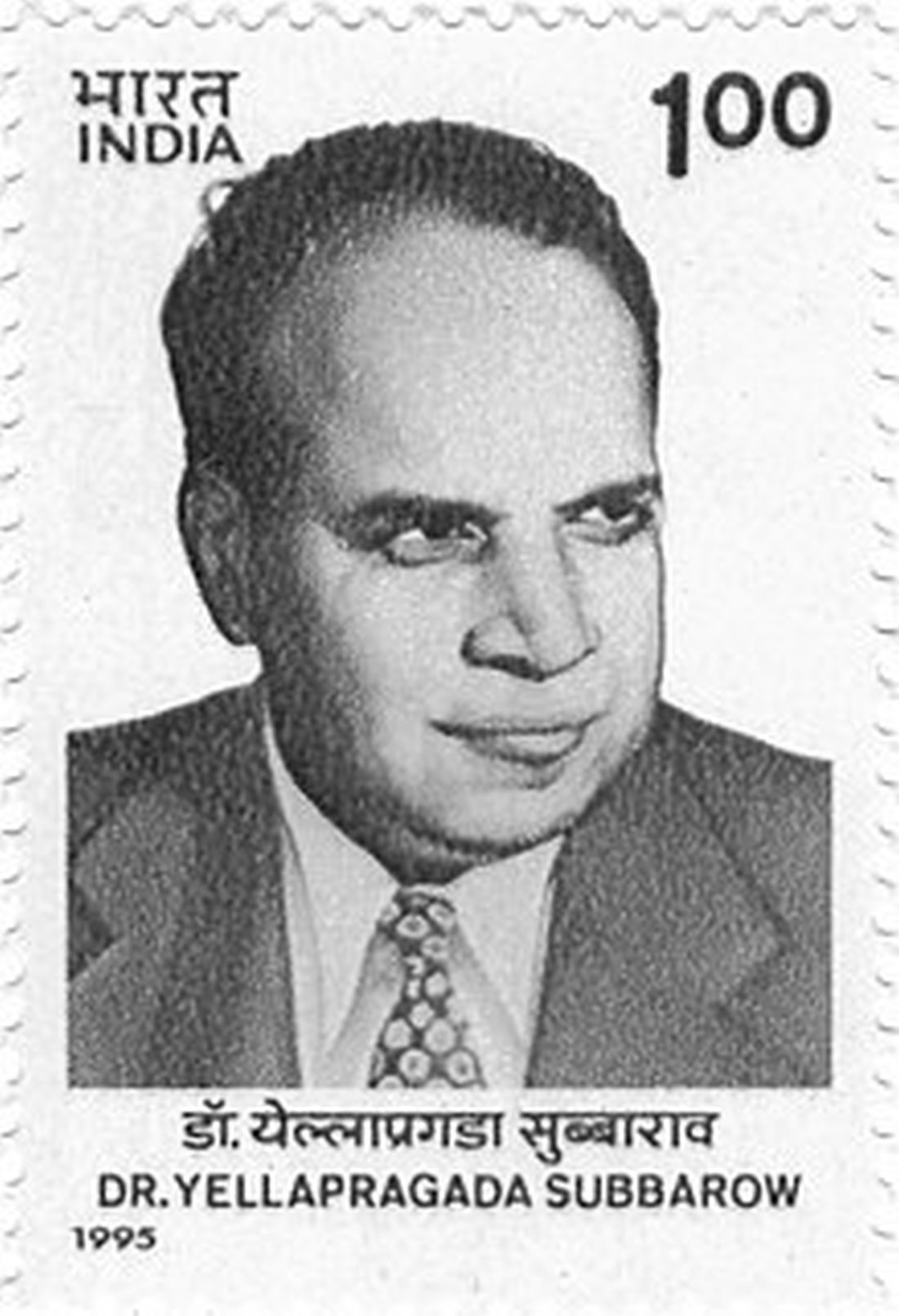
Dr. Yellapragada SubbaRow has been called the “father of chemotherapy” for developing methotrexate, a powerful, inexpensive therapy for leukemia and other diseases, and he is celebrated for additional scientific achievements. Yet Dr. SubbaRow’s life was marked more by struggle than glory.
Born poor in southeastern India, he nearly succumbed to a tropical disease that killed two older brothers, and he didn’t focus on schoolwork until his father died. Later, prejudice dogged his years as an immigrant to the United States, and a blood clot took his life at the age of 53.
Scientifically, however, Dr. SubbaRow (pronounced sue-buh-rao) triumphed, despite mammoth challenges and a lack of recognition that persists to this day. National Cancer Research Month is a fitting time to look back on his extraordinary life and work and pay tribute to his accomplishments.
‘Yella,’ folic acid, and a paradigm shift
No one appreciates Dr. SubbaRow more than a cadre of Indian-born physicians who have kept his legacy alive in journal articles, presentations, and a Pulitzer Prize-winning book. Among them is author and oncologist Siddhartha Mukherjee, MD, who chronicled Dr. SubbaRow’s achievements in his New York Times No. 1 bestseller, “The Emperor of All Maladies: A Biography of Cancer.”
As Dr. Mukherjee wrote, Dr. SubbaRow was a “pioneer in many ways, a physician turned cellular physiologist, a chemist who had accidentally wandered into biology.” (Per Indian tradition, SubbaRow is the doctor’s first name, and Yellapragada is his surname, but medical literature uses SubbaRow as his cognomen, with some variations in spelling. Dr. Mukherjee wrote that his friends called him “Yella.”)
Dr. SubbaRow came to the United States in 1923, after enduring a difficult childhood and young adulthood. He’d survived bouts of religious fervor, childhood rebellion (including a bid to run away from home and become a banana trader), and a failed arranged marriage. His wife bore him a child who died in infancy. He left it all behind.
In Boston, medical officials rejected his degree. Broke, he worked for a time as a night porter at Brigham and Women’s Hospital in Boston, changing sheets and cleaning urinals. To a poor but proud high-caste Indian Brahmin, the culture shock of carrying out these tasks must have been especially jarring.
Dr. SubbaRow went on to earn a diploma from Harvard Medical School, also in Boston, and became a junior faculty member. As a foreigner, Dr. Mukherjee wrote, Dr. SubbaRow was a “reclusive, nocturnal, heavily accented vegetarian,” so different from his colleagues that advancement seemed impossible. Despite his pioneering biochemistry work, Harvard later declined to offer Dr. SubbaRow a tenured faculty position.
By the early 1940s, he took a job at an upstate New York pharmaceutical company called Lederle Labs (later purchased by Pfizer). At Lederle, Dr. SubbaRow strove to synthesize the vitamin known as folic acid. He ended up creating a kind of antivitamin, a lookalike that acted like folic acid but only succeeded in gumming up the works in receptors. But what good would it do to stop the body from absorbing folic acid? Plenty, it turned out.
Discoveries pile up, but credit and fame prove elusive
Dr. SubbaRow was no stranger to producing landmark biological work. He’d previously codiscovered phosphocreatine and ATP, which are crucial to muscular contractions. However, “in 1935, he had to disown the extent of his role in the discovery of the color test related to phosphorus, instead giving the credit to his co-author, who was being considered for promotion to a full professorship at Harvard,” wrote author Gerald Posner in his 2020 book, “Pharma: Greed, Lies and the Poisoning of America.”
Houston-area oncologist Kirtan Nautiyal, MD, who paid tribute to Dr. SubbaRow in a 2018 article, contended that “with his Indian instinct for self-effacement, he had irreparably sabotaged his own career.”
Dr. SubbaRow and his team also developed “the first effective treatment of filariasis, which causes elephantiasis of the lower limbs and genitals in millions of people, mainly in tropical countries,” Dr. Nautiyal wrote. “Later in the decade, his antibiotic program generated polymyxin, the first effective treatment against the class of bacteria called Gram negatives, and aureomycin, the first “broad-spectrum’ antibiotic.” (Aureomycin is also the first tetracycline antibiotic.)
Dr. SubbaRow’s discovery of a folic acid antagonist would again go largely unheralded. But first came the realization that folic acid made childhood leukemia worse, not better, and the prospect that this process could potentially be reversed.
Rise of methotrexate and fall of leukemia
In Boston, Sidney Farber, MD, a Boston pathologist, was desperate to help Robert Sandler, a 2-year-old leukemia patient. Dr. Farber contacted his ex-colleague Dr. SubbaRow to request a supply of aminopterin, an early version of methotrexate that Dr. SubbaRow and his team had developed. Dr. Farber injected Robert with the substance and within 3 days, the toddler’s white blood count started falling – fast. He stopped bleeding, resumed eating, and once again seemed almost identical to his twin brother, as Dr. Mukherjee wrote in his book.
Leukemia had never gone into remission before. Unfortunately, the treatment only worked temporarily. Robert, like other children treated with the drug, relapsed and died within months. But Dr. Farber “saw a door open” – a chemical, a kind of chemotherapy, that could turn back cancer. In the case of folic acid antagonists, they do so by stopping cancer cells from replicating.
Methotrexate, a related agent synthesized by Dr. SubbaRow, would become a mainstay of leukemia treatment and begin to produce long-term remission from acute lymphoblastic leukemia in 1970, when combination chemotherapy was developed.
Other cancers fell to methotrexate treatment. “Previous assumptions that cancer was nearly always fatal were revised, and the field of medical oncology (treatment of cancer with chemotherapy), which had not previously existed, was formally established in 1971,” according to the National Cancer Institute’s history of methotrexate. This account does not mention Dr. SubbaRow.
Death takes the doctor, but his legacy remains
In biographies, as well as his own words, Dr. SubbaRow comes across as a prickly, hard-driving workaholic who had little interest in intimate human connections. “It is not good to ask in every letter when I will be back,” he wrote to his wife back in India, before cutting off ties completely in the early 1930s. “I will come as early as possible. ... I do not want to write anything more.”
It seems, as his biographer S.P.K. Gupta noted, that “he was quite determined that the time allotted to him on Earth should be completely devoted to finding cures for ailments that plagued mankind.”
Still, Dr. SubbaRow’s research team was devoted to him, and he had plenty of reasons to be bitter, such as the prejudice and isolation he encountered in the United States and earlier, in British-run India. According to Mr. Posner’s book, even as a young medical student, Dr. SubbaRow heeded the call of Indian independence activist Mohandas Gandhi. He “refused the British surgical gown given him at school and instead donned a traditional and simple cotton Khadi. That act of defiance cost SubbaRow the college degree that was necessary for him to get into the State Medical College.”
During the last year of his life, Dr. SubbaRow faced yet another humiliation: In his landmark 1948 study about aminopterin as a treatment for leukemia, his colleague Dr. Farber failed to credit him, an “astonishing omission” as Yaddanapudi Ravindranath, MBBS, a pediatric hematologist/oncologist at Wayne State University, Detroit, put it. “From everything I know, Dr. Farber spent the rest of his career apologizing and trying to make amends for it,” Dr. Ravindranath said in an interview.
A career cut short, and a lasting legacy
In 1948, at the age of 53, Dr. SubbaRow suddenly died. “Many think Dr. SubbaRow would have won [the] Nobel Prize had he lived a few years longer,” said Dr. Ravindranath.
Like Dr. SubbaRow, Dr. Ravindranath was born in Andhra Pradesh state, near the city of Chennai formerly known as Madras. “Being a compatriot, in a way I continue his legacy, and I am obviously proud of him,” said Dr. Ravindranath, who has conducted his own landmark research regarding methotrexate and leukemia.
Nearly 75 years after Dr. SubbaRow’s death, Indian-born physicians like Dr. Ravindranath continue to honor him in print, trying to ensure that he’s not forgotten. Methotrexate remains a crucial treatment for leukemia, along with a long list of other ailments, including psoriasis.
Recognition for “Yella” may have come late and infrequently, but a Lederle Laboratories research library named after him offered Dr. SubbaRow a kind of immortality. A plaque there memorialized him in stone as a scientist, teacher, philosopher, and humanitarian, featuring the quote: “Science simply prolongs life. Religion deepens it.”
By all accounts, Dr. SubbaRow was a man of science and faith who had faith in science.
We have a half-forgotten Indian immigrant to thank – a hospital night porter turned biochemist –for revolutionizing treatment of leukemia, the once deadly childhood scourge that is still the most common pediatric cancer.
Dr. Yellapragada SubbaRow has been called the “father of chemotherapy” for developing methotrexate, a powerful, inexpensive therapy for leukemia and other diseases, and he is celebrated for additional scientific achievements. Yet Dr. SubbaRow’s life was marked more by struggle than glory.
Born poor in southeastern India, he nearly succumbed to a tropical disease that killed two older brothers, and he didn’t focus on schoolwork until his father died. Later, prejudice dogged his years as an immigrant to the United States, and a blood clot took his life at the age of 53.
Scientifically, however, Dr. SubbaRow (pronounced sue-buh-rao) triumphed, despite mammoth challenges and a lack of recognition that persists to this day. National Cancer Research Month is a fitting time to look back on his extraordinary life and work and pay tribute to his accomplishments.
‘Yella,’ folic acid, and a paradigm shift
No one appreciates Dr. SubbaRow more than a cadre of Indian-born physicians who have kept his legacy alive in journal articles, presentations, and a Pulitzer Prize-winning book. Among them is author and oncologist Siddhartha Mukherjee, MD, who chronicled Dr. SubbaRow’s achievements in his New York Times No. 1 bestseller, “The Emperor of All Maladies: A Biography of Cancer.”
As Dr. Mukherjee wrote, Dr. SubbaRow was a “pioneer in many ways, a physician turned cellular physiologist, a chemist who had accidentally wandered into biology.” (Per Indian tradition, SubbaRow is the doctor’s first name, and Yellapragada is his surname, but medical literature uses SubbaRow as his cognomen, with some variations in spelling. Dr. Mukherjee wrote that his friends called him “Yella.”)
Dr. SubbaRow came to the United States in 1923, after enduring a difficult childhood and young adulthood. He’d survived bouts of religious fervor, childhood rebellion (including a bid to run away from home and become a banana trader), and a failed arranged marriage. His wife bore him a child who died in infancy. He left it all behind.
In Boston, medical officials rejected his degree. Broke, he worked for a time as a night porter at Brigham and Women’s Hospital in Boston, changing sheets and cleaning urinals. To a poor but proud high-caste Indian Brahmin, the culture shock of carrying out these tasks must have been especially jarring.
Dr. SubbaRow went on to earn a diploma from Harvard Medical School, also in Boston, and became a junior faculty member. As a foreigner, Dr. Mukherjee wrote, Dr. SubbaRow was a “reclusive, nocturnal, heavily accented vegetarian,” so different from his colleagues that advancement seemed impossible. Despite his pioneering biochemistry work, Harvard later declined to offer Dr. SubbaRow a tenured faculty position.
By the early 1940s, he took a job at an upstate New York pharmaceutical company called Lederle Labs (later purchased by Pfizer). At Lederle, Dr. SubbaRow strove to synthesize the vitamin known as folic acid. He ended up creating a kind of antivitamin, a lookalike that acted like folic acid but only succeeded in gumming up the works in receptors. But what good would it do to stop the body from absorbing folic acid? Plenty, it turned out.
Discoveries pile up, but credit and fame prove elusive
Dr. SubbaRow was no stranger to producing landmark biological work. He’d previously codiscovered phosphocreatine and ATP, which are crucial to muscular contractions. However, “in 1935, he had to disown the extent of his role in the discovery of the color test related to phosphorus, instead giving the credit to his co-author, who was being considered for promotion to a full professorship at Harvard,” wrote author Gerald Posner in his 2020 book, “Pharma: Greed, Lies and the Poisoning of America.”
Houston-area oncologist Kirtan Nautiyal, MD, who paid tribute to Dr. SubbaRow in a 2018 article, contended that “with his Indian instinct for self-effacement, he had irreparably sabotaged his own career.”
Dr. SubbaRow and his team also developed “the first effective treatment of filariasis, which causes elephantiasis of the lower limbs and genitals in millions of people, mainly in tropical countries,” Dr. Nautiyal wrote. “Later in the decade, his antibiotic program generated polymyxin, the first effective treatment against the class of bacteria called Gram negatives, and aureomycin, the first “broad-spectrum’ antibiotic.” (Aureomycin is also the first tetracycline antibiotic.)
Dr. SubbaRow’s discovery of a folic acid antagonist would again go largely unheralded. But first came the realization that folic acid made childhood leukemia worse, not better, and the prospect that this process could potentially be reversed.
Rise of methotrexate and fall of leukemia
In Boston, Sidney Farber, MD, a Boston pathologist, was desperate to help Robert Sandler, a 2-year-old leukemia patient. Dr. Farber contacted his ex-colleague Dr. SubbaRow to request a supply of aminopterin, an early version of methotrexate that Dr. SubbaRow and his team had developed. Dr. Farber injected Robert with the substance and within 3 days, the toddler’s white blood count started falling – fast. He stopped bleeding, resumed eating, and once again seemed almost identical to his twin brother, as Dr. Mukherjee wrote in his book.
Leukemia had never gone into remission before. Unfortunately, the treatment only worked temporarily. Robert, like other children treated with the drug, relapsed and died within months. But Dr. Farber “saw a door open” – a chemical, a kind of chemotherapy, that could turn back cancer. In the case of folic acid antagonists, they do so by stopping cancer cells from replicating.
Methotrexate, a related agent synthesized by Dr. SubbaRow, would become a mainstay of leukemia treatment and begin to produce long-term remission from acute lymphoblastic leukemia in 1970, when combination chemotherapy was developed.
Other cancers fell to methotrexate treatment. “Previous assumptions that cancer was nearly always fatal were revised, and the field of medical oncology (treatment of cancer with chemotherapy), which had not previously existed, was formally established in 1971,” according to the National Cancer Institute’s history of methotrexate. This account does not mention Dr. SubbaRow.
Death takes the doctor, but his legacy remains
In biographies, as well as his own words, Dr. SubbaRow comes across as a prickly, hard-driving workaholic who had little interest in intimate human connections. “It is not good to ask in every letter when I will be back,” he wrote to his wife back in India, before cutting off ties completely in the early 1930s. “I will come as early as possible. ... I do not want to write anything more.”
It seems, as his biographer S.P.K. Gupta noted, that “he was quite determined that the time allotted to him on Earth should be completely devoted to finding cures for ailments that plagued mankind.”
Still, Dr. SubbaRow’s research team was devoted to him, and he had plenty of reasons to be bitter, such as the prejudice and isolation he encountered in the United States and earlier, in British-run India. According to Mr. Posner’s book, even as a young medical student, Dr. SubbaRow heeded the call of Indian independence activist Mohandas Gandhi. He “refused the British surgical gown given him at school and instead donned a traditional and simple cotton Khadi. That act of defiance cost SubbaRow the college degree that was necessary for him to get into the State Medical College.”
During the last year of his life, Dr. SubbaRow faced yet another humiliation: In his landmark 1948 study about aminopterin as a treatment for leukemia, his colleague Dr. Farber failed to credit him, an “astonishing omission” as Yaddanapudi Ravindranath, MBBS, a pediatric hematologist/oncologist at Wayne State University, Detroit, put it. “From everything I know, Dr. Farber spent the rest of his career apologizing and trying to make amends for it,” Dr. Ravindranath said in an interview.
A career cut short, and a lasting legacy
In 1948, at the age of 53, Dr. SubbaRow suddenly died. “Many think Dr. SubbaRow would have won [the] Nobel Prize had he lived a few years longer,” said Dr. Ravindranath.
Like Dr. SubbaRow, Dr. Ravindranath was born in Andhra Pradesh state, near the city of Chennai formerly known as Madras. “Being a compatriot, in a way I continue his legacy, and I am obviously proud of him,” said Dr. Ravindranath, who has conducted his own landmark research regarding methotrexate and leukemia.
Nearly 75 years after Dr. SubbaRow’s death, Indian-born physicians like Dr. Ravindranath continue to honor him in print, trying to ensure that he’s not forgotten. Methotrexate remains a crucial treatment for leukemia, along with a long list of other ailments, including psoriasis.
Recognition for “Yella” may have come late and infrequently, but a Lederle Laboratories research library named after him offered Dr. SubbaRow a kind of immortality. A plaque there memorialized him in stone as a scientist, teacher, philosopher, and humanitarian, featuring the quote: “Science simply prolongs life. Religion deepens it.”
By all accounts, Dr. SubbaRow was a man of science and faith who had faith in science.
‘Agony of choice’ for clinicians treating leukemia
“Targeted therapies have outnumbered chemoimmunotherapy-based treatment approaches, demonstrating superior efficacy and tolerability profiles across nearly all CLL patient subgroups in the frontline and relapsed disease treatment setting,” author Jan-Paul Bohn, MD, PhD, of the department of internal medicine V, hematology and oncology, at Medical University of Innsbruck (Austria), reported in the review published in Memo, the Magazine of European Medical Oncology.
The options leave clinicians “spoilt for choice when selecting optimal therapy,” he said.
The three major drug classes to emerge – inhibitors of Bruton tyrosine kinase (BTK), antiapoptotic protein B-cell lymphoma 2 (BCL2) and phosphoinositide 3’-kinase (PI3K) – all appear similar in efficacy and tolerability.
Particularly in high-risk patients, the drugs have been so effective that the less desirable previous standard of “chemoimmunotherapy has widely faded into the background in the Western hemisphere,” Dr. Bohn wrote.
However, with caveats of the newer drugs including acquired resistances and potential toxicities, challenges have shifted to determining how to best juggle and/or combine the agents.
Frontline therapy
In terms of frontline options for CLL therapy, the BTK inhibitors, along with the BCL2 inhibitor venetoclax have been key in negating the need for chemotherapy, with some of the latest data showing superiority of venetoclax in combination with obinutuzumab (GVe) over chemotherapy even in the higher-risk subset of patients with mutated IGHV status and without TP53 disruption.
Hence, “chemoimmunotherapy may now even be questioned in the remaining subset of CLL patients with mutated IGHV status and without TP53 disruption,” Dr. Bohn reported.
That being said, the criteria for treatment choices in the frontline setting among the newer drug classes can often come down to the key issues of patients’ comorbidities and treatment preferences.
For example, in terms of patients who have higher risk because of tumor lysis syndrome (TLS), or issues including declining renal function, continuous BTK inhibitor treatment may be the preferred choice over the combination of venetoclax plus obinutuzumab (GVe), Dr. Bohn noted.
Conversely, for patients with cardiac comorbidities or a higher risk of bleeding, the GVe combination may be preferred over ibrutinib, with recent findings showing ibrutinib to be associated with as much as an 18-times higher risk of sudden unexplained death or cardiac death in young and fit patients who had preexisting arterial hypertension and/or a history of cardiac disorders requiring therapy.
For those with cardiac comorbidities, the more selective second-generation BTK inhibitor acalabrutinib is a potentially favorable alternative, as the drug is “at least similarly effective and more favorable in terms of tolerability, compared with ibrutinib, particularly as far as cardiac and bleeding side effects are considered,” Dr. Bohn said.
And in higher-risk cases involving TP53 dysfunction, a BTK inhibitor may be superior to GVe for frontline treatment, Dr. Bohn noted, with data showing progression-free survival in patients with and without deletion 17p to be significantly reduced with GVe versus the BTK inhibitor ibrutinib.
Relapsed and refractory disease
With similarly high efficacy observed with the new drug classes among relapsed and/or refractory patients, chemoimmunotherapy has likewise “become obsolete in nearly all patients naive to novel agents at relapse who typically present with genetically high-risk disease,” Dr. Bohn noted.
He wrote that most of the recommendations for frontline therapy hold true in the relapsed and refractory patients, with comorbidities and personal preferences again key drivers of treatment choices.
While data is currently limited regarding benefits of venetoclax-based regimens over BTK inhibitors in relapsed/refractory patients, there is “growing evidence suggesting similar clinical outcomes achievable with these agents in either order,” Dr. Bohn wrote.
Further recommendations regarding relapsed or refractory patients include:
- Among patients who do experience disease progression while on continuous treatment with BTK inhibitors, venetoclax-based regimes seem most effective. However, with relapse after venetoclax-based regimes, some growing evidence supports retreatment with the drug “depending on depth and duration of response achieved after first venetoclax exposure,” Dr. Bohn noted.
- For patients with deletion 17p, venetoclax shows promising efficacy during relapse when given as monotherapy until disease progression or occurrence of unacceptable toxicity.
- And for patients with TP53 abnormalities, the considerations are the same as for frontline therapy, with venetoclax showing promising efficacy when given in monotherapy until disease progression or occurrence of unacceptable toxicity.
Of note, PI3K inhibitors are generally not used in CLL patients naive to BTK and BCL2 inhibitors because of the higher risk of immune-mediated toxicities and infectious complications associated with the currently approved PI3K inhibitors idelalisib and duvelisib, he reported.
Nevertheless, “PI3K inhibitors remain a valuable therapeutic addition in patients refractory or intolerant to BTK inhibitors and venetoclax-based regimens,” Dr. Bohn said.
Newer agents, fixed duration
Commenting on the review, hematologist Seema A. Bhat, MD, an assistant professor with the Ohio State University Comprehensive Cancer Center, Columbus, said that the advances with targeted therapies in CLL are paying off with improved survival.
“With these recent advances in the treatment of CLL, especially the availability of targeted therapies, there has been an improvement in survival of patients with CLL, as the CLL-related death rate steadily reduced by approximately 3% per year between 2006 and 2015,” she said in an interview.
She added that even-newer agents in development, including the reversibly binding BTK inhibitor–like pirtobrutinib and nemtabrutinib, when approved, will further add to the treatment choices for patients.
Meanwhile, a key area of focus is the combination of BTK inhibitors and BCL2 inhibitors, specifically for a fixed duration of time to obtain a deeper response and hence possibility a time-limited therapy, she noted. “We are also excited about the possibility of having more fixed-duration treatments available for our patients, which will make their treatment journey less troublesome, both physically as well as financially.”
Dr. Bohn reported receiving personal fees from AbbVie, AstraZeneca and Janssen for advisory board participation. Dr. Bhat has served on advisory board for AstraZeneca and received honorarium from them.
FROM MEMO – MAGAZINE OF EUROPEAN MEDICAL ONCOLOGY
Zanubrutinib shows worth against standard CLL drugs
MANCHESTER, ENGLAND – A new treatment option may soon be available for patients with chronic lymphocytic leukemia (CLL)/small lymphocytic lymphoma (SLL).
Zanubrutinib (Brukinsa), an irreversible, next-generation Bruton tyrosine kinase (BTK) inhibitor, is designed to minimize the off-target cardiovascular toxicities, such as atrial fibrillation and hypertension, seen with the first-generation ibrutinib (Imbruvica).
Zanubrutinib is already approved for use in mantle cell and marginal zone lymphomas and Waldenström’s macroglobulinemia.
Now it has also shown efficacy in CLL.
However, experts question whether the drug will find its place in an increasingly crowded space for the management of CLL.
Data from two phase 3 trials
The new data from two phase 3 clinical trials were presented recently at the British Society for Haematology 62nd annual scientific meeting, held in Manchester, England.
The ALPINE trial compared zanubrutinib with ibrutinib in 415 patients with CLL/SLL and showed that the novel drug was associated with a significant improvement in overall response rate, at 78% versus 63%.
This first interim analysis also showed that there was an increase in progression-free survival (PFS) with zanubrutinib, and crucially, it was associated with a lower atrial fibrillation/flutter rate than ibrutinib.
“These data support that more selective BTK inhibition, with more complete and sustained BTK occupancy, results in improved efficacy and safety outcomes,” said lead author Peter Hillmen, MBChB, FRCP, PhD, St. James’s University Hospital, Leeds, England.
The SEQUOIA study looked at zanubrutinib versus bendamustine plus rituximab in patients with untreated CLL/SLL with a 17p deletion and showed that PFS was improved with zanubrutinib by 58%.
Zanubrutinib was also associated with improved overall response rates and was well tolerated.
The results therefore “support the potential utility of zanubrutinib in the frontline management of patients with previously untreated CLL/SLL,” said lead author Talha Munir, MBBS, also of St. James’s University Hospital.
Improvement over ibrutinib
Ibrutinib, the first BTK inhibitor, “truly revolutionized the way we treat CLL,” commented Renata Walewska, MRCP, PhD, consultant hematologist at the Royal Bournemouth (England) Hospital and chair of the UKCLL Forum.
“But it has got quite a lot of, especially cardiac, problems, with atrial fibrillation and hypertension,” she said in an interview. The problem is that it acts not only as an inhibitor of Bruton kinase, but also affects other kinases, she explained.
Zanubrutinib is “much cleaner,” continued Dr. Walewska, who was lead author of the recently published British Society of Haematology guideline for the treatment of CLL.
However, the drug “is not that groundbreaking,” she commented, as acalabrutinib (Calquence), another next-generation BTK inhibitor, is already available for use in the clinic.
“We’re really lucky in CLL,” Dr. Walewska said, “we’ve got so many new drugs available, and it’s getting quite crowded. Trying to find a place for zanubrutinib is going be tricky.”
Lee Greenberger, PhD, chief scientific officer at the Leukemia & Lymphoma Society, commented that he “gives a lot of credit” to BeiGene, the company behind zanubrutinib, for “taking on these big studies.”
He said that, with the improvements in PFS and reduced atrial fibrillation with the drug, “there will be many clinicians paying attention to this and zanubrutinib could be preferred over conventional options.”
However, he agreed that it will have to compete with acalabrutinib, adding that, beyond BTK inhibitors, there are “a lot of options” for patients with CLL.
“That makes it very difficult for physicians to figure out what is the best type of therapy” to use in these patients, he added.
Dr. Greenberger told this news organization that further studies will need to demonstrate that zanubrutinib is associated with extended survival, which is “just not possible to show” at the moment with the current follow-up period.
He also noted that, in 10 years, ibrutinib will be off-patent, but zanubrutinib will not, at which point the “substantial” cost of the medication, which is a source of “hardship to patients,” will be increasingly relevant.
Study details
The phase 3 ALPINE study involved 415 adults with CLL/SLL refractory to one or more prior systemic therapies and measurable lymphadenopathy on imaging.
They were randomized 1:1 to zanubrutinib or ibrutinib until disease progression or withdrawal from the study.
Most patients had Binet stage A/B or Ann Arbor stage I/II disease, and 7.3% of patients treated with zanubrutinib and 10.1% of those assigned to ibrutinib had received more than three prior lines of therapy.
Over 60% of patients were aged 65 years or older and around 70% were men, with no significant differences between treatment groups.
Patients were randomized 1:1 to zanubrutinib or ibrutinib until disease progression or study withdrawal.
After a median follow-up of 15 months, the overall response rate was significantly higher with zanubrutinib than ibrutinib, at 78.3% versus 62.5% (P = .0006).
Subgroup analysis confirmed that the effect was seen regardless of age, sex, disease stage, number of prior lines of therapy, mutation status, or bulky disease.
Over a median follow-up of 14 months, the investigator-assessed 12-month PFS was 94.9% for zanubrutinib and 84.0% for ibrutinib (P = .0007). Overall survival at 12 months was 97% versus 92.7%, but the difference was not significant (P = .1081).
Patients treated with zanubrutinib experienced more grade 3 or higher adverse events than those given ibrutinib, at 55.9% versus 51.2%, although they had fewer adverse events leading to treatment discontinuation, at 7.8% versus 13.0%.
More importantly, there were fewer cardiac disorders of any grade with zanubrutinib versus ibrutinib, and any-grade atrial fibrillation was significantly less common, at 2.5% versus 10.1% (P = .0014).
Rates of hypertension and hemorrhage were similar between the two treatments, while rates of neutropenia were higher with zanubrutinib versus ibrutinib, at 28.4% versus 21.7%.
The phase 3 SEQUOIA study looked at an earlier stage of disease and included patients with previously untreated CLL/SLL (without 17p depletion) who were unsuitable for treatment with fludarabine, cyclophosphamide, and rituximab.
This trial involved 479 patients randomized to zanubrutinib or bendamustine (days 1 and 2) plus rituximab for six cycles of 28 days each (B+R).
The median age of patients was 70 years, and approximately 80% were at least 65 years old. Just over 60% were men and most (over 70%) were from Europe.
After a median of 26.2 months, independent review committee–assessed PFS was significantly longer with zanubrutinib versus B+R (hazard ratio, 0.42; P < .0001), with an estimated 24-month PFS of 85.5% versus 69.5%.
These results held whether patients were stratified by age, Binet stage, bulky disease, or 11q deletion status, and for patients with an unmutated, but not mutated, immunoglobulin heavy chain gene.
The overall response rate with zanubrutinib was 94.6% versus 85.3% with B+R, and estimated 24-month overall survival was 94.3% versus 94.6%.
Rates of adverse events of any grade were similar between the two treatment groups, although B+R was associated with a higher (grade ≥ 3) adverse event rate, at 79.7%, versus 52.5% for zanubrutinib, and a higher rate of treatment discontinuation because of adverse events, at 13.7% versus 8.3%.
Interestingly, any-grade hypertension was more common with zanubrutinib versus B+R, at 14.2% versus 10.6%, but much lower rates of neutropenia were more common with zanubrutinib, at 15.8% versus 56.8%.
The studies were sponsored by BeiGene. Dr. Hillmen has reported relationships with Janssen, AbbVie, Pharmacyclics, Roche, Gilead, AstraZeneca, SOBI, and BeiGene. Dr. Munir has reported relationships with AbbVie, AstraZeneca, Roche, Alexion, Janssen, MorphoSys, and SOBI. Other authors have also declared numerous relationships.
A version of this article first appeared on Medscape.com.
When CPI fails, HL patients should get timely allo-HCT
In fact, prior treatment with PD-1–directed therapies nivolumab (Opdivo) and pembrolizumab (Keytruda) appears to improve outcomes in allo-HCT patients, said Miguel-Angel Perales, MD, chief of the adult bone marrow transplant service at Memorial Sloan Kettering Cancer Center in New York.
“The use of allogeneic HCT is decreasing for Hodgkin even though it is a curative option, and we see patients referred after they have had multiple lines of therapy,” Dr. Perales said in an interview. “The lymphoma MDs have a perception that outcomes are poor, and therefore don’t refer.”

To illustrate his point, Dr. Perales shared data from the EBMT database. In 2014, the registry accrued approximately 450 allo-HCT cases; by 2021 this had fallen to fewer than 200 procedures.
Ironically, this declining enthusiasm for transplantation coincides with a steady improvement in transplant outcomes following PD-1 blockade, Dr. Perales noted. For example, an analysis, published in Nature, yielded an 82% overall survival (OS) at 3 years in patients who underwent allo-HCT after CPI treatment (n =209).
“Results of allo-HCT in patients with Hodgkin show a remarkable cure rate,” said Dr. Perales. “Part of that is probably driven by lower relapse due to enhanced graft-versus-lymphoma effect due to long CPI half-life.” (The half-lives of pembrolizumab and nivolumab are 22 and 25 days, respectively.)
At the EBMT meeting, Dr. Perales presented a new retrospective analysis that tested the hypothesis that CPIs might actually improve outcomes for allo-HCT patients. An international team of clinicians from EBMT and the Center for International Blood and Marrow Transplant Research (CIBMTR) compared allo-HCT outcomes with (n = 347) and without (n = 1,382) prior treatment with a checkpoint inhibitor.
They found that prior CPI therapy was, indeed, associated with lower relapse (hazard ratio, 0.53; P = .00023) and longer progression-free survival (PFS) (HR, 0.75; P = .0171).
However, prior PD-1 drugs provided no survival advantage, Dr. Perales said. “The easiest explanation for a study showing a difference in PFS/relapse, not OS, is that we have good treatments that can treat patients who relapse and so their overall survival ends up being the same.”
The researchers also confirmed previous reports that patients who received PD-1 inhibitors prior to transplant had a higher incidence of GVHD. Prevalence of acute grades 2-4 GVHD was significantly higher (P = .027); however, acute grades 3-4 GVHD and chronic GVHD were not significantly different between the two groups.
Dr. Perales speculated that the use of posttransplant cyclophosphamide for GVHD prophylaxis would mitigate the risk of GVHD associated with PD-1 inhibitors, “we have not yet proven that formally ... [we] are still analyzing our data.”
Commenting on the results of the new analysis, Dr. Perales expressed concern that patients are being recruited to early-phase clinical trials after failing on a checkpoint inhibitor, instead of being offered allo-HCT – a potentially curative treatment – because treaters are misinformed about the safety of transplant after these drugs.
The NIH clinical-trials database backs up Dr. Perales’ worries. In the United States, for example, there are currently 19 trials recruiting for relapsed/refractory Hodgkin lymphoma patients prior to transplant. Of these, 15 studies permit enrollment of patients who have failed on CPIs, and 8 are phase 1 or 2 studies.
“The good news is that new drugs, including CPIs, have dramatically changed outcomes in this disease and that fewer patients now need an allo-HCT,” said Dr. Perales. And if a transplant is needed, “it is safe to perform allo-HCT in patients treated with prior CPI.”
However, time is of the essence. “Patients with Hodgkin lymphoma should be referred to allo-HCT if they are not responding or tolerating CPI, rather than go on a series of phase 1 trials,” Dr. Perales said. “Median age is 32, and we should be going for a cure, nothing less.”
Dr. Perales reported receiving honoraria from numerous pharmaceutical companies; serving on the data and safety monitoring boards of Cidara Therapeutics, Medigene, Sellas Life Sciences, and Servier; and serving on the scientific advisory board of NexImmune. He has ownership interests in NexImmune and Omeros, and has received institutional research support for clinical trials from Incyte, Kite/Gilead, Miltenyi Biotec, Nektar Therapeutics, and Novartis.
In fact, prior treatment with PD-1–directed therapies nivolumab (Opdivo) and pembrolizumab (Keytruda) appears to improve outcomes in allo-HCT patients, said Miguel-Angel Perales, MD, chief of the adult bone marrow transplant service at Memorial Sloan Kettering Cancer Center in New York.
“The use of allogeneic HCT is decreasing for Hodgkin even though it is a curative option, and we see patients referred after they have had multiple lines of therapy,” Dr. Perales said in an interview. “The lymphoma MDs have a perception that outcomes are poor, and therefore don’t refer.”
To illustrate his point, Dr. Perales shared data from the EBMT database. In 2014, the registry accrued approximately 450 allo-HCT cases; by 2021 this had fallen to fewer than 200 procedures.
Ironically, this declining enthusiasm for transplantation coincides with a steady improvement in transplant outcomes following PD-1 blockade, Dr. Perales noted. For example, an analysis, published in Nature, yielded an 82% overall survival (OS) at 3 years in patients who underwent allo-HCT after CPI treatment (n =209).
“Results of allo-HCT in patients with Hodgkin show a remarkable cure rate,” said Dr. Perales. “Part of that is probably driven by lower relapse due to enhanced graft-versus-lymphoma effect due to long CPI half-life.” (The half-lives of pembrolizumab and nivolumab are 22 and 25 days, respectively.)
At the EBMT meeting, Dr. Perales presented a new retrospective analysis that tested the hypothesis that CPIs might actually improve outcomes for allo-HCT patients. An international team of clinicians from EBMT and the Center for International Blood and Marrow Transplant Research (CIBMTR) compared allo-HCT outcomes with (n = 347) and without (n = 1,382) prior treatment with a checkpoint inhibitor.
They found that prior CPI therapy was, indeed, associated with lower relapse (hazard ratio, 0.53; P = .00023) and longer progression-free survival (PFS) (HR, 0.75; P = .0171).
However, prior PD-1 drugs provided no survival advantage, Dr. Perales said. “The easiest explanation for a study showing a difference in PFS/relapse, not OS, is that we have good treatments that can treat patients who relapse and so their overall survival ends up being the same.”
The researchers also confirmed previous reports that patients who received PD-1 inhibitors prior to transplant had a higher incidence of GVHD. Prevalence of acute grades 2-4 GVHD was significantly higher (P = .027); however, acute grades 3-4 GVHD and chronic GVHD were not significantly different between the two groups.
Dr. Perales speculated that the use of posttransplant cyclophosphamide for GVHD prophylaxis would mitigate the risk of GVHD associated with PD-1 inhibitors, “we have not yet proven that formally ... [we] are still analyzing our data.”
Commenting on the results of the new analysis, Dr. Perales expressed concern that patients are being recruited to early-phase clinical trials after failing on a checkpoint inhibitor, instead of being offered allo-HCT – a potentially curative treatment – because treaters are misinformed about the safety of transplant after these drugs.
The NIH clinical-trials database backs up Dr. Perales’ worries. In the United States, for example, there are currently 19 trials recruiting for relapsed/refractory Hodgkin lymphoma patients prior to transplant. Of these, 15 studies permit enrollment of patients who have failed on CPIs, and 8 are phase 1 or 2 studies.
“The good news is that new drugs, including CPIs, have dramatically changed outcomes in this disease and that fewer patients now need an allo-HCT,” said Dr. Perales. And if a transplant is needed, “it is safe to perform allo-HCT in patients treated with prior CPI.”
However, time is of the essence. “Patients with Hodgkin lymphoma should be referred to allo-HCT if they are not responding or tolerating CPI, rather than go on a series of phase 1 trials,” Dr. Perales said. “Median age is 32, and we should be going for a cure, nothing less.”
Dr. Perales reported receiving honoraria from numerous pharmaceutical companies; serving on the data and safety monitoring boards of Cidara Therapeutics, Medigene, Sellas Life Sciences, and Servier; and serving on the scientific advisory board of NexImmune. He has ownership interests in NexImmune and Omeros, and has received institutional research support for clinical trials from Incyte, Kite/Gilead, Miltenyi Biotec, Nektar Therapeutics, and Novartis.
Survivor’s story foreshadows one of oncology’s greatest successes
At 32 years old, the world was at Larry Unger’s feet. He was vice president at one of Wall Street’s most successful investment management firms, selling mutual funds to more than 1,000 brokers across New York. His clients relied on him for good advice, great jokes, and superlative Yankees tickets. His recent memories included fraternity days at Cornell University and a Harvard law degree. His childhood on the Lower East Side was behind him. He had his own apartment and a beautiful girlfriend.
Then his back started hurting, and he was drenched in sweat at night. His physician suggested it was a basketball injury. Weeks of tests followed, and he changed doctors. Mr. Unger met with an oncologist at Memorial Sloan Kettering Cancer Center who wouldn’t let him go home after the appointment. The next day brought exploratory surgery and an answer to all the questions.
Mr. Unger was diagnosed with stage IIIB Hodgkin lymphoma.
Thirty years later, Mr. Unger credited his survival to the late Subhash Gulati, MD, PhD, then MSKCC’s director of stem cell transplantation. He still recalls Dr. Gulati’s words to him: “Radical situations call for radical solutions.” In 1992, that “radical solution” was an autologous bone-marrow transplant.
“Mr. Unger was a patient pioneer,” said Kenneth Offit, MD, another MSKCC oncologist who also cared for him at that time.
Transplantation for Hodgkin: The early 90s
Hodgkin lymphoma is fairly rare, accounting for just 0.5% of all cancers and 15% of lymphomas. It tends to target young, male adults like Mr. Unger. Today 88% of patients with Hodgkin survive at least 5 years.
When Dr. Gulati offered Mr. Unger his “radical solution” 3 decades ago, the idea of autologous bone marrow transplantation in Hodgkin lymphoma was not new. The first attempt appeared in the literature in the 1950s, but it was still unclear how patients could survive the procedure. It involved destroying the patient’s own immune system prior to the transplant, a huge risk in itself. Worse, the patient was pummeled with chemotherapy and/or radiation to clear out the cancerous bone marrow – a process called “conditioning.”
However, throughout the 1980s, MSKCC had been running clinical trials to perfect the conditioning mix, so by 1992 Dr. Gulati was well-placed to help Mr. Unger.
It is unclear what conditioning Mr. Unger received because his records were not made available. However, around the time that Mr. Unger underwent his transplant, Dr. Gulati and colleagues published the conditioning regimens in use at MSKCC. Patients with refractory or relapsed Hodgkin disease received a conditioning mix of total nodal irradiation (TNI), etoposide (Vepesid) and cyclophosphamide. Patients who had already been through radiotherapy were given carmustine instead of TNI.
In that early publication, Dr. Gulati and the MSKCC team reported 0 “toxic deaths” with the TNI mix, and at the 2-year point 75% of the patients were still alive (n = 28). Patients who had already received radiation treatment did less well, with 55% survival at 2 years, at a cost of 14% toxic deaths (n = 22).
Mr. Unger’s experience, 30 years ago
According to Mr. Unger, the initial treatment for his stage IIIB Hodgkin lymphoma was MOPP (mechlorethamine hydrochloride, vincristine sulfate, procarbazine hydrochloride, and prednisone) plus ABVD (doxorubicin hydrochloride, bleomycin sulfate, vinblastine sulfate, and dacarbazine).
“They wanted to give me two chemo programs at once because they said I was very sick,” Mr. Unger recalled. “I wound up staying in the hospital quite a bit because every time I got these [treatments] I’d get a fever. This went on for month after month after month. Finally, they said: ‘The tumors are starting to shrink. ... I want you to meet Dr. Gulati.’ ”
Mr. Unger said that Dr. Gulati told him: “There is another procedure called the bone marrow transplant which we’ve been doing. This would be like hitting it with a nuclear weapon. We would really wipe it out and make sure that you never come back.”
The alternative was high-dose radiotherapy. However, Dr. Gulati shared MSKCC’s hard-won knowledge that an autologous transplant was less successful after radiation. Dr. Gulati also told Mr. Unger that surgery was needed before the transplant: a laparotomy to restage his tumors.
After discussing the situation with his father, Mr. Unger decided to undergo the transplant.
The night before treatment started, he was laughing and joking with a friend in his room at MSKCC. The next day, the laughing stopped. The conditioning, he said, “was harrowing beyond belief ... 100 times worse than the chemo.”
Chemotherapy came first, followed 2 days later by radiation, presumably TNI. Mr. Unger experienced constant vomiting, intraocular bleeding and high fevers; the soft tissue of his throat “fell apart,” he said.
“I couldn’t move. It was like being dead,” he said. “Finally, maybe after a month or so, I could finally have a little water.” Mr. Unger said his immune system took 6-8 weeks to recover. He concluded, with heroic understatement, “it was rough.”
The battle against Hodgkin was over, but fallout from the chemotherapy lingered. Although Mr. Unger was able to return to his family and the job he loved, in the following years he was never entirely well. He contracted shingles soon after his transplant, then diabetes within 15 years. A heart attack followed in 2008 then, in 2015, an autoimmune disease that still affects his mobility.
However, Mr. Unger remains grateful: “The fact that we did these cutting-edge techniques with me got me to the point where – although I had some problems afterward, and I have problems now – it gave me well over 30 years of a really great life.”
“There are a lot of good doctors out there,” he added. “Some of them go to extraordinary lengths to help people. I try to do the same with the extra 30 years I’ve been given, try to be nice to people and make people feel good. I don’t really see any other reason to be on earth.”
Treatment for Hodgkin lymphoma: 2022
For a comparison of Mr. Unger’s experience with the current approach to Hodgkin lymphoma, this news organization spoke to Miguel-Angel Perales, MD, current chief of the adult bone marrow transplant service at MSKCC. Although Dr. Perales could not comment specifically on Mr. Unger’s case without his records, Dr. Perales was able to review the revolutions in treatment for all patients over the past 30 years.

Certainly, physicians no longer need to inflict a laparotomy on patients just to stage the disease, Dr. Perales said. “This sounds barbaric today. Nowadays we have PET scans.”
Another key change, Dr. Perales said, is in the up-front management of the disease.
For example, MOPP “is going back to the prehistory of chemotherapy,” Dr. Perales said. He was not surprised to learn that Mr. Unger later developed complications such as diabetes and heart disease.
“We’ve completely revolutionized the treatment,” Dr. Perales said. “We [now] use combinations that are much less toxic than MOPP, [and] we’re curing more patients up front.” Treatment is tailored by stage and the likelihood of response to therapy. Aggressive approaches are reserved for patients more likely to fail treatment.
Pretransplant conditioning has also changed for the better, with less toxicity and fewer long-term complications. Total body irradiation has “fallen by the wayside,” said Dr. Perales. Instead, patients get BEAM, a combination of carmustine, etoposide, cytarabine (Cytosar-U, Ara-C), and melphalan (Alkeran), 1 week before the transplant.
Perhaps the most profound change, which began in the 1990s shortly after Larry’s transplant, was that peripheral-blood stem cells gradually replaced bone marrow for both autologous and allogeneic transplant. In 2022, nearly all autologous transplants use peripheral-blood stem cells.
Instead of onerous bone-marrow aspiration in the operating room, the stem cells are collected from the patient’s blood. First, the patient’s bone marrow is hyperstimulated with high doses of filgrastim (G-CSF, Neupogen, Granix) for several days. Stem cells spill into the patient’s blood. Once blood is collected from the patient, the stem cells are separated and stored ready for the transplant. (In theory, stem cell products are “cancer free”; in practice there may be some contaminating cells, said Dr. Perales.)
Nowadays “transplanting” the stem cells back into the body bears no relation to what happened in 1992. The stem-cell infusion is typically an outpatient procedure, and one-third of patients may never be admitted to the hospital at all.
In contrast to Mr. Unger’s excruciating 8-week hospital stay, immune recovery currently takes 12-14 days, often entirely in the patient’s own home, with the option of extra filgrastim to speed things up.
Despite these profound changes, said Dr. Perales, the real quantum leap has occurred post transplant.
In 2015, a multinational team led by MSKCC’s Dr. Craig Moskowitz published a trial in the Lancet showing that brentuximab vedotin halved the risk of relapse after autologous transplantation in high-risk Hodgkin lymphoma patients versus placebo (hazard ratio, 0.57; P = .0013; n = 329). The CD30-directed antibody-drug conjugate was so successful that the placebo patients were encouraged to cross over into the treatment group; many of them were salvaged.
As a result, Dr. Perales said, brentuximab vedotin has now become the standard in high-risk Hodgkin patients following a transplant.
The checkpoint inhibitors nivolumab (Opdivo) and pembrolizumab (Keytruda) have also been “transformational” in Hodgkin lymphoma, Dr. Perales said. He explained that Hodgkin lymphoma is “exquisitely sensitive” to these therapies because the disease expresses high levels of the binding proteins for these drugs. This allows the immunotherapies to hit both the immune system and the disease.
Most cancers have response rates for checkpoint inhibitors below 40%, according to a recent analysis by Anas Younes, former chief of lymphoma at MSKCC, and his colleague Eri Matsuki, then a visiting fellow. By contrast, in Hodgkin lymphoma response to these drugs is 66%-87%.
Dr. Perales said: “It tells you how effective these drugs are, that we could move from somebody getting MOPP, which is like throwing a nuclear bomb at somebody, to a combination of two drugs that can easily be given out-patient and that have very little, if any, side effects.”
The future: No chemo, no transplants?
“One of the holy grails in Hodgkin would be if we could treat patients with the combination of a checkpoint inhibitor and brentuximab and what is being termed the ‘chemotherapy-free’ approach to Hodgkin disease,” said Dr. Perales.
What else remains to be done in the world of transplants for Hodgkin lymphoma?
Dr. Perales didn’t hesitate: “To eliminate the need for them. If we can have better targeted therapy up front that cures more patients, then we never even have to consider transplant. Basically, to put me out of work. I’m sure I’ll find other things to do.”
The current treatment of Hodgkin lymphoma “is really what we all consider one of the successes in oncology,” said Dr. Perales. “It’s a beautiful story.”
Dr. Perales reported receiving honoraria from numerous pharmaceutical companies; serves on data and safety monitoring boards for Cidara Therapeutics, Medigene, Sellas Life Sciences, and Servier; and serves on the scientific advisory board of NexImmune. He has ownership interests in NexImmune and Omeros, and has received institutional research support for clinical trials from Incyte, Kite/Gilead, Miltenyi Biotec, Nektar Therapeutics, and Novartis.
At 32 years old, the world was at Larry Unger’s feet. He was vice president at one of Wall Street’s most successful investment management firms, selling mutual funds to more than 1,000 brokers across New York. His clients relied on him for good advice, great jokes, and superlative Yankees tickets. His recent memories included fraternity days at Cornell University and a Harvard law degree. His childhood on the Lower East Side was behind him. He had his own apartment and a beautiful girlfriend.
Then his back started hurting, and he was drenched in sweat at night. His physician suggested it was a basketball injury. Weeks of tests followed, and he changed doctors. Mr. Unger met with an oncologist at Memorial Sloan Kettering Cancer Center who wouldn’t let him go home after the appointment. The next day brought exploratory surgery and an answer to all the questions.
Mr. Unger was diagnosed with stage IIIB Hodgkin lymphoma.
Thirty years later, Mr. Unger credited his survival to the late Subhash Gulati, MD, PhD, then MSKCC’s director of stem cell transplantation. He still recalls Dr. Gulati’s words to him: “Radical situations call for radical solutions.” In 1992, that “radical solution” was an autologous bone-marrow transplant.
“Mr. Unger was a patient pioneer,” said Kenneth Offit, MD, another MSKCC oncologist who also cared for him at that time.
Transplantation for Hodgkin: The early 90s
Hodgkin lymphoma is fairly rare, accounting for just 0.5% of all cancers and 15% of lymphomas. It tends to target young, male adults like Mr. Unger. Today 88% of patients with Hodgkin survive at least 5 years.
When Dr. Gulati offered Mr. Unger his “radical solution” 3 decades ago, the idea of autologous bone marrow transplantation in Hodgkin lymphoma was not new. The first attempt appeared in the literature in the 1950s, but it was still unclear how patients could survive the procedure. It involved destroying the patient’s own immune system prior to the transplant, a huge risk in itself. Worse, the patient was pummeled with chemotherapy and/or radiation to clear out the cancerous bone marrow – a process called “conditioning.”
However, throughout the 1980s, MSKCC had been running clinical trials to perfect the conditioning mix, so by 1992 Dr. Gulati was well-placed to help Mr. Unger.
It is unclear what conditioning Mr. Unger received because his records were not made available. However, around the time that Mr. Unger underwent his transplant, Dr. Gulati and colleagues published the conditioning regimens in use at MSKCC. Patients with refractory or relapsed Hodgkin disease received a conditioning mix of total nodal irradiation (TNI), etoposide (Vepesid) and cyclophosphamide. Patients who had already been through radiotherapy were given carmustine instead of TNI.
In that early publication, Dr. Gulati and the MSKCC team reported 0 “toxic deaths” with the TNI mix, and at the 2-year point 75% of the patients were still alive (n = 28). Patients who had already received radiation treatment did less well, with 55% survival at 2 years, at a cost of 14% toxic deaths (n = 22).
Mr. Unger’s experience, 30 years ago
According to Mr. Unger, the initial treatment for his stage IIIB Hodgkin lymphoma was MOPP (mechlorethamine hydrochloride, vincristine sulfate, procarbazine hydrochloride, and prednisone) plus ABVD (doxorubicin hydrochloride, bleomycin sulfate, vinblastine sulfate, and dacarbazine).
“They wanted to give me two chemo programs at once because they said I was very sick,” Mr. Unger recalled. “I wound up staying in the hospital quite a bit because every time I got these [treatments] I’d get a fever. This went on for month after month after month. Finally, they said: ‘The tumors are starting to shrink. ... I want you to meet Dr. Gulati.’ ”
Mr. Unger said that Dr. Gulati told him: “There is another procedure called the bone marrow transplant which we’ve been doing. This would be like hitting it with a nuclear weapon. We would really wipe it out and make sure that you never come back.”
The alternative was high-dose radiotherapy. However, Dr. Gulati shared MSKCC’s hard-won knowledge that an autologous transplant was less successful after radiation. Dr. Gulati also told Mr. Unger that surgery was needed before the transplant: a laparotomy to restage his tumors.
After discussing the situation with his father, Mr. Unger decided to undergo the transplant.
The night before treatment started, he was laughing and joking with a friend in his room at MSKCC. The next day, the laughing stopped. The conditioning, he said, “was harrowing beyond belief ... 100 times worse than the chemo.”
Chemotherapy came first, followed 2 days later by radiation, presumably TNI. Mr. Unger experienced constant vomiting, intraocular bleeding and high fevers; the soft tissue of his throat “fell apart,” he said.
“I couldn’t move. It was like being dead,” he said. “Finally, maybe after a month or so, I could finally have a little water.” Mr. Unger said his immune system took 6-8 weeks to recover. He concluded, with heroic understatement, “it was rough.”
The battle against Hodgkin was over, but fallout from the chemotherapy lingered. Although Mr. Unger was able to return to his family and the job he loved, in the following years he was never entirely well. He contracted shingles soon after his transplant, then diabetes within 15 years. A heart attack followed in 2008 then, in 2015, an autoimmune disease that still affects his mobility.
However, Mr. Unger remains grateful: “The fact that we did these cutting-edge techniques with me got me to the point where – although I had some problems afterward, and I have problems now – it gave me well over 30 years of a really great life.”
“There are a lot of good doctors out there,” he added. “Some of them go to extraordinary lengths to help people. I try to do the same with the extra 30 years I’ve been given, try to be nice to people and make people feel good. I don’t really see any other reason to be on earth.”
Treatment for Hodgkin lymphoma: 2022
For a comparison of Mr. Unger’s experience with the current approach to Hodgkin lymphoma, this news organization spoke to Miguel-Angel Perales, MD, current chief of the adult bone marrow transplant service at MSKCC. Although Dr. Perales could not comment specifically on Mr. Unger’s case without his records, Dr. Perales was able to review the revolutions in treatment for all patients over the past 30 years.
Certainly, physicians no longer need to inflict a laparotomy on patients just to stage the disease, Dr. Perales said. “This sounds barbaric today. Nowadays we have PET scans.”
Another key change, Dr. Perales said, is in the up-front management of the disease.
For example, MOPP “is going back to the prehistory of chemotherapy,” Dr. Perales said. He was not surprised to learn that Mr. Unger later developed complications such as diabetes and heart disease.
“We’ve completely revolutionized the treatment,” Dr. Perales said. “We [now] use combinations that are much less toxic than MOPP, [and] we’re curing more patients up front.” Treatment is tailored by stage and the likelihood of response to therapy. Aggressive approaches are reserved for patients more likely to fail treatment.
Pretransplant conditioning has also changed for the better, with less toxicity and fewer long-term complications. Total body irradiation has “fallen by the wayside,” said Dr. Perales. Instead, patients get BEAM, a combination of carmustine, etoposide, cytarabine (Cytosar-U, Ara-C), and melphalan (Alkeran), 1 week before the transplant.
Perhaps the most profound change, which began in the 1990s shortly after Larry’s transplant, was that peripheral-blood stem cells gradually replaced bone marrow for both autologous and allogeneic transplant. In 2022, nearly all autologous transplants use peripheral-blood stem cells.
Instead of onerous bone-marrow aspiration in the operating room, the stem cells are collected from the patient’s blood. First, the patient’s bone marrow is hyperstimulated with high doses of filgrastim (G-CSF, Neupogen, Granix) for several days. Stem cells spill into the patient’s blood. Once blood is collected from the patient, the stem cells are separated and stored ready for the transplant. (In theory, stem cell products are “cancer free”; in practice there may be some contaminating cells, said Dr. Perales.)
Nowadays “transplanting” the stem cells back into the body bears no relation to what happened in 1992. The stem-cell infusion is typically an outpatient procedure, and one-third of patients may never be admitted to the hospital at all.
In contrast to Mr. Unger’s excruciating 8-week hospital stay, immune recovery currently takes 12-14 days, often entirely in the patient’s own home, with the option of extra filgrastim to speed things up.
Despite these profound changes, said Dr. Perales, the real quantum leap has occurred post transplant.
In 2015, a multinational team led by MSKCC’s Dr. Craig Moskowitz published a trial in the Lancet showing that brentuximab vedotin halved the risk of relapse after autologous transplantation in high-risk Hodgkin lymphoma patients versus placebo (hazard ratio, 0.57; P = .0013; n = 329). The CD30-directed antibody-drug conjugate was so successful that the placebo patients were encouraged to cross over into the treatment group; many of them were salvaged.
As a result, Dr. Perales said, brentuximab vedotin has now become the standard in high-risk Hodgkin patients following a transplant.
The checkpoint inhibitors nivolumab (Opdivo) and pembrolizumab (Keytruda) have also been “transformational” in Hodgkin lymphoma, Dr. Perales said. He explained that Hodgkin lymphoma is “exquisitely sensitive” to these therapies because the disease expresses high levels of the binding proteins for these drugs. This allows the immunotherapies to hit both the immune system and the disease.
Most cancers have response rates for checkpoint inhibitors below 40%, according to a recent analysis by Anas Younes, former chief of lymphoma at MSKCC, and his colleague Eri Matsuki, then a visiting fellow. By contrast, in Hodgkin lymphoma response to these drugs is 66%-87%.
Dr. Perales said: “It tells you how effective these drugs are, that we could move from somebody getting MOPP, which is like throwing a nuclear bomb at somebody, to a combination of two drugs that can easily be given out-patient and that have very little, if any, side effects.”
The future: No chemo, no transplants?
“One of the holy grails in Hodgkin would be if we could treat patients with the combination of a checkpoint inhibitor and brentuximab and what is being termed the ‘chemotherapy-free’ approach to Hodgkin disease,” said Dr. Perales.
What else remains to be done in the world of transplants for Hodgkin lymphoma?
Dr. Perales didn’t hesitate: “To eliminate the need for them. If we can have better targeted therapy up front that cures more patients, then we never even have to consider transplant. Basically, to put me out of work. I’m sure I’ll find other things to do.”
The current treatment of Hodgkin lymphoma “is really what we all consider one of the successes in oncology,” said Dr. Perales. “It’s a beautiful story.”
Dr. Perales reported receiving honoraria from numerous pharmaceutical companies; serves on data and safety monitoring boards for Cidara Therapeutics, Medigene, Sellas Life Sciences, and Servier; and serves on the scientific advisory board of NexImmune. He has ownership interests in NexImmune and Omeros, and has received institutional research support for clinical trials from Incyte, Kite/Gilead, Miltenyi Biotec, Nektar Therapeutics, and Novartis.
Some leukemias detectable up to 16 years before diagnosis?
Previous analyses showed that monoclonal B-cell lymphocytosis (MBL), a CLL precursor state, has been detected up to 6 years before CLL diagnosis, the investigators explained, noting that “[a]nother prognostically relevant immunogenetic feature of CLL concerns the stereotype of the B-cell receptor immunoglobulins (BcR IG).”
“Indeed, distinct stereotyped subsets can be defined by the expression of shared sequence motifs and are associated with particular presentation and outcomes,” P. Martijn Kolijn, PhD, a researcher in the department of immunology at Erasmus Medical Center, Rotterdam, the Netherlands, and colleagues wrote in a brief report published online in Blood. In an effort to “gain insight into the composition of the BcR IG repertoire during the early stages of CLL,” the investigators utilized next-generation sequencing to analyze 124 blood samples taken from healthy individuals up to 22 years before they received a diagnosis of CLL or small lymphocytic leukemia (SLL). An additional 118 matched control samples were also analyzed.
Study subjects were participants in the European Prospective Investigation into Cancer and Nutrition (EPIC) cohort.
“First, unsurprisingly, we observed a significant difference in the frequency of the dominant clonotype in CLL patients versus controls with a median frequency of 54.9%, compared to only 0.38% in controls,” they wrote.
Among 28 patients whose lymphocyte counts were measured at baseline, 10 showed evidence of lymphocytosis up to 8 years before CLL diagnosis.
This suggests undiagnosed instances of high-count MBL (cases with a cell count above 0.5x 10⁹ cells/L, which can progress to CLL) or asymptomatic CLL, they explained.
“In contrast, next-generation sequencing results showed detectable skewing of the IGH gene repertoire in 21/28 patients up to 15 years before CLL diagnosis, often in the absence of elevated lymphocyte counts,” they wrote. “Remarkably, some patients with CLL requiring treatment and clinical transformation to an aggressive B-cell lymphoma displayed considerable skewing in the IGH gene repertoire even 16 years before CLL diagnosis.”
Patients with a prediagnostic IGHV-unmutated dominant clonotype had significantly shorter overall survival after CLL diagnosis than did those with an IGHV-mutated clonotype, they noted.
“Furthermore, at early timepoints (>10 years before diagnosis), patients with a high dominant clonotype frequency were more likely to be IGHV mutated, whereas closer to diagnosis this tendency was lost, indicating that the prediagnostic phase may be even longer than 16 years for [mutated] CLL patients,” they added.
The investigators also found that:
- Twenty-five patients carried stereotyped BcR IG up to 17 years prior to CLL diagnosis, and of these, 10 clonotypes were assigned to minor subsets and 15 to major CLL subsets. Among the latter, 14 of the 15 belonged to high-risk subsets, and most of those showed a trend for faster disease evolution.
- High frequency of the dominant clonotype was evident in samples obtained less than 6 years before diagnosis, whereas high-risk stereotyped clonotypes found longer before diagnosis (as early as 16 years) tended to have a lower dominant clonotype frequency (<20% of IGH gene repertoire)
- The stereotyped BcR IG matched the clonotype at diagnosis for both patients with diagnostic material.
- No stereotyped subsets were identified among the dominant clonotypes of the healthy controls.
“To our knowledge, the dynamics of the emergence of biclonality in an MBL patient and subsequent progression to CLL have never been captured in such a convincing manner,” they noted.
The findings “extend current knowledge on the evolution of the IGH repertoire prior to CLL diagnosis, highlighting that even high-risk CLL subtypes may display a prolonged indolent preclinical stage,” they added, speculating that “somatic genetic aberrations, (auto)stimulation, epigenetic and/or microenvironmental influences are required for the transformation into overt CLL.”
The investigators also noted that since the observed skewing in the IGH gene repertoire often occurs prior to B-cell lymphocytosis, they consider the findings “a novel extension to the characterization of MBL.”
“Further studies may prove invaluable in the clinical distinction between ‘progressing’ MBL versus ‘stable’ MBL. Notwithstanding the above, we emphasize that early detection is only warranted if it provides clear benefits to patient care,” they concluded.
In a related commentary, Gerald Marti, MD, PhD, of the National Heart, Lung, and Blood Institute, emphasized that the findings “represent the earliest detection of a clonotypic precursor cell for CLL.” .
They also raise new questions and point to new directions for research, Dr. Marti noted.
“Where do we go from here? CLL has a long evolutionary history in which early branching may start as an oligoclonal process (antigen stimulation) and include driver mutations,” he wrote. “A long-term analysis of the B-cell repertoire in familial CLL might shed light on this process. Further clarification of the mechanisms of age-related immune senescence is also of interest.”
The study authors and Dr. Marti reported having no competing financial interests.
FROM BLOOD
A little-known offshoot of hem/onc opens pathway for professional development
Only a small number of pediatric hematologist oncologists and even fewer of our adult counterparts feel comfortable evaluating and treating vascular anomalies.
While admittedly rare, these conditions are still common enough that clinicians in many disciplines encounter them. Hematologist/oncologists are most likely to see vascular malformations, which often present as mass lesions. Complications of these disorders occur across the hematology-oncology spectrum and include clots, pulmonary emboli, cancer predisposition, and an array of functional and psychosocial disorders.
Vascular anomalies are broadly categorized as vascular tumors or malformations. The tumors include hemangiomas, locally aggressive lesions, and true cancers. Malformations can be isolated disorders of one or more blood vessel types (veins, arteries, capillaries or lymphatics), or they can be one part of syndromic disorders. Lymphedema also falls under the heading of vascular anomalies. To make the terminology less confusing, in 2018 the International Society for the Study of Vascular Anomalies refined its classification scheme.
Vascular malformations are thought to be congenital. Although some are obvious at birth, others aren’t apparent until adulthood. In most cases, they grow with a child and may do so disproportionately at puberty and with pregnancies. The fact that vascular malformations persist into adulthood is one reason why their care should be integral to medical hematology-oncology.
Although the cause of a vascular malformation is not always known, a wide range of genetic mutations thought to be pathogenic have been reported. These mutations are usually somatic (only within the involved tissues, not in the blood or germ cells and therefore, not heritable) and tend to cluster in the VEGF-PIK3CA and RAS-MAP signaling pathways.
These genes and pathways will be familiar to any oncologist who cares for patients with solid tumors, notably breast cancer or melanoma. However, unlike the clonal expansion seen in cancers, most vascular malformations will express pathogenic mutations in less than 20% of vascular endothelium within a malformation.
Since 2008, medical management has been limited to sirolimus (rapamycin), a mammalian target of rapamycin inhibitor, which can be effective even when mTOR mutations aren’t apparent. In a seminal phase 2 trial of 57 patients with complex vascular anomalies who were aged 0-29 years, 47 patients had a partial response, 3 patients had stable disease, and 7 patients had progressive disease. None had complete responses. These data highlight the need for more effective treatments.
Recently, vascular anomalists have begun to repurpose drugs from adult oncology that specifically target pathogenic mutations. Some studies underway include Novartis’ international Alpelisib (Piqray) clinical trial for adults and children with PIK3CA-related overgrowth syndromes (NCT04589650) and Merck’s follow-up study of the AKT inhibitor miransertib for PROS and Proteus syndrome. Doses tend to be lower than those used to treat cancers. To date, these have been generally well-tolerated, with sometimes striking but preliminary evidence of efficacy.
During the past 2 years, symposia on vascular anomalies at the annual meeting of the American Society of Hematology have launched what we are hoping is just the start of a broader discussion. In 2020, Fran Blei, MD, chaired Vascular Anomalies 101: Case-Based Discussion on the Diagnosis, Treatment and Lifelong Care of These Patients, and in 2021, Adrienne Hammill, MD, PhD, and Dr. Raj Kasthuri, MBBS, MD, chaired a more specialized symposium: Hereditary Hemorrhagic Telangiectasia (HHT): A Practical Guide to Management.
Dr. Blatt is in the division of pediatric hematology oncology at the University of North Carolina at Chapel Hill. She disclosed no relevant financial relationships.