User login
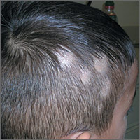
Hair loss on scalp
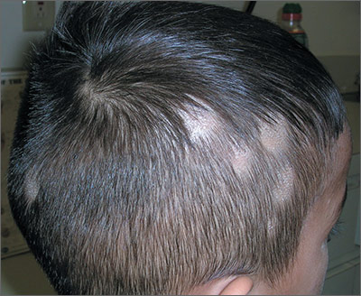
The FP noted patchy alopecia with scaling of the scalp and made the presumptive diagnosis of tinea capitis. A woods lamp examination did not demonstrate fluorescence. The child was very cooperative and the doctor was able to perform a potassium hydroxide (KOH) preparation by scraping the areas of alopecia with the edge of one glass slide while catching the scale on another slide. Microscopic examination revealed branching hyphae and some broken hairs with fungal elements within the hair shaft. (See video on how to perform a KOH preparation.) This microscopic picture was consistent with Trichophyton tonsurans, the most common cause of tinea capitis in the United States. The reason that the infected area did not fluoresce was that the dermatophyte was within the hair shaft (endothrix) rather than external to the hair (exothrix).
Topical antifungal therapy is not adequate for tinea capitis; oral treatment is needed. Oral antifungal choices include griseofulvin, terbinafine, and fluconazole. Griseofulvin comes in an oral suspension making it a desirable option for children who can’t swallow pills. However, at least 6 to 8 weeks of treatment (20 mg/kg/d) is required. Oral terbinafine tablets are less expensive and shorter courses of therapy may be used. Tablets of 250 mg terbinafine (most affordable of all the choices) can be broken in half for younger children and the dose should always be calculated based on weight. Fluconazole comes in various tablets, strengths, and liquid formulations and can be prescribed for 3 to 6 weeks, as needed.
Photos and text for Photo Rounds Friday courtesy of Richard P. Usatine, MD. This case was adapted from: Usatine R, Yao C. Tinea capitis. In: Usatine R, Smith M, Mayeaux EJ, et al, eds. Color Atlas of Family Medicine. 2nd ed. New York, NY: McGraw-Hill;2013:782-787.
To learn more about the Color Atlas of Family Medicine, see: www.amazon.com/Color-Family-Medicine-Richard-Usatine/dp/0071769641/
You can now get the second edition of the Color Atlas of Family Medicine as an app by clicking on this link: usatinemedia.com
Dark rash under breasts
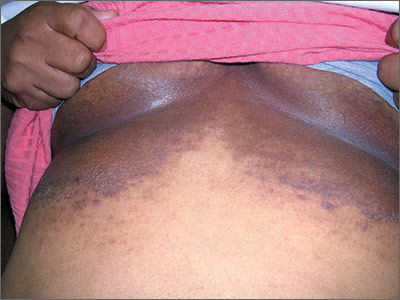
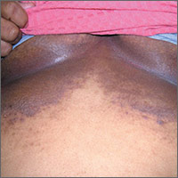
The family physician (FP) suspected that the patient had some type of fungal infection under her breasts. The FP scraped the edge and performed a potassium hydroxide (KOH) preparation that showed pseudohyphae and budding yeast forms, indicating a candida infection. (See video on how to perform a KOH preparation.) The FP also noted that there appeared to be some satellite lesions around the edge of the hyperpigmented area. Under the left breast was a small fissure and some white coloration at the skinfold. The differential diagnosis included tinea corporis and inverse psoriasis.
The FP prescribed topical clotrimazole cream 1% over the counter to be applied twice daily until at least one week after the rash resolved. While nystatin would be another option, it required a prescription and would not cover any possible dermatophyte in the area. The FP also explained that the dark pigmentation is the result of a longstanding inflammatory process related to the infection and would not go away immediately with treatment. In fact, the postinflammatory hyperpigmentation might remain for life.
The FP also spent time counseling the patient on an improved diet, increased exercise, and adherence to her diabetes medicines. A follow-up appointment for one month was set to review the patient's general health and to see if the candida infection resolved.
In cases where intertriginous fungal infections are treated without KOH or fungal culture confirmation, be suspicious for inverse psoriasis if the patient does not respond to antifungal medications. With such a large area involved, an oral antifungal such as fluconazole might occasionally be needed. Terbinafine is not an effective medicine for cutaneous candidiasis. If psoriasis is suspected, it helps to look for clues, such as nail pitting or cutaneous involvement with plaques on the elbows, knees, or scalp. If all else fails, a punch biopsy can be used to determine the correct diagnosis.
Photos and text for Photo Rounds Friday courtesy of Richard P. Usatine, MD. This case was adapted from: Usatine R. Candidiasis. In: Usatine R, Smith M, Mayeaux EJ, et al, eds. Color Atlas of Family Medicine. 2nd ed. New York, NY: McGraw-Hill; 2013:777-781.
To learn more about the Color Atlas of Family Medicine, see: www.amazon.com/Color-Family-Medicine-Richard-Usatine/dp/0071769641/
You can now get the second edition of the Color Atlas of Family Medicine as an app by clicking on this link: usatinemedia.com
White coating on infant’s tongue

The FP diagnosed thrush or candidiasis of the oral mucosa. To make sure it was not just milk, the FP gently drew a tongue blade over the tongue and the white exudate was mostly adherent (milk wipes away rather easily). The tongue blade was then rubbed onto a glass slide and 2 drops of potassium hydroxide (KOH) solution were applied. Microscopic evaluation revealed pseudohyphae and budding yeasts consistent with Candida albicans. (See video on how to perform a KOH preparation.)
Thrush is a common condition in infants with normal immune systems and does not require work-up for immunosuppression if this is the only finding at this age. Of course, thrush is seen in people who are immunosuppressed from various diseases (such as human immunodeficiency virus) and medications (like chemotherapy).
The FP chose to treat the child with oral nystatin suspension 2 mL 4 times a day. The directions were to give 1 mL in each side of the infant’s mouth and to continue this until 48 hours after signs and symptoms resolved. The mother was not having any symptoms or erythema of the nipples, but if she was, a topical antifungal agent (to be washed off before breastfeeding) could be used.
Photos and text for Photo Rounds Friday courtesy of Richard P. Usatine, MD. This case was adapted from: Usatine R. Candidiasis. In: Usatine R, Smith M, Mayeaux EJ, et al, eds. Color Atlas of Family Medicine. 2nd ed. New York, NY: McGraw-Hill;2013:777-781.
To learn more about the Color Atlas of Family Medicine, see: www.amazon.com/Color-Family-Medicine-Richard-Usatine/dp/0071769641/
You can now get the second edition of the Color Atlas of Family Medicine as an app by clicking on this link: usatinemedia.com

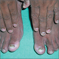
Rash on left hand, feet

The FP diagnosed this patient with two foot one hand syndrome, which is a fungal infection involving both feet and one hand. To be sure of the diagnosis, the FP performed a potassium hydroxide (KOH) preparation from the scale on the left hand and found typical branching hyphae caused by a dermatophyte. (See video on how to perform a KOH preparation.) The dermatophyte was most likely Trichophyton rubrum, but it wasn’t necessary to determine the exact species in order to treat this condition.
Two foot one hand syndrome is not rare, but it is somewhat puzzling as to why it involves only one hand while involving both feet. Also, it does not preferentially involve the dominant hand. In this case, the patient’s 2 large toenails had visible onychomycosis and 3 of the nails on his left hand had fungal changes (his dominant right hand was not affected). Tinea pedis was seen between the toes in a moccasin distribution, as well.
Treatment for this condition usually requires an oral antifungal agent. (If, however, the nails were not involved and there were contraindications to an oral antifungal, then a topical antifungal cream could be tried first.)
The patient described here had no history of liver disease or alcohol abuse. So the FP prescribed terbinafine 250 mg daily for one month to fully clear the skin infection and a follow-up appointment was set for 4 weeks. At that appointment, the FP planned to discuss continuing oral treatment for 2 additional months to clear the fungal infection from the nails. If ongoing oral terbinafine was needed, liver function tests could be performed at that time.
Photos and text for Photo Rounds Friday courtesy of Richard P. Usatine, MD. This case was adapted from: Usatine R. Fungal overview. In: Usatine R, Smith M, Mayeaux EJ, et al, eds. Color Atlas of Family Medicine. 2nd ed. New York, NY: McGraw-Hill;2013:771-776.
To learn more about the Color Atlas of Family Medicine, see: www.amazon.com/Color-Family-Medicine-Richard-Usatine/dp/0071769641/
You can now get the second edition of the Color Atlas of Family Medicine as an app by clicking on this link: usatinemedia.com
Painful ulcers in mouth
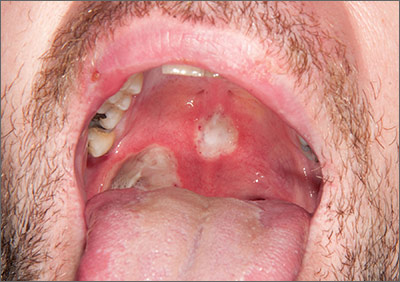
The patient was diagnosed with Behçet’s disease (BD) based on his clinical presentation. BD is a rare multisystem inflammatory disorder of unknown cause. The development of ulceration at the site of superficial skin injury (pathergy) is typical of BD. (The patient underwent multiple venipunctures while being investigated for a presumed infective illness prior to this presentation.)
There are no diagnostic laboratory tests for BD; laboratory findings usually reflect systemic inflammation. The International Study Group for BD, however, has derived classification criteria for use in clinical research studies. Their criteria include oral ulceration (that has recurred at least 3 times in a 12-month period), plus 2 of the following: recurrent genital ulceration, eye lesions, cutaneous lesions, or a positive pathergy test.
Recurrent mouth ulcers frequently involve the soft palate and oropharynx. Genital ulceration is the second most common manifestation of BD and is present in 57% to 93% of patients. The scrotum is most commonly involved, although the shaft and glans penis may also be affected. Ocular involvement is also seen in 30% to 70% of patients and is more frequent and severe in men.
There is no curative treatment for BD. The goals of treatment are to prevent organ damage and alleviate symptoms. Mucocutaneous disease is treated with potent topical corticosteroids. Severe attacks are treated with oral corticosteroids—1 mg/kg of prednisolone. The drug is tapered and discontinued once the disease is under control. Colchicine or dapsone is also an option. In refractory cases, consider thalidomide (50 mg once a day) or azathioprine (1-3 mg/kg). An anti-tumor necrosis factor agent may also be considered.
In this case, the patient was prescribed prednisolone 60 mg once a day, but relapsed once he was weaned off of it. He was then given thalidomide 50 mg once a day for 4 months and the disease resolved completely. The thalidomide was then reduced to 50 mg 3 times a week for 4 weeks, and then stopped completely. Nearly 2 years later, the patient has remained disease free.
Adapted from: Aslam A, Chalmers R. Mucocutaneous ulceration in a previously healthy man. J Fam Pract. 2014;63:97-100.
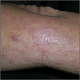
Pruritus since childhood
A 48-year-old woman experiencing homelessness presented to our clinic with a 4-week history of an intensely pruritic rash on her upper back and bilateral upper extremities. She reported that she had experienced exacerbations and remissions of the rash in similar locations for the past several years and during childhood. Factors that exacerbated the rash included being outdoors and being exposed to heat. Her pruritus was intensified by scratching the skin and was significantly worse at night. Previous doctors had diagnosed her with psoriasis and prescribed a short trial of hydrocortisone cream and oral antihistamines, but they provided minimal relief.
The patient indicated that the itching interrupted her sleep and her skin’s appearance made it difficult to get a job. The physical exam revealed excoriated and erythematous papules and patches on her upper back, the extensor and flexor aspects of her bilateral forearms, and the dorsal surface of her bilateral wrists, hands, and fingers (FIGURE 1). Her skin was dry and scaly with pigmentary changes and skin thickening (FIGURE 2). She denied any other systemic symptoms. Her hair and nails were normal, she had no palpable lymph nodes, and she was afebrile. She reported suffering from seasonal allergies, but wasn’t aware of a family history of skin disorders.


WHAT IS YOUR DIAGNOSIS?
HOW WOULD YOU TREAT THIS PATIENT?
Diagnosis: Chronic atopic dermatitis
Although the patient was told she had psoriasis by previous doctors, we diagnosed her condition as atopic dermatitis based on its clinical appearance. There is no single test that can establish a diagnosis of atopic dermatitis. While serum total IgE levels are often elevated, testing is not currently recommended.
The United Kingdom working group on atopic dermatitis published diagnostic criteria based on clinical history and physical exam that include pruritic skin in addition to the presence of 3 or more of the following: skin crease involvement, chronically dry skin, symptom onset before 2 years of age, and visible evidence of dermatitis involving flexural surfaces.1 Our patient fulfilled all but one condition, as she wasn’t sure if her symptoms began before age 2.
Atopic dermatitis is a chronic and inflammatory cutaneous disease that affects approximately 10% to 12% of children and less than 1% of adults in the United States.2 Approximately 90% of cases present before the age of 5 and the literature demonstrates a slight female predominance.3,4
Disease severity is classified as mild, moderate, or severe.5 Mild disease is characterized by dry skin and minimal itching with little impairment of the patient’s physical and psychological wellbeing. Moderate disease includes frequent pruritus and erythema with or without secondary skin changes and a moderate impact on physical and mental health. In severe disease, extensive secondary skin changes exist and the patient’s daily activities, sleep, and mental health may be severely impaired.
Etiology is multifactorial. Causes of atopic dermatitis include abnormalities in the epidermal stratum corneum and tight junctions, a heightened type-2 helper T-cell response to environmental antigens, innate immunity defects, and altered microbial skin flora.6,7
Genetic influences appear to play a substantial role in disease development. Approximately 70% of patients have a positive family history of an atopic disease such as eczema, asthma, or allergic rhinitis.8 Genetic defects are believed to be related to defective proteins and lipids in the epidermis that lead to disruption of the epidermal barrier and subsequent cutaneous inflammation.6,7
Clinical presentation: Lesion distribution varies with age
Intense pruritus and dry scaly skin occur in both children and adults, although the distribution of lesions may vary with age. Children typically exhibit erythematous patches with papules and crusting on the face, scalp, extremities, or trunk. In adults, lesions are primarily located on the hands and feet, but may also present on the face, wrists, forearms, and flexural areas.3
Adults also frequently present with secondary skin changes such as thickened skin, pigmentation changes, lichenification, and excoriated papules due to chronic rubbing or scratching.3 Our patient presented with significant lichenification and hyperpigmentation of the skin that was most prominent on the wrists and forearms.
Additional clinical features consistent with atopic dermatitis include a personal history of allergic conditions and a disease course characterized by exacerbations and remissions. Exacerbations may be caused by heat exposure, dry climates, anxiety, rapid temperature variations, contact with certain chemical substances, or microbial infections.8
Differential Dx includes psoriasis and scabies
The differential diagnosis of chronic atopic dermatitis consists of allergic or irritant contact dermatitis, plaque psoriasis, seborrheic dermatitis, scabies, and drug eruptions. Early diagnosis of atopic dermatitis is imperative to prevent sleep disturbances, chronic secondary skin changes, scarring, and the development of skin infections.
Allergic or irritant contact dermatitis is a cutaneous inflammation occurring after contact with an allergen or irritant. The lesions include erythematous, scaling areas with marked borders that are commonly pruritic. Acute cases often present with vesicles and bullae, while lichenification with cracks and fissures are common among chronic cases. Patch testing may be performed if the diagnosis is suspected.
Plaque psoriasis is characterized by areas of dry, erythematous, and well-demarcated plaques with silver scales that are most commonly found on the knees, elbows, scalp, and lower back. Systemic manifestations can include joint pain and joint swelling. Nail pitting and onycholysis are also common. While our patient had skin thickening, it was from the lichenification that is common in atopic dermatitis.
Psoriasis and atopic dermatitis are often confused. Psoriasis has discrete plaques on extensor surfaces and is often associated with nail changes, while the thickening of the skin that comes with chronic itch and scratching of atopic dermatitis is often less well circumscribed and found on flexor surfaces. Family physicians are frequently the first to encounter patients with atopic dermatitis and psoriasis and must be able to distinguish these conditions, as their treatments differ.
Seborrheic dermatitis is a chronic, relapsing inflammatory condition characterized by pruritic, erythematous, greasy, scaly patches on sebum-rich skin such as the scalp, face, and upper trunk. Seborrheic dermatitis is a clinical diagnosis.
Scabies is a pruritic skin condition caused by Sarcoptes scabiei var hominis. Characteristic linear burrows often appear as serpiginous, gray, threadlike elevations in the webbed spaces of the fingers, scrotum, areolae, elbows, axillae, feet, and wrist flexors. Secondary lesions from scratching or inflammation include excoriations, erythema, and hyperpigmentation. The diagnosis is made clinically and confirmed by dermoscopy, when available. Alternatively, mites or eggs may be observed on skin scrapings using light microscopy.
Drug eruptions should be considered in individuals taking medications who develop acute, symmetric cutaneous eruptions that may be morbilliform, urticarial, papulosquamous, pustular, or bullous in nature.
Treatment depends on severity, area of involvement, and patient’s age
Components of atopic dermatitis treatment include skin hydration, negative stimuli avoidance, pharmacologic modalities, and patient education. Improved skin hydration can be achieved by applying thick emollients containing little to no water at least twice daily and after bathing.
Topical corticosteroids are added when emollient use alone fails. Potency selection is based upon the patient’s age, involved body region, and the severity of skin inflammation.8 In order to reduce cutaneous atrophy, only low-potency corticosteroids should be applied to the face, groin, and axillae. Patients with mild disease may apply desonide 0.05% or hydrocortisone 2.5% cream or ointment once or twice daily for 2 to 4 weeks.8 Patients without improvement or with moderate disease may need medium- to high-potency steroids such as fluocinolone 0.025% or triamcinolone 0.1%.
Patients with atopic dermatitis on the face, eyelids, neck, and skin folds or those who do not obtain relief from combined emollients and topical corticosteroids may benefit from topical calcineurin inhibitors such as pimecrolimus or tacrolimus.9 However, these agents should be utilized only for short periods of time and avoided in immunocompromised patients and those younger than 2 years of age.9
Patients with uncontrolled moderate to severe refractory disease may consider a trial of phototherapy or cyclosporine if phototherapy is ineffective or unavailable.10 A meta-analysis has shown that once remission is achieved, intermittent therapy with moderate- to high-potency corticosteroids or tacrolimus may be effective in reducing subsequent flares.11
In all cases, sedating antihistamines such as diphenhydramine or hydroxyzine can be utilized for pruritic relief, particularly at night. Additionally, signs suggestive of infection should prompt antibiotic treatment with agents that provide coverage for Staphylococcus and Streptococcus species. Lastly, patients must be adequately educated on stimuli avoidance (eg, hot water, wool) and counseled on the medical and psychological issues that often accompany atopic dermatitis.
Our patient was placed on triamcinolone 0.1% for 4 weeks and her condition improved.
CORRESPONDENCE
Andrea Richardson, MD, MPH, 7414 Carriage Bay, San Antonio, TX 78249; [email protected].
1. Williams HC, Burney PG, Pembroke AC, et al. The U.K. Working Party’s Diagnostic Criteria for Atopic Dermatitis. III. Independent hospital validation. Br J Dermatol. 1994;131:406-416.
2. Horii KA, Simon SD, Liu DY, et al. Atopic dermatitis in children in the United States, 1997-2004: visit trends, patient and provider characteristics, and prescribing patterns. Pediatrics. 2007;120:e527-e534.
3. Rudikoff D, Lebwohl M. Atopic dermatitis. Lancet. 1998;351:1715-1721.
4. Kang K, Polster AM, Nedorost ST, et al. Atopic dermatitis. In: Dermatology. Bolognia JL, Jorizzo JL, Rapini RP, et al, eds. Mosby, New York;2003:199.
5. Lewis-Jones S, Mugglestone MA; Guideline Development Group. Management of atopic eczema in children aged up to 12 years: summary of NICE guidance. BMJ. 2007;335:1263-1264.
6. Kuo IH, Yoshida T, De Benedetto A, et al. The cutaneous innate immune response in patients with atopic dermatitis. J Allergy Clin Immunol. 2013;131:266-278.
7. Boguniewicz M, Leung DY. Atopic dermatitis: a disease of altered skin barrier and immune dysregulation. Immunol Rev. 2011;242:233-246.
8. Eichenfield LF, Tom WL, Chamlin SL, et al. Guidelines of care for the management of atopic dermatitis: section 1. Diagnosis and assessment of atopic dermatitis. J Am Acad Dermatol. 2014;70:338-351.
9. Ashcroft DM, Dimmock P, Garside R, et al. Efficacy and tolerability of topical pimecrolimus and tacrolimus in the treatment of atopic dermatitis: meta-analysis of randomised controlled trials. BMJ. 2005;330:516.
10. Garritsen FM, Brouwer MW, Limpens J, et al. Photo(chemo)therapy in the management of atopic dermatitis: an updated systematic review with implications for practice and research. Br J Dermatol. 2014;170:501-513.
11. Schmitt J, von Kobyletzki L, Svensson A, et al. Efficacy and tolerability of proactive treatment with topical corticosteroids and calcineurin inhibitors for atopic eczema: systematic review and meta-analysis of randomized controlled trials. Br J Dermatol. 2011;164:415-428.
A 48-year-old woman experiencing homelessness presented to our clinic with a 4-week history of an intensely pruritic rash on her upper back and bilateral upper extremities. She reported that she had experienced exacerbations and remissions of the rash in similar locations for the past several years and during childhood. Factors that exacerbated the rash included being outdoors and being exposed to heat. Her pruritus was intensified by scratching the skin and was significantly worse at night. Previous doctors had diagnosed her with psoriasis and prescribed a short trial of hydrocortisone cream and oral antihistamines, but they provided minimal relief.
The patient indicated that the itching interrupted her sleep and her skin’s appearance made it difficult to get a job. The physical exam revealed excoriated and erythematous papules and patches on her upper back, the extensor and flexor aspects of her bilateral forearms, and the dorsal surface of her bilateral wrists, hands, and fingers (FIGURE 1). Her skin was dry and scaly with pigmentary changes and skin thickening (FIGURE 2). She denied any other systemic symptoms. Her hair and nails were normal, she had no palpable lymph nodes, and she was afebrile. She reported suffering from seasonal allergies, but wasn’t aware of a family history of skin disorders.
WHAT IS YOUR DIAGNOSIS?
HOW WOULD YOU TREAT THIS PATIENT?
Diagnosis: Chronic atopic dermatitis
Although the patient was told she had psoriasis by previous doctors, we diagnosed her condition as atopic dermatitis based on its clinical appearance. There is no single test that can establish a diagnosis of atopic dermatitis. While serum total IgE levels are often elevated, testing is not currently recommended.
The United Kingdom working group on atopic dermatitis published diagnostic criteria based on clinical history and physical exam that include pruritic skin in addition to the presence of 3 or more of the following: skin crease involvement, chronically dry skin, symptom onset before 2 years of age, and visible evidence of dermatitis involving flexural surfaces.1 Our patient fulfilled all but one condition, as she wasn’t sure if her symptoms began before age 2.
Atopic dermatitis is a chronic and inflammatory cutaneous disease that affects approximately 10% to 12% of children and less than 1% of adults in the United States.2 Approximately 90% of cases present before the age of 5 and the literature demonstrates a slight female predominance.3,4
Disease severity is classified as mild, moderate, or severe.5 Mild disease is characterized by dry skin and minimal itching with little impairment of the patient’s physical and psychological wellbeing. Moderate disease includes frequent pruritus and erythema with or without secondary skin changes and a moderate impact on physical and mental health. In severe disease, extensive secondary skin changes exist and the patient’s daily activities, sleep, and mental health may be severely impaired.
Etiology is multifactorial. Causes of atopic dermatitis include abnormalities in the epidermal stratum corneum and tight junctions, a heightened type-2 helper T-cell response to environmental antigens, innate immunity defects, and altered microbial skin flora.6,7
Genetic influences appear to play a substantial role in disease development. Approximately 70% of patients have a positive family history of an atopic disease such as eczema, asthma, or allergic rhinitis.8 Genetic defects are believed to be related to defective proteins and lipids in the epidermis that lead to disruption of the epidermal barrier and subsequent cutaneous inflammation.6,7
Clinical presentation: Lesion distribution varies with age
Intense pruritus and dry scaly skin occur in both children and adults, although the distribution of lesions may vary with age. Children typically exhibit erythematous patches with papules and crusting on the face, scalp, extremities, or trunk. In adults, lesions are primarily located on the hands and feet, but may also present on the face, wrists, forearms, and flexural areas.3
Adults also frequently present with secondary skin changes such as thickened skin, pigmentation changes, lichenification, and excoriated papules due to chronic rubbing or scratching.3 Our patient presented with significant lichenification and hyperpigmentation of the skin that was most prominent on the wrists and forearms.
Additional clinical features consistent with atopic dermatitis include a personal history of allergic conditions and a disease course characterized by exacerbations and remissions. Exacerbations may be caused by heat exposure, dry climates, anxiety, rapid temperature variations, contact with certain chemical substances, or microbial infections.8
Differential Dx includes psoriasis and scabies
The differential diagnosis of chronic atopic dermatitis consists of allergic or irritant contact dermatitis, plaque psoriasis, seborrheic dermatitis, scabies, and drug eruptions. Early diagnosis of atopic dermatitis is imperative to prevent sleep disturbances, chronic secondary skin changes, scarring, and the development of skin infections.
Allergic or irritant contact dermatitis is a cutaneous inflammation occurring after contact with an allergen or irritant. The lesions include erythematous, scaling areas with marked borders that are commonly pruritic. Acute cases often present with vesicles and bullae, while lichenification with cracks and fissures are common among chronic cases. Patch testing may be performed if the diagnosis is suspected.
Plaque psoriasis is characterized by areas of dry, erythematous, and well-demarcated plaques with silver scales that are most commonly found on the knees, elbows, scalp, and lower back. Systemic manifestations can include joint pain and joint swelling. Nail pitting and onycholysis are also common. While our patient had skin thickening, it was from the lichenification that is common in atopic dermatitis.
Psoriasis and atopic dermatitis are often confused. Psoriasis has discrete plaques on extensor surfaces and is often associated with nail changes, while the thickening of the skin that comes with chronic itch and scratching of atopic dermatitis is often less well circumscribed and found on flexor surfaces. Family physicians are frequently the first to encounter patients with atopic dermatitis and psoriasis and must be able to distinguish these conditions, as their treatments differ.
Seborrheic dermatitis is a chronic, relapsing inflammatory condition characterized by pruritic, erythematous, greasy, scaly patches on sebum-rich skin such as the scalp, face, and upper trunk. Seborrheic dermatitis is a clinical diagnosis.
Scabies is a pruritic skin condition caused by Sarcoptes scabiei var hominis. Characteristic linear burrows often appear as serpiginous, gray, threadlike elevations in the webbed spaces of the fingers, scrotum, areolae, elbows, axillae, feet, and wrist flexors. Secondary lesions from scratching or inflammation include excoriations, erythema, and hyperpigmentation. The diagnosis is made clinically and confirmed by dermoscopy, when available. Alternatively, mites or eggs may be observed on skin scrapings using light microscopy.
Drug eruptions should be considered in individuals taking medications who develop acute, symmetric cutaneous eruptions that may be morbilliform, urticarial, papulosquamous, pustular, or bullous in nature.
Treatment depends on severity, area of involvement, and patient’s age
Components of atopic dermatitis treatment include skin hydration, negative stimuli avoidance, pharmacologic modalities, and patient education. Improved skin hydration can be achieved by applying thick emollients containing little to no water at least twice daily and after bathing.
Topical corticosteroids are added when emollient use alone fails. Potency selection is based upon the patient’s age, involved body region, and the severity of skin inflammation.8 In order to reduce cutaneous atrophy, only low-potency corticosteroids should be applied to the face, groin, and axillae. Patients with mild disease may apply desonide 0.05% or hydrocortisone 2.5% cream or ointment once or twice daily for 2 to 4 weeks.8 Patients without improvement or with moderate disease may need medium- to high-potency steroids such as fluocinolone 0.025% or triamcinolone 0.1%.
Patients with atopic dermatitis on the face, eyelids, neck, and skin folds or those who do not obtain relief from combined emollients and topical corticosteroids may benefit from topical calcineurin inhibitors such as pimecrolimus or tacrolimus.9 However, these agents should be utilized only for short periods of time and avoided in immunocompromised patients and those younger than 2 years of age.9
Patients with uncontrolled moderate to severe refractory disease may consider a trial of phototherapy or cyclosporine if phototherapy is ineffective or unavailable.10 A meta-analysis has shown that once remission is achieved, intermittent therapy with moderate- to high-potency corticosteroids or tacrolimus may be effective in reducing subsequent flares.11
In all cases, sedating antihistamines such as diphenhydramine or hydroxyzine can be utilized for pruritic relief, particularly at night. Additionally, signs suggestive of infection should prompt antibiotic treatment with agents that provide coverage for Staphylococcus and Streptococcus species. Lastly, patients must be adequately educated on stimuli avoidance (eg, hot water, wool) and counseled on the medical and psychological issues that often accompany atopic dermatitis.
Our patient was placed on triamcinolone 0.1% for 4 weeks and her condition improved.
CORRESPONDENCE
Andrea Richardson, MD, MPH, 7414 Carriage Bay, San Antonio, TX 78249; [email protected].
1. Williams HC, Burney PG, Pembroke AC, et al. The U.K. Working Party’s Diagnostic Criteria for Atopic Dermatitis. III. Independent hospital validation. Br J Dermatol. 1994;131:406-416.
2. Horii KA, Simon SD, Liu DY, et al. Atopic dermatitis in children in the United States, 1997-2004: visit trends, patient and provider characteristics, and prescribing patterns. Pediatrics. 2007;120:e527-e534.
3. Rudikoff D, Lebwohl M. Atopic dermatitis. Lancet. 1998;351:1715-1721.
4. Kang K, Polster AM, Nedorost ST, et al. Atopic dermatitis. In: Dermatology. Bolognia JL, Jorizzo JL, Rapini RP, et al, eds. Mosby, New York;2003:199.
5. Lewis-Jones S, Mugglestone MA; Guideline Development Group. Management of atopic eczema in children aged up to 12 years: summary of NICE guidance. BMJ. 2007;335:1263-1264.
6. Kuo IH, Yoshida T, De Benedetto A, et al. The cutaneous innate immune response in patients with atopic dermatitis. J Allergy Clin Immunol. 2013;131:266-278.
7. Boguniewicz M, Leung DY. Atopic dermatitis: a disease of altered skin barrier and immune dysregulation. Immunol Rev. 2011;242:233-246.
8. Eichenfield LF, Tom WL, Chamlin SL, et al. Guidelines of care for the management of atopic dermatitis: section 1. Diagnosis and assessment of atopic dermatitis. J Am Acad Dermatol. 2014;70:338-351.
9. Ashcroft DM, Dimmock P, Garside R, et al. Efficacy and tolerability of topical pimecrolimus and tacrolimus in the treatment of atopic dermatitis: meta-analysis of randomised controlled trials. BMJ. 2005;330:516.
10. Garritsen FM, Brouwer MW, Limpens J, et al. Photo(chemo)therapy in the management of atopic dermatitis: an updated systematic review with implications for practice and research. Br J Dermatol. 2014;170:501-513.
11. Schmitt J, von Kobyletzki L, Svensson A, et al. Efficacy and tolerability of proactive treatment with topical corticosteroids and calcineurin inhibitors for atopic eczema: systematic review and meta-analysis of randomized controlled trials. Br J Dermatol. 2011;164:415-428.
Pruritic, lightly-scaled patches on wrists
A 19-year-old man presented to our clinic with erythematous, pruritic, lightly-scaled, and annular patches on his dorsal wrists. The rash had first appeared 3 weeks earlier on the patient’s left wrist, which is where he’d been wearing a chrome-colored watch for a couple of years. After the rash appeared on his left wrist, the patient began wearing the watch on his right wrist. Soon after the switch, the rash appeared on his right wrist. The patient was otherwise healthy and denied any previous rashes, had no body piercings or allergies of any kind, and was not on any medications.
On physical exam, we noted 2 erythematous, scaly, annular, and slightly raised plaques on the distal dorsal aspects of both forearms/wrists with a few erythematous papular lesions (FIGURE). There was also scaling on the soles of the patient’s feet and white, moist scaling in the web space between his 4th and 5th toes bilaterally.
WHAT IS YOUR DIAGNOSIS?
HOW WOULD YOU TREAT THIS PATIENT?
Diagnosis: Tinea corporis (ringworm)
The patient and physician initially considered the possibility of allergic contact dermatitis due to nickel because of the history of redness, scaling, and itching underneath the watch when it was worn on the left wrist, and then when it was worn on the right wrist. Nickel dermatitis is common and it is easy to attribute the cause of a condition like this to the most obvious diagnosis without considering a more complete differential diagnosis.1
However, there were clues that prompted us to suspect tinea corporis (ringworm). The red, scaly rash spread centrifugally over several weeks, and fomites, such as a watch, can spread infectious diseases. Also, our patient had a few erythematous papular lesions, and the presence of papules in addition to scaling rings is typical of fungal infections involving hair follicles (Majocchi’s granuloma).
A positive potassium hydroxide (KOH) preparation confirmed the diagnosis and eliminated the need for nickel patch testing.2
Warmth and moisture could explain tinea on the wrists
Dermatophytes are fungi that can cause infections in the skin, hair, and nails. They are classified by where they are found—anthropophilic (humans), geophilic (soil), or zoophilic (animals). Anthropophilic and zoophilic dermatophytes from the genera Trichophyton, Microsporum, and Epidermophyton are primarily responsible for human fungal infections.3 It is estimated that superficial fungal infections affect up to a quarter of the world’s population.3
Tinea corporis mainly occurs in prepubertal children, presenting as a red, annular, scaly, pruritic patch with central clearing and an active border.4 Tinea corporis includes all superficial dermatophyte infections of the glabrous skin and is particularly common in areas of excessive heat and moisture.5 Patients can pick up tinea corporis via fomites at the gym, through soil in the garden, or by touching a pet’s fur or a child’s scalp when either has the fungal infection.
The wrists are not a common place for tinea corporis, but the condition can occur anywhere on the body. This patient may well have contracted tinea from his own interdigital tinea pedis. Warmth and moisture under the watch could also explain the predilection for fungus to grow on the wrists.
Distinguish between contact dermatitis and tinea corporis
The differential diagnosis for tinea corporis includes allergic contact dermatitis, granuloma annulare, annular elastolytic granuloma, and erythema chronicum migrans.
Allergic contact dermatitis is caused by an allergy to a substance, such as the metal nickel. A preliminary diagnosis of contact dermatitis could easily be made in error if one were to assume that a patient was having a type IV hypersensitivity response to nickel from a watch.6
Granuloma annulare produces slowly expanding annular plaques that are not itchy and do not scale. This commonly occurs over the joints and is of unknown etiology.7
Annular elastolytic granuloma is a variant of granuloma annulare that occurs on skin that has been exposed to the sun. It presents with a red, ring-like pattern and is associated with little scaling or pruritus.8
Erythema chronicum migrans produces annular lesions at the site of a tick bite and is the primary sign of Lyme disease. The tick must be in place for 24 hours for infection to occur.9 (Our patient did not notice a tick attached at either site.)
In this case, a KOH preparation of skin scrapings identified septate hyphae, which supported our diagnosis of tinea corporis.2 A history of red, scaly, itchy, and expanding round/oval patches or plaques and evidence of “athlete’s foot” can also help one to make the diagnosis.
Antifungal agents will clear the rash
Proper treatment of tinea corporis consists of antifungal creams containing ketoconazole, econazole, or naftifine on non-hair-bearing areas. The creams should be applied twice daily and rarely cause adverse effects. Bandages are not usually necessary, but may be used if contact with others is anticipated. For the scalp and other hair-bearing areas, systemic treatment with terbinafine 250 mg daily for one month in an adult is necessary. Oral agents will usually clear the rash within 4 to 6 weeks.10,11
Because our patient had bilateral involvement and some papule formation indicating Majocchi’s granuloma, we prescribed oral terbinafine 250 mg daily for 2 weeks in addition to econazole cream. The patient was to apply the cream for a total of 4 weeks to ensure the rash did not recur.
CORRESPONDENCE
Stephen E. Helms, MD, Department of Dermatology, University of Mississippi Medical Center, 2500 North State Street, Jackson, MS 39216; [email protected].
1. Groopman J. How Doctors Think. Houghton Mifflin Co: Boston, Massachusetts; 2007.
2. Panasiti V, Borroni RG, Devirgiliis V, et al. Comparison of diagnostic methods in the diagnosis of dermatomycoses and onychomycoses. Mycoses. 2006;49:26-29.
3. Havlickova B, Czaika VA, Friedrich M. Epidemiological trends in skin mycoses worldwide. Mycoses. 2008;51:2-15.
4. Ely JW, Rosenfeld S, Seabury Stone M. Diagnosis and management of tinea infections. Am Fam Physician. 2014;90:702-710.
5. Gupta AK, Chaudhry M, Elewski B. Tinea corporis, tinea cruris, tinea nigra, and piedra. Dermatol Clin. 2003;21:395-400.
6. Lidén C, Menné T, Burrows D. Nickel-containing alloys and platings and their ability to cause dermatitis. Br J Dermatol. 1996;134:193-198.
7. Barron DF, Cootauco MH, Cohen BA. Granuloma annulare. A clinical review. Lippincotts Prim Care Pract. 1997;1:33-39.
8. Ventura F, Vilarinho C, da Luz Duarte M, et al. Two cases of annular elastolytic giant cell granuloma: Different response to the treatment. Dermatol Online J. 2010;16:11.
9. Feder HM Jr, Abeles M, Bernstein M, et al. Diagnosis, treatment, and prognosis of erythema migrans and Lyme arthritis. Clin Dermatol. 2006;24:509-520.
10. Kelly BP. Superficial fungal infections. Pediatr Rev. 2012;33:e22-e37.
11. Rotta I, Sanchez A, Gonçalves PR, et al. Efficacy and safety of topical antifungals in the treatment of dermatomycosis: a systematic review. Br J Dermatol. 2012;166:927-933.
A 19-year-old man presented to our clinic with erythematous, pruritic, lightly-scaled, and annular patches on his dorsal wrists. The rash had first appeared 3 weeks earlier on the patient’s left wrist, which is where he’d been wearing a chrome-colored watch for a couple of years. After the rash appeared on his left wrist, the patient began wearing the watch on his right wrist. Soon after the switch, the rash appeared on his right wrist. The patient was otherwise healthy and denied any previous rashes, had no body piercings or allergies of any kind, and was not on any medications.
On physical exam, we noted 2 erythematous, scaly, annular, and slightly raised plaques on the distal dorsal aspects of both forearms/wrists with a few erythematous papular lesions (FIGURE). There was also scaling on the soles of the patient’s feet and white, moist scaling in the web space between his 4th and 5th toes bilaterally.
WHAT IS YOUR DIAGNOSIS?
HOW WOULD YOU TREAT THIS PATIENT?
Diagnosis: Tinea corporis (ringworm)
The patient and physician initially considered the possibility of allergic contact dermatitis due to nickel because of the history of redness, scaling, and itching underneath the watch when it was worn on the left wrist, and then when it was worn on the right wrist. Nickel dermatitis is common and it is easy to attribute the cause of a condition like this to the most obvious diagnosis without considering a more complete differential diagnosis.1
However, there were clues that prompted us to suspect tinea corporis (ringworm). The red, scaly rash spread centrifugally over several weeks, and fomites, such as a watch, can spread infectious diseases. Also, our patient had a few erythematous papular lesions, and the presence of papules in addition to scaling rings is typical of fungal infections involving hair follicles (Majocchi’s granuloma).
A positive potassium hydroxide (KOH) preparation confirmed the diagnosis and eliminated the need for nickel patch testing.2
Warmth and moisture could explain tinea on the wrists
Dermatophytes are fungi that can cause infections in the skin, hair, and nails. They are classified by where they are found—anthropophilic (humans), geophilic (soil), or zoophilic (animals). Anthropophilic and zoophilic dermatophytes from the genera Trichophyton, Microsporum, and Epidermophyton are primarily responsible for human fungal infections.3 It is estimated that superficial fungal infections affect up to a quarter of the world’s population.3
Tinea corporis mainly occurs in prepubertal children, presenting as a red, annular, scaly, pruritic patch with central clearing and an active border.4 Tinea corporis includes all superficial dermatophyte infections of the glabrous skin and is particularly common in areas of excessive heat and moisture.5 Patients can pick up tinea corporis via fomites at the gym, through soil in the garden, or by touching a pet’s fur or a child’s scalp when either has the fungal infection.
The wrists are not a common place for tinea corporis, but the condition can occur anywhere on the body. This patient may well have contracted tinea from his own interdigital tinea pedis. Warmth and moisture under the watch could also explain the predilection for fungus to grow on the wrists.
Distinguish between contact dermatitis and tinea corporis
The differential diagnosis for tinea corporis includes allergic contact dermatitis, granuloma annulare, annular elastolytic granuloma, and erythema chronicum migrans.
Allergic contact dermatitis is caused by an allergy to a substance, such as the metal nickel. A preliminary diagnosis of contact dermatitis could easily be made in error if one were to assume that a patient was having a type IV hypersensitivity response to nickel from a watch.6
Granuloma annulare produces slowly expanding annular plaques that are not itchy and do not scale. This commonly occurs over the joints and is of unknown etiology.7
Annular elastolytic granuloma is a variant of granuloma annulare that occurs on skin that has been exposed to the sun. It presents with a red, ring-like pattern and is associated with little scaling or pruritus.8
Erythema chronicum migrans produces annular lesions at the site of a tick bite and is the primary sign of Lyme disease. The tick must be in place for 24 hours for infection to occur.9 (Our patient did not notice a tick attached at either site.)
In this case, a KOH preparation of skin scrapings identified septate hyphae, which supported our diagnosis of tinea corporis.2 A history of red, scaly, itchy, and expanding round/oval patches or plaques and evidence of “athlete’s foot” can also help one to make the diagnosis.
Antifungal agents will clear the rash
Proper treatment of tinea corporis consists of antifungal creams containing ketoconazole, econazole, or naftifine on non-hair-bearing areas. The creams should be applied twice daily and rarely cause adverse effects. Bandages are not usually necessary, but may be used if contact with others is anticipated. For the scalp and other hair-bearing areas, systemic treatment with terbinafine 250 mg daily for one month in an adult is necessary. Oral agents will usually clear the rash within 4 to 6 weeks.10,11
Because our patient had bilateral involvement and some papule formation indicating Majocchi’s granuloma, we prescribed oral terbinafine 250 mg daily for 2 weeks in addition to econazole cream. The patient was to apply the cream for a total of 4 weeks to ensure the rash did not recur.
CORRESPONDENCE
Stephen E. Helms, MD, Department of Dermatology, University of Mississippi Medical Center, 2500 North State Street, Jackson, MS 39216; [email protected].
1. Groopman J. How Doctors Think. Houghton Mifflin Co: Boston, Massachusetts; 2007.
2. Panasiti V, Borroni RG, Devirgiliis V, et al. Comparison of diagnostic methods in the diagnosis of dermatomycoses and onychomycoses. Mycoses. 2006;49:26-29.
3. Havlickova B, Czaika VA, Friedrich M. Epidemiological trends in skin mycoses worldwide. Mycoses. 2008;51:2-15.
4. Ely JW, Rosenfeld S, Seabury Stone M. Diagnosis and management of tinea infections. Am Fam Physician. 2014;90:702-710.
5. Gupta AK, Chaudhry M, Elewski B. Tinea corporis, tinea cruris, tinea nigra, and piedra. Dermatol Clin. 2003;21:395-400.
6. Lidén C, Menné T, Burrows D. Nickel-containing alloys and platings and their ability to cause dermatitis. Br J Dermatol. 1996;134:193-198.
7. Barron DF, Cootauco MH, Cohen BA. Granuloma annulare. A clinical review. Lippincotts Prim Care Pract. 1997;1:33-39.
8. Ventura F, Vilarinho C, da Luz Duarte M, et al. Two cases of annular elastolytic giant cell granuloma: Different response to the treatment. Dermatol Online J. 2010;16:11.
9. Feder HM Jr, Abeles M, Bernstein M, et al. Diagnosis, treatment, and prognosis of erythema migrans and Lyme arthritis. Clin Dermatol. 2006;24:509-520.
10. Kelly BP. Superficial fungal infections. Pediatr Rev. 2012;33:e22-e37.
11. Rotta I, Sanchez A, Gonçalves PR, et al. Efficacy and safety of topical antifungals in the treatment of dermatomycosis: a systematic review. Br J Dermatol. 2012;166:927-933.
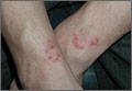
Pruritic rash in axilla
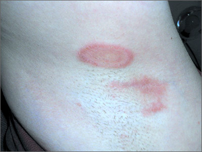
The FP diagnosed tinea corporis by recognizing the pattern of the concentric rings, which have a high specificity for tinea infections. To confirm the diagnosis, the FP performed a scraping of the area and found branching septate hyphae. This is the microscopic appearance of dermatophytes, which come in 3 genera: microsporum, epidermophyton, and trichophyton. There are approximately 40 species in these 3 genera and these fungi cause tinea pedis and manus, tinea capitis, tinea corporis, tinea cruris, tinea faciei, and onychomycosis.
Potassium hydroxide (KOH) test characteristics (without fungal stains) have a sensitivity of 77% to 88% and a specificity of 62% to 95%.1 The sensitivity and specificity is higher with fungal stains and the experience of the person performing the test. Fungal infections of the skin and mucous membranes are ubiquitous and common. There are many types of fungus that grow on humans, but they all share a predilection for warm and moist areas. Consequently, hot and humid climates promote fungal infections. The FP also asked to check the patient’s feet and found a mild case of tinea pedis between the 4th and 5th toes bilaterally.
The FP explained the diagnoses to the patient and recommended that she purchase topical terbinafine over the counter. He explained that there are no better topical antifungals by prescription and that she could find this medicine in the athlete’s foot area of any pharmacy or large grocery store. He told her that she did not have to stop running and that anyone can get a fungal infection. The FP recommended that the patient use the cream for at least one week after clinical clearance and to do her best to keep her feet and axillae dry to avoid new fungal infections.
1. Thomas B. Clear choices in managing epidermal tinea infections. J Fam Pract. 2003;52:850-862.
Photos and text for Photo Rounds Friday courtesy of Richard P. Usatine, MD. This case was adapted from: Usatine R. Fungal overview. In: Usatine R, Smith M, Mayeaux EJ, et al, eds. Color Atlas of Family Medicine. 2nd ed. New York, NY: McGraw-Hill; 2013:771-776.
To learn more about the Color Atlas of Family Medicine, see: www.amazon.com/Color-Family-Medicine-Richard-Usatine/dp/0071769641/
You can now get the second edition of the Color Atlas of Family Medicine as an app by clicking on this link: usatinemedia.com
