User login
Choosing a career in health care administration
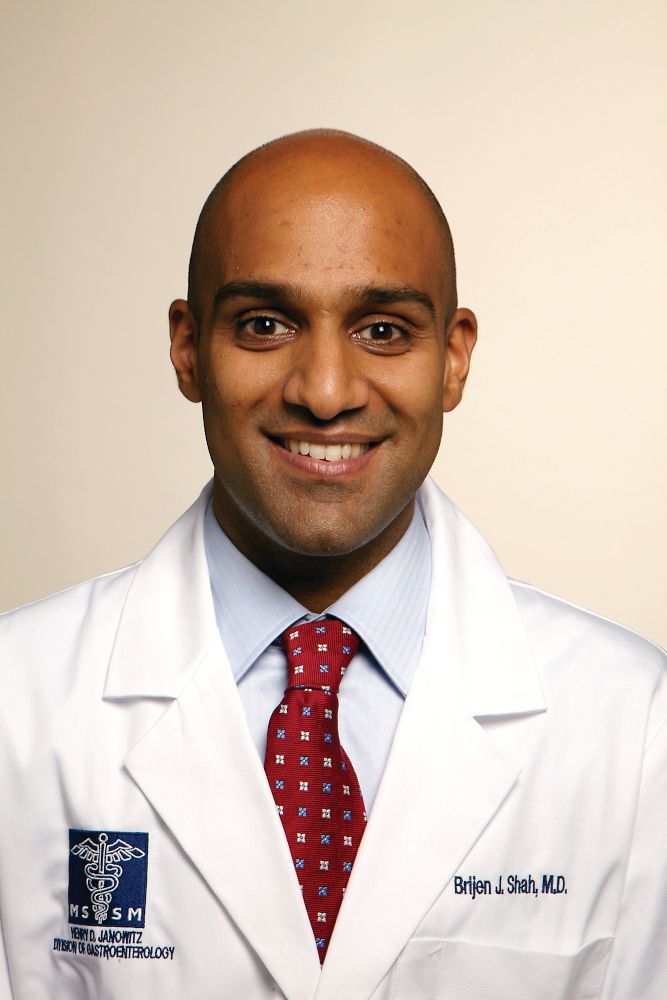
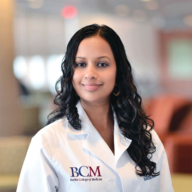
Dr. Shah is chief medical officer, Mount Sinai Queens, director of quality and patient safety education, Mount Sinai Health System, associate professor of medicine/GI and geriatrics/palliative medicine, Icahn School of Medicine at Mount Sinai, New York, N.Y.
How did your career pathway lead you into hospital administration?
My career pathway into hospital administration was not by design. Since college, I have always enjoyed building and managing programs. As chief resident, I was exposed to operational and managerial aspects of running an internal medicine residency program as well as an outpatient clinic. This program first exposed me to quality improvement and patient-safety functions within a hospital. I continued to build on this during my GI fellowship and in my first few years as faculty in our division. Patient safety and quality improvement involve the critical thinking and quantitative aspects of research applied to problems in the workplace. This interest and skill, along with my growing experience in management, led me to explore opportunities in hospital administration.
What are your responsibilities in a typical week?
As a chief medical officer at a smaller hospital, my scope of responsibilities includes overseeing health care quality and patient safety, building our outpatient practice and new clinical service lines, building a cohesive medical staff, handling disciplinary and professionalism issues, and overseeing six support departments. I work closely with our chief operating officer, chief nursing officer, executive director, and site chiefs.
What do you enjoy most about working in hospital administration?
I enjoy the work I do related to making care safer for our patients and for those who work in our hospital. I take each patient-safety event personally. The teamwork necessary to understand these events and develop creative solutions is extremely rewarding. I enjoy mentoring our faculty and hospital managers to achieve their own goals. Lastly, I have enjoyed working with professionals with backgrounds that differ from my own. I have learned an incredible amount about hospital operations, leadership, financing, as well as legal and labor-relations issues from our staff in other departments.
What do you find most challenging about working in hospital administration?
The scope of work in my role at my current hospital is very broad. It can be a challenge to focus on long-term goals given the number of “fires” that creep up during the week. I always try to keep patients and staff at the center of my decision making. However, a hospital is a complex organism and decisions also have a financial and operational impact. One challenge is to know and understand this impact and then work with other leaders to develop a solution that works for all.
The other challenge is managing conflict in a way that leads to creative solutions satisfying the needs of multiple stakeholders. We are constantly challenged by limited resources (including time). These challenges are inherent to any leadership position. I am fortunate to work within a leadership team that is collaborative; they are an invaluable resource when these issues arise.
What are the different hospital administration positions that are available to GIs?
More than ever, the options for administrative work in health care have expanded. There are hospital-based roles within a department (e.g., director of endoscopy, clinical chief, director of quality improvement/patient safety for GI) and larger roles in the hospital-at-large (e.g., chief operating officer, chief medical officer, chief quality officer). These roles require certain technical skills and knowledge as well as experience within patient care. Other opportunities include the medical board, credentials committee, or serving as a member of a hospital committee (e.g., pharmacy, perioperative, infection control, process improvement). Since hospitals and health systems have expanded into the outpatient setting, additional positions include medical director for a practice, director of population health, or a leadership role in clinical operations.
Physicians from many different specialties have entered these roles based on their local hospital needs. In addition to clinical experience, leadership and interpersonal skills are critical for success.
How would a fellow or early-career GI who is interested in hospital medicine pursue this career pathway?
My first suggestion is to get involved in local efforts based on your interest. For example, if you are interested in quality improvement, seek to be a member of your department or hospital quality improvement committee. In GI and hepatology, natural places to get involved are around the development of care pathways, readmission committees, and initiatives to increase screening and treatment of hepatitis C or colon cancer. If you are interested in operations, look to see if there is an endoscopy or clinic operations committee you can get involved in. Get to know your medical board and medical staff structure. I gained exposure to this world by observing some of these meetings and then being asked to join them. These are valuable groups that help to create policy, raise important issues, and work with administration in the management of the hospital.
I am also a big fan of informational interviewing. If there are leaders who do the type of work you are interested in, consider reaching out with a call or email and asking to meet with them to talk about their role and career path.
As a fellow, there is an Accreditation Council for Graduate Medical Education requirement to incorporate residents and fellows onto hospital committees. This requirement has been a great way to have fellows incorporated into hospital work. You will find that those in hospital administration are eager to have interested and collegial partners in the work that is being done.
Are there any advanced training options available for those interested in hospital administration?
Depending on the position, there are numerous certificate and master’s programs that can provide formal education. CEOs and COOs may seek an MBA or master’s in Health Care Administration. There are programs that focus on Health Care Leadership or Quality and Patient Safety that are applicable to many leadership positions. These are offered in in-person and online formats. However, many physicians in these positions have a combination of informal and experiential learning programs that developed their skill set.
Some hospital systems offer an internal physician leadership training program to develop early and midcareer physician executives. There are professional organizations that offer courses for leadership development (e.g., American College of Physician Executives). Some business schools offer shorter-format programs that are geared toward health care leaders and focus on finance, operations, or quality.
I received some of my training through the Clinical Quality Fellowship Program, which is a 14-month experiential learning program in quality and patient safety that is run locally in New York City. In addition, I had some leadership training through the Association of American Medical Colleges and through the AGA Future Leaders Program (http://www.gastro.org/about/initiatives/aga-future-leaders-program).
Hospitals, outpatient practices, and health systems offer career paths including patient safety, quality improvement, or hospital management. I have enjoyed stretching my existing skill set in these roles while learning about how health facilities work, gaining knowledge of health care financing, and making care safer while ensuring high quality. These roles require teamwork across professions and specialties. As a gastroenterologist or hepatologist, we bring our own clinical and professional experience, which can be invaluable to the overall health care management team.
Gastroenterology debuts editorial fellowship program
The readership of Gastroenterology includes a broad distribution of stakeholders in digestive health, including those with vested interests in clinical practice, education, policy, clinical investigation, and basic research. One of our most critical constituencies, however, is trainees and early-career GIs. In an effort to support such individuals, our editorial team has developed a freshly minted 1-year editorial fellowship for Gastroenterology. The overarching purpose of this fellowship is to mentor an outstanding trainee for future editorial leadership roles in scientific publishing, as a means to promote the interests of trainee and early-career GI constituencies within the AGA and Gastroenterology. This fellowship is available to exceptional second- or third-year fellows through an application process. The intent of this training is to allow the selected applicant to become intimately involved with Gastroenterology’s entire editorial process, including peer review, editorial oversight, manuscript selection for publication, production, and postpublication activities. Our first fellow, Eric Shah, MD, MBA, was selected from a highly competitive pool of exceptional applicants, and began his fellowship on July 1, 2017.
This year, we have been delighted to work with Dr. Shah as our inaugural Gastroenterology fellow. Dr. Shah has a unique background, having pursued a joint MD and MBA (earning both concurrently), while also following venture-oriented interests in developing GI technology from academia. Dr. Shah began his research career under the mentorship of Mark Pimentel, MD, and Gil Melmed, MD, at Cedars-Sinai as part of a Research Honors Program. Since that time, he has focused on evaluating the comparative efficacy, durability, and harm associated with pharmacotherapy in functional bowel disorders. Dr. Shah was accepted into the GI fellowship training program at the University of Michigan and received a slot on the T32 training grant to study cost-effectiveness and qualitative research techniques to address gaps in the care of functional bowel disorders. His work under the mentorship of William Chey, MD, Ryan Stidham, MD, and Philip S. Schoenfeld, MD, has flourished and culminated in an oral presentation and several posters for DDW 2017, as well as several first-author manuscripts that have been submitted. Dr. Shah has fully embraced the Gastroenterology fellowship and has far surpassed our high expectations for this position.
The video associated with this article is no longer available on this site. Please view all of our videos on the MDedge YouTube channel
VIDEO SOURCE: AMERICAN GASTROENTEROLOGICAL ASSOCIATION
In addition to creating an editorial fellowship, our team has also developed other components within the journal that specifically target trainees and early-career GIs. The Mentoring, Education and Training section – initiated in 2011 through the vision and insight of Bishr Omary MD, PhD, and John Del Valle, MD, at the University of Michigan – has been extremely effective in highlighting critical issues relevant to trainees, young faculty, and early-career GIs. Topics have included mentoring advice not only for individuals in academic or private practice careers but also industry careers and midlevel providers. Other topics have included Accreditation Council for Graduate Medical Education milestones, career advancement for clinician-educators, sex and ethnic diversity, and maintenance of certification, as well as guidance regarding nontraditional funding mechanisms such as philanthropy. Potential future topics will include information about major new public and private funding initiatives, comments and input from National Institutes of Health officials, and reports of funding trends relevant to both physician scientists and clinicians. We are fortunate to have Prateek Sharma, MD, lead this section, and his depth of experience as an exceptional mentor has provided the requisite expertise.
Additionally, we offer a reduction in page charges to junior investigators (within 7 years of fellowship) who are the corresponding authors of exceedingly important original Gastroenterology manuscripts. These manuscripts from junior investigators will be highlighted in both print and online versions of Gastroenterology. We are using the journal to expand electronic access to educational offerings for new technologies, training, self-assessment, and practice improvement to establish the AGA as the ultimate resource for junior academicians and practicing physicians. We are also currently integrating Gastroenterology more closely into other AGA educational efforts that target young physicians, such as the AGA Education and Training Committee.
At Gastroenterology, we are acutely aware of the needs and obstacles facing trainees, young faculty, and early-career GIs. We have boldly adopted a multidimensional approach to provide guidance and opportunities to overcome these challenges, including the creation of the nascent Editorial Fellowship. We welcome applications for the next fellowship, which will be announced by the AGA in the spring of 2018!
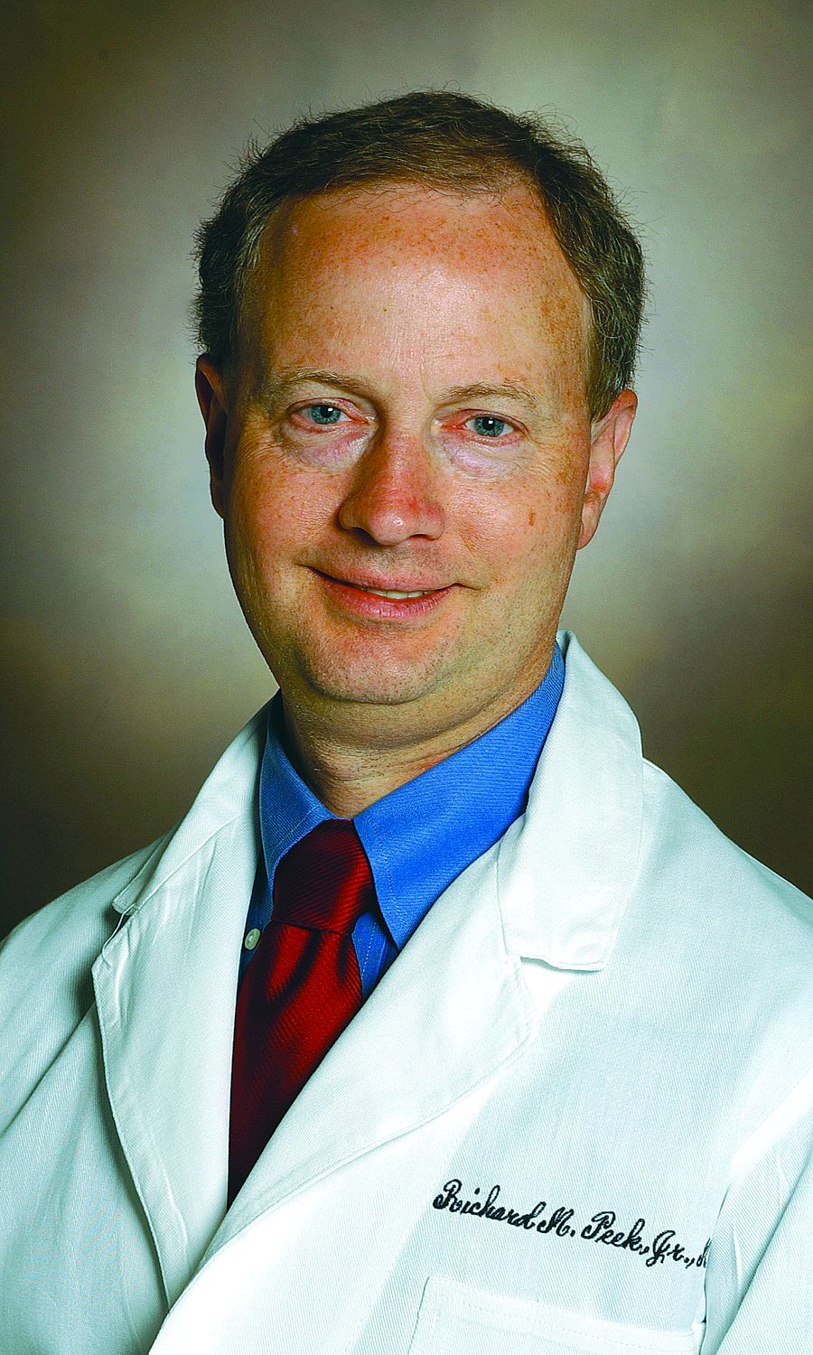
Dr. Peek is the Mina Wallace Professor of Medicine, Cancer Biology, and Pathology, Microbiology, and Immunology, and director, division of gastroenterology, hepatology and nutrition, Vanderbilt University Medical Center, Nashville, Tenn. He has no conflicts of interest.
2017 GI thought leaders
Trainee and early-career members made an impact in the GI field this past year, especially through contributing to and engaging in various collaborations via the American Gastroenterological Association’s member-only online discussion forum – the AGA Community.
We’re proud to announce that the title of 2017 top contributor is held by an early-career member: Meet Dmitriy Kedrin, MD, PhD, of Elliot Hospital in Manchester, N.H., and find out a little more about how the AGA Community is an important part of his routine in this brief Q&A session.
Thanks for being such an active member of the AGA Community! Why do you contribute?
“I think it is important for GI docs to be a part of a larger community, stay informed on the latest guidelines, research publications, and approaches to difficult cases, where more than one road can be taken. I feel that it is a great forum for someone like me, a relatively junior gastroenterologist.”
Why do you enjoy being part of the AGA Community?
“I find the case discussions informative. I learn a great deal about current trends and opinions on important topics in the GI world.”
What do you like to do in your free time?
“I bake bread and run a gastroenterology literature review podcast called ‘GI Pearls.’ ”
What’s your approach to handling a difficult patient case you come across in your practice?
“I often seek advice of other clinicians, some with more expertise in a particular area. I also go to the literature and try to learn more that way, to help expand my differential as well as figure out the best therapeutic approach.”
Was there a conversation in the AGA Community in 2017 that was your favorite?
“Oh, there were several. I recall a patient case where there were several thought leaders in the field who had a disagreement about the best approach to treatment. The work-life balance conversation was also very good. I also enjoyed reading about different opinions regarding the values of clinical versus observational trials that happened a while back.”
Here are other trainee and early-career members who made the list of top contributors in the AGA Community last year:
- Avinash Ketwaroo, MD
- Hüseyin Bozkurt, MD
- Peter Liang, MD, MPH
- Fola May, MD, PhD
- Richa Shukla, MD
- Arthur Beyder, MD, PhD
- Shazia Siddique, MD
- Brigid Boland, MD
Check the achievements section of your AGA Community profile to see whether you made the list. We look forward to seeing you all in the forum in 2018!
Topics rarely discussed during fellowship
Each year, the American Gastroenterological Association travels throughout the country, hosting Regional Practice Skills Workshops to answer tough and practical questions that are rarely addressed during fellowship. Presentations from several of the 2017 workshops are now available on the AGA website for you to watch at your leisure (login required). Each presentation is 20 minutes or less and provides expert advice to help you succeed in your career.
Here’s a selection of what you’ll find at www.gastro.org/education:
- Physician Contracts and Negotiations – Jon Appino from Contract Diagnostics discusses the anatomy of physician employment agreements. He shares questions that are important for you to ask when reviewing a contract.
- Career in Hybrid Practice – John S. Hanson, MD, AGAF, from Carolinas HealthCare System reviews different practice types and provides insight into “hybrid practice” and how it compares to private practice and academic practice.
- Career in Private Practice – Rig S. Patel, MD, the president of Digestive Healthcare PA, builds a clear picture of what life is like in private practice and shares nine things to consider when reviewing a private practice.
- Wealth Management Perspectives – Two financial advisors from Morgan Stanley provide tips and strategies to help early career physicians stay “financially fit.”
See all of AGA’s on-demand education for trainees and early career GIs by visiting www.gastro.org/education and searching for the series Trainees and Early Career GIs.
The AGA Regional Practice Skills Workshops are organized by the AGA Trainee & Early Career Committee in support of early career GIs. In December 2017, workshops were held in California, Iowa, and New York. The spring 2018 workshops will take place in Columbus, Ohio (Feb. 24), and Philadelphia, Penn. (April 11).
Register early for the AGA Postgraduate Course
We held a contest in the AGA Community forum asking members to share a piece of advice they learned or heard in the past year for a chance to win free registration to this year’s AGA Postgraduate Course in Washington, D.C., on June 2 and 3.
Here are some of our favorite responses from early career members, including our winner, Hüseyin Bozkurt from Medical Park Private Tarsus Hospital, Turkey:
- “We can provide healthy gut microbiome, don’t lose your hope. The future is changeable.” Hüseyin Bozkurt Sr., MD, contest winner.
- “Ambulatory reflux monitoring modalities can help phenotype GERD and guide optimal management.” Amit Patel, MD, 2018 AGA Postgraduate Course faculty.
- “One tip I heard from an attending in the first year of fellowship, which is particularly useful for IBS patients, is to set realistic expectations from the outset, for e.g. – when diagnosing patients with IBS and giving them advice, telling them ‘We are not looking to cure your GI symptoms, but to control them – you will have GI symptoms off and on despite treatment, my goal is to make sure you have more good days, than bad.’” Aakash Aggarwal, MD
Registration for the popular 1.5-day course is now open. Learn more at pgcourse.gastro.org and save $75 by registering before April 18.
The AGA Postgraduate Course takes place during AGA’s annual meeting Digestive Disease Week®, which is cosponsored by AASLD, ASGE, and SSAT. Learn more and register at www.ddw.org.
Five reasons to pursue a career in IBD research or patient care
A career in inflammatory bowel disease promises to be challenging, exciting, at times frustrating, and always educational. We asked our expert faculty at the inaugural Crohn’s & Colitis Congress, which took place Jan. 18-20 in Las Vegas, to reflect on why a career in IBD is an excellent path to take.
Why pursue a career in IBD
1. “IBD is the fastest moving area of GI to integrate science (genomics, microbiome, immunology) into care that will change the natural history of disease. The physicians and scientists have an unusually collegial culture, and the patients really care.” – Jonathan G. Braun, MD, PhD
2. “Managing patients with IBD is becoming ever more complex. When patients move beyond having mild disease, complex decisions need to be made. Choosing the right medication at the right time for the right patient will lead to the best outcomes for patients with IBD. I have every reason to believe that specializing in the clinical care of patients with IBD will be intellectually challenging while offering great personal satisfaction in taking care of these ill patients.” – Francis A. Farraye, MD, MSc
3. “IBD research findings and the implications for patient care are evolving rapidly. Many recommendations that we made 5-10 years ago have changed as we are learning more about IBD every day. There are so many opportunities to participate in the expansion of that knowledge base and help us reach our goal of a cure for IBD in the lifetime of many of our patients. Take the challenge.” – Teri Lynn Jackson, MSN, ARNP
4. “IBD is an outstanding field led by great people who want to see fellows and junior faculty succeed. Identify a mentor and listen to them, meet and engage with new people, be curious, think big, and work hard!” – Michael J. Rosen, MD, MSCI
5. “The best career in the world! Such variety. A home for everyone with any interest. It won’t always be smooth, but it will be incredibly rewarding with hardly a dull moment.” – Dermot McGovern, MD, PhD, FRCP
For additional tips and advice, visit the AGA Community.
Calendar
For more information about upcoming events and award deadlines, please visit http://www.gastro.org/education and http://www.gastro.org/research-funding.
UPCOMING EVENTS
Feb. 22, 2018; March 22, 2018
Reimbursement, Coding and Compliance for Gastroenterology
Improve the efficiency and performance of your practice by staying current on the latest reimbursement, coding, and compliance changes.
2/22 (Edison, NJ); 3/22 (St. Charles, MO)
AGA Regional Practice Skills Workshop – Ohio
During this free workshop, senior and junior GI leaders will guide you through various practice options and address topics rarely discussed during fellowship, such as employment models, partnerships, hospital politics, billing and coding, compliance, contracts and more. Find out more at http://www.gastro.org/in-person/aga-regional-practice-skills-workshop-ohio.
Columbus, OH
March 12, 2018; March 14, 2018
Advancing Collaborative Approaches in IBD Treatment Decision-Making
This is a unique opportunity for payers and providers to gather in the same room to discuss inflammatory bowel disease therapy selection, disease monitoring, treatment criteria, and access.
3/12 (Pittsburgh); 3/14 (Chicago)
March 21-23, 2018
2018 AGA Tech Summit: Connecting Stakeholders in GI Innovation
Join leaders in the physician, investor, regulatory, and medtech communities as they examine the issues surrounding the development and delivery of new GI medical technologies.
Boston, MA
April 11, 2018
AGA Regional Practice Skills Workshop – Pennsylvania
During this free workshop, senior and junior GI leaders will guide you through various practice options and address topics rarely discussed during fellowship, such as employment models, partnerships, hospital politics, billing and coding, compliance, contracts, and more. Find out more at http://www.gastro.org/in-person/regional-practice-skills-workshop-philadelphia.
Philadelphia
May 10-11, 2018
HIV and Hepatitis Management: THE NEW YORK COURSE
This advanced CME activity will provide participants with state-of-the-art information and practical guidance on progress in managing HIV, hepatitis B, and hepatitis C and will enable practitioners to deliver the highest-quality care in all practice settings.
New York City
Jun. 2-5, 2018
DIGESTIVE DISEASE WEEK® (DDW) 2018 – WASHINGTON, DC
AGA Trainee and Early-Career GI Sessions
Join your colleagues at special sessions to meet the unique needs of physicians who are new to the field. Participants will learn about all aspects of starting a career in clinical practice or research, have the opportunity to network with mentors and peers, and review board material.
• June 2, 8:15 a.m.-5:30 p.m.; June 3, 8:30 a.m.-12:35 p.m.
AGA Postgraduate Course: From Abstract to Reality
Attend this multi-topic course to get practical, applicable information to push your practice to the next level. The 2018 course will provide a comprehensive look at the latest medical, surgical, and technological advances over the past 12 months that aim to keep you up to date in a rapidly changing field. Each presenter will turn abstract ideas into concrete action items that you can implement in your practice immediately. AGA member trainees and early-career GIs receive discounted pricing for this course.
• June 3, 4-5:30 p.m.
Difficult Conversations: Navigating People, Negotiations, Promotions, and Complications
During this session, attendees will obtain effective negotiation techniques and learn how to navigate difficult situations in clinical and research environments.
• June 4, 4-5:30 p.m.
Advancing Clinical Practice: Gastroenterology Fellow–Directed Quality-Improvement Projects
This trainee-focused session will showcase selected abstracts from GI fellows based on quality improvement with a state-of-the-art lecture. Attendees will be provided with information that defines practical approaches to quality improvement from start to finish. A limited supply of coffee and tea will be provided during the session.
• June 5, 1:30-5:30 p.m.
Board Review Course
This session, designed around content from DDSEP® 8, serves as a primer for third-year fellows preparing for the board exam as well as a review course for others wanting to test their knowledge. Session attendees will receive a $50 coupon to use at the AGA Store at DDW to purchase DDSEP 8.
• TBD
AGA Early-Career Networking Hour
Date, time, and location to be announced soon.
June 4-8, 2018
Exosomes/Microvesicles: Heterogeneity, Biogenesis, Function, and Therapeutic Developments (E2)
Deepen your understanding of the structural and functional complexity of extracellular vesicles, their biogenesis and function in health and disease, cargo enrichment, potential as ideal biomarkers, and breakthroughs in their use as therapeutic targets/agents.
Breckenridge, CO
AWARDS APPLICATION DEADLINES
AGA Fellow Abstract Award
This travel award provides $500 and one $1,000 prize to recipients who are MD and/or PhD postdoctoral fellows presenting posters/oral sessions at DDW.
Application Deadline: Feb. 16, 2018
AGA-Moti L. & Kamla Rustgi International Travel Awards
This travel award provides $750 to recipients who are young basic, translational, or clinical investigators residing outside North America to support travel and related expenses to attend DDW.
Application Deadline: Feb. 16, 2018
AGA Student Abstract Award
This travel award provides $500 and one $1,000 prize to recipients who are high school, undergraduate, graduate, medical students, or residents (residents up to year 3 postgraduates) presenting posters/oral sessions at DDW.
Application Deadline: Feb. 16, 2018
February 2018
Gastroenterology:
Living like an academic athlete: How to improve clinical and academic productivity as a gastroenterologist. Benchimol E et al.
2018 Jan;154(1):8-14. doi: 10.1053/j.gastro.2017.11.017.
“Spending your life wisely”: How to create an asset management plan. Adams MA et al.
2017 Dec;153(6):1469-72. doi: 10.1053/j.gastro.2017.10.032.
How to balance clinical work and research in the current era of academic medicine. Katzka DA.
2017 Nov;153(5):1177-80. doi: 10.1053/j.gastro.2017.09.024.
Clin Gastroenterol Hepatol.:
New models of gastroenterology practice. Allen JI et al.
2018 Jan;16(1):3-6. doi: 10.1016/j.cgh.2017.10.003.
Cracking the clinician educator code in gastroenterology. Shapiro JM et al.
2017 Dec;15(12):1828-32. doi: 10.1016/j.cgh.2017.08.040.
Cell Mol Gastroenterol Hepatol.:
Setting up a lab: The early years. Habtezion A.
2017 Nov; 4(3): 445-6. doi: 10.1016/j.jcmgh.2017.08.003.
Understanding Social Media in GI Practice: Influence, Learn, and Prosper
Gone are the days when social media was primarily used by millennials and those early adopters on the diffusion-of-innovation curve. Now, baby boomers and laggards alike are using social media to communicate with the world around them. Furthermore, health and healthcare issues are common topics in the social media universe. Eight in 10 Internet users seek health information online and 74% of these health information seekers use social media.1,2 Additionally, when they look online, they are more likely to trust information from doctors (61%) than from hospitals (55%), insurers (42%), or pharmaceutical companies (37%).3 Therefore, there is tremendous opportunity for physicians to engage patients, policy makers, advocacy groups, and other health care influencers in order to share reliable information. Yet, we must do so responsibly. There is a considerable degree of misinformation circulated in social media and we believe that physicians should help combat this by providing accurate information.
On a more individual level, social media can help you stay up-to-date on best practices, breakthroughs, and controversies in medicine. It can help you take control of your online reputation rather than letting it be the default Google search results. Social media can also be a vehicle through which you build your offline network of potential colleagues, collaborators, and supporters as well as facilitate speaking, consulting, research, and other professional opportunities.
We that we have convinced you to actively participate in social media professionally. Next, we would like to share our top six best practices for responsible use.
1. Understand and define your goals. We have broadly laid out our rationale but that is different from your specific, desired outcomes. If you do not know what you are trying to accomplish you will have no idea if you are successful or if what you are doing is working versus whether you should try different strategies. Social media does take time; therefore, you should be strategic and goal oriented.
3. Share reliable/vetted information in your area of expertise and interest. Do not try to be all things to all people. Focus on content that distinguishes you and meets your goals. On the other hand, this should not be all about you; this can be boring, difficult, and give the impression that all you care about is self-promotion. No more than a quarter to a third of the content should be about you and the rest should be curated content from other reliable sources. Sharing with attribution helps you build your community. Also, people appreciate vetted content in the great web of misinformation available. You can facilitate audience engagement by including graphics, photos, and videos and by engaging and responding to other posts. Importantly, having a disclaimer on your account (e.g., retweets are not endorsements, posts are not medical advice) is never a substitute for knowing/vetting what you are sharing.
4. Exercise caution when responding to medical questions on social media and/or sharing patient information. While we encourage engagement, you should never answer specific medical questions. This develops a doctor-patient relationship and creates legal “duty.” It could even constitute practicing without a license, if the person asking the question lives in another state. Instead, provide general information about a condition, especially as a link to a reliable site (www.gastro.org/patient-care/patientinfo-center) and suggest seeking care from their local medical professional. Along these lines, do not share any potentially identifiable patient information without documented permission. In addition to the obvious (e.g., patient name, photos, medical documents with identifiers), avoid stories of care, complications, rare conditions, or identifiable specimens. With an approximate date and the location of your practice, it may be very easy for someone to determine the patient’s identity.
6. Know and adhere to the social media policies of your practice, institution, organization, or employer. Most academic institutions have social media policies and they are becoming more widely adapted to other settings. While you may just get a metaphorical slap on the wrist for not following the rules, I think we all would agree that it would be a tragedy to get fired over a social media post.
However, none of the above best practices are a substitute for being intentional and mindful when sharing information on social media, whether it be Facebook, Twitter, Instagram, Youtube, or another platform. What does being intentional and mindful on social media mean? Absolutely avoid commenting/posting about patients, colleagues, or your workplace in any way that could be perceived to be negative. Declare conflicts of interest where applicable (i.e., if you’re a consultant for a pharma company, avoid endorsing a drug without declaring your conflict). Above all else, don’t post anything that you wouldn’t mind being on a billboard or mainstream news.
Participation is an investment of your most valuable resource: time. Therefore, know your goals and revisit these goals and your success in reaching them regularly. Start small and expand as your time and interest allows. Finally, minimize your exposure to risk by keeping our guidance and your institutional policies in mind and always pause before you post.
You can find out more about the AGA, its programs, and publications via our social media outlets, including:
Twitter:
@amergastroassn
@AGA_CGH
@AGA_CMGH
@AGA_Gastro
Facebook:
@AmerGastroAssn
@cghjournal
@cmghjournal
@gastrojournal
Dr. Fisher is associate professor in the department of medicine, division of gastroenterology, Duke University, Durham, N.C. VA Medical Center. Twitter: @DrDeborahFisher. Dr. Gray is assistant professor, department of medicine, division of gastroenterology, hepatology and nutrition, Ohio State University College of Medicine. Twitter: @DMGrayMD.
This information was presented at a Meet-the-Professor Luncheon at DDW® 2017. More for in-depth details than are described in this article, refer to this session on DDW on Demand (http://www.ddw.org/education/session-recordings).
References
1 Von Muhlen M., Ohno-Machado L. J Am Med Inform Assoc. 2012;19(5):777-81.
2 Childs L.M., Martin C.Y. Am J Health System Pharm. 2012;69(23):2044-50.
3. Social media “likes” healthcare. From marketing to social business – 2012 Report
https://www.pwc.com/us/en/health-industries/health-research-institute/publications/health-care-social-media.html.
4. www.martinsights.com/social-media-marketing/social-media-strategy/new-global-social-media-research/.
5. Online Medical Professionalism: Patient and Public Relationships: Policy Statement From the American College of Physicians and the Federation of State Medical Boards. Ann Intern Med. 2013;158(8):620-7.
Advocacy in Action: Meeting Congressman Gene Green
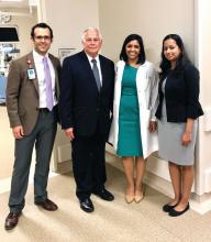
The hospital is often the intersection between a patient’s medical illness and their social and financial issues. As physicians, it is important to recognize that patient care encompasses not only prescribing medications and performing procedures but also practicing systems-based medicine; ensuring social and financial barriers do not impede access to, and delivery of, care. Some of these barriers cannot be eliminated by one individual practitioner; they can only be improved by working with government representatives and policy makers to make systemic changes. For gastroenterologists, advocacy involves educating patients, practitioners, and our government representatives about issues related to GI illnesses and the importance of ensuring access to GI specialty care and treatment for all patients who require it.
As physicians, we are uniquely positioned to represent the needs of our patients. We appreciate the AGA facilitating that voice by providing updates on legislation and coordinating meetings between senators and members of Congress and practicing gastroenterologists and GI fellows. These meetings are an important opportunity to network and share our experiences. Congressman Green was very interested to hear our perspectives as health care providers. It was enlightening to hear about his experiences on the Health Subcommittee and learn about its procedures. We would strongly encourage other AGA members to take advantage of this important program. n
Dr. Natarajan is assistant professor, Dr. Shukla is assistant professor, and Dr. Shapiro is a second-year fellow; all are in the section of gastroenterology and hepatology, Baylor College of Medicine, Houston.
References
1. Ehrlich E. (2017). NIH’S Role in Sustaining the U.S. Economy. United for Medical Research. Accessed at http://www.unitedformedicalresearch.com/wp-content/uploads/2017/03/NIH-Role-in-the-Economy-FY2016.
2. AGA Position Statement on Research Funding. Accessed at http://www.gastro.org/take-action/top-issues/research-funding.
3. El-Serag H.B., Kanwal F., Davila J.A., Kramer J., Richardson P. A new laboratory-based algorithm to predict development of hepatocellular carcinoma in patients with hepatitis C and cirrhosis. Gastroenterology. 2014;May146(5):1249-55.
4. White D.L., Richardson P., Tayoub N., Davila J.A., Kanwal F., El-Serag H.B. The updated model: An adjusted serum alpha-fetoprotein-based algorithm for hepatocellular carcinoma detection with hepatitis C virus-related cirrhosis. Gastroenterology. 2015;Dec 149(7):1986-7.
5. AGA Position Statement on Patient Cost-Sharing for Screening Colonoscopy. Accessed a: http://www.gastro.org/take-action/top-issues/patient-cost-sharing-for-screening-colonoscopy.
