User login
Malpractice Chronicle
Reprinted with permission from Medical Malpractice Verdicts, Settlements and Experts, Lewis Laska, Editor, (800) 298-6288.
Delayed Treatment for Preeclampsia
A woman experiencing her first pregnancy had an expected due date of December 5. She had been attending her prenatal appointments regularly with the defendant obstetrician/gynecologist. In October, she presented to the defendant hospital with complaints of left upper quadrant abdominal pain. Her blood pressure was elevated. She was discharged the next day.
Two days later, the patient made a scheduled visit to the office where she was receiving her prenatal care. Because she was noted to have protein in her urine, she was sent to the hospital for hypertension testing and a 24-hour urine test. It was determined that she had either pregnancy-induced hypertension or mild preeclampsia. She was sent home with instructions for biweekly fetal nonstress testing and a repeat 24-hour urine test.
A few days later, the patient presented to the hospital for a biophysical profile test. Her blood pressure was elevated. Physical examination revealed 2+ edema, and she was admitted to the hospital for monitoring.
Two days later, a resident documented a plan to repeat lab work for pregnancy-induced hypertension and continue the current care plan. The attending physician, however, discharged the woman with a diagnosis of preeclampsia. She was told to follow up on an outpatient basis.
Two days later, the patient returned to the hospital with a headache and elevated blood pressure. A biophysical profile test was nonreassuring, with fetal heart rate decelerations noted. A cesarean delivery was performed. Blood gas measurements after the child’s birth indicated metabolic acidosis. The infant was transferred with records indicating neonatal depression. Head CT revealed intraventricular hemorrhage.
The defendants claimed that this was a prematurity-related injury that could not have been prevented. The defendants also argued that the child had a normal IQ and mild cerebral palsy and was functioning well with few residual effects.
According to a published account, a $1.9 million settlement was reached.
Difficult Bead Removal Blamed for Ear Damage
When a woman discovered that her 5-year-old son had a bead stuck in his ear, she took him to a hospital emergency department, where he was seen by the defendant emergency physician. He made two attempts to remove the bead without sedating the child; the mother tried to hold him still, but he wriggled and screamed. Nurses also tried unsuccessfully to hold the child still, until blood was noted coming from his ear.
The emergency physician and the defendant family practitioner then sedated the child, and the latter spent more than an hour attempting to remove the bead. Eventually, he succeeded. As a result of the incident, however, the child suffered hearing loss that, despite reconstructive surgeries to repair the damage, will require hearing aids.
The emergency physician settled before trial for $40,000. The plaintiff claimed that the defendant family physician tried for too long to remove the bead and that his visualization was obscured by blood in the ear, resulting in rupture of the tympanic membrane and severe damage to the ossicular structure of the middle ear.
The defendant argued that he used suction to clear the field and did not use any instruments without having good visualization of the bead. He maintained that the main problem in removing the bead was a stricture in the ear canal where the temporal bone meets the cartilage.
The defendant also argued that the mother failed to return the child to his office in 10 days, as he had requested, in order for him to assess the extent of damage to the ear—an assessment he was unable to make immediately after removing the bead due to blood in the child’s ear.
According to a published report, a defense verdict was returned.
Alcohol and Hydromorphone a Bad Mix
After drinking alcohol all day, an obese 37-year-old man began to experience severe abdominal pain. Paramedics were called to his home, and he was transported to the defendant hospital emergency department (ED). Breath analysis indicated a blood alcohol level of 0.207, indicating very serious intoxication. The patient was noted to be alert, oriented, and cooperative, but he was anxious about his pain, which he rated as 10 on a 1-to-10 scale.
The defendant physician ordered IV fluids, including IV hydromorphone, 1 to 2 mg every 20 minutes as needed, with a note to keep the systolic blood pressure at 100 mm Hg or higher. The defendant made that order without evaluating the patient.
Over the course of one hour, the patient was administered 6 mg of hydromorphone. According to the patient’s record, only one full set of vital signs was taken during that time. Continuous monitoring was not performed, as the patient had been placed in an overflow room.
About two hours after his arrival at the hospital, the patient was found in cardiopulmonary arrest, and a code was called. Extensive resuscitation efforts were unsuccessful.
At autopsy, the cause of death was found to be anoxic encephalopathy resulting from mixed alcohol and hydromorphone. A physician contacted the medical examiner’s office over concern about the amount of hydromorphone given to the decedent.
The defendant physician testified that she was unaware of the decedent’s blood alcohol level. She said she assumed that nurses would monitor the decedent appropriately and would use their judgment regarding how much hydromorphone to administer, based on the decedent’s response. The nurses, however, maintained that no monitoring was ordered and claimed that the doctor never questioned them regarding the decedent’s status.
The defendants claimed that the amount of hydromorphone ordered was appropriate for an obese man with severe pain and that continuous monitoring was not required. The defendants also claimed that the man’s death was actually the result of severe hemorrhagic pancreatitis. The defendants also disputed the decedent’s life expectancy.
A $500,000 settlement was reached.
Paps are “normal” despite bleeding and cervical cancer… and more
Paps are “normal” despite bleeding and cervical cancer
A ROUTINE PAP SMEAR of a 27-year-old woman showed atypical squamous cells of undetermined significance. Over the next 3 years, the same gynecologist obtained annual Pap smears; pathologists and cytotechnologists interpreted these as being within normal limits. Then the patient reported postcoital bleeding to her gynecologist. Assuming the bleeding to be due to low estrogen associated with her oral contraceptive (OC), he switched her to another OC. Over the next 7 months, the patient reported on six occasions that she was still experiencing significant postcoital bleeding, tenderness during intercourse, and abdominal cramping. A Pap smear on one of those visits indicated no evidence of malignancy. Nine months after the change in OC, cervical cancer was diagnosed. Ten months later, the patient began radiation and chemotherapy because she was found to have metastatic cervical cancer of the rectum, pelvis, and colon. She died 9 months later at age 32.
PLAINTIFF’S CLAIM The first Pap smear actually showed evidence of a low-grade squamous intraepithelial lesion, so further testing was needed to rule out cervical cancer. When the patient reported postcoital bleeding, colposcopy and cervical biopsy should have been performed to determine whether she indeed had cervical cancer.
PHYSICIAN’S DEFENSE Not reported.
VERDICT $1.3 million Massachusetts settlement.
Ectopic pregnancy with IUD leads to fallopian tube removal
A FEW WEEKS AFTER IUD PLACEMENT, a 26-year-old woman reported to a hospital complaining of abdominal pain and bleeding. An ObGyn diagnosed an ectopic pregnancy, recommended removal of both fallopian tubes, and then proceeded to remove them.
PATIENT’S CLAIM It was negligent to perform nonemergent surgery when she was unable to consent to it.
PHYSICIAN’S DEFENSE The procedure was proper, as the patient was highly likely to have another ectopic pregnancy. Also, the patient could undergo in vitro fertilization if she wanted to become pregnant.
VERDICT Tennessee defense verdict.
Could retractors have caused right-leg femoral nerve neuropathy?
A 66-YEAR-OLD WOMAN with endometrial cancer underwent hysterectomy and surgical staging. Following the procedure, she suffered complete neuropathy of the femoral nerve in her right leg.
PATIENT’S CLAIM Retractors were used improperly during surgery, causing the injury.
PHYSICIAN’S DEFENSE The neuropathy was not a result of the type of retraction, but was probably due to the patient’s modified lithotomy position during surgery. Such an injury is a known risk of the procedure.
VERDICT $750,000 New York verdict. As the verdict was for all past pain and suffering, the court increased the judgment to $900,000 after the trial to include future pain and suffering.
Was retained clip the reason for kidney failure 12 years later?
BECAUSE OF A TUMOR on her left ovary, a woman’s left ovary and fallopian tube were removed. During surgery, Dr. A found and lysed adhesions around her right ovary. Seventeen months later, the patient underwent laparoscopy and lysis of adhesions as well as biopsy of the right ovary. Dr. B, who performed the procedure, did not note any clip on the left ureter. Three months after that, the patient underwent exploratory laparotomy with lysis of adhesions and right ovarian cystectomy and partial omentectomy—performed by Dr. C. Upon visual inspection, the left kidney appeared to be larger than the right kidney. When the patient complained of left-sided abdominal pain 10 years later, she underwent a CT urogram, which showed a chronically obstructed left kidney—probably related to a surgical clip obstructing the distal third of the left ureter. She was diagnosed with hydronephrosis of the left kidney, which was essentially nonfunctioning. The urologist believed the clip had been left there during the first surgery 12 years earlier.
PATIENT’S CLAIM Dr. A was negligent for placing the clip on the ureter, causing kidney damage.
PHYSICIAN’S DEFENSE Because of the statute of limitation and state of repose, Dr. A moved for dismissal and summary judgment, but the motions were denied. He also claimed that clipping the ureter during the first surgery would have caused immediate excruciating pain. However, he admitted that partial obstruction could occur without pain and in fact lead to total obstruction and death of the kidney years later.
VERDICT $450,000 Massachusetts arbitration award.
Nurses reassure new mother, who then dies from PE
A 25-YEAR-OLD WOMAN GAVE BIRTH to a healthy boy. She did not feel well during the week after hospital discharge. When she called her ObGyn’s office to discuss her complaints, the nurses reassured her. Ten days after delivery, she was taken to the emergency room, where she died from a pulmonary embolism.
PLAINTIFF’S CLAIM The physician and nurses failed to respond properly to the patient’s complaints, which were consistent with a pulmonary embolism.
PHYSICIAN’S DEFENSE The patient was monitored properly. An embolism is a sudden event.
VERDICT $867,273 Tennessee verdict. The physician group was found 70% at fault and the hospital 30% at fault.
Despite US results, birth delayed to 41 weeks
ULTRASONOGRAPHY SHOWED a shortened cervix, a subchorionic hematoma, and a choroid plexus cyst in the fetal brain during a patient’s prenatal care. The ObGyns induced labor at 41 weeks’ gestation and then performed emergent cesarean delivery. The child suffered birth asphyxia, thrombocytopenia, hypocalcemia, and cerebral palsy.
PATIENT’S CLAIM The ObGyns should have induced labor and/or performed cesarean delivery before 39 weeks’ gestation, but they failed to recognize the significance of the mother’s condition.
PHYSICIANS’ DEFENSE Not reported.
VERDICT $1.1 million Michigan settlement.
Sponge emerges 7 months after cesarean delivery
A LAPAROTOMY SPONGE was unknowingly left in the abdomen of a 29-year-old woman who underwent cesarean delivery. Seven months later, she was examined for a stitch abscess. Thinking he was removing a retained stitch, the surgeon pulled out a 12-inch sponge. She was awake at the time and experienced severe pain. The next day, she underwent laparotomy and drains were placed. She remained hospitalized for several days and drainage continued for another 6 days. The patient suffered no permanent injury; incisions for both operations were made at the same site, and she later gave birth without complication.
PATIENT’S CLAIM Leaving a sponge inside her was negligent.
PHYSICIAN’S DEFENSE The nurses who assisted in the surgery were responsible for the retained sponge.
VERDICT $110,410 Illinois verdict against the surgeon. Confidential settlement with the hospital prior to trial.
Would an earlier birth have saved this stillborn child?
WHEN 32 WEEKS’ PREGNANT, a 16-year-old patient repeatedly told her ObGyn she was experiencing bleeding. Later, she reported decreased fetal movement, but a sonogram indicated nothing abnormal. Twenty-three days later, her infant was delivered stillborn.
PATIENT’S CLAIM Placental abruption, which occurred 24 to 96 hours before delivery, caused the stillbirth. Because of her risk factors—bleeding, age, smoking, decreased fetal movement—labor should have been induced or a cesarean delivery performed earlier.
PHYSICIAN’S DEFENSE Ultrasonography did not indicate placental abruption, so delivery at that time was not warranted. An umbilical cord accident—which was unforeseeable and unpreventable—caused the stillbirth.
VERDICT Kentucky defense verdict.
Mother claims she wasn’t told test results for Down syndrome
A TRIPLE SCREEN BLOOD TEST ordered for a patient under prenatal care indicated that she had a 1:37 chance of giving birth to a child with Down syndrome. Six months later, her infant was born with Down syndrome.
PATIENT’S CLAIM The obstetrician failed to inform her that the triple screen test indicated a risk of having a child with Down syndrome. If she had known, she would have undergone an abortion.
PHYSICIAN’S DEFENSE The patient was informed three times of the test results. She was advised to undergo amniocentesis to obtain a definitive diagnosis, but she refused.
VERDICT Maryland defense verdict.
These cases were selected by the editors of OBG Management from Medical Malpractice Verdicts, Settlements & Experts, with permission of the editor, Lewis Laska (www.verdictslaska.com). The information available to the editors about the cases presented here is sometimes incomplete. Moreover, the cases may or may not have merit. Nevertheless, these cases represent the types of clinical situations that typically result in litigation and are meant to illustrate nationwide variation in jury verdicts and awards.
Paps are “normal” despite bleeding and cervical cancer
A ROUTINE PAP SMEAR of a 27-year-old woman showed atypical squamous cells of undetermined significance. Over the next 3 years, the same gynecologist obtained annual Pap smears; pathologists and cytotechnologists interpreted these as being within normal limits. Then the patient reported postcoital bleeding to her gynecologist. Assuming the bleeding to be due to low estrogen associated with her oral contraceptive (OC), he switched her to another OC. Over the next 7 months, the patient reported on six occasions that she was still experiencing significant postcoital bleeding, tenderness during intercourse, and abdominal cramping. A Pap smear on one of those visits indicated no evidence of malignancy. Nine months after the change in OC, cervical cancer was diagnosed. Ten months later, the patient began radiation and chemotherapy because she was found to have metastatic cervical cancer of the rectum, pelvis, and colon. She died 9 months later at age 32.
PLAINTIFF’S CLAIM The first Pap smear actually showed evidence of a low-grade squamous intraepithelial lesion, so further testing was needed to rule out cervical cancer. When the patient reported postcoital bleeding, colposcopy and cervical biopsy should have been performed to determine whether she indeed had cervical cancer.
PHYSICIAN’S DEFENSE Not reported.
VERDICT $1.3 million Massachusetts settlement.
Ectopic pregnancy with IUD leads to fallopian tube removal
A FEW WEEKS AFTER IUD PLACEMENT, a 26-year-old woman reported to a hospital complaining of abdominal pain and bleeding. An ObGyn diagnosed an ectopic pregnancy, recommended removal of both fallopian tubes, and then proceeded to remove them.
PATIENT’S CLAIM It was negligent to perform nonemergent surgery when she was unable to consent to it.
PHYSICIAN’S DEFENSE The procedure was proper, as the patient was highly likely to have another ectopic pregnancy. Also, the patient could undergo in vitro fertilization if she wanted to become pregnant.
VERDICT Tennessee defense verdict.
Could retractors have caused right-leg femoral nerve neuropathy?
A 66-YEAR-OLD WOMAN with endometrial cancer underwent hysterectomy and surgical staging. Following the procedure, she suffered complete neuropathy of the femoral nerve in her right leg.
PATIENT’S CLAIM Retractors were used improperly during surgery, causing the injury.
PHYSICIAN’S DEFENSE The neuropathy was not a result of the type of retraction, but was probably due to the patient’s modified lithotomy position during surgery. Such an injury is a known risk of the procedure.
VERDICT $750,000 New York verdict. As the verdict was for all past pain and suffering, the court increased the judgment to $900,000 after the trial to include future pain and suffering.
Was retained clip the reason for kidney failure 12 years later?
BECAUSE OF A TUMOR on her left ovary, a woman’s left ovary and fallopian tube were removed. During surgery, Dr. A found and lysed adhesions around her right ovary. Seventeen months later, the patient underwent laparoscopy and lysis of adhesions as well as biopsy of the right ovary. Dr. B, who performed the procedure, did not note any clip on the left ureter. Three months after that, the patient underwent exploratory laparotomy with lysis of adhesions and right ovarian cystectomy and partial omentectomy—performed by Dr. C. Upon visual inspection, the left kidney appeared to be larger than the right kidney. When the patient complained of left-sided abdominal pain 10 years later, she underwent a CT urogram, which showed a chronically obstructed left kidney—probably related to a surgical clip obstructing the distal third of the left ureter. She was diagnosed with hydronephrosis of the left kidney, which was essentially nonfunctioning. The urologist believed the clip had been left there during the first surgery 12 years earlier.
PATIENT’S CLAIM Dr. A was negligent for placing the clip on the ureter, causing kidney damage.
PHYSICIAN’S DEFENSE Because of the statute of limitation and state of repose, Dr. A moved for dismissal and summary judgment, but the motions were denied. He also claimed that clipping the ureter during the first surgery would have caused immediate excruciating pain. However, he admitted that partial obstruction could occur without pain and in fact lead to total obstruction and death of the kidney years later.
VERDICT $450,000 Massachusetts arbitration award.
Nurses reassure new mother, who then dies from PE
A 25-YEAR-OLD WOMAN GAVE BIRTH to a healthy boy. She did not feel well during the week after hospital discharge. When she called her ObGyn’s office to discuss her complaints, the nurses reassured her. Ten days after delivery, she was taken to the emergency room, where she died from a pulmonary embolism.
PLAINTIFF’S CLAIM The physician and nurses failed to respond properly to the patient’s complaints, which were consistent with a pulmonary embolism.
PHYSICIAN’S DEFENSE The patient was monitored properly. An embolism is a sudden event.
VERDICT $867,273 Tennessee verdict. The physician group was found 70% at fault and the hospital 30% at fault.
Despite US results, birth delayed to 41 weeks
ULTRASONOGRAPHY SHOWED a shortened cervix, a subchorionic hematoma, and a choroid plexus cyst in the fetal brain during a patient’s prenatal care. The ObGyns induced labor at 41 weeks’ gestation and then performed emergent cesarean delivery. The child suffered birth asphyxia, thrombocytopenia, hypocalcemia, and cerebral palsy.
PATIENT’S CLAIM The ObGyns should have induced labor and/or performed cesarean delivery before 39 weeks’ gestation, but they failed to recognize the significance of the mother’s condition.
PHYSICIANS’ DEFENSE Not reported.
VERDICT $1.1 million Michigan settlement.
Sponge emerges 7 months after cesarean delivery
A LAPAROTOMY SPONGE was unknowingly left in the abdomen of a 29-year-old woman who underwent cesarean delivery. Seven months later, she was examined for a stitch abscess. Thinking he was removing a retained stitch, the surgeon pulled out a 12-inch sponge. She was awake at the time and experienced severe pain. The next day, she underwent laparotomy and drains were placed. She remained hospitalized for several days and drainage continued for another 6 days. The patient suffered no permanent injury; incisions for both operations were made at the same site, and she later gave birth without complication.
PATIENT’S CLAIM Leaving a sponge inside her was negligent.
PHYSICIAN’S DEFENSE The nurses who assisted in the surgery were responsible for the retained sponge.
VERDICT $110,410 Illinois verdict against the surgeon. Confidential settlement with the hospital prior to trial.
Would an earlier birth have saved this stillborn child?
WHEN 32 WEEKS’ PREGNANT, a 16-year-old patient repeatedly told her ObGyn she was experiencing bleeding. Later, she reported decreased fetal movement, but a sonogram indicated nothing abnormal. Twenty-three days later, her infant was delivered stillborn.
PATIENT’S CLAIM Placental abruption, which occurred 24 to 96 hours before delivery, caused the stillbirth. Because of her risk factors—bleeding, age, smoking, decreased fetal movement—labor should have been induced or a cesarean delivery performed earlier.
PHYSICIAN’S DEFENSE Ultrasonography did not indicate placental abruption, so delivery at that time was not warranted. An umbilical cord accident—which was unforeseeable and unpreventable—caused the stillbirth.
VERDICT Kentucky defense verdict.
Mother claims she wasn’t told test results for Down syndrome
A TRIPLE SCREEN BLOOD TEST ordered for a patient under prenatal care indicated that she had a 1:37 chance of giving birth to a child with Down syndrome. Six months later, her infant was born with Down syndrome.
PATIENT’S CLAIM The obstetrician failed to inform her that the triple screen test indicated a risk of having a child with Down syndrome. If she had known, she would have undergone an abortion.
PHYSICIAN’S DEFENSE The patient was informed three times of the test results. She was advised to undergo amniocentesis to obtain a definitive diagnosis, but she refused.
VERDICT Maryland defense verdict.
These cases were selected by the editors of OBG Management from Medical Malpractice Verdicts, Settlements & Experts, with permission of the editor, Lewis Laska (www.verdictslaska.com). The information available to the editors about the cases presented here is sometimes incomplete. Moreover, the cases may or may not have merit. Nevertheless, these cases represent the types of clinical situations that typically result in litigation and are meant to illustrate nationwide variation in jury verdicts and awards.
These cases were selected by the editors of OBG Management from Medical Malpractice Verdicts, Settlements & Experts, with permission of the editor, Lewis Laska (www.verdictslaska.com). The information available to the editors about the cases presented here is sometimes incomplete. Moreover, the cases may or may not have merit. Nevertheless, these cases represent the types of clinical situations that typically result in litigation and are meant to illustrate nationwide variation in jury verdicts and awards.
OBG Management ©2010 Dowden Health Media
After a patient’s unexpected death, First Aid for the emotionally wounded
2 CASES: Situations that stun
Your postop patient suffers acute chest pain and dyspnea on the second hospital day, becomes moribund, and dies. Her family, agitated and upset, has gathered in her hospital room. The unit nurse telephones you and asks you to come to the hospital.
Your brother calls to tell you that his teenaged daughter has just been killed in an automobile crash. He is at the emergency room of your local hospital and asks you to join him there.
In both cases, how can you prepare yourself for what you will face, and what you will say, when you arrive at the hospital?
Like all ObGyns, you have experience telling parents about an expected, or unexpected, perinatal death, and bringing news to a family when death comes finally to a patient who has metastatic cancer. But how well are you prepared to handle the two hypothetical scenarios above? Granted, they represent infrequent, if not rare, occurrences for most of us, in or outside our practices—but they happen.
In this article, we offer suggestions—based on extensive experience we’ve gained working with a national organization, the Trauma Intervention Program (TIP)—on how you can provide emotional first aid to family and other loved ones in the hours after your patient has died unexpectedly. We also briefly explain how TIP citizen volunteers can extend the comfort and counsel that you and other hospital team members provide immediately after the death.
Be present in a caring manner. Caregivers often believe that they must do something. But survivors have repeatedly reported that what they appreciated most was just the person’s caring presence. This includes reaching out physically and emotionally: positioning one’s self at the survivor’s physical level of standing or sitting; a gentle touch; use of the person’s name; a soft voice; and acknowledging the reality of the experience.
Protect the survivors from unnecessary and inappropriate emotional and physical intrusions and behaviors. This skill includes redirecting survivors from making impulsive and, in particular, major decisions—most of which can wait. It also involves paying attention to the person’s physical needs, such as food, water, prescription medicine, and rest. Last, it means helping survivors find a safe place, where they can be protected from being pressured or victimized by others who may not have their best interests in mind.
Provide survivors with timely, clear, valid, and understandable information about what is happening; convey it in an affirming and useful manner. Doing so can be greatly reassuring to loved ones because there is often an urgent need to have answers to questions such as “What happened?” and “Why did this happen?” Preferably, this task falls to medical personnel but, at times, it’s necessary for another member of the team to act as the information advocate and to focus on what the survivors specifically want to know. The more accurate the information that survivors have, the less apt they are to blame themselves for the death of the patient or the circumstances of that death.
Help organize a simple plan that will facilitate survivors’ regaining a sense of control of the situation. Focusing on what needs to be done now mitigates the paralysis that causes a person to lose the capacity to deal with the novel demands created by tragedy.
Reinforce survivors’ source of strength. This is an essential step. Survivors will seek to find something or someone to hold onto in the first hours in an effort to survive emotionally and regain a sense of control. The task of the caregiver is to help them find that source of strength and then support its value once found.
After a death, a window opens briefly for crucial action and care
When a patient dies, your role is usually limited to the notification of death and whatever comfort you can provide in the short time you spend with the family and other loved ones (we’ll simply call them all “survivors” here). Most of us have not interacted with grieving survivors beyond that—in the several hours after the immediate time of the death.
But what does, or does not, happen during that subsequent interval has the potential to be healing for survivors or to cause them pain (and, it’s worth noting, to have a positive or a negative impact on your emotional health). In those hours, many thoughts crowd in for survivors: What happened to their loved one; what they were doing and how they were informed; the attitude, behavior, language, and tone of first responders and health care professionals. And all these thoughts become everlasting memories.
After such a traumatic event, those closest to the person who died often feel helpless and confused. Confronted with circumstances for which they are probably wholly unprepared, they are in emotional shock. Their lives have been irreparably altered and their priorities for the upcoming period have shifted.
Shock and confusion notwithstanding, the hours after a death require decision making by survivors. Being organized and decisive can be emotionally challenging and disruptive, and can bring repetitive stress for both family and health care professionals.
A period of turmoil calls for emotional First Aid
Immediately after a death, the family often finds itself surrounded by people who—to be blunt—soon have a job to return to. You and the other health care professionals on your team have other patients; you must get back on service and concentrate on their care.
The coroner or medical examiner’s office may need to determine if an autopsy is mandated.
The survivors have work to do, too: notify extended family and friends; make plans for a service; choose a mortuary for the burial or cremation; and care for young children, to name a few tasks.
Some families call for a personal pastor or a hospital chaplain to be present at this time. Well-meaning friends and family members arrive, too, and they often hold strong opinions about what should or shouldn’t be done next.
All of these activities and personalities have the potential to add unwanted emotional turmoil.
5 skills to master. Whether the caregiver who provided the notification of death is a physician, nurse, social worker, chaplain, or trained citizen volunteer, we have determined that five general skills form the basis for providing emotional first aid to survivors (see “5 basic skills for providing emotional First Aid,”).
We recommend that a comprehensive manual to inform and counsel grieving families contain these key sections. The manual should also contain a chapter on resources for families who speak any language other than English that is spoken widely in the community.
- I. Coping after a tragedy
In considering the purpose of those five skills, however, consider this overarching tenet: A broken heart cannot be “fixed.” Don’t try! What you can offer to someone who is emotionally devastated is a caring presence. Just being there is powerful and will be experienced by survivors as deeply helpful. It is best, therefore, not to “overcare”—to do too much for them.
Benefits of an expanded team approach. We have found that a hospital crisis response team approach, with an identified role for each team member, can be of great value to survivors. In addition to the deceased patient’s attending physician and primary nurse, the team typically includes a social worker and hospital chaplain.
In many instances, however, these professionals have so many responsibilities that they are precluded from assisting survivors and from being present for more than a short time after the death. Furthermore, shift changes mean team members come and go during the hours crucial for the survivors; and few hospitals employ social workers and chaplains around the clock.
That is why our repeated experience supports an essential role for a certified, trained citizen volunteer whose only responsibility is to assist and support survivors at all times of the day, all week. This caregiver serves as a guide and a buffer to enable survivors to act on their wishes, feelings, values, and beliefs—not according to what others think should be done. The volunteer provides this necessary temporary support until survivors are able to depend reliably on family, friends, neighbors, and others.
As noted, coauthor Dr. Chez is a certified citizen volunteer for the Trauma Intervention Program (TIP), established by coauthor Mr. Fortin in 1989 as a nonprofit organization
The national TIP organization, TIPNational, reports that it comprises 18 regional chapters in the United States that serve more than 75 cities, 100 hospitals, 67 police departments, and 55 fire departments. Mr. Fortin describes TIP as the largest operator of emergency services volunteer programs in the nation.
To learn more about TIP, how to become certified as a citizen volunteer, and other ways to participate in the organization’s work, visit www.tipnational.org/home1.htm.
This model of a trained volunteer was developed by the Trauma Intervention Program (TIP) with which we work. You can learn about TIP in the box, “Want to learn more about TIP? About becoming certified as a TIP citizen volunteer?,” and at www.tipnational.org/home1.htm.
The value of training and a manual
We strongly recommend that you encourage the administration at your hospital to create a response to expected and unanticipated adult death that includes education—for physicians, nurses, social workers, and hospital clergy—in emotional First Aid. The suggested reading list below forms a good basis for that education.
Last, we encourage hospitals to publish a resource manual for distribution to grieving families as an ongoing source of information. Our recommendations for the contents of such a resource manual appear in “Sample ‘Table of contents’ for a hospital’s resource manual”.
You can obtain a copy of the resource manual that we have found most useful in our work in Orange County (California) by writing to us in care of the Editors at [email protected]. Please provide your name and mailing address with your request.
Suggested Reading
Bub B. Strategies for breaking bad news to patients. OBG Management. 2008;20(9):21-30.
Maciejewski PK, Zhang B, Block SD, Prigerson HG. An empirical examination of the stage theory of grief. JAMA. 2007;297(7):716-723.
Gold KJ, Kuznia AL, Hayward RA. How physicians cope with stillbirth or neonatal death: a national survey of obstetricians. Obstet Gynecol. 2008;112(1):29-34.
Iverson KV. Grave words: notifying survivors about sudden, unexpected deaths. Tucson, AZ: Galen Press; 1998. Resources. Trauma Intervention Programs, Inc. TIPNational Web site. www.tipnational.org/resources.htm.
2 CASES: Situations that stun
Your postop patient suffers acute chest pain and dyspnea on the second hospital day, becomes moribund, and dies. Her family, agitated and upset, has gathered in her hospital room. The unit nurse telephones you and asks you to come to the hospital.
Your brother calls to tell you that his teenaged daughter has just been killed in an automobile crash. He is at the emergency room of your local hospital and asks you to join him there.
In both cases, how can you prepare yourself for what you will face, and what you will say, when you arrive at the hospital?
Like all ObGyns, you have experience telling parents about an expected, or unexpected, perinatal death, and bringing news to a family when death comes finally to a patient who has metastatic cancer. But how well are you prepared to handle the two hypothetical scenarios above? Granted, they represent infrequent, if not rare, occurrences for most of us, in or outside our practices—but they happen.
In this article, we offer suggestions—based on extensive experience we’ve gained working with a national organization, the Trauma Intervention Program (TIP)—on how you can provide emotional first aid to family and other loved ones in the hours after your patient has died unexpectedly. We also briefly explain how TIP citizen volunteers can extend the comfort and counsel that you and other hospital team members provide immediately after the death.
Be present in a caring manner. Caregivers often believe that they must do something. But survivors have repeatedly reported that what they appreciated most was just the person’s caring presence. This includes reaching out physically and emotionally: positioning one’s self at the survivor’s physical level of standing or sitting; a gentle touch; use of the person’s name; a soft voice; and acknowledging the reality of the experience.
Protect the survivors from unnecessary and inappropriate emotional and physical intrusions and behaviors. This skill includes redirecting survivors from making impulsive and, in particular, major decisions—most of which can wait. It also involves paying attention to the person’s physical needs, such as food, water, prescription medicine, and rest. Last, it means helping survivors find a safe place, where they can be protected from being pressured or victimized by others who may not have their best interests in mind.
Provide survivors with timely, clear, valid, and understandable information about what is happening; convey it in an affirming and useful manner. Doing so can be greatly reassuring to loved ones because there is often an urgent need to have answers to questions such as “What happened?” and “Why did this happen?” Preferably, this task falls to medical personnel but, at times, it’s necessary for another member of the team to act as the information advocate and to focus on what the survivors specifically want to know. The more accurate the information that survivors have, the less apt they are to blame themselves for the death of the patient or the circumstances of that death.
Help organize a simple plan that will facilitate survivors’ regaining a sense of control of the situation. Focusing on what needs to be done now mitigates the paralysis that causes a person to lose the capacity to deal with the novel demands created by tragedy.
Reinforce survivors’ source of strength. This is an essential step. Survivors will seek to find something or someone to hold onto in the first hours in an effort to survive emotionally and regain a sense of control. The task of the caregiver is to help them find that source of strength and then support its value once found.
After a death, a window opens briefly for crucial action and care
When a patient dies, your role is usually limited to the notification of death and whatever comfort you can provide in the short time you spend with the family and other loved ones (we’ll simply call them all “survivors” here). Most of us have not interacted with grieving survivors beyond that—in the several hours after the immediate time of the death.
But what does, or does not, happen during that subsequent interval has the potential to be healing for survivors or to cause them pain (and, it’s worth noting, to have a positive or a negative impact on your emotional health). In those hours, many thoughts crowd in for survivors: What happened to their loved one; what they were doing and how they were informed; the attitude, behavior, language, and tone of first responders and health care professionals. And all these thoughts become everlasting memories.
After such a traumatic event, those closest to the person who died often feel helpless and confused. Confronted with circumstances for which they are probably wholly unprepared, they are in emotional shock. Their lives have been irreparably altered and their priorities for the upcoming period have shifted.
Shock and confusion notwithstanding, the hours after a death require decision making by survivors. Being organized and decisive can be emotionally challenging and disruptive, and can bring repetitive stress for both family and health care professionals.
A period of turmoil calls for emotional First Aid
Immediately after a death, the family often finds itself surrounded by people who—to be blunt—soon have a job to return to. You and the other health care professionals on your team have other patients; you must get back on service and concentrate on their care.
The coroner or medical examiner’s office may need to determine if an autopsy is mandated.
The survivors have work to do, too: notify extended family and friends; make plans for a service; choose a mortuary for the burial or cremation; and care for young children, to name a few tasks.
Some families call for a personal pastor or a hospital chaplain to be present at this time. Well-meaning friends and family members arrive, too, and they often hold strong opinions about what should or shouldn’t be done next.
All of these activities and personalities have the potential to add unwanted emotional turmoil.
5 skills to master. Whether the caregiver who provided the notification of death is a physician, nurse, social worker, chaplain, or trained citizen volunteer, we have determined that five general skills form the basis for providing emotional first aid to survivors (see “5 basic skills for providing emotional First Aid,”).
We recommend that a comprehensive manual to inform and counsel grieving families contain these key sections. The manual should also contain a chapter on resources for families who speak any language other than English that is spoken widely in the community.
- I. Coping after a tragedy
In considering the purpose of those five skills, however, consider this overarching tenet: A broken heart cannot be “fixed.” Don’t try! What you can offer to someone who is emotionally devastated is a caring presence. Just being there is powerful and will be experienced by survivors as deeply helpful. It is best, therefore, not to “overcare”—to do too much for them.
Benefits of an expanded team approach. We have found that a hospital crisis response team approach, with an identified role for each team member, can be of great value to survivors. In addition to the deceased patient’s attending physician and primary nurse, the team typically includes a social worker and hospital chaplain.
In many instances, however, these professionals have so many responsibilities that they are precluded from assisting survivors and from being present for more than a short time after the death. Furthermore, shift changes mean team members come and go during the hours crucial for the survivors; and few hospitals employ social workers and chaplains around the clock.
That is why our repeated experience supports an essential role for a certified, trained citizen volunteer whose only responsibility is to assist and support survivors at all times of the day, all week. This caregiver serves as a guide and a buffer to enable survivors to act on their wishes, feelings, values, and beliefs—not according to what others think should be done. The volunteer provides this necessary temporary support until survivors are able to depend reliably on family, friends, neighbors, and others.
As noted, coauthor Dr. Chez is a certified citizen volunteer for the Trauma Intervention Program (TIP), established by coauthor Mr. Fortin in 1989 as a nonprofit organization
The national TIP organization, TIPNational, reports that it comprises 18 regional chapters in the United States that serve more than 75 cities, 100 hospitals, 67 police departments, and 55 fire departments. Mr. Fortin describes TIP as the largest operator of emergency services volunteer programs in the nation.
To learn more about TIP, how to become certified as a citizen volunteer, and other ways to participate in the organization’s work, visit www.tipnational.org/home1.htm.
This model of a trained volunteer was developed by the Trauma Intervention Program (TIP) with which we work. You can learn about TIP in the box, “Want to learn more about TIP? About becoming certified as a TIP citizen volunteer?,” and at www.tipnational.org/home1.htm.
The value of training and a manual
We strongly recommend that you encourage the administration at your hospital to create a response to expected and unanticipated adult death that includes education—for physicians, nurses, social workers, and hospital clergy—in emotional First Aid. The suggested reading list below forms a good basis for that education.
Last, we encourage hospitals to publish a resource manual for distribution to grieving families as an ongoing source of information. Our recommendations for the contents of such a resource manual appear in “Sample ‘Table of contents’ for a hospital’s resource manual”.
You can obtain a copy of the resource manual that we have found most useful in our work in Orange County (California) by writing to us in care of the Editors at [email protected]. Please provide your name and mailing address with your request.
Suggested Reading
Bub B. Strategies for breaking bad news to patients. OBG Management. 2008;20(9):21-30.
Maciejewski PK, Zhang B, Block SD, Prigerson HG. An empirical examination of the stage theory of grief. JAMA. 2007;297(7):716-723.
Gold KJ, Kuznia AL, Hayward RA. How physicians cope with stillbirth or neonatal death: a national survey of obstetricians. Obstet Gynecol. 2008;112(1):29-34.
Iverson KV. Grave words: notifying survivors about sudden, unexpected deaths. Tucson, AZ: Galen Press; 1998. Resources. Trauma Intervention Programs, Inc. TIPNational Web site. www.tipnational.org/resources.htm.
OBG Management ©2010 Dowden Health Media
Texas-Sized Tort Reform
Advocates have written open letters to politicians describing it as “the least-expensive and best-known way to lower healthcare costs.” Detractors have blogged that it has saved almost no money and instead “gutted patient rights.” Among the recent templates for whether and how to proceed on the contentious issue of tort reform, Texas has become a prime example of either the wisdom or the folly of capping medical liability payouts, depending on your vantage point.
Tort reform is backed by most doctors and the insurance industry but opposed by lawyers and consumer advocates. The Congressional Budget Office has documented increases in both medical liability premiums and average malpractice claim payments that have significantly outpaced inflation. Congress itself has largely punted on the issue, however, leaving most of the wrangling over specifics to individual states.
Which brings us to Texas. In reaction to the perception that unsustainable medical liability costs were driving away doctors and driving up healthcare costs, state voters in 2003 approved Proposition 12. Among its provisions, the state constitutional amendment capped noneconomic medical liability payouts at $250,000 in nearly all cases.
Much of the ensuing debate over whether Texas did the right thing has focused on cost: For example, will the reduction in malpractice claims translate into significant savings within the healthcare system? Is tort reform relevant in recouping the perceived waste from “defensive medicine,” in which physicians are presumed to order unnecessary tests and procedures out of fear of lawsuits?
More centrally, however, the question boils down to this: Does tort reform improve the ability of doctors to do their jobs, and the opportunity for patients to benefit from that care? So far, statistics, reports, and anecdotal information suggest that Texas has achieved the first goal but not necessarily the latter, highlighting the extreme difficulty in striking the right balance.
Mission: Predictable
Kirk A. Calhoun, MD, who became president of the University of Texas Health Science Center at Tyler in 2002, points to two principal benefits of the state’s tort reform. First, the package of reforms led to a significant number of physicians migrating to Texas and helping to deal with the state’s chronic doctor shortage. For doctors, part of that attraction was the second big benefit, a significant decrease in liability insurance premiums.
“It has resulted in making Texas a more attractive state in which to practice medicine,” Dr. Calhoun says. “As a result of those expenses going down, we are able to better invest in our primary mission, and on patient care.”
Kenneth McDaniel, a program specialist in professional liability in the Texas Department of Insurance, says the dearth of affordable or available malpractice insurance in the state had spiraled into a crisis. “In Texas, we were staring at the brink of a chasm so deep that we virtually had to do something,” he says. “We were within probably some months or a year of having almost no malpractice insurance industry at all. It had become very dire.”
McDaniel stresses that the new liability cap is only for intangibles or pain and suffering, and it leaves intact the potential for higher economic damages. “But those can be predicted,” he says. “As soon as claims became more predictable, insurers started coming back into the field.” A summary of 17 companies’ rates supplied by McDaniel includes four new arrivals to the medical liability market and the return of a fifth.
At the very least, tort reform appears to have dramatically curbed the number and cost of claims in Texas. From 2003 to 2007, malpractice payments to patients dropped by two-thirds. Liability premiums paid by doctors also have fallen, by an average of 27.5%, and more insurers have rejoined the market. “We are now back to, I would say, a pretty healthy environment,” McDaniel says.
Physician-Friendly Environs
Hospitalist Gregory Johnson, MD, chair of the Texas Medical Association’s Young Physicians Section, moved to Texas in 2002, just before the reforms were approved. “The best part about Prop 12 passing is the fact that Texas is now seen as a very physician-friendly environment,” says Johnson, who now serves as a Houston-based regional chief medical officer for Tacoma, Wash.-based Sound Physicians. The significant drops in malpractice insurance rates and lawsuits have made it far easier for him to recruit out-of-state doctors. “That basically comes off any physician’s radar as a particular concern.”
Most Texas hospitals and healthcare systems do not employ physicians directly. Instead, they contract or affiliate with private or nonprofit physicians groups. Due to that arrangement, Dr. Johnson explains, the cost of insurance premiums “becomes a much more individually based and personal issue because it’s coming out of an individual’s pocket, or a group’s pocket.”
From his own experience, Dr. Johnson says, he believes hospitalists are more willing to go to underserved parts of the state because of tort reform. Three years ago, he helped start Amarillo Hospitalist Services, a program that began with three doctors and has since grown to eight, all affiliated with Northwest Texas Hospital.
Of course, hospitalists appear to be thriving in major metropolitan areas, too. Dr. Johnson’s new employer, Sound Physicians, now operates three HM programs within Houston’s Memorial Hermann Healthcare System and employs about two-dozen physicians in all. More are on the way. “We’re actively hiring,” he says.
Mixed Outcomes
Statistics from the Texas Medical Board and Department of State Health Services confirm the anecdotal evidence that a more doctor-friendly Texas is paying dividends. Even so, they paint a somewhat more complicated picture than some commentators have portrayed in recent editorials. Doctors have indeed flocked to the state—some 11,000 since 2002 alone, an increase of 31%. That rate has far outpaced the state’s overall population growth of 14.2%.
But not all areas of the state have benefited equally from the influx.
Starr County, the third-poorest county in the U.S. based on per capita income, is among those that have fared well since 2002. Overall, its number of doctors increased from 14 to 24, a net increase of 71%, as its population rose by a projected 17%. But the next five poorest counties in Texas, accounting for nearly 86,000 residents in 2002, lost six doctors during the same time period—a 12.5% decline, even as their collective population rose by a projected 10.2%. Contrary to some public pronouncements, tort reform alone has not solved the chronic shortage of doctors in poor rural areas.
A withering report released in December by Washington, D.C.-based Public Citizen, a nonprofit consumer advocacy group, offers a harsher assessment, concluding that Texas’ “experiment with medical liability caps has failed” (www.citizen.org/publications/release.cfm?ID=7721). The report suggests that Texas’ dead-last ranking in percentage of uninsured residents (25%) and the doctor shortage in rural areas have actually grown worse since tort reform. Meanwhile, the cost of health insurance has more than doubled, while the cost of healthcare also has increased at nearly double the national average, other metrics that led to the organization’s vote of no confidence.
The impact on quality of care has been harder to assess. But Dr. Johnson and other observers say they haven’t seen any dip within hospitals. “I think that we as physicians and we as hospitalists really want to focus on our patients, and we can help to drive down those costs if we’re given the freedom to do our job,” he says. Tort reform, he adds, has helped doctors do precisely that.
Dr. Calhoun agrees. “No one wants to be sued. Everyone wants to do a good job,” he says. The threat of a lawsuit alone is only one of many factors influencing quality, he adds. But creating a more inviting environment for doctors can make a big difference by encouraging the increased use of hospitalists. “Having a hospitalist in the hospital all the time,” he says, “is an obvious quality improvement.” TH
Bryn Nelson is a freelance medical writer based in Seattle.
IMAGE SOURCE: DRASCHWARTZ/ISTOCK.COM
Admit Documentation
In light of the recent elimination of consultation codes from the Medicare Physician Fee Schedule, physicians of all specialties are being asked to report initial hospital care services (99221-99223) for their first encounter with a patient.1 This leaves hospitalists with questions about the billing and financial implications of reporting admissions services.
Here’s a typical scenario: Dr. A admits a Medicare patient to the hospital from the ED for hyperglycemia and dehydration in the setting of uncontrolled diabetes. He performs and documents an initial hospital-care service on day one of the admission. On day two, another hospitalist, Dr. B, who works in the same HM group, sees the patient for the first time. What should each of the physicians report for their first encounter with the patient?
Each hospitalist should select the CPT code that best fits the service and their role in the case. Remember, only one physician is named “attending of record” or “admitting physician.”
When billing during the course of the hospitalization, consider all physicians of the same specialty in the same provider group as the “admitting physician/group.”
Admissions Service
On day one, Dr. A admits the patient. He performs and documents a comprehensive history, a comprehensive exam, and medical decision-making of high complexity. The documentation corresponds to the highest initial admission service, 99223. Given the recent Medicare billing changes, the attending of record is required to append modifier “AI” (principal physician of record) to the admission service (e.g., 99223-AI).
The purpose of this modifier is “to identify the physician who oversees the patient’s care from all other physicians who may be furnishing specialty care.”2 This modifier has no financial implications. It does not increase or decrease the payment associated with the reported visit level (i.e., 99223 is reimbursed at a national rate of approximately $190, with or without modifier AI).
Initial Encounter by Team Members
As previously stated, the elimination of consultation services requires physicians to report their initial hospital encounter with an initial hospital-care code (i.e., 99221-99223). However, Medicare states that “physicians in the same group practice who are in the same specialty must bill and be paid as though they were a single physician.”3 This means followup services performed on days subsequent to a group member’s initial admission service must be reported with subsequent hospital-care codes (99231-99233). Therefore, in the scenario above, Dr. B is obligated to report the appropriate subsequent hospital-care code for his patient encounter on day two.
Incomplete Documentation
Initial hospital-care services (99221-99223) require the physician to obtain, perform, and document the necessary elements of history, physical exam, and medical decision-making in support of the code reported on the claim. There are occasions when the physician’s documentation does not support the lowest code (i.e., 99221). A reasonable approach is to report the service with an unlisted E&M code (99499). “Unlisted” codes do not have a payor-recognized code description or fee. When reporting an unlisted code, the biller must manually enter a charge description (e.g., expanded problem-focused admissions service) and a fee. A payor-prompted request for documentation is likely before payment is made.
Some payors have more specific references to the situation and allow for options. Two options exist for coding services that do not meet the work and/or medical necessity requirements of 99221-99223: report an unlisted E&M service (99499); or report a subsequent hospital care code (99231-99233) that appropriately reflects physician work and medical necessity for the service, and avoids mandatory medical record submission and manual medical review.4
In fact, Medicare Administrator Contractor TrailBlazer Health’s Web site (www.trailblazerhealth.com) offers guidance to physicians who are unsure if subsequent hospital care is an appropriate choice for this dilemma: “TrailBlazer recognizes provider reluctance to miscode initial hospital care as subsequent hospital care. However, doing so is preferable in that it allows Medicare to process and pay the claims much more efficiently. For those concerned about miscoding these services, please understand that TrailBlazer will not find fault with providers who choose this option when records appropriately demonstrate the work and medical necessity of the subsequent code chosen.”4 TH
Carol Pohlig is a billing and coding expert with the University of Pennsylvania Medical Center, Philadelphia. She also is faculty for SHM’s inpatient coding course.
References
- CMS announces payment, policy changes for physicians services to Medicare beneficiaries in 2010. Centers for Medicare and Medicaid Services Web site. Available at: www.cms.hhs.gov/apps/media/ press/release.asp?Counter=3539&intNumPerPage=10&checkDate=&checkKey=&srchType=1&numDays=3500&srchOpt=0&srchData=&keywordType=All&chkNewsType=1%2C+2%2C+3%2C+4%2C+5&intPage=&showAll=&pYear=&year=&desc=&cboOrder=date. Accessed Nov. 12, 2009.
- Revisions to Consultation Services Payment Policy. Medicare Learning Network Web site. Available at: www.cms.hhs.gov/MLNMattersArticles/downloads/ MM6740.pdf. Accessed Jan. 16, 2010.
- Medicare Claims Processing Manual: Chapter 12, Section 30.6.5. CMS Web site. Available at: www.cms.hhs.gov/manuals/downloads/clm104c12.pdf. Accessed Jan. 16, 2010.
- Update-evaluation and management services formerly coded as consultations. Trailblazer Health Enterprises Web site. Available at: www.trailblazerhealth.com/Tools/Notices.aspx?DomainID=1. Accessed Jan. 17, 2010.
- Beebe M, Dalton J, Espronceda M, Evans D, Glenn R. Current Procedural Terminology Professional Edition. Chicago: American Medical Association Press; 2009;14-15.
When Should an IVC Filter Be Used to Treat a DVT?
Case
A 67-year-old man with a history of hypertension presents with a swollen right lower extremity. An ultrasound reveals a DVT, and he is commenced on low-molecular-weight heparin and warfarin. Two days later, he develops slurred speech and right-sided weakness. A head CT reveals an intracranial hemorrhage. When should an inferior vena cava (IVC) filter be utilized for treatment of DVT?
Overview
It is estimated that 350,000 to 600,000 Americans develop a VTE each year.1 Patients with a DVT are at high risk of developing a pulmonary embolism (PE). In a multicenter study, nearly 40% of patients admitted with a DVT had evidence of a PE on ventilation perfusion scan.2 Treatment of a DVT is aimed at preventing the extension of the DVT and embolization.3 The American College of Chest Physicians (ACCP) recommends anticoagulation as the primary DVT treatment (Grade 1A).4 However, IVC filters might be considered when anticoagulation is contraindicated.
In 1868, Trousseau created the conceptual model of surgical interruption of the IVC to prevent PE. However, it wasn’t until 1959 by Bottini that the surgical interruption was successfully performed.5 The Mobin-Uddin filter was introduced in 1967 as the first mechanical IVC filter.6 IVC filters mechanically trap the DVT, preventing emboli from traveling into the pulmonary vasculature.7
There are two classes of IVC filters: permanent filters and removable filters. Removable filters include both temporary filters and retrievable filters. Temporary filters are attached to a catheter that exits the skin and therefore must be removed due to the risk of infection and embolization.7 Retrievable filters are similar in design to permanent filters but are designed to be removed. However, this must be done with caution, as neointimal hyperplasia can prevent removal or cause vessel wall damage upon removal.8
IVC filters are inserted into the vena cava percutaneously via the femoral or jugular approach under fluoroscopy or ultrasound guidance (see Figure 1, p. 16). The filters typically are placed infrarenally, unless there is an indication for a suprarenal filter (e.g., renal vein thrombosis or IVC thrombus extending above the renal veins).7 Complete IVC thrombosis is an absolute contraindication to IVC filter placement, and the relative contraindications include significant coagulopathy and bacteremia.9
The incidence of complications related to IVC filter placement is 4% to 11%. Complications include:
- Insertion-site thrombosis;
- IVC thrombosis;
- Recurrent DVT postphlebitic syndrome;
- Filter migration;
- Erosion of the filter through the vessel wall; and
- Vena caval obstruction.10
A review of the National Hospital Discharge Survey database for trends in IVC filter use in the U.S. found a dramatic increase in the use of IVC filters from 1979 to 1999—to 49,000 patients from 2,000 patients with IVC filters in place. The indications for IVC filter use vary such that it is imperative there are well-designed trials and guidelines to guide appropriate use.11
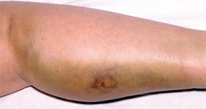
The Evidence
The 2008 ACCP guidelines on VTE management follow a grading system that classifies recommendations as Grade 1 (strong) or Grade 2 (weak), and classifies the quality of evidence as A (high), B (moderate), or C (low).12 The ACCP guidelines’ recommended first-line treatment for a confirmed DVT is anticoagulation with subcutaneous low-molecular-weight heparin, intravenous unfractionated heparin, monitored subcutaneous heparin, fixed-dose subcutaneous unfractionated heparin, or subcutaneous fondaparinux (all Grade 1A recommendations). The ACCP recommends against the routine use of an IVC filter in addition to anticoagulants (Grade 1A). However, for patients with acute proximal DVT, if anticoagulant therapy is not possible because of the risk of bleeding, IVC filter placement is recommended (Grade 1C). If a patient requires an IVC filter for treatment of an acute DVT as an alternative to anticoagulation, it is recommended to start anticoagulant therapy once the risk of bleeding resolves (Grade 1C).4
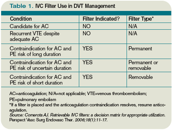
The 2008 ACCP guidelines for IVC filter use have a few important changes from the 2004 version. First, the IVC filter placement recommendation for patients with contraindications to anticoagulation was strengthened from Grade 2C to Grade 1C. Second, the 2008 guidelines omitted the early recommendation of IVC filter use for recurrent VTE, despite adequate anticoagulation (Grade 2C).13
Only one randomized study has evaluated the efficacy of IVC filters. All other studies of IVC filters are retrospective or prospective case series.
The PREPIC study randomized 400 patients with proximal DVT considered to be at high risk for PE to receive either an IVC filter or no IVC filter. Additionally, patients were randomized to receive enoxaparin or unfractionated heparin as a bridge to warfarin therapy, which was continued for at least three months. The primary endpoints were recurrent DVT, PE, major bleeding, or death. The patients were followed up at day 12, two years, and then annually up to eight years following randomization.14 At day 12, there were fewer PEs in the group that received filters (OR 0.22, 95% CI, 0.05-0.90). However, at year two, there was no significant difference in PE development in the filter group compared with the no-filter group (OR 0.50, 95% CI, 0.19-1.33).
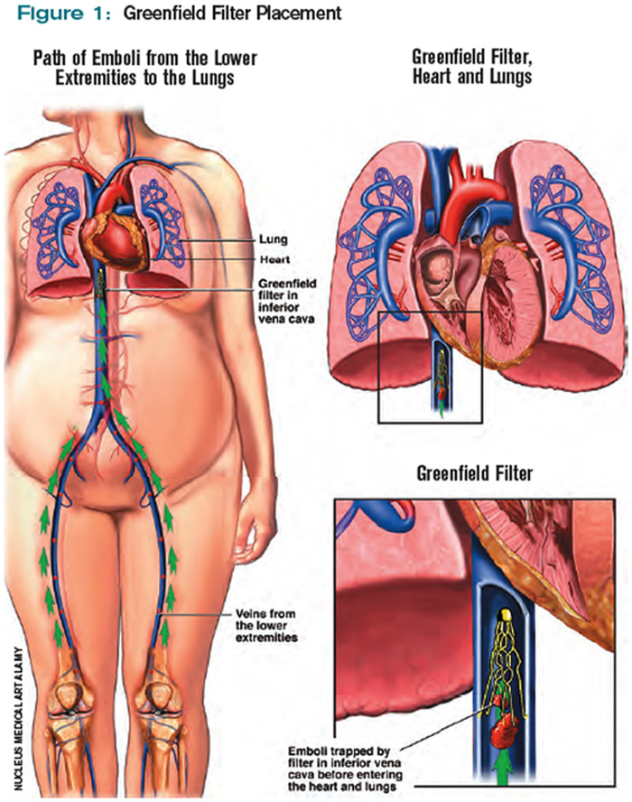
Additionally, at year two, the filter group was more likely to develop recurrent DVT (OR 1.87, 95% CI, 1.10-3.20). At year eight, there was a significant reduction in the number of PEs in the filter group versus the no-filter group (6.2% vs.15.1%, P=0.008). However, at eight-year followup, IVC filter use was associated with increased DVT (35.7% vs. 27.5%, P=0.042). There was no difference in mortality between the two groups.
In summary, the use of IVC filters was associated with decreased incidence of PE at eight years, offset by higher rates of recurrent DVT and no overall mortality benefit.14,15 Importantly, the indications for IVC filter use in this study differ from the current ACCP guidelines; all patients were given concomitant anticoagulation for at least three months, which might not be possible in patients for whom the ACCP recommends IVC filters.
There are no randomized studies to compare the efficacy of permanent IVC filters and retrievable filters for PE prevention. A retrospective study comparing the clinical effectiveness of the two filter types reported no difference in the rates of symptomatic PE (permanent filter 4% vs. retrievable filter 4.7%, P=0.67) or DVT (11.3% vs. 12.6%, P=0.59). In addition, the frequency of symptomatic IVC thrombosis was similar (1.1% vs. 0.5%, p=0.39).16 A paper reviewing the efficacy of IVC filters reported that permanent filters were associated with a 0%-6.2% rate of PE versus a 0%-1.9% rate with retrievable filters.7 Notably, these studies were not randomized controlled trials—rather, case series—and the indications for IVC filters were not necessarily those currently recommended by the ACCP.
Due to the long-term complications of permanent IVC filters, it is suggested that a retrievable IVC filter be used for patients with temporary contraindications to anticoagulation.17 Comerata et al created a clinical decision-making tool for picking the type of filter to employ. If the duration of contraindication to anticoagulation is short or uncertain, a retrievable filter is recommended.18 Table 1 (p. 15) outlines the recommendations for IVC filter placement.

There are no randomized controlled trials to guide the use of concomitant anticoagulation after filter insertion, although this intervention may be beneficial to prevent DVT propagation, recurrence, or IVC filter thrombosis.5 A meta-analysis of 14 studies evaluating the rates of VTE after IVC filter placement demonstrated a non-statistically significant trend toward fewer VTE events in the patients with an IVC filter and concomitant anticoagulation in comparison with those who solely had an IVC filter (OR 0.64, 95% CI, 0.35-1.2). The duration and degree of anticoagulation was not presented in all of the studies in the meta-analysis, therefore limiting the analysis.19
In addition to the ACCP guidelines, there have been other proposed indications for IVC filter use, including recurrent VTE despite anticoagulation, chronic recurrent PE with pulmonary hypertension, extensive free-floating iliofemoral thrombus, and thrombolysis of ilio-caval thrombus.20 The ACCP guidelines do not specifically address these individual indications, and at this time there are no randomized controlled trials to guide IVC filter use in these cases.
Back to the Case
Our patient developed a significant complication from anticoagulation. Current ACCP guidelines recommend an IVC filter if anticoagulant therapy is contraindicated (Grade 1C). The anticoagulation was discontinued and a retrievable IVC filter was placed. Once a patient no longer has a contraindication for anticoagulation, the ACCP recommends restarting a conventional course of anticoagulation. Thus, once the patient can tolerate anticoagulation, consideration will be given to removal of the retrievable filter.
Bottom Line
An IVC filter should be considered in patients with a DVT who have a contraindication to anticoagulation. Other indications for IVC filter use are not supported by the current literature. TH
Drs. Bhogal and Eid are hospitalist fellows and instructors at Johns Hopkins Bayview Medical Center in Baltimore. Dr. Kantsiper is a hospitalist and assistant professor at Bayview Medical Center.
References
- The Surgeon General’s Call to Action to Prevent Deep Vein Thrombosis and Pulmonary Embolism. U.S. Department of Health & Human Services Web site. Available at: www.surgeongeneral.gov/topics/deepvein/. Accessed Jan. 25, 2010.
- Moser KM, Fedullo PR, LitteJohn JK, Crawford R. Frequent asymptomatic pulmonary embolism in patients with deep venous thrombosis. JAMA. 1994;271(3):223-225.
- Bates SM, Ginsberg JS. Treatment of deep vein thrombosis. N Engl J Med. 2004;351:268-277.
- Kearon C, Kahn SR, Agnelli G, Goldhaber S, Raskob GE, Comerota AJ, American College of Chest Physicians. Antithrombotic therapy for venous theomboembolic disease: American College of Chest Physicians Evidence-Based Clinical Practice Guidelines (8th Edition). Chest. 2008;133(6 Suppl):454S-545S.
- Becker DM, Philbrick JT, Selby JB. Inferior vena cava filters. Indications, safety, effectiveness. Arch Intern Med. 1992;152(10):1985-1994.
- Streiff MB. Vena caval filters: a comprehensive review. Blood. 2000;95(12):3669-3677.
- Chung J, Owen RJ. Using inferior vena cava filters to prevent pulmonary embolism. Can Fam Physician. 2008;54(1):49-55.
- Ku GH. Billett HH. Long lives, short indications. The case for removable inferior cava filters. Thromb Haemost. 2005;93(1):17-22.
- Stavropoulos WS. Inferior vena cava filters. Tech Vasc Interv Radiol. 2004;7(2):91-95.
- Crowther MA. Inferior vena cava filters in the management of venous thromboembolism. Am J Med. 2007;120(10 Suppl 2):S13–S17.
- Stein PD, Kayali F, Olson RE. Twenty-one-year trends in the use of inferior vena cava filters. Arch Intern Med. 2004;164(14):1541-1545.
- Guyatt G, Gutterman D, Baumann MH, et al. Grading strength of recommendations and quality of evidence in clinical guidelines: report from an American College of Chest Physicians task force. Chest. 2006;129(1):174-181.
- Büller HR, Agnelli G, Hull RD, Hyers TM, Prins MH, Raskob GE. Antithrombotic therapy for venous thromboembolic disease: the Seventh ACCP Conference on Antithrombotic and Thrombolytic Therapy. Chest. 2004;126(3 Suppl):401S-428S.
- Decousus H, Leizorovicz A, Parent F, et al. A clinical trial of vena caval filters in the prevention of pulmonary embolism in patients with proximal deep-vein thrombosis. Prévention du Risque d’Embolie Pulmonaire par Interruption Cave Study Group. N Engl J Med. 1998;338(7):409-415.
- Decousus H, Barral F, Buchmuller-Cordier A, et al. Participating centers eight-year follow-up of patients with permanent vena cava filters in the prevention of pulmonary embolism: the PREPIC randomization croup. Circulation. 2005;112:416-422.
- Kim HS, Young MJ, Narayan AK, Liddell RP, Streiff MB. A comparison of clinical outcomes with retrievable and permanent inferior vena cava filters. J Vasc Interv Radiol. 2008:19(3):393-399.
- Houman Fekrazad M, Lopes RD, Stashenko GJ, Alexander JH, Garcia D. Treatment of venous thromboembolism: guidelines translated for the clinician. J Thromb Thrombolysis. 2009; 28(3):270–275.
- Comerota AJ. Retrievable IVC filters: a decision matrix for appropriate utilization. Perspect Vasc Surg Endovasc Ther. 2006;18(1):11-17.
- Ray CE Jr, Prochazka A. The need for anticoagulation following inferior vena cava filter placement: systematic review. Cardiovasc Intervent Radiol. 2008; 31(2):316-324.
- Hajduk B, Tomkowski WZ, Malek G, Davidson BL. Vena cava filter occlusion and venous thromboembolism risk in persistently anticoagulated patients: A prospective, observational cohort study. Chest. 2009.
Case
A 67-year-old man with a history of hypertension presents with a swollen right lower extremity. An ultrasound reveals a DVT, and he is commenced on low-molecular-weight heparin and warfarin. Two days later, he develops slurred speech and right-sided weakness. A head CT reveals an intracranial hemorrhage. When should an inferior vena cava (IVC) filter be utilized for treatment of DVT?
Overview
It is estimated that 350,000 to 600,000 Americans develop a VTE each year.1 Patients with a DVT are at high risk of developing a pulmonary embolism (PE). In a multicenter study, nearly 40% of patients admitted with a DVT had evidence of a PE on ventilation perfusion scan.2 Treatment of a DVT is aimed at preventing the extension of the DVT and embolization.3 The American College of Chest Physicians (ACCP) recommends anticoagulation as the primary DVT treatment (Grade 1A).4 However, IVC filters might be considered when anticoagulation is contraindicated.
In 1868, Trousseau created the conceptual model of surgical interruption of the IVC to prevent PE. However, it wasn’t until 1959 by Bottini that the surgical interruption was successfully performed.5 The Mobin-Uddin filter was introduced in 1967 as the first mechanical IVC filter.6 IVC filters mechanically trap the DVT, preventing emboli from traveling into the pulmonary vasculature.7
There are two classes of IVC filters: permanent filters and removable filters. Removable filters include both temporary filters and retrievable filters. Temporary filters are attached to a catheter that exits the skin and therefore must be removed due to the risk of infection and embolization.7 Retrievable filters are similar in design to permanent filters but are designed to be removed. However, this must be done with caution, as neointimal hyperplasia can prevent removal or cause vessel wall damage upon removal.8
IVC filters are inserted into the vena cava percutaneously via the femoral or jugular approach under fluoroscopy or ultrasound guidance (see Figure 1, p. 16). The filters typically are placed infrarenally, unless there is an indication for a suprarenal filter (e.g., renal vein thrombosis or IVC thrombus extending above the renal veins).7 Complete IVC thrombosis is an absolute contraindication to IVC filter placement, and the relative contraindications include significant coagulopathy and bacteremia.9
The incidence of complications related to IVC filter placement is 4% to 11%. Complications include:
- Insertion-site thrombosis;
- IVC thrombosis;
- Recurrent DVT postphlebitic syndrome;
- Filter migration;
- Erosion of the filter through the vessel wall; and
- Vena caval obstruction.10
A review of the National Hospital Discharge Survey database for trends in IVC filter use in the U.S. found a dramatic increase in the use of IVC filters from 1979 to 1999—to 49,000 patients from 2,000 patients with IVC filters in place. The indications for IVC filter use vary such that it is imperative there are well-designed trials and guidelines to guide appropriate use.11
The Evidence
The 2008 ACCP guidelines on VTE management follow a grading system that classifies recommendations as Grade 1 (strong) or Grade 2 (weak), and classifies the quality of evidence as A (high), B (moderate), or C (low).12 The ACCP guidelines’ recommended first-line treatment for a confirmed DVT is anticoagulation with subcutaneous low-molecular-weight heparin, intravenous unfractionated heparin, monitored subcutaneous heparin, fixed-dose subcutaneous unfractionated heparin, or subcutaneous fondaparinux (all Grade 1A recommendations). The ACCP recommends against the routine use of an IVC filter in addition to anticoagulants (Grade 1A). However, for patients with acute proximal DVT, if anticoagulant therapy is not possible because of the risk of bleeding, IVC filter placement is recommended (Grade 1C). If a patient requires an IVC filter for treatment of an acute DVT as an alternative to anticoagulation, it is recommended to start anticoagulant therapy once the risk of bleeding resolves (Grade 1C).4
The 2008 ACCP guidelines for IVC filter use have a few important changes from the 2004 version. First, the IVC filter placement recommendation for patients with contraindications to anticoagulation was strengthened from Grade 2C to Grade 1C. Second, the 2008 guidelines omitted the early recommendation of IVC filter use for recurrent VTE, despite adequate anticoagulation (Grade 2C).13
Only one randomized study has evaluated the efficacy of IVC filters. All other studies of IVC filters are retrospective or prospective case series.
The PREPIC study randomized 400 patients with proximal DVT considered to be at high risk for PE to receive either an IVC filter or no IVC filter. Additionally, patients were randomized to receive enoxaparin or unfractionated heparin as a bridge to warfarin therapy, which was continued for at least three months. The primary endpoints were recurrent DVT, PE, major bleeding, or death. The patients were followed up at day 12, two years, and then annually up to eight years following randomization.14 At day 12, there were fewer PEs in the group that received filters (OR 0.22, 95% CI, 0.05-0.90). However, at year two, there was no significant difference in PE development in the filter group compared with the no-filter group (OR 0.50, 95% CI, 0.19-1.33).
Additionally, at year two, the filter group was more likely to develop recurrent DVT (OR 1.87, 95% CI, 1.10-3.20). At year eight, there was a significant reduction in the number of PEs in the filter group versus the no-filter group (6.2% vs.15.1%, P=0.008). However, at eight-year followup, IVC filter use was associated with increased DVT (35.7% vs. 27.5%, P=0.042). There was no difference in mortality between the two groups.
In summary, the use of IVC filters was associated with decreased incidence of PE at eight years, offset by higher rates of recurrent DVT and no overall mortality benefit.14,15 Importantly, the indications for IVC filter use in this study differ from the current ACCP guidelines; all patients were given concomitant anticoagulation for at least three months, which might not be possible in patients for whom the ACCP recommends IVC filters.
There are no randomized studies to compare the efficacy of permanent IVC filters and retrievable filters for PE prevention. A retrospective study comparing the clinical effectiveness of the two filter types reported no difference in the rates of symptomatic PE (permanent filter 4% vs. retrievable filter 4.7%, P=0.67) or DVT (11.3% vs. 12.6%, P=0.59). In addition, the frequency of symptomatic IVC thrombosis was similar (1.1% vs. 0.5%, p=0.39).16 A paper reviewing the efficacy of IVC filters reported that permanent filters were associated with a 0%-6.2% rate of PE versus a 0%-1.9% rate with retrievable filters.7 Notably, these studies were not randomized controlled trials—rather, case series—and the indications for IVC filters were not necessarily those currently recommended by the ACCP.
Due to the long-term complications of permanent IVC filters, it is suggested that a retrievable IVC filter be used for patients with temporary contraindications to anticoagulation.17 Comerata et al created a clinical decision-making tool for picking the type of filter to employ. If the duration of contraindication to anticoagulation is short or uncertain, a retrievable filter is recommended.18 Table 1 (p. 15) outlines the recommendations for IVC filter placement.
There are no randomized controlled trials to guide the use of concomitant anticoagulation after filter insertion, although this intervention may be beneficial to prevent DVT propagation, recurrence, or IVC filter thrombosis.5 A meta-analysis of 14 studies evaluating the rates of VTE after IVC filter placement demonstrated a non-statistically significant trend toward fewer VTE events in the patients with an IVC filter and concomitant anticoagulation in comparison with those who solely had an IVC filter (OR 0.64, 95% CI, 0.35-1.2). The duration and degree of anticoagulation was not presented in all of the studies in the meta-analysis, therefore limiting the analysis.19
In addition to the ACCP guidelines, there have been other proposed indications for IVC filter use, including recurrent VTE despite anticoagulation, chronic recurrent PE with pulmonary hypertension, extensive free-floating iliofemoral thrombus, and thrombolysis of ilio-caval thrombus.20 The ACCP guidelines do not specifically address these individual indications, and at this time there are no randomized controlled trials to guide IVC filter use in these cases.
Back to the Case
Our patient developed a significant complication from anticoagulation. Current ACCP guidelines recommend an IVC filter if anticoagulant therapy is contraindicated (Grade 1C). The anticoagulation was discontinued and a retrievable IVC filter was placed. Once a patient no longer has a contraindication for anticoagulation, the ACCP recommends restarting a conventional course of anticoagulation. Thus, once the patient can tolerate anticoagulation, consideration will be given to removal of the retrievable filter.
Bottom Line
An IVC filter should be considered in patients with a DVT who have a contraindication to anticoagulation. Other indications for IVC filter use are not supported by the current literature. TH
Drs. Bhogal and Eid are hospitalist fellows and instructors at Johns Hopkins Bayview Medical Center in Baltimore. Dr. Kantsiper is a hospitalist and assistant professor at Bayview Medical Center.
References
- The Surgeon General’s Call to Action to Prevent Deep Vein Thrombosis and Pulmonary Embolism. U.S. Department of Health & Human Services Web site. Available at: www.surgeongeneral.gov/topics/deepvein/. Accessed Jan. 25, 2010.
- Moser KM, Fedullo PR, LitteJohn JK, Crawford R. Frequent asymptomatic pulmonary embolism in patients with deep venous thrombosis. JAMA. 1994;271(3):223-225.
- Bates SM, Ginsberg JS. Treatment of deep vein thrombosis. N Engl J Med. 2004;351:268-277.
- Kearon C, Kahn SR, Agnelli G, Goldhaber S, Raskob GE, Comerota AJ, American College of Chest Physicians. Antithrombotic therapy for venous theomboembolic disease: American College of Chest Physicians Evidence-Based Clinical Practice Guidelines (8th Edition). Chest. 2008;133(6 Suppl):454S-545S.
- Becker DM, Philbrick JT, Selby JB. Inferior vena cava filters. Indications, safety, effectiveness. Arch Intern Med. 1992;152(10):1985-1994.
- Streiff MB. Vena caval filters: a comprehensive review. Blood. 2000;95(12):3669-3677.
- Chung J, Owen RJ. Using inferior vena cava filters to prevent pulmonary embolism. Can Fam Physician. 2008;54(1):49-55.
- Ku GH. Billett HH. Long lives, short indications. The case for removable inferior cava filters. Thromb Haemost. 2005;93(1):17-22.
- Stavropoulos WS. Inferior vena cava filters. Tech Vasc Interv Radiol. 2004;7(2):91-95.
- Crowther MA. Inferior vena cava filters in the management of venous thromboembolism. Am J Med. 2007;120(10 Suppl 2):S13–S17.
- Stein PD, Kayali F, Olson RE. Twenty-one-year trends in the use of inferior vena cava filters. Arch Intern Med. 2004;164(14):1541-1545.
- Guyatt G, Gutterman D, Baumann MH, et al. Grading strength of recommendations and quality of evidence in clinical guidelines: report from an American College of Chest Physicians task force. Chest. 2006;129(1):174-181.
- Büller HR, Agnelli G, Hull RD, Hyers TM, Prins MH, Raskob GE. Antithrombotic therapy for venous thromboembolic disease: the Seventh ACCP Conference on Antithrombotic and Thrombolytic Therapy. Chest. 2004;126(3 Suppl):401S-428S.
- Decousus H, Leizorovicz A, Parent F, et al. A clinical trial of vena caval filters in the prevention of pulmonary embolism in patients with proximal deep-vein thrombosis. Prévention du Risque d’Embolie Pulmonaire par Interruption Cave Study Group. N Engl J Med. 1998;338(7):409-415.
- Decousus H, Barral F, Buchmuller-Cordier A, et al. Participating centers eight-year follow-up of patients with permanent vena cava filters in the prevention of pulmonary embolism: the PREPIC randomization croup. Circulation. 2005;112:416-422.
- Kim HS, Young MJ, Narayan AK, Liddell RP, Streiff MB. A comparison of clinical outcomes with retrievable and permanent inferior vena cava filters. J Vasc Interv Radiol. 2008:19(3):393-399.
- Houman Fekrazad M, Lopes RD, Stashenko GJ, Alexander JH, Garcia D. Treatment of venous thromboembolism: guidelines translated for the clinician. J Thromb Thrombolysis. 2009; 28(3):270–275.
- Comerota AJ. Retrievable IVC filters: a decision matrix for appropriate utilization. Perspect Vasc Surg Endovasc Ther. 2006;18(1):11-17.
- Ray CE Jr, Prochazka A. The need for anticoagulation following inferior vena cava filter placement: systematic review. Cardiovasc Intervent Radiol. 2008; 31(2):316-324.
- Hajduk B, Tomkowski WZ, Malek G, Davidson BL. Vena cava filter occlusion and venous thromboembolism risk in persistently anticoagulated patients: A prospective, observational cohort study. Chest. 2009.
A Time to Be Recognized
Like so many things in HM, the story of how hospitalists first learned about the focused practice program is a modern one.
It started with a text message, which led to a blog post, which reached thousands of readers, many of them hospitalists interested in how to bolster their bona fides in a specialty known for its explosive growth in recent years.
Now, hospitalists certified in internal medicine have the opportunity to reinforce their commitment to the specialty by maintaining their certification through the Focused Practice in Hospital Medicine pathway offered by the American Board of Internal Medicine (ABIM). The Focused Practice in Hospital Medicine (FPHM) Maintenance of Certification (MOC) program enables hospitalists to distinguish their practice within the larger specialty of internal medicine.
The Evolution of FPHM
The new pathway has been years in the making, and it reflects the growing influence of HM in healthcare, according to ABIM Chief Medical Officer Eric Holmboe, MD. He sees the FPHM as the result of a combination of factors, including the fact that the specialty now has more than 30,000 hospitalists practicing nationwide. “If you look at the past years, this has been a viable and vibrant practice,” he says. “If you look at the number of people doing hospital medicine, it’s a factor.”
For Holmboe, it also is a shift in how individuals are recognized based on their practice areas. “This is an acknowledgement by ABIM and the American Board of Medical Specialties to look at Maintenance of Certification in terms of what the individual actually does,” he explains. “Hospitalists play a very important role in the hospital.”
He also credits the leadership of the HM movement—especially pioneers like Robert Wachter, MD, FHM. One of HM’s most ardent champions, Dr. Wachter, chief of the hospital medicine division, professor, and associate chair of the Department of Medicine at the University of California at San Francisco, worked with ABIM to find a way to recognize hospitalists’ specialized skill sets and their commitment to inpatient medicine. After more than a decade of advocating for a board-certified process to recognize the field, Dr. Wachter, an ABIM board member, began receiving multiple text messages from colleagues announcing that ABIM had approved the focused-practice program. He wrote a post on his blog, Wachter’s World (www.wachtersworld.com), that outlined the need for the FPHM and the significance for aspiring hospitalists.
“In any case, this is an important milestone for the field,” Dr. Wachter wrote in his Sept. 23, 2009, blog entry, “Board Certification for Hospitalists: It’s Heeeere!” “In fact, when I first began speaking to groups of hospitalists nearly 15 years ago, I often showed a slide listing the elements of a true specialty, and one by one we’ve ticked them off,” wrote Dr. Wachter, a former SHM president. “The only unchecked box was recognition of the field as a legitimate ‘specialty,’ as codified by the ABMS board certification process.”
Unchecked, that is, until now.
In early 2011, the medical world will be introduced to the first internists recognized for their focus in HM. For Holmboe, the FPHM is the beginning of an even larger movement.
“The goal is continued interest: getting people involved in quality in their hospital and encouraging people to change behaviors and be recognized by patients and credentialists as valuable,” he says. “That’s the primary mission of ABIM: using certification to improve care.”
Fellow in Hospital Medicine Spotlight
O’Neil Pyke, MD, FHM

Dr. Pyke is a clinical instructor at Commonwealth Medical College and a medical director at the Wyoming Valley Health Care System in Wilkes-Barre, Pa. He also serves as a consultant for various hospitalist programs, most actively for his own private consulting company, AMP Hospitalist Consulting, which partners with Salem, N.H.-based physician staffing company Medicus Healthcare Solutions.
Undergraduate Education: Queens College, City University of New York, Flushing, N.Y.
Medical School: Ohio State University College of Medicine and Public Health, Columbus
Notable: Dr. Pyke was born in Jamaica and moved to New York during high school. He says he owes everything to his parents. His parents, who had no education beyond high school, pushed Dr. Pyke and his siblings to achieve more than they did. His sister is an OB-GYN and his brother is pursuing a medical degree.
FYI: Dr. Pyke enjoys playing golf, cheering for his beloved Ohio State Buckeyes, and spends every Friday night with his wife and two daughters—he even admits to watching “chick flicks” on family night.
For more information about the FHM designation, visit www.hospitalmedicine.org/fellows.
Requirements and Process
Shortly after the program’s approval, ABIM, which administers the FPHM program, went to work in defining the process for the FPHM application and building infrastructure to support the tests. Holmboe expects ABIM will be ready to process pre-applications by April or May. While some details may change, the FPHM application will dovetail with ABIM’s MOC process.
Although hospitalists’ MOC must be current in order to apply for FPHM, hospitalists can begin the FPHM application process at any time. Hospitalists do not need to wait until their next MOC renewal.
Before beginning the application process, hospitalists should ensure that they are eligible. ABIM requires FPHM candidates to have:
- A current or previous ABIM certification in internal medicine;
- A valid, unrestricted medical license and confirmation of good standing in the local practice community;
- ACLS certification; and
- At least three years of hospital medicine practice experience.
Candidates who meet the requirements can then begin the enrollment process by:
- Submitting attestations. Both the hospitalist and a senior officer at the hospital must provide attestations that demonstrate the hospitalist’s experience in HM and his or her commitment to the principles of the specialty.
- Performing a self-assessment. Hospitalists must quantify their experience in HM through an MOC self-assessment. Candidates must achieve at least 100 MOC points. Successful applicants must submit a new self-assessment every three years. The self-assessment can be conducted before or after the exam.
- Taking the MOC examination in Hospital Medicine. Registration for the first HM examination will begin in May. The exam will be conducted in October, and diplomates can take the exam at any time in the process.
Passing the exam and completing the other requirements will earn ABIM diplomats recognition as “Board Certified in Internal Medicine with a Focused Practice in Hospital Medicine.” ABIM will notify successful applicants in late 2010 and ship personalized certificates in early 2011. TH
Brendon Shank is a freelance writer based in Philadelphia.
Like so many things in HM, the story of how hospitalists first learned about the focused practice program is a modern one.
It started with a text message, which led to a blog post, which reached thousands of readers, many of them hospitalists interested in how to bolster their bona fides in a specialty known for its explosive growth in recent years.
Now, hospitalists certified in internal medicine have the opportunity to reinforce their commitment to the specialty by maintaining their certification through the Focused Practice in Hospital Medicine pathway offered by the American Board of Internal Medicine (ABIM). The Focused Practice in Hospital Medicine (FPHM) Maintenance of Certification (MOC) program enables hospitalists to distinguish their practice within the larger specialty of internal medicine.
The Evolution of FPHM
The new pathway has been years in the making, and it reflects the growing influence of HM in healthcare, according to ABIM Chief Medical Officer Eric Holmboe, MD. He sees the FPHM as the result of a combination of factors, including the fact that the specialty now has more than 30,000 hospitalists practicing nationwide. “If you look at the past years, this has been a viable and vibrant practice,” he says. “If you look at the number of people doing hospital medicine, it’s a factor.”
For Holmboe, it also is a shift in how individuals are recognized based on their practice areas. “This is an acknowledgement by ABIM and the American Board of Medical Specialties to look at Maintenance of Certification in terms of what the individual actually does,” he explains. “Hospitalists play a very important role in the hospital.”
He also credits the leadership of the HM movement—especially pioneers like Robert Wachter, MD, FHM. One of HM’s most ardent champions, Dr. Wachter, chief of the hospital medicine division, professor, and associate chair of the Department of Medicine at the University of California at San Francisco, worked with ABIM to find a way to recognize hospitalists’ specialized skill sets and their commitment to inpatient medicine. After more than a decade of advocating for a board-certified process to recognize the field, Dr. Wachter, an ABIM board member, began receiving multiple text messages from colleagues announcing that ABIM had approved the focused-practice program. He wrote a post on his blog, Wachter’s World (www.wachtersworld.com), that outlined the need for the FPHM and the significance for aspiring hospitalists.
“In any case, this is an important milestone for the field,” Dr. Wachter wrote in his Sept. 23, 2009, blog entry, “Board Certification for Hospitalists: It’s Heeeere!” “In fact, when I first began speaking to groups of hospitalists nearly 15 years ago, I often showed a slide listing the elements of a true specialty, and one by one we’ve ticked them off,” wrote Dr. Wachter, a former SHM president. “The only unchecked box was recognition of the field as a legitimate ‘specialty,’ as codified by the ABMS board certification process.”
Unchecked, that is, until now.
In early 2011, the medical world will be introduced to the first internists recognized for their focus in HM. For Holmboe, the FPHM is the beginning of an even larger movement.
“The goal is continued interest: getting people involved in quality in their hospital and encouraging people to change behaviors and be recognized by patients and credentialists as valuable,” he says. “That’s the primary mission of ABIM: using certification to improve care.”
Fellow in Hospital Medicine Spotlight
O’Neil Pyke, MD, FHM
Dr. Pyke is a clinical instructor at Commonwealth Medical College and a medical director at the Wyoming Valley Health Care System in Wilkes-Barre, Pa. He also serves as a consultant for various hospitalist programs, most actively for his own private consulting company, AMP Hospitalist Consulting, which partners with Salem, N.H.-based physician staffing company Medicus Healthcare Solutions.
Undergraduate Education: Queens College, City University of New York, Flushing, N.Y.
Medical School: Ohio State University College of Medicine and Public Health, Columbus
Notable: Dr. Pyke was born in Jamaica and moved to New York during high school. He says he owes everything to his parents. His parents, who had no education beyond high school, pushed Dr. Pyke and his siblings to achieve more than they did. His sister is an OB-GYN and his brother is pursuing a medical degree.
FYI: Dr. Pyke enjoys playing golf, cheering for his beloved Ohio State Buckeyes, and spends every Friday night with his wife and two daughters—he even admits to watching “chick flicks” on family night.
For more information about the FHM designation, visit www.hospitalmedicine.org/fellows.
Requirements and Process
Shortly after the program’s approval, ABIM, which administers the FPHM program, went to work in defining the process for the FPHM application and building infrastructure to support the tests. Holmboe expects ABIM will be ready to process pre-applications by April or May. While some details may change, the FPHM application will dovetail with ABIM’s MOC process.
Although hospitalists’ MOC must be current in order to apply for FPHM, hospitalists can begin the FPHM application process at any time. Hospitalists do not need to wait until their next MOC renewal.
Before beginning the application process, hospitalists should ensure that they are eligible. ABIM requires FPHM candidates to have:
- A current or previous ABIM certification in internal medicine;
- A valid, unrestricted medical license and confirmation of good standing in the local practice community;
- ACLS certification; and
- At least three years of hospital medicine practice experience.
Candidates who meet the requirements can then begin the enrollment process by:
- Submitting attestations. Both the hospitalist and a senior officer at the hospital must provide attestations that demonstrate the hospitalist’s experience in HM and his or her commitment to the principles of the specialty.
- Performing a self-assessment. Hospitalists must quantify their experience in HM through an MOC self-assessment. Candidates must achieve at least 100 MOC points. Successful applicants must submit a new self-assessment every three years. The self-assessment can be conducted before or after the exam.
- Taking the MOC examination in Hospital Medicine. Registration for the first HM examination will begin in May. The exam will be conducted in October, and diplomates can take the exam at any time in the process.
Passing the exam and completing the other requirements will earn ABIM diplomats recognition as “Board Certified in Internal Medicine with a Focused Practice in Hospital Medicine.” ABIM will notify successful applicants in late 2010 and ship personalized certificates in early 2011. TH
Brendon Shank is a freelance writer based in Philadelphia.
Group Leaders Can Shift the HM Negotiation Paradigm
Whether hospitalists like it or not, the art of negotiation has a significant impact on their daily activities. Negotiations take place with consultants over what the perceived optimal plan of care should be. Discussions are held with patients on how best to overcome the social, financial, and psychological barriers that may impede their health. Hospitalists negotiate with administrators over schedules, benefits, and responsibilities.
Quite frequently, negotiation is viewed as a process where one party wins and the other loses, a zero-sum game, like chess. The spoils may be financial (e.g., better reimbursements) or they may be cognitive (e.g., success in convincing someone of your particular viewpoint). Significant value that could potentially benefit both parties may be lost if the negotiation is approached with a win-loss mentality. However, with proper preparation and insight, a hospitalist can create value in a negotiation that otherwise may be lost by shifting their negotiation paradigm to a collaborative strategy.
A collaborative strategy is when the relationship—and not just the outcome—is important. This would apply to most negotiations that hospitalists take part in.
A significant part of this strategy involves listening and allowing the other side to divulge their interests and positions. Information must flow freely. Once the problem is identified, it must then be detailed further, ensuring both parties understand each other.
Only once both party’s issues are presented can an alternative solution be contemplated that will be win-win in nature. The parties then must both agree to choose that solution and move forward.
The optimal result is that the chosen solution appeases both parties and has a greater total value than if both sides were solely vying for their own interests.
Riyad Fares, MD,
hospitalist,
Adventist Hospital, Portland, Ore.
Malpractice Chronicle
Reprinted with permission from Medical Malpractice Verdicts, Settlements and Experts, Lewis Laska, Editor, (800) 298-6288.
Was Surgery to Save Patient—or to Teach Residents?
A laparoscopic hysterectomy was performed on a 48-year-old woman. After the procedure, when the patient’s pain was disproportionate to what might be expected, it was discovered that the bowel had been perforated. She was transferred to another facility, where it was initially assumed that she would not survive.
During subsequent surgeries, almost all of the patient’s large and small intestines were removed. Although she has survived, she currently requires total parenteral nutrition 16 hours a day and is on a waiting list for transplantation of the intestines, stomach, and liver. The patient has been hospitalized 25 times since the initial event for dialysis, blood transfusions, and treatment of infections.
The plaintiff claimed that her survival was unexpected and that the physician at the receiving hospital performed surgery on the bowel as a teaching tool for his residents. The possibility of transplantation surgery was a factor in reaching a settlement, as there were concerns that a transplant could affect the plaintiff’s survival and her ability to participate at trial. Also in the settlement considerations was the plaintiff’s having gone on a public health insurance plan, which would have created a large lien in the event of a transplant.
A $2,975,000 settlement was reached.
Internal Bleeding Goes Undetected After Car Accident
A man who fell asleep while driving was involved in a rollover accident and was rushed to the emergency department (ED) of the defendant hospital. He was treated in the ED by Dr. R. for cuts and bruises. He had bruising on his stomach and was complaining of pain that he described as 10 on a pain scale of 10; he said he could not lie flat because of the pain. He expressed concern to Dr. R. that his accident might have dislodged a pain pump previously implanted in his stomach to relieve chronic back pain.
X-rays were taken, but neither CT nor MRI was ordered. The man was released to home.
His wife found him the next morning lying on the bathroom floor, barely alive. He was rushed back to the hospital but died shortly after his arrival there. Based on findings at autopsy, which included fractured ribs and a collapsed lung, the man’s death was attributed to internal bleeding.
Plaintiff for the decedent alleged negligence in a failure to perform the proper testing to diagnose the extent of the decedent’s injuries.
A confidential settlement was reached.
Recognized Complication of Epidural Anesthesia
A man complaining of abdominal pain was given a diagnosis of an inguinal hernia and was referred to the defendant surgeon for a surgical repair the following month. The surgeon decided to use an epidural anesthetic in order to test the strength of the repair at the conclusion of the surgery.
The patient reportedly became unresponsive during the surgery, but the procedure was continued. The patient awoke in the postoperative area with numbness and a burning sensation in both legs. He was also unable to urinate.
The man’s numbness and inability to urinate persisted after his discharge. He later presented to an ED, where he underwent catheterization for urinary retention. He was sent for a neurologic consult and was subsequently treated at a research center.
The plaintiff claimed that the defendant surgeon had chosen epidural anesthesia without obtaining his informed consent and against his wishes. He also claimed that the defendant anesthesiologist stated to the patient’s family that he might have inserted the epidural needle too far, thus introducing the anesthetic to an inappropriate area. Additionally, the plaintiff claimed he had been told that something had struck his spinal cord during the epidural. The plaintiff charged the defendant anesthesiologist with negligence in piercing the dura and injuring the spinal cord. The litigation ultimately went forward against the anesthesiologist alone.
The plaintiff claimed that the injection of an excessive dose of anesthetic beyond the dura caused weakness in the legs, as well as neurogenic bowel and bladder.
The defendant claimed that the anesthetic intended for the epidural space spread to the subarachnoid space, causing a neurotoxic reaction that damaged the spinal cord. The defendant maintained that this was a recognized complication and not the result of any negligence. The defendant also claimed that the plaintiff had been told of the potential risks of the epidural.
According to a published account, a defense verdict was returned.
Severe Abdominal Pain After Tubal Ligation
A woman, 28, underwent tubal ligation and was released the same day. She returned to the hospital’s ED that night complaining of severe abdominal pain. She was seen by an ED physician, Dr. M., who ordered oxycodone with acetaminophen but did not specify the dosage; the woman was given between 5 and 10 mg. When her pain persisted, Dr. M. ordered hydromorphone and promethazine, to be administered by injection.
Dr. M. saw her an hour later. At that time, she had minimal abdominal distention and normal vital signs with no tenderness or guarding. Dr. M. made a diagnosis of postoperative pain, and the patient was soon discharged.
She returned to the ED the next day with continuing complaints of severe abdominal pain. She was seen by another physician and admitted for emergency abdominal surgery, during which a perforated small bowel was discovered, complicated by peritonitis. The patient developed pulmonary insufficiency and renal failure resulting from sepsis. She remained on life support for one month until her death, attributed to multiorgan failure and septic shock.
The plaintiff claimed that Dr. M. should have obtained laboratory and radiology studies and a surgical consult. The plaintiff further claimed that the medications administered masked the decedent’s abdominal pain.
The defendants claimed that the decedent did not have a “through-and-through” perforation when she initially presented to the ED, and that pain medication does not mask a bowel perforation; the defendants claimed that the perforation occurred later.
A defense verdict was returned, with the plaintiff receiving an undisclosed amount under the terms of a high/low agreement.
Sterilized woman skips f/u HSG test, becomes pregnant…and more
Sterilized woman skips f/u HSG test, becomes pregnant
A 31-YEAR-OLD MOTHER OF THREE underwent a hysteroscopic tubal sterilization procedure because she wanted to avoid pregnancy. She neglected to return 3 months later for a hysterosalpingogram (HSG) to determine whether the procedure was effective. A few months after that, she discovered she was pregnant.
PATIENT’S CLAIM The physician was negligent for failing to inform her that postoperative testing was necessary, and importantly, to confirm her inability to become pregnant. He also failed to advise her to use alternative contraceptive methods.
PHYSICIAN’S DEFENSE The patient was told several times that the HSG test was needed and important, and that she should use alternative contraception. Also, five appointments were scheduled for her to have the HSG test, but each time she either canceled or failed to come for the test.
VERDICT Connecticut defense verdict.
Preeclampsia leads to infant’s death and 24/7 care for mother
A WOMAN AT 30 WEEKS’ GESTATION complained of headaches to her ObGyn during a routine examination. Her blood pressure, although within normal limits, was above her baseline level, and a dipstick urine demonstrated proteinuria. The next morning, she had a seizure and was taken to the hospital. Preeclampsia was diagnosed. An emergent cesarean delivery was performed. The infant, who suffered neurologic impairments, died at 18 months. The mother was hospitalized for 7 months due to hypoxia-induced neurologic injuries, sepsis, lung problems, a ministroke, and a second hypoxic episode. Wheelchair-bound, she is speech-impaired and requires 24-hour care.
PATIENT’S CLAIM The ObGyn’s failure to diagnose preeclampsia was negligent.
PHYSICIAN’S DEFENSE The patient showed no signs of preeclampsia. Despite protein in her urine and elevated blood pressure, the levels were not in a range indicating preeclampsia.
VERDICT South Carolina defense verdict.
Did emboli in utero from deceased twin cause other twin’s postnatal problems?
A WOMAN PREGNANT WITH TWINS was admitted to the hospital because of premature contractions. Tests performed 3 weeks later indicated the intrauterine demise of one twin. The other twin showed signs of distress 8 days later and was delivered by cesarean later that day. The infant experienced hypoxic-ischemic brain damage, which caused severe mental retardation, cerebral palsy, and spastic quadriparesis.
PATIENT’S CLAIM Prior to delivery, emboli of thromboplastin material from the deceased fetus traveled to the surviving fetus, contributing to that fetus’s injuries. The delivery should have been performed sooner.
PHYSICIAN’S DEFENSE The plaintiff’s proposed theory for the injuries was not supported scientifically.
VERDICT All defendants except the hospital settled for a total of $1.5 million prior to trial. A $4 million Florida verdict against the hospital for injuries to the surviving twin was reduced to $2.95 million.
Was excessive force applied in case of shoulder dystocia?
THE OBGYN WHO HAD PROVIDED ALL PRENATAL CARE encountered shoulder dystocia during the infant’s delivery. The problem was resolved with the McRoberts maneuver with suprapubic pressure, the Wood’s screw maneuver, and attempted delivery of the posterior arm. Born with a severe left brachial plexus injury, the child underwent surgery but still has limited use of his left arm and hand.
PATIENT’S CLAIM Among relatives present at the delivery were two nurses. They testified at the trial that the physician used excessive downward lateral traction and also allowed a nurse to apply fundal pressure.
PHYSICIAN’S DEFENSE Shoulder dystocia was unexpected, excessive traction was not used, and once dystocia was evident, fundal pressure was not used.
VERDICT $80,000 Illinois settlement with the hospital prior to trial. Although the jury delivered a defense verdict for the physician, a $1,000,000/$200,000 high/low agreement was in place.
These cases were selected by the editors of OBG Management from Medical Malpractice Verdicts, Settlements & Experts, with permission of the editor, Lewis Laska (www.verdictslaska.com). The information available to the editors about the cases presented here is sometimes incomplete. Moreover, the cases may or may not have merit. Nevertheless, these cases represent the types of clinical situations that typically result in litigation and are meant to illustrate nationwide variation in jury verdicts and awards.
Sterilized woman skips f/u HSG test, becomes pregnant
A 31-YEAR-OLD MOTHER OF THREE underwent a hysteroscopic tubal sterilization procedure because she wanted to avoid pregnancy. She neglected to return 3 months later for a hysterosalpingogram (HSG) to determine whether the procedure was effective. A few months after that, she discovered she was pregnant.
PATIENT’S CLAIM The physician was negligent for failing to inform her that postoperative testing was necessary, and importantly, to confirm her inability to become pregnant. He also failed to advise her to use alternative contraceptive methods.
PHYSICIAN’S DEFENSE The patient was told several times that the HSG test was needed and important, and that she should use alternative contraception. Also, five appointments were scheduled for her to have the HSG test, but each time she either canceled or failed to come for the test.
VERDICT Connecticut defense verdict.
Preeclampsia leads to infant’s death and 24/7 care for mother
A WOMAN AT 30 WEEKS’ GESTATION complained of headaches to her ObGyn during a routine examination. Her blood pressure, although within normal limits, was above her baseline level, and a dipstick urine demonstrated proteinuria. The next morning, she had a seizure and was taken to the hospital. Preeclampsia was diagnosed. An emergent cesarean delivery was performed. The infant, who suffered neurologic impairments, died at 18 months. The mother was hospitalized for 7 months due to hypoxia-induced neurologic injuries, sepsis, lung problems, a ministroke, and a second hypoxic episode. Wheelchair-bound, she is speech-impaired and requires 24-hour care.
PATIENT’S CLAIM The ObGyn’s failure to diagnose preeclampsia was negligent.
PHYSICIAN’S DEFENSE The patient showed no signs of preeclampsia. Despite protein in her urine and elevated blood pressure, the levels were not in a range indicating preeclampsia.
VERDICT South Carolina defense verdict.
Did emboli in utero from deceased twin cause other twin’s postnatal problems?
A WOMAN PREGNANT WITH TWINS was admitted to the hospital because of premature contractions. Tests performed 3 weeks later indicated the intrauterine demise of one twin. The other twin showed signs of distress 8 days later and was delivered by cesarean later that day. The infant experienced hypoxic-ischemic brain damage, which caused severe mental retardation, cerebral palsy, and spastic quadriparesis.
PATIENT’S CLAIM Prior to delivery, emboli of thromboplastin material from the deceased fetus traveled to the surviving fetus, contributing to that fetus’s injuries. The delivery should have been performed sooner.
PHYSICIAN’S DEFENSE The plaintiff’s proposed theory for the injuries was not supported scientifically.
VERDICT All defendants except the hospital settled for a total of $1.5 million prior to trial. A $4 million Florida verdict against the hospital for injuries to the surviving twin was reduced to $2.95 million.
Was excessive force applied in case of shoulder dystocia?
THE OBGYN WHO HAD PROVIDED ALL PRENATAL CARE encountered shoulder dystocia during the infant’s delivery. The problem was resolved with the McRoberts maneuver with suprapubic pressure, the Wood’s screw maneuver, and attempted delivery of the posterior arm. Born with a severe left brachial plexus injury, the child underwent surgery but still has limited use of his left arm and hand.
PATIENT’S CLAIM Among relatives present at the delivery were two nurses. They testified at the trial that the physician used excessive downward lateral traction and also allowed a nurse to apply fundal pressure.
PHYSICIAN’S DEFENSE Shoulder dystocia was unexpected, excessive traction was not used, and once dystocia was evident, fundal pressure was not used.
VERDICT $80,000 Illinois settlement with the hospital prior to trial. Although the jury delivered a defense verdict for the physician, a $1,000,000/$200,000 high/low agreement was in place.
These cases were selected by the editors of OBG Management from Medical Malpractice Verdicts, Settlements & Experts, with permission of the editor, Lewis Laska (www.verdictslaska.com). The information available to the editors about the cases presented here is sometimes incomplete. Moreover, the cases may or may not have merit. Nevertheless, these cases represent the types of clinical situations that typically result in litigation and are meant to illustrate nationwide variation in jury verdicts and awards.
These cases were selected by the editors of OBG Management from Medical Malpractice Verdicts, Settlements & Experts, with permission of the editor, Lewis Laska (www.verdictslaska.com). The information available to the editors about the cases presented here is sometimes incomplete. Moreover, the cases may or may not have merit. Nevertheless, these cases represent the types of clinical situations that typically result in litigation and are meant to illustrate nationwide variation in jury verdicts and awards.