User login
VA MRSA Prevention Initiative reports continued health care–associated infection declines
The U.S. Department of Veterans Affairs MRSA Prevention Initiative, implemented in October 2007, has shown progress at limiting health care–associated infections of methicillin-resistant Staphylococcus aureus through 2011 and 2012.
A new report published in the January 2017 issue of the American Journal of Infection Control tracks continued declines in infection through September 2015.
Monthly rates of health care–associated infections fell significantly in all settings from October 2007 to September 2015: an 87% decrease in ICUs, 80.1% in non-ICUs, 80.9% in spinal cord injury units, and 49.4% in long-term care facilities (P for all less than .0001).
“The VA data suggest that active surveillance followed by contact precautions (with or without decolonization) may be most useful when MRSA [health care–associated infection] rates are unacceptably high (as they were in VA facilities during 2007) or to decrease infections in high-risk units such as ICUs,” Dr. Evans and his colleagues concluded.
Details about the implementation of the initiative were previously published in the New England Journal of Medicine in 2011, including the initiative’s goal to promote “a change in the institutional culture whereby infection control would become the responsibility of everyone who had contact with patients” (N Engl J Med. 2011;364:1419-30).
Dr. Evans and his colleagues had no relevant financial disclosures.
FROM THE AMERICAN JOURNAL OF INFECTION CONTROL
Do not overtreat febrile neutropenia
Clinical question: Does emergency department management of patients with febrile neutropenia (FN) follow current guidelines?
Background: Chemotherapy-related FN is an oncologic emergency frequently leading to hospitalization and intravenous antibiotics. Familiarity with FN guidelines allows risk stratification for inpatient versus outpatient therapy.
Study design: Single-center, retrospective, cohort study.
Setting: Large, urban, tertiary-care academic hospital.
Synopsis: Of 173 patient visits, 25% were risk stratified as eligible for outpatient treatment and 75% as inpatient care. All patient care was assessed for guideline concordance at the time of ED disposition and therapy.
Primary outcome analysis demonstrated management was guideline discordant in 98% of low-risk patients versus 7% of high-risk patients. Secondary 30-day clinical outcomes showed high-risk patients were more likely to have positive blood cultures (54%), sepsis-induced hypotension (9.3%), and death (5.4%). Seventeen percent of all patients who received IV antibiotics were prescribed vancomycin without guideline support.
Bottom line: Low-risk FN patients in the ED received more aggressive treatment than recommended. Further research is needed to strategize means of better aligning FN management with standards of care.
Citation: Baugh CW, Wang TJ, Caterino JM, et al. ED management of patients with febrile neutropenia: guideline concordant or overly aggressive [published online ahead of print Sept. 9, 2016]? Acad Emerg Med. doi: 10.1111/acem.13079.
Dr. Zuleta is an assistant professor and associate program director of the Jackson Memorial/University of Miami Internal Medicine residency training program and the site director of the program at University of Miami Hospital.
Cancer risk six times higher in children with congenital heart defects
NEW ORLEANS – Children with congenital heart defects have a five- to sixfold increased risk of developing pediatric cancer, Matthew Oster, MD, reported at the American Heart Association scientific sessions.
“The absolute risk is small, but it’s still a five- to sixfold risk compared to the general pediatric population, and it does warrant monitoring and a high index of suspicion in our children with congenital heart defects,” said Dr. Oster of Children’s Healthcare of Atlanta.

The increased cancer risk in adults with congenital heart defects has recently been the focus of research attention. However, little is known about the risk of cancer during the childhood of patients with congenital heart defects.
This was the impetus for Dr. Oster’s nationwide retrospective study of 6.1 million children and adolescents continuously enrolled in private, employer-sponsored health insurance plans during 2009-2015. The data came from the Truven Health MarketScan administrative database.
Children with Down syndrome were excluded from the study because their condition is known to be associated with increased rates of both congenital heart defects and pediatric cancers.
Among 88,493 individuals under age 18 with a diagnosed congenital heart defect, the incidence of any neoplasm diagnosed at least 30 days after diagnosis of the heart defect was 3.91/1,000, compared with 0.79/1,000 in more than 6 million children and adolescents without congenital heart disease.
Thus, children with a congenital heart defect were at a 4.9-fold increased risk for developing a childhood cancer. The risk for bone tumors was 11.2-fold greater than in the general pediatric population, and their neuroblastoma risk was 9.8-fold greater. Their risks of lymphoma and leukemia were increased 5.2- and 2.8-fold, respectively. Of note, they had no increased risk of brain tumors.
To confirm their results, Dr. Oster and his coinvestigators also conducted a sensitivity analysis limited to the 55,079 children and adolescents with at least two outpatient or one inpatient ICD-9 diagnostic code for congenital heart disease. Here the incidence of childhood malignancies was 5.1/1,000 patients, for an overall 6.4-fold increased relative risk.
“Bedside-to-bench work is now needed to determine potential mechanisms,” he said. “We believe the increased risk is related more to a common genetic pathway than to an exposure or treatment pathway because it occurs so early. But there may be some impact of exposures or treatment as well. We think that radiation exposure is more of a longer-term risk.”
He and his coinvestigators next plan to look at the impact on pediatric cancer risk of specific types of congenital heart defects.
Dr. Oster reported having no financial conflicts of interest regarding this study, which received Centers for Disease Control and Prevention funding.
NEW ORLEANS – Children with congenital heart defects have a five- to sixfold increased risk of developing pediatric cancer, Matthew Oster, MD, reported at the American Heart Association scientific sessions.
“The absolute risk is small, but it’s still a five- to sixfold risk compared to the general pediatric population, and it does warrant monitoring and a high index of suspicion in our children with congenital heart defects,” said Dr. Oster of Children’s Healthcare of Atlanta.
The increased cancer risk in adults with congenital heart defects has recently been the focus of research attention. However, little is known about the risk of cancer during the childhood of patients with congenital heart defects.
This was the impetus for Dr. Oster’s nationwide retrospective study of 6.1 million children and adolescents continuously enrolled in private, employer-sponsored health insurance plans during 2009-2015. The data came from the Truven Health MarketScan administrative database.
Children with Down syndrome were excluded from the study because their condition is known to be associated with increased rates of both congenital heart defects and pediatric cancers.
Among 88,493 individuals under age 18 with a diagnosed congenital heart defect, the incidence of any neoplasm diagnosed at least 30 days after diagnosis of the heart defect was 3.91/1,000, compared with 0.79/1,000 in more than 6 million children and adolescents without congenital heart disease.
Thus, children with a congenital heart defect were at a 4.9-fold increased risk for developing a childhood cancer. The risk for bone tumors was 11.2-fold greater than in the general pediatric population, and their neuroblastoma risk was 9.8-fold greater. Their risks of lymphoma and leukemia were increased 5.2- and 2.8-fold, respectively. Of note, they had no increased risk of brain tumors.
To confirm their results, Dr. Oster and his coinvestigators also conducted a sensitivity analysis limited to the 55,079 children and adolescents with at least two outpatient or one inpatient ICD-9 diagnostic code for congenital heart disease. Here the incidence of childhood malignancies was 5.1/1,000 patients, for an overall 6.4-fold increased relative risk.
“Bedside-to-bench work is now needed to determine potential mechanisms,” he said. “We believe the increased risk is related more to a common genetic pathway than to an exposure or treatment pathway because it occurs so early. But there may be some impact of exposures or treatment as well. We think that radiation exposure is more of a longer-term risk.”
He and his coinvestigators next plan to look at the impact on pediatric cancer risk of specific types of congenital heart defects.
Dr. Oster reported having no financial conflicts of interest regarding this study, which received Centers for Disease Control and Prevention funding.
AT THE AHA SCIENTIFIC SESSIONS
Key clinical point:
Major finding: Children with congenital heart defects are at a roughly sixfold increased risk of developing a pediatric cancer.
Data source: A retrospective analysis of administrative health insurance plan data on 6.1 million U.S. subjects under age 18 years, more than 88,000 of whom had congenital heart defects.
Disclosures: The presenter reported having no financial conflicts of interest regarding this study, which received Centers for Disease Control and Prevention funding.
VA Program Rewards Innovators and Best Practices
In December 2016, Federal Practitioner sat down with Shereef M. Elnahal, MD, MBA, VHA chief quality & safety officer, to discuss the VA Diffusion of Excellence (VADOE) program, which seeks to change VA culture and reward employees who have developed innovative programs that increase patient satisfaction, access to care, and clinical results, among other VA priorities. A recent JAMA article by Dr. Elnahal, VA Secretary Nominee David Shulkin, MD, and Deputy Under Secretary for Health for Organizational Excellence Carolyn Clancy, MD, highlights how VADOE can be a model for excellence at other large-scale health care systems.
The video associated with this article is no longer available on this site. Please view all of our videos on the MDedge YouTube channel
According to Dr. Elnahal, in 1 year, more than 400 VADOE programs have been submitted, more than 20 have been replicated at multiple sites, and 8 are targeted nationally, including a virtual tumor board and clinical pharmacy best practices. “We really empower and recognize the frontline employees who not only contribute the best practices but who replicate them,” Dr. Elnahal told Federal Practitioner. “Essentially, we give them a systemwide leadership role… This is part of many different initiatives that are trying to recognize and elevate the great work that physicians do and really improve their morale and reduce burnout.”
Ireland lifts lifetime ban on MSM blood donors
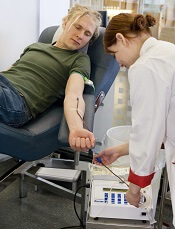
Photo by Marja Helander
The Irish Blood Transfusion Service (IBTS) has lifted the lifetime ban on blood donations from men who have sex with men (MSM).
However, prospective MSM blood donors are still subject to deferral.
Now, MSMs are allowed to donate blood in Ireland if it has been more than 12 months since their last sexual contact with a man and if they meet the other blood donor selection criteria.
The IBTS has also introduced new regulations relating to individuals with a history of specific, notifiable sexually transmitted infections (STIs).
These individuals are now allowed to donate blood 5 years after they have completed treatment for their STIs.
“In June of last year, I accepted the recommendations of the IBTS to change their blood donation deferral policies for men who have sex with men, as well as for donors who have had a sexually transmitted infection,” said Ireland’s Health Minister, Simon Harris.
“I would like to take this opportunity to thank the IBTS for their work over the past 6 months, which, today, sees these recommendations brought to fruition within the timescale agreed. [T]he IBTS will continue to keep all deferral policies under active review in the light of scientific evidence, emerging infections, and international experience.”
MSM deferral
The change in deferral policy relating to MSMs follows a 2-year review of the issues by the IBTS.
The agency hosted an international symposium on the topic in April 2016. Experts from 7 countries who had either lifted, or were in the process of lifting, their lifetime ban on MSM blood donors presented their respective stances, research, and the rationale behind their decisions.
The IBTS said its change to a 1-year deferral period for MSMs is supported by the most current scientific evidence available and brings Ireland into line with similar policies in the UK, Canada, and the US.
STI-related deferral
The IBTS said the 1-year deferral policy for MSMs will protect against the risk of HIV transmission. However, there is concern that it may not be sufficient to deal with an emerging infection.
Therefore, the board of the IBTS decided that individuals who have had a notifiable STI, such as chlamydia or genital herpes, should be deferred from donating blood for 5 years after completing treatment for that STI.
Individuals who have had syphilis, gonorrhea, lymphogranuloma venereum, or granuloma inguinale are (and have been) permanently banned from donating blood.
Individuals who have taken medication to prevent HIV infection are also deferred from donating blood for 5 years after they take the medication.
Safety of the blood supply
“The IBTS provides a safe, reliable, and robust blood service to the Irish health system and has the necessary program and procedures in place to protect both donors and recipients of blood and blood products,” Harris said.
All prospective blood donors in Ireland undergo nucleic acid testing for a number of diseases, including HIV, hepatitis B, and hepatitis C. This is the most sensitive method of testing available.
The risk of transmitted infection of blood is at its highest when individuals donate blood during the 5- to 15-day period following exposure to a virus.
There is no biological measure to detect infectivity during this period and, as a consequence, the IBTS temporarily or permanently defers, on average, 1 in 10 people from giving blood. ![]()
Delayed cord clamping reduces anemia risk
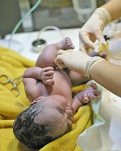
Photo by Meutia Chaerani
and Indradi Soemardjan
Delaying umbilical cord clamping by a few minutes can reduce the risk of anemia several months after birth, according to research published in JAMA Pediatrics.
A randomized clinical trial showed that delaying cord clamping by 3 minutes or more after birth—rather than clamping within 1 minute of birth—reduced the prevalence of anemia, iron deficiency, and iron deficiency anemia in infants at 8 months and 12 months of age.
Ola Andersson, MD, PhD, of Uppsala University in Uppsala, Sweden, and his colleagues conducted this research in Nepal, a country with a high prevalence of anemia.
The study included 540 infants—281 boys and 259 girls—with a mean gestational age of 39.2 weeks. Half of the subjects were randomized to delayed cord clamping (3 minutes or more after birth) or early cord clamping (within 1 minute of birth).
At 8 months of age, 78.5% of infants from the delayed clamping group and 69.6% from the early clamping group returned for blood sampling.
Results showed that infants in the delayed clamping group had higher levels of hemoglobin than infants in the early clamping group—10.4 g/dL and 10.2 g/dL, respectively (P=0.008).
Infants in the delayed clamping group also had a lower incidence of:
- Anemia (hemoglobin level <11.0 g/dL)—73.0% and 82.2%, respectively (P=0.01)
- Iron deficiency—22.2% and 38.1%, respectively (P<0.001)
- Iron deficiency anemia—19.3% and 33.3%, respectively (P<0.001).
The relative risk (RR) of anemia was 0.89, the RR of iron deficiency was 0.58, and the RR of iron deficiency anemia was 0.58.
Results were similar when the infants reached 12 months of age, although all the between-group differences were not statistically significant.
Again, infants in the delayed clamping group had higher levels of hemoglobin than infants in the early clamping group—10.3 g/dL and 10.1 g/dL, respectively (P=0.02).
And infants in the delayed clamping group had a lower incidence of:
- Anemia—77.8% and 85.9%, respectively (P=0.02)
- Iron deficiency—35.6% and 43%, respectively (P=0.09)
- Iron deficiency anemia—30.4% and 37.8%, respectively (P=0.08).
The RR of anemia was 0.91, the RR of iron deficiency was 0.83, and the RR of iron deficiency anemia was 0.80.
The researchers said this study shows that delayed cord clamping was an effective intervention to reduce anemia in a high-risk population, with minimal cost and without apparent adverse effects.
The team believes that, if this intervention were implemented on a global scale, this could translate to 5 million fewer infants with anemia at 8 months of age. ![]()
EZH2 may be therapeutic target for multiple myeloma

multiple myeloma
Preclinical research published in Oncotarget has provided new insights regarding how the protein EZH2 affects the development of multiple myeloma (MM) and reinforces the idea that EZH2 inhibition may be a way to treat MM.
Previous research by the same group suggested that EZH2 is a potential therapeutic target in MM.
With the current study, the group further investigated the anti-myeloma mechanisms mediated by EZH2 inhibition.
They found that targeting EZH2 with an agent known as UNC1999 reduces the expression of 4 MM-associated oncogenes—IRF-4, XBP-1, PRDM1/BLIMP-1, and c-MYC.
“The role of oncogenes in the development of cancer is to potentiate the survival of the cancer cell, which, instead of undergoing cell death, as is usually the case when the cell is not functioning properly, continues to divide and proliferate,” said study author Helena Jernberg-Wiklund, PhD, of Uppsala University in Uppsala, Sweden.
“In our study, we identified 4 oncogenes that showed lower activity in cells treated with the EZH2 inhibitor as compared to control-treated cells. All 4 genes have previously been shown to be associated with the development of multiple myeloma. This confirms our previous findings that inhibition of EZH2 could be used as a means to treat multiple myeloma.”
However, the researchers were puzzled by the fact that inhibition of EZH2 could decrease the activity of the oncogenes.
The chemical histone modification performed by EZH2 leads to decreased activity of affected genes. Therefore, inhibition of EZH2 should result in a reduced level of chemical modifications, which, in turn, should result in increased gene activity.
“The answer is that there are other genetic factors involved, called microRNAs,” Dr Jernberg-Wiklund said. “In the cells treated with the EZH2 inhibitor, we found 2 microRNA genes with increased activity, and we believe that the oncogenes are regulated by these microRNAs.”
“What happens then is that, when EZH2 is inhibited, there is a reduced histone modification at the microRNA genes. This leads to an increased synthesis of the microRNAs, which, in turn, decreases the activity of the oncogenes. This is a completely new mechanism for EZH2 action.”
The microRNAs the researchers identified are miR-125a-3p and miR-320c. The team found that miR-125a-3p and miR-320c were targets of EZH2 and H3K27me3 in MM cell lines and primary cells.
The researchers said these results support their previous work suggesting EZH2 could be a therapeutic target in MM. ![]()
Considering cattle could help fight malaria

Photo by Ilya Mauter
The goal of eliminating malaria in countries like India could be more achievable if mosquito-control efforts take into account the relationship between mosquitoes and cattle, according to an international team of researchers.
The group analyzed 2 mosquito
species found in Odisha, the state with the highest number of malaria cases in
India, and found that both species fed on cattle as well as humans.
“In many parts of the world, the mosquitoes responsible for transmitting malaria are specialist feeders on humans and often rest within human houses,” said Matthew Thomas, PhD, of Pennsylvania State University in University Park, Pennsylvania.
“We found that, in an area of India that has a high burden of malaria, most of the mosquitoes that are known to transmit malaria rest in cattle sheds and feed on both cows and humans.”
Dr Thomas and his colleagues reported these findings in Scientific Reports.
According to the researchers, cattle sheds in India are often next to, and sometimes even share a wall with, human houses. However, current malaria control efforts are restricted to domestic dwellings only.
“Given this cattle-shed ‘refuge’ for mosquitoes, focusing only on humans with regard to malaria control is a bit like treating the tip of an iceberg,” said study author Jessica Waite, PhD, also of Pennsylvania State University.
She, Dr Thomas, and their colleagues determined the importance of cows in the malaria-control problem by capturing adult mosquitoes in different habitats within 6 villages in Odisha state—which has the highest number of malaria cases in the country—and noting where the mosquitoes had been resting.
The team then used molecular techniques to determine which species of mosquitoes had been captured and which hosts they had been feeding on.
The researchers collected 1774 Anopheles culicifacies mosquitoes and 169 Anopheles fluviatilis mosquitoes across all study sites.
Both species were denser in cattle sheds than in human dwellings, and both were feeding on humans as well as cattle.
Next, the researchers used their field-collected data to help build a computer model that simulated the life of an adult mosquito. The team used the model to explore how best to control the mosquitoes to have maximum impact on malaria transmission in these villages.
“Our model analysis suggests that conventional control tools—such as insecticide-treated bed nets and indoor insecticide sprays—are less effective when mosquitoes exhibit ‘zoophilic’ behaviors (having an attraction to nonhuman animals),” Dr Thomas said.
“However, extending controls to better target the zoophilic mosquitoes—for example, by broadening coverage of non-repellant insecticide sprays to include cattle sheds—could help reduce transmission dramatically.”
Dr Waite added that the model suggests very little cattle-based vector control effort would be required to drive malaria transmission in the region to elimination.
“We show that directing even modest amounts of effort to specifically increase mosquito mortality associated with zoophilic behavior can shift the balance towards elimination,” she said.
“Understanding the dynamic between humans, cattle, and mosquitoes could have major implications for malaria control policy and practice, not only in India, but in other areas where transmission is sustained by zoophilic vectors,” Dr Thomas said.
“Specifically, optimizing use of existing tools will be essential to achieving the ambitious 2030 elimination target set by the World Health Organization, which aims to decrease malaria cases globally by 90% compared to 2015 levels and eliminate malaria in at least 35 additional countries by 2030.” ![]()
Electronic Medical Records Can Keep Pace With Military Life
People in the military are somewhat nomadic by necessity: They move around a lot, and their medical records should be just as mobile, says Army Brig. Gen. John Cho, deputy chief of staff for support for the Army’s Medical Command, in an article for Health.mil. Cho is the functional champion on development of MHS Genesis, the new electronic health record that is replacing less agile DoD legacy systems.
Related: IHS Pilots Improved Version of Health Records
Developed with input from more than 850 experts in the field around the world, MHS Genesis is intended to fix shortcomings in the current system. It will integrate medical and dental records throughout the continuum of care, from point of injury to the military treatment facility where the care is provided.
Benefits include the ability to monitor a beneficiary’s health status through health data, tracking, and alerting capabilities; improved ability to monitor patient safety, outcomes, and operational and medical readiness; improved access to integrated, evidence-based health care delivery and decision-making; and increased sharing of health information across the spectrum of military operations, the VA, and civilian health care organizations.
Related: How Safe Are Patients’ Electronic Records?
MHS Genesis will support the availability of electronic health records for > 9.4 million DoD beneficiaries and approximately 205,000 MHS personnel globally. The new system is currently being tested, with rollout in the Pacific Northwest scheduled for early 2017. The system expected to be ready throughout the MHS in 5 years.
What to do with isolated calf DVT
Clinical question: Does therapeutic anticoagulation of isolated calf deep vein thrombosis (DVT) decrease risk for proximal DVT or PE?
Background: Optimal management of isolated calf DVT lacks consensus.
Study design: Single-center, retrospective, cohort study.
Setting: Large academic hospital.
Nevertheless, 9.2% of control patients and 3.3% of exposure patients developed a proximal DVT or PE. The anticoagulation group was associated with lower likelihood of proximal DVT or PE (risk ratio 0.36; 95% CI, 0.15-0.84) but an increased risk of bleeding (8.6%), compared with the nonexposure group (2.2%). Sensitivity analysis did not alter the observed association.
Bottom line: Therapeutic anticoagulation for isolated calf DVT may be warranted to decrease the risk for proximal DVT or PE but with an increased risk of bleeding. Randomized trials are needed to clarify the risk versus benefit.
Citation: Utter GH, Dhillon TS, Salcedo ES, et al. Therapeutic anticoagulation for isolated calf deep vein thrombosis. JAMA Surg. 2016;151(9):e161770. doi: 10.1001/jamasurg.2016.1770.
Dr. Zuleta is an assistant professor and associate program director of the Jackson Memorial/University of Miami Internal Medicine residency training program and the site director of the program at University of Miami Hospital.