User login
Young adult stroke survivors have distinct risk profile
SAN DIEGO – Young adults who have suffered a stroke are at greater risk of a second stroke than other cardiovascular events, at least in the first year. That’s the conclusion drawn from a new analysis of 2013 data drawn from the Nationwide Readmissions Database.
The results suggest that younger adults who have a first-time stroke have a different risk profile than older adults and could require different management to improve long-term outcomes. The incidence of stroke has increased in recent years to the point that this population now accounts for about 10% of all strokes.
The analysis looked at all admissions for ischemic stroke in patients aged 18-45. The researchers found a cumulative risk of rehospitalization of 5.5% at 300 days, compared with 3.6% for cardiovascular disease.
The study can’t explain the association, nor can it prove causation. “Our thought was that the effects of hypertension might take longer to manifest in terms of cardiovascular outcomes as compared to hypercholesterolemia and diabetes,” said Dr. Jin, who is chief resident in the department of neurology at Icahn School of Medicine at Mount Sinai, New York. He presented the research at the annual meeting of the American Neurological Association.
The result sends a clear message for physicians caring for young adults who have experienced a first-time stroke. “They should be rigorously worked up and managed for any disorders in blood sugar and lipid disorders,” Dr. Jin said. Their care is vital because these younger adults have more time to accumulate second, third, or fourth strokes that could dramatically increase the burden of disease. “It’s just a matter of time. Optimizing their secondary prevention is crucial,” Dr. Jin said.
The study included data from 12,392 young adults in the Nationwide Readmissions Database who had suffered a first-time stroke. The researchers identified a higher readmission rate for stroke than cardiovascular events at 90 days (2,913.3 vs. 1,132.4 per 100,000 index hospitalizations). This pattern held when the analysis was restricted to patients who had no cardiovascular risk factors prior to the index hospitalization (2,534.9 vs. 676 per 100,000 index hospitalizations).
At 100 days, the cumulative risks were 3.2% for stroke and 2.5% for cardiovascular events. The risks were 4.3% and 3.2% at 200 days, and 5.5% and 3.6% at 300 days.
A multivariate analysis showed that patients with baseline diabetes were at a heightened risk of cardiovascular events (hazard ratio, 1.49; 95% confidence interval, 1.17-1.88), as were patients with hypercholesterolemia (HR, 1.43; 95% CI, 1.15-1.79) and those with atrial fibrillation or flutter (HR, 3.86; 95% CI, 2.74-5.43). Only diabetes was significantly associated with increased risk for hospitalization for recurrent stroke (HR, 1.5; 95% CI, 1.22-1.84).
Dr. Jin is eager to see if future research might establish a causative link between these risk factors and outcomes. The current work grew out of an administrative data set, but registry data or a prospective study could be more robust.
The study received no external funding. Dr. Jin reported having no financial disclosures.
AT ANA 2017
Key clinical point:
Major finding: Hospitalization for cardiovascular disease was associated with baseline diabetes (HR, 1.49) and hypercholesterolemia (HR, 1.43).
Data source: A retrospective analysis of data from the Nationwide Readmissions Database (n = 12,392).
Disclosures: The study received no external funding. Dr. Jin reported having no financial disclosures.
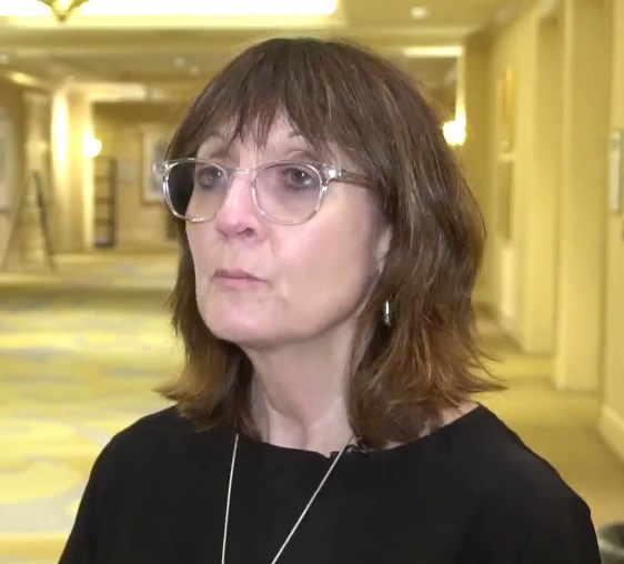
VIDEO: Rethinking deep brain stimulation for depression
SAN DIEGO – Earlier this month, an article in Lancet Psychiatry reported the results of a prospective, randomized, sham-controlled trial that tested deep brain stimulation of the Brodmann area 25 within the subcallosal cingulate white matter in 90 patients with treatment-resistant depression. Unfortunately, the study showed no significant benefit at 6 months.
The approach had shown promise in some previous open-label studies, which prompted the multicenter trial (Lancet Psychiatry. 2017 Oct 4. doi: 10.1016/S2215-0366(17)30371-1).
Although the 6-month results were disappointing, the open-label phase of the study told a different story. At 2 years, 48% of patients in the stimulation group achieved an antidepressant response, higher than what would be expected from treatment as usual in this difficult population.
In this video interview at the annual meeting of the American Neurological Association, Helen Mayberg, MD, one of the study authors and professor of psychiatry, neurology, and radiology at Emory University, Atlanta, discusses these long-term results and their implications, as well as lessons learned and how they might inform future research.
AT ANA 2017
Bringing critical care training to hospitalists
It’s 9 p.m., and the ER calls you to admit a 60-year-old woman with COPD and multilobar pneumonia. She’s hypoxemic, intubated, and hypotensive after 3 L of crystalloid.
You’re asked to evaluate a patient who has developed stridor after an anterior cervical decompression and fusion. He seems to have responded to racemic epinephrine. Is he okay or not? What should you do next?
A patient develops dyspnea and chest pain after a total knee replacement. Chest CT shows extensive bilateral PE and a dilated right ventricle. She’s normotensive, but tachycardic and tachypneic. Now what?
Recognizing this growing phenomenon, SHM convened a task force of hospitalists and intensivists to quantify the problem and to develop tools and curricula to support hospitalists who provide critical care services. Our mission is make sure that every hospitalist who cares for critically ill patients has the skills and knowledge necessary to do so safely and competently. We’re working to define the scale and scope of the problem, advocate for hospitalists who provide critical care services, and develop educational content to fill gaps in knowledge and skill. We hope to offer a comprehensive but flexible critical care curriculum to meet the needs of hospitalists across the range of knowledge and skills.
When we can, we’ll leverage existing critical care courses and content and build that into our curriculum. When we can’t find material that is appropriate for hospitalists, we’ll develop our own. As a first step, we have produced targeted CME-eligible web-based education modules on the SHM Learning Portal covering high-risk clinical scenarios that hospitalists commonly encounter:
• Airway management for the hospitalist
• Noninvasive positive pressure ventilation
• Arrhythmias
• High-risk pulmonary embolism
This is an ambitious project, and we still have a long way to go. Have a scenario that we haven’t covered? Like what you see? We’d love to hear your feedback and ideas. Please contact [email protected].
Dr. Aymond is associate clinical professor of medicine at Louisiana State University, Alexandria, and a hospitalist ICU provider at Byrd Regional Hospital, Leesville, La., and Lake Charles (La.) Memorial Hospital. Dr. Siegal is adjunct clinical professor of medicine at the University of Wisconsin–Madison and an intensivist at Aurora Health Care.
Transbronchial cryobiopsy, updated guidelines for chronic cough in children, PD-1 inhibition
Interventional Chest/Diagnostic Procedures
Cryobiopsy for ILD: Careful stewardship needed
Interest in transbronchial cryobiopsy has accelerated rapidly in recent years. This procedure is performed by advancing a cryoprobe into the peripheral lung via flexible bronchoscopy, where lung tissue freezes and adheres to the probe and is subsequently extracted as a cryobiopsy. The number of cryobiopsy-related publications has increased exponentially since it was described in 2009 (Babiak A, et al. Respiration. 2009;78[2]:203). This interest stems from reports of high diagnostic yields in patients with interstitial lung disease (ILD) while maintaining complication rates similar to that of conventional bronchoscopic biopsy.
Traditional bronchoscopic biopsies are notoriously insensitive; a specific diagnosis can be established in fewer than a third of cases (Sheth JS, et al. Chest. 2017;151[2]:389). As such, surgical lung biopsy continues to be recommended but is associated with significant mortality (2%) and morbidity (30%) in patients with ILD (Hutchinson JP, et al. ARJCCM. 2016;193[10]:1161). Cryobiopsy, which appears to rival surgical lung biopsy in terms of ability to contribute to a specific diagnosis, is, therefore, a highly promising alternative (Tomassetti S, et al. AJRCCM. 2016;193[7]:745).
As cryobiopsy is increasingly adopted around the world, however, troubling reports of serious complications have surfaced. Most notable is the recently reported experience of the initial 25 cases performed at the University of Pennsylvania, in which almost one in four patients suffered serious complications (DiBardino DM, et al. Ann Am Thorac Soc. 2017;14[6]:851). The authors pointed to lack of a predefined procedural protocol, as well as several choices relating to the specific technique used, including inconsistent use of fluoroscopy, lack of prophylactic bronchial blocker placement, and predominant use of laryngeal mask airways as potential contributing factors. Indeed, many variations of the basic cryobiopsy procedure have been described (Lentz RJ, et al. J Thoracic Dis. 2017;9[7]:2186), with no formal guidance or training available to inform advanced bronchoscopists interested in this procedure.
It is incumbent on the interventional pulmonology and ILD specialist communities to be responsible stewards of this promising procedure. Implementation of three parallel efforts to standardize and rigorously study this procedure should be considered as soon as possible: creation of expert consensus guidelines establishing best-practices for safe and effective biopsy technique; a training requirement before independent performance of the procedure; and creation of an international cryobiopsy registry to facilitate higher-quality research into optimal technique and outcomes. We owe this to our patients.
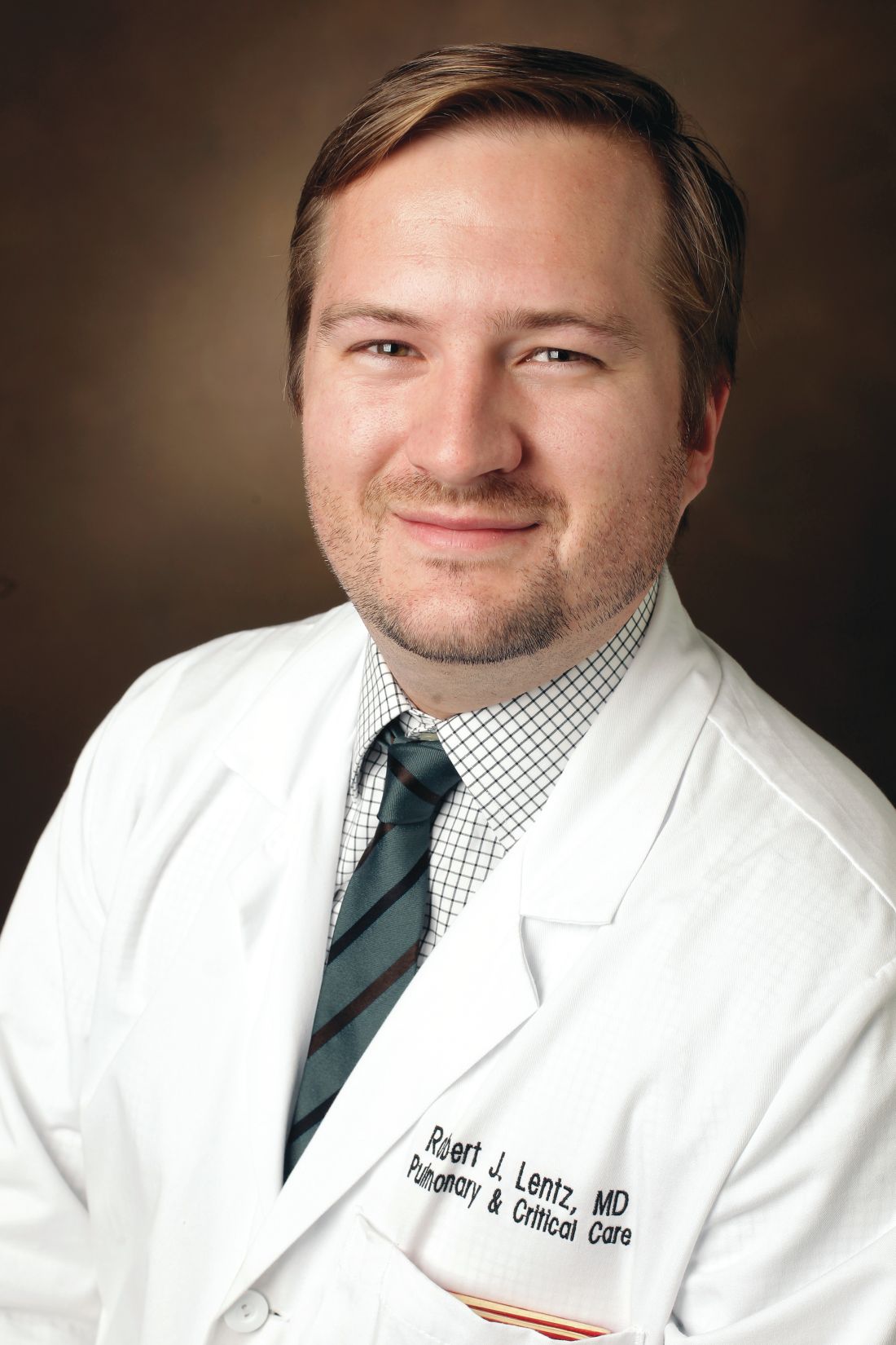
Robert J. Lentz, MD
NetWork Member
Fabien Maldonado, MD, FCCP
NetWork Member
Pediatric Chest Medicine
Chronic cough in children: New guidelines
A chronic cough is a common complaint among children whose parents seek medical evaluation. Chronic wet cough can indicate an underlying illness; therefore, an early diagnosis can lead to prevention of complications of the disease and improvement in quality of life.
CHEST is a leading resource in evidence and consensus-based guidelines on important topics affecting children. The most recent guidelines entitled Management of Children with Chronic Wet Cough and Protracted Bacterial Bronchitis (Chest. 2017;151(4):884-890) and Use of Management Pathways or Algorithms in Children with Chronic Cough (Chest. 2017;151(4):875-873) are updates from the 2006 CHEST guidelines on chronic cough in children.
The present updates utilized the CHEST methodological guidelines with chronic wet or productive cough and Grading of Recommendations Assessment, Development, and Evaluation framework and also performed a systematic review addressing key questions concerning the management of childhood disease for children 14 years and younger.
Guidance provided by the expert panel focused on recommendations to answer six key questions concerning the management of children 14 years and younger with a chronic wet cough unrelated to established chronic lung disease. The recommendations are:
1. Chronic cough is defined as the presence of a cough 4 weeks or longer in duration.
2. Assessment of the effect of the cough on the child and the family be undertaken as part of clinical consultation.
3. Evaluation of a chronic cough should be done with a systematic approach with pediatric-specific cough management protocols or algorithms.
4. Chest radiograph and, when age appropriate, spirometry with bronchodilator be undertaken as evaluation; tests for pertussis infection only to be performed if clinically suspected.
5. Chronic wet cough with no specific clinical features should receive antibiotics for 2 weeks targeted for common respiratory bacteria (Streptococcus pneumoniae, Haemophilus influenzae, and Moraxella catarrhalis).
6. When cough persists despite 2 weeks of appropriate antibiotics, it is recommended to continue for an additional 2 weeks.
7. Additional tests (eg skin prick test, Mantoux, bronchoscopy, chest CT scan) should be individualized in accordance with the clinical setting and child’s clinical symptoms and signs.
The panel recognizes the need for prospective studies to assess current algorithms outcomes of children with chronic cough. Both articles can be found on the guidelines section of the CHEST site.
John Bishara, DO
Fellow-in-Training Member
Pulmonary Physiology, Function, and Rehabilitation
Functional imaging of the lung
Quantifying heterogeneity of ventilation and gas exchange in lung diseases remains a clinical challenge. Conventional pulmonary function test is insensitive to regional changes. The multiple inert gas elimination technique can quantify ventilation-perfusion distribution, but it requires invasive instrumentation (eg, pulmonary artery catheterization) and is not practical for clinical use. Computed tomography (CT) scans delineate spatial changes in lung structures but do not directly measure changes in ventilation and gas exchange. With its radiation, it is difficult to apply CT scanning repeatedly in patients. More recently, MR imaging techniques have been developed to directly “visualize” and quantify regional lung function (Kruger SJ, et al. J Magn Reson Imaging. 2016;43(2):295; Roos JE, et al. Magn Reson Imaging Clin N Am. 2015;23(2):217). These techniques employ inhalation of gases, such as oxygen, perfluorinated gases, and hyperpolarized 3He and 129Xe. Hyperpolarized 3He has been studied the most; however, the dwindling supply of 3He gas and its rising cost have prevented its further development. 129Xe has abundant supply and has emerged to be the inert gas of choice for MR imaging. Hyperpolarized 129Xe can measure ventilation, like hyperpolarized 3He. In addition, Xe diffuses into alveolar barrier (interstitium and plasma) and red blood cells, where it exhibits distinct resonant frequency shifts that can be captured by MR. Therefore, in one test, information on pulmonary ventilation and gas transfer can be obtained. To date, the results from MR imaging studies have provided new insights into the pathophysiology of obstructive and restrictive lung diseases. With continuous development, MR imaging of the lung could become a clinically useful tool in the near future.
Yuh-Chin T. Huang, MD, MHS, FCCP
Steering Committee Member
Thoracic Oncology
Immune-mediated pneumonitis and PD-1 inhibition
Inhibitors of the programmed cell death 1 receptor (PD-1) have shown significant promise in the treatment of advanced stage malignancy. With the recent expansion of indications for use of these agents, the number of patients treated will continue to grow. Clinicians must be aware of their potential for serious adverse side effects, including dermatitis, colitis, and potentially life-threatening pneumonitis.
The development of pneumonitis secondary to PD-1 inhibitions is reported to occur in 2% to 5% of patients and can present at any time during therapy, with 1% of patients developing grade 3 or higher pneumonitis.1,2 The most common symptoms are dyspnea and cough, though one-third of patients are asymptomatic at presentation.2 Radiographic and pathologic features vary greatly and include organizing pneumonia, interstitial pneumonitis, hypersensitivity pneumonitis, or diffuse alveolar damage.3 While pneumonitis due to PD-1 inhibition is reportedly uncommon, the increasing number of patients expected to receive these medications will predictably result in increasing overall frequency of pneumonitis cases. In addition, the lack of large prospective randomized trials and reliance on radiographic rather than pathologic data in diagnosing immune-mediated pneumonitis gives one pause. Given the variability of presentation, lack of routine pathologic data, and increasing use of dual agents (eg, PD-1 and CTLA-4), chest physicians and medical oncologists should have a high index of suspicion yet practice equipoise in patients receiving immunotherapy who develop unexplained pulmonary symptoms or infiltrates. More research is needed to help improve the multidisciplinary diagnosis and treatment of this potentially serious complication.
David Maurice Chambers, MD
Fellow-in-Training Member
Jason Atticus Akulian, MD, MPH
Steering Committee Member
References
1. Nishino M, et al. Incidence of programmed cell death 1 inhibitor-related pneumonitis in patients with advanced cancer: a systematic review and meta-analysis. JAMA Oncology. 2016;2(12):1607.
2. Naidoo J, et al. Pneumonitis in patients treated with anti-programmed death-1/programmed death ligand 1 therapy. J Clin Oncol. 2017;35(7):709.
3. Nishino M, et al. PD-1 inhibitor-related pneumonitis in advanced cancer patients: radiographic patterns and clinical course. Clin Cancer Res. 2016;22(24):6051.
Pulmonary Vascular Disease
Pulmonary Arterial Hypertension Associated With SLE
While pulmonary arterial hypertension (PAH) commonly complicates scleroderma (SSc), it is a rare complication of other connective tissue diseases (CTD), such as systemic lupus erythematosus (SLE). In the few prospective studies that utilize right-sided heart catheterization (RHC), the estimated prevalence of PAH in SLE is about 4%. However, since the prevalence of SLE is 10 to 15 times greater than SSc in the United States, the true prevalence of SLE-PAH may be higher than previously thought, and, thus, clinically relevant. Despite this, little is known about SLE-PAH.
A recent retrospective study from the French Pulmonary Hypertension Registry has added significantly to our understanding of this complication of SLE. Hachulla and colleagues studied 51 patients with RHC-proven SLE-PAH compared with 101 SLE control subjects without PAH. While the authors did not find any relevant differences in the demographics between groups, they did find a significantly higher prevalence of SSA and SSB antibodies in SLE-PAH. Interestingly, the presence of anti-U1 RNP antibody appeared to be less common in SLE-PAH patients; this lack of association is in contrast to prior studies in mixed CTD patients with anti-U1 RNP antibodies in which the prevalence of PAH can be as high as 60%. Further, none of the SLE-PAH patients demonstrated an acute response to vasodilator challenge during RHC, emphasizing that this maneuver does not need to be performed in SLE patients at risk of PAH. Trends toward improved survival in SLE-PAH patients treated with hydroxychloroquine are preliminary and hypothesis-generating but require confirmation in larger clinical studies.
Stephen Mathai, MD, FCCP
Chair
Leena Palwar, MD
Fellow-in-Training Member
References
Hachulla E, Jais X, Cinquetti G, et al. Pulmonary arterial hypertension associated with SLE: Results from the French pulmonary hypertension registry. Chest. 2017 Aug 26. pii: S0012-3692(17)31430-7. doi: 10.1016/j.chest.2017.08.014. [Epub ahead of print]
Chung L, Liu J, Parsons L, et al. Characterization of connective tissue disease-associated pulmonary arterial hypertension from REVEAL: identifying systemic sclerosis as a unique phenotype. Chest. 2010;138:1383-1394.
Shirai Y, Yasuoka H, Okano Y, Takeuchi T, Satoh T, Kuwana M. Clinical characteristics and survival of Japanese patients with connective tissue disease and pulmonary arterial hypertension: a singlecentre cohort. Rheumatology. 2012;51:1846-1854.
Hao YJ, Jiang X, Zhou W, et al. Connective tissue disease-associated pulmonary arterial hypertension in Chinese patients. Eur Respir J. 2014;44: 963-972.
Huang C, Li M, Liu Y, et al. Baseline characteristics and risk factors of pulmonary arterial hypertension in systemic lupus erythematosus patients. Medicine. 2016;95:e2761.
Pérez-Peñate GM, Rúa-Figueroa I, Juliá- Serdá G, et al. Pulmonary arterial hypertension in systemic lupus erythematosus: prevalence and predictors. J Rheumatol. 2016;43:323-329.
Alpert MA, Goldberg SH, Sindem BH, et al. Cardiovascular manifestations of mixed connective tissue disease in adults. Circulation. 1983;63:1182-1193.
Interventional Chest/Diagnostic Procedures
Cryobiopsy for ILD: Careful stewardship needed
Interest in transbronchial cryobiopsy has accelerated rapidly in recent years. This procedure is performed by advancing a cryoprobe into the peripheral lung via flexible bronchoscopy, where lung tissue freezes and adheres to the probe and is subsequently extracted as a cryobiopsy. The number of cryobiopsy-related publications has increased exponentially since it was described in 2009 (Babiak A, et al. Respiration. 2009;78[2]:203). This interest stems from reports of high diagnostic yields in patients with interstitial lung disease (ILD) while maintaining complication rates similar to that of conventional bronchoscopic biopsy.
Traditional bronchoscopic biopsies are notoriously insensitive; a specific diagnosis can be established in fewer than a third of cases (Sheth JS, et al. Chest. 2017;151[2]:389). As such, surgical lung biopsy continues to be recommended but is associated with significant mortality (2%) and morbidity (30%) in patients with ILD (Hutchinson JP, et al. ARJCCM. 2016;193[10]:1161). Cryobiopsy, which appears to rival surgical lung biopsy in terms of ability to contribute to a specific diagnosis, is, therefore, a highly promising alternative (Tomassetti S, et al. AJRCCM. 2016;193[7]:745).
As cryobiopsy is increasingly adopted around the world, however, troubling reports of serious complications have surfaced. Most notable is the recently reported experience of the initial 25 cases performed at the University of Pennsylvania, in which almost one in four patients suffered serious complications (DiBardino DM, et al. Ann Am Thorac Soc. 2017;14[6]:851). The authors pointed to lack of a predefined procedural protocol, as well as several choices relating to the specific technique used, including inconsistent use of fluoroscopy, lack of prophylactic bronchial blocker placement, and predominant use of laryngeal mask airways as potential contributing factors. Indeed, many variations of the basic cryobiopsy procedure have been described (Lentz RJ, et al. J Thoracic Dis. 2017;9[7]:2186), with no formal guidance or training available to inform advanced bronchoscopists interested in this procedure.
It is incumbent on the interventional pulmonology and ILD specialist communities to be responsible stewards of this promising procedure. Implementation of three parallel efforts to standardize and rigorously study this procedure should be considered as soon as possible: creation of expert consensus guidelines establishing best-practices for safe and effective biopsy technique; a training requirement before independent performance of the procedure; and creation of an international cryobiopsy registry to facilitate higher-quality research into optimal technique and outcomes. We owe this to our patients.
Robert J. Lentz, MD
NetWork Member
Fabien Maldonado, MD, FCCP
NetWork Member
Pediatric Chest Medicine
Chronic cough in children: New guidelines
A chronic cough is a common complaint among children whose parents seek medical evaluation. Chronic wet cough can indicate an underlying illness; therefore, an early diagnosis can lead to prevention of complications of the disease and improvement in quality of life.
CHEST is a leading resource in evidence and consensus-based guidelines on important topics affecting children. The most recent guidelines entitled Management of Children with Chronic Wet Cough and Protracted Bacterial Bronchitis (Chest. 2017;151(4):884-890) and Use of Management Pathways or Algorithms in Children with Chronic Cough (Chest. 2017;151(4):875-873) are updates from the 2006 CHEST guidelines on chronic cough in children.
The present updates utilized the CHEST methodological guidelines with chronic wet or productive cough and Grading of Recommendations Assessment, Development, and Evaluation framework and also performed a systematic review addressing key questions concerning the management of childhood disease for children 14 years and younger.
Guidance provided by the expert panel focused on recommendations to answer six key questions concerning the management of children 14 years and younger with a chronic wet cough unrelated to established chronic lung disease. The recommendations are:
1. Chronic cough is defined as the presence of a cough 4 weeks or longer in duration.
2. Assessment of the effect of the cough on the child and the family be undertaken as part of clinical consultation.
3. Evaluation of a chronic cough should be done with a systematic approach with pediatric-specific cough management protocols or algorithms.
4. Chest radiograph and, when age appropriate, spirometry with bronchodilator be undertaken as evaluation; tests for pertussis infection only to be performed if clinically suspected.
5. Chronic wet cough with no specific clinical features should receive antibiotics for 2 weeks targeted for common respiratory bacteria (Streptococcus pneumoniae, Haemophilus influenzae, and Moraxella catarrhalis).
6. When cough persists despite 2 weeks of appropriate antibiotics, it is recommended to continue for an additional 2 weeks.
7. Additional tests (eg skin prick test, Mantoux, bronchoscopy, chest CT scan) should be individualized in accordance with the clinical setting and child’s clinical symptoms and signs.
The panel recognizes the need for prospective studies to assess current algorithms outcomes of children with chronic cough. Both articles can be found on the guidelines section of the CHEST site.
John Bishara, DO
Fellow-in-Training Member
Pulmonary Physiology, Function, and Rehabilitation
Functional imaging of the lung
Quantifying heterogeneity of ventilation and gas exchange in lung diseases remains a clinical challenge. Conventional pulmonary function test is insensitive to regional changes. The multiple inert gas elimination technique can quantify ventilation-perfusion distribution, but it requires invasive instrumentation (eg, pulmonary artery catheterization) and is not practical for clinical use. Computed tomography (CT) scans delineate spatial changes in lung structures but do not directly measure changes in ventilation and gas exchange. With its radiation, it is difficult to apply CT scanning repeatedly in patients. More recently, MR imaging techniques have been developed to directly “visualize” and quantify regional lung function (Kruger SJ, et al. J Magn Reson Imaging. 2016;43(2):295; Roos JE, et al. Magn Reson Imaging Clin N Am. 2015;23(2):217). These techniques employ inhalation of gases, such as oxygen, perfluorinated gases, and hyperpolarized 3He and 129Xe. Hyperpolarized 3He has been studied the most; however, the dwindling supply of 3He gas and its rising cost have prevented its further development. 129Xe has abundant supply and has emerged to be the inert gas of choice for MR imaging. Hyperpolarized 129Xe can measure ventilation, like hyperpolarized 3He. In addition, Xe diffuses into alveolar barrier (interstitium and plasma) and red blood cells, where it exhibits distinct resonant frequency shifts that can be captured by MR. Therefore, in one test, information on pulmonary ventilation and gas transfer can be obtained. To date, the results from MR imaging studies have provided new insights into the pathophysiology of obstructive and restrictive lung diseases. With continuous development, MR imaging of the lung could become a clinically useful tool in the near future.
Yuh-Chin T. Huang, MD, MHS, FCCP
Steering Committee Member
Thoracic Oncology
Immune-mediated pneumonitis and PD-1 inhibition
Inhibitors of the programmed cell death 1 receptor (PD-1) have shown significant promise in the treatment of advanced stage malignancy. With the recent expansion of indications for use of these agents, the number of patients treated will continue to grow. Clinicians must be aware of their potential for serious adverse side effects, including dermatitis, colitis, and potentially life-threatening pneumonitis.
The development of pneumonitis secondary to PD-1 inhibitions is reported to occur in 2% to 5% of patients and can present at any time during therapy, with 1% of patients developing grade 3 or higher pneumonitis.1,2 The most common symptoms are dyspnea and cough, though one-third of patients are asymptomatic at presentation.2 Radiographic and pathologic features vary greatly and include organizing pneumonia, interstitial pneumonitis, hypersensitivity pneumonitis, or diffuse alveolar damage.3 While pneumonitis due to PD-1 inhibition is reportedly uncommon, the increasing number of patients expected to receive these medications will predictably result in increasing overall frequency of pneumonitis cases. In addition, the lack of large prospective randomized trials and reliance on radiographic rather than pathologic data in diagnosing immune-mediated pneumonitis gives one pause. Given the variability of presentation, lack of routine pathologic data, and increasing use of dual agents (eg, PD-1 and CTLA-4), chest physicians and medical oncologists should have a high index of suspicion yet practice equipoise in patients receiving immunotherapy who develop unexplained pulmonary symptoms or infiltrates. More research is needed to help improve the multidisciplinary diagnosis and treatment of this potentially serious complication.
David Maurice Chambers, MD
Fellow-in-Training Member
Jason Atticus Akulian, MD, MPH
Steering Committee Member
References
1. Nishino M, et al. Incidence of programmed cell death 1 inhibitor-related pneumonitis in patients with advanced cancer: a systematic review and meta-analysis. JAMA Oncology. 2016;2(12):1607.
2. Naidoo J, et al. Pneumonitis in patients treated with anti-programmed death-1/programmed death ligand 1 therapy. J Clin Oncol. 2017;35(7):709.
3. Nishino M, et al. PD-1 inhibitor-related pneumonitis in advanced cancer patients: radiographic patterns and clinical course. Clin Cancer Res. 2016;22(24):6051.
Pulmonary Vascular Disease
Pulmonary Arterial Hypertension Associated With SLE
While pulmonary arterial hypertension (PAH) commonly complicates scleroderma (SSc), it is a rare complication of other connective tissue diseases (CTD), such as systemic lupus erythematosus (SLE). In the few prospective studies that utilize right-sided heart catheterization (RHC), the estimated prevalence of PAH in SLE is about 4%. However, since the prevalence of SLE is 10 to 15 times greater than SSc in the United States, the true prevalence of SLE-PAH may be higher than previously thought, and, thus, clinically relevant. Despite this, little is known about SLE-PAH.
A recent retrospective study from the French Pulmonary Hypertension Registry has added significantly to our understanding of this complication of SLE. Hachulla and colleagues studied 51 patients with RHC-proven SLE-PAH compared with 101 SLE control subjects without PAH. While the authors did not find any relevant differences in the demographics between groups, they did find a significantly higher prevalence of SSA and SSB antibodies in SLE-PAH. Interestingly, the presence of anti-U1 RNP antibody appeared to be less common in SLE-PAH patients; this lack of association is in contrast to prior studies in mixed CTD patients with anti-U1 RNP antibodies in which the prevalence of PAH can be as high as 60%. Further, none of the SLE-PAH patients demonstrated an acute response to vasodilator challenge during RHC, emphasizing that this maneuver does not need to be performed in SLE patients at risk of PAH. Trends toward improved survival in SLE-PAH patients treated with hydroxychloroquine are preliminary and hypothesis-generating but require confirmation in larger clinical studies.
Stephen Mathai, MD, FCCP
Chair
Leena Palwar, MD
Fellow-in-Training Member
References
Hachulla E, Jais X, Cinquetti G, et al. Pulmonary arterial hypertension associated with SLE: Results from the French pulmonary hypertension registry. Chest. 2017 Aug 26. pii: S0012-3692(17)31430-7. doi: 10.1016/j.chest.2017.08.014. [Epub ahead of print]
Chung L, Liu J, Parsons L, et al. Characterization of connective tissue disease-associated pulmonary arterial hypertension from REVEAL: identifying systemic sclerosis as a unique phenotype. Chest. 2010;138:1383-1394.
Shirai Y, Yasuoka H, Okano Y, Takeuchi T, Satoh T, Kuwana M. Clinical characteristics and survival of Japanese patients with connective tissue disease and pulmonary arterial hypertension: a singlecentre cohort. Rheumatology. 2012;51:1846-1854.
Hao YJ, Jiang X, Zhou W, et al. Connective tissue disease-associated pulmonary arterial hypertension in Chinese patients. Eur Respir J. 2014;44: 963-972.
Huang C, Li M, Liu Y, et al. Baseline characteristics and risk factors of pulmonary arterial hypertension in systemic lupus erythematosus patients. Medicine. 2016;95:e2761.
Pérez-Peñate GM, Rúa-Figueroa I, Juliá- Serdá G, et al. Pulmonary arterial hypertension in systemic lupus erythematosus: prevalence and predictors. J Rheumatol. 2016;43:323-329.
Alpert MA, Goldberg SH, Sindem BH, et al. Cardiovascular manifestations of mixed connective tissue disease in adults. Circulation. 1983;63:1182-1193.
#Winning: Are you in it to win it at CHEST 2017?
CHEST 2017 offers several contests and opportunities to win great prizes! Are you ready to take home the prize?
CHEST Events App Game: Click Game
![]()
Rules of participation
Are you a VITweep?
Get active on Twitter, and share your latest highlights for #CHEST2017! Sitting in on an interesting session? Having a great time visiting the posters? Let us know! The most active tweeters for the day will receive a special prize!
Share your selfies!
See a selfie spot and take advantage of it! We know there’s more to your trip than lectures and keynote speakers, and we want to see it!
Throughout the convention center, you’ll find many designated areas to snap and share photos of yourself and colleagues! Be sure to find them all and share your images on Twitter or Instagram using our #CHEST2017 hashtag. We’ll choose our favorite photo of the day and reshare your picture with our social media followers. Don’t miss your chance to be featured!
Don’t miss out on CHEST Bingo
Take advantage of one of the many opportunities in the Exhibit Hall during CHEST. Play CHEST Bingo daily, starting Monday, October 30, through Wednesday, November 1, for a chance to win a prize!
How to play:
Find your bingo card in the program guide that you will receive during registration. Get each bingo letter to spell out C-H-E-S-T as you visit each of the five sponsors’ booths. You will then have a chance to win a $75 gift certificate to the CHEST bookstore. There will be a winner drawn every night!
Win an iPad®!
This year, attendees will have the opportunity to win a refurbished iPad for playing one of our Simulation Center’s arcade style GAMEs (Games Augmenting Medical Education). Last year, we gave away 15 refurbished iPad 2s; this year, we hope to give away 30 refurbished iPad 2s!
iPads will be awarded for the following:
- One each day for the fastest time on Aspirated!
- One each day for whoever has played the most games and Virtual Patient Tours (VPTs).
- Several for playing the games Peer Pressure and Nodal Nemesis.
Please refer to the program schedule in the CHEST Events app for dates and times of the GAMEs and VPTs.
An interview with incoming CHEST President, John Studdard, MD, FCCP
John Studdard, MD, FCCP, has been a member of the American College of Chest Physicians for 36 years, and, this November, he will be inaugurated as CHEST President. This will not be Dr. Studdard’s first time in a presidential role for CHEST, as he served as CHEST Foundation President in 2013 and 2014. Currently, Dr. Studdard serves as a pulmonologist at Jackson Pulmonary in Jackson, Mississippi. Being a physician and being as heavily involved with an organization as Dr. Studdard is takes a lot of prioritizing, hard work, and dedication. Get to know CHEST’s new President through this interview.
Born and raised in Mississippi, Dr. Studdard says there were four factors that inspired him to become a physician:
1. I have always loved people and working with them, and I always admired the respect that physicians received in my community.

3. I am competitive and decided if it was going to be hard to get into medical school, then I wanted to go to medical school.
4. My dad always told my brother and me that we would be doctors when we grew up, because we were going to be our own boss. I have been in private practice for 36 years, and that is not the case, not if you are doing it right. I obviously love medicine, and my dad was great in that he paid for our education…but he called the shots.
What are some of the biggest challenges you have encountered throughout your career?
Private practice makes you gain more independence and autonomy; you have to become more agile, more efficient, and you have awfully big workloads. However, you give up the academic stimulation of being in an academic center. It is a tough discipline in the private practice of medicine to try to stay up to date. Whether going to the CHEST Annual Meeting, reading our journal CHEST, or looking at CHEST education online products, those of us in the clinical practice of pulmonary, critical care, and sleep medicine are more dependent than any group on what our clinical educators write and teach.
How do/did you balance work and your personal life?
We are busy in practice, particularly when taking on volunteer opportunities, and that time comes out of something: time with family, hobbies, it has to come from somewhere. But it is not unique to those of us in medicine. My daughter is a 33-year-old mother to a 20-month-old beautiful granddaughter of ours and is pregnant with another child, and she and her husband both work full time. Our son and his wife also both work and must find ways to balance work/life issues. So work-life balance, particularly in today’s world, is more difficult than ever for everyone. I am blessed that my wife is the daughter of a general surgeon, and she understood a little bit about stressors in a physician’s life – sometimes she seems to understand more than others—she is a unique person. Work-life balance is all about priorities – our priority was our family. We spent a ton of time with our children, great vacations, rarely missed a program or ballgame (there were lots of them), and frequently that involved going to work early in the morning, coming home early in the evening, and going back to the hospital to finish up late at night. A lot of being a good parent is being lucky. We either did a lot of things right, or were lucky, or a combination of both, because I think our kids turned out pretty darn well.
What has been your favorite project throughout your involvement with CHEST?
Early in my days as a member of CHEST, a mentor of mine from training at the Mayo Clinic, Dr. Doug Gracey, gave me the opportunity to join the CHEST Government Relations Committee, which he chaired. After a few years, I was given the opportunity to serve as its Chair. We became heavily involved in the tobacco wars, as some people called them. Our Attorney General in Mississippi at the time, Mike Moore, and a plaintiff’s attorney in Mississippi, Dick Scruggs, whom I knew from some work I had done from the defense side of asbestos litigation, took a lead role in the Attorney General’s Master Settlement - a group of attorney generals suing the tobacco industry (basically, state’s Medicaid was suing the tobacco industry for reimbursement of funds). It was a completely different approach. The tobacco industry turned its nose up at it at first – they did not think it had a chance to fly, but it did. CHEST got involved early on, and then a big a group of people, including Tobacco Free Kids, the American Cancer Society, and many others in the public health space, got involved. CHEST represented the public health community during part of the negotiations that led to the Attorney General’s Master Settlement. We should be very proud of the role CHEST played in this critical public health effort. If I can look back at my time spent in CHEST leadership, and see it as fondly as I do when I look back at my time just being a part of our CHEST Foundation, I will feel incredibly fulfilled.
What made you want to be President of CHEST?
I believe it is always important to give back to the people who gave you something. CHEST has given me a ton over the last 36 years, so giving back to CHEST is easy.
What are you looking forward to as President of CHEST?
On a personal level, I am looking forward to what we are doing right now, meeting new people, and learning from young people.
Because of my background and upbringing, I have a passion for diversity and inclusion; I think we need to continue to talk about, learn about, care about, be open about, and be transparent about diversity of thought, inclusion, and care disparity. The word “diversity” means something different to every person, and, for that, we have to have respect.
As Dr. Studdard prepares to take on his new role in CHEST leadership this October, he is optimistic about what the future will bring and about the things that he will learn. He considers himself incredibly lucky to be in the position that he is in, and he values each relationship he has made during his involvement with CHEST. He is looking forward to all that is in store during his time as President. He left us with a quote from Wyatt Cooper:
“The only immortality we can be sure of is that part of ourselves we invest in others—the contribution we make to the totality of man, the knowledge we have shared, the truths we have found, the causes we have served, the lessons we have lived.”
John Studdard, MD, FCCP, has been a member of the American College of Chest Physicians for 36 years, and, this November, he will be inaugurated as CHEST President. This will not be Dr. Studdard’s first time in a presidential role for CHEST, as he served as CHEST Foundation President in 2013 and 2014. Currently, Dr. Studdard serves as a pulmonologist at Jackson Pulmonary in Jackson, Mississippi. Being a physician and being as heavily involved with an organization as Dr. Studdard is takes a lot of prioritizing, hard work, and dedication. Get to know CHEST’s new President through this interview.
Born and raised in Mississippi, Dr. Studdard says there were four factors that inspired him to become a physician:
1. I have always loved people and working with them, and I always admired the respect that physicians received in my community.
3. I am competitive and decided if it was going to be hard to get into medical school, then I wanted to go to medical school.
4. My dad always told my brother and me that we would be doctors when we grew up, because we were going to be our own boss. I have been in private practice for 36 years, and that is not the case, not if you are doing it right. I obviously love medicine, and my dad was great in that he paid for our education…but he called the shots.
What are some of the biggest challenges you have encountered throughout your career?
Private practice makes you gain more independence and autonomy; you have to become more agile, more efficient, and you have awfully big workloads. However, you give up the academic stimulation of being in an academic center. It is a tough discipline in the private practice of medicine to try to stay up to date. Whether going to the CHEST Annual Meeting, reading our journal CHEST, or looking at CHEST education online products, those of us in the clinical practice of pulmonary, critical care, and sleep medicine are more dependent than any group on what our clinical educators write and teach.
How do/did you balance work and your personal life?
We are busy in practice, particularly when taking on volunteer opportunities, and that time comes out of something: time with family, hobbies, it has to come from somewhere. But it is not unique to those of us in medicine. My daughter is a 33-year-old mother to a 20-month-old beautiful granddaughter of ours and is pregnant with another child, and she and her husband both work full time. Our son and his wife also both work and must find ways to balance work/life issues. So work-life balance, particularly in today’s world, is more difficult than ever for everyone. I am blessed that my wife is the daughter of a general surgeon, and she understood a little bit about stressors in a physician’s life – sometimes she seems to understand more than others—she is a unique person. Work-life balance is all about priorities – our priority was our family. We spent a ton of time with our children, great vacations, rarely missed a program or ballgame (there were lots of them), and frequently that involved going to work early in the morning, coming home early in the evening, and going back to the hospital to finish up late at night. A lot of being a good parent is being lucky. We either did a lot of things right, or were lucky, or a combination of both, because I think our kids turned out pretty darn well.
What has been your favorite project throughout your involvement with CHEST?
Early in my days as a member of CHEST, a mentor of mine from training at the Mayo Clinic, Dr. Doug Gracey, gave me the opportunity to join the CHEST Government Relations Committee, which he chaired. After a few years, I was given the opportunity to serve as its Chair. We became heavily involved in the tobacco wars, as some people called them. Our Attorney General in Mississippi at the time, Mike Moore, and a plaintiff’s attorney in Mississippi, Dick Scruggs, whom I knew from some work I had done from the defense side of asbestos litigation, took a lead role in the Attorney General’s Master Settlement - a group of attorney generals suing the tobacco industry (basically, state’s Medicaid was suing the tobacco industry for reimbursement of funds). It was a completely different approach. The tobacco industry turned its nose up at it at first – they did not think it had a chance to fly, but it did. CHEST got involved early on, and then a big a group of people, including Tobacco Free Kids, the American Cancer Society, and many others in the public health space, got involved. CHEST represented the public health community during part of the negotiations that led to the Attorney General’s Master Settlement. We should be very proud of the role CHEST played in this critical public health effort. If I can look back at my time spent in CHEST leadership, and see it as fondly as I do when I look back at my time just being a part of our CHEST Foundation, I will feel incredibly fulfilled.
What made you want to be President of CHEST?
I believe it is always important to give back to the people who gave you something. CHEST has given me a ton over the last 36 years, so giving back to CHEST is easy.
What are you looking forward to as President of CHEST?
On a personal level, I am looking forward to what we are doing right now, meeting new people, and learning from young people.
Because of my background and upbringing, I have a passion for diversity and inclusion; I think we need to continue to talk about, learn about, care about, be open about, and be transparent about diversity of thought, inclusion, and care disparity. The word “diversity” means something different to every person, and, for that, we have to have respect.
As Dr. Studdard prepares to take on his new role in CHEST leadership this October, he is optimistic about what the future will bring and about the things that he will learn. He considers himself incredibly lucky to be in the position that he is in, and he values each relationship he has made during his involvement with CHEST. He is looking forward to all that is in store during his time as President. He left us with a quote from Wyatt Cooper:
“The only immortality we can be sure of is that part of ourselves we invest in others—the contribution we make to the totality of man, the knowledge we have shared, the truths we have found, the causes we have served, the lessons we have lived.”
Keep Up to Date with VESAP4
Don’t forget how valuable the Vascular Educational and Self-Assessment Program can be in keeping with all things vascular-related. And the mobile app (Apple products only) for off-line use is expected to be available within days.
The fourth edition launched just two months ago. Besides the app, VESAP4 also offers syncing between the companion app and desktop version; expanded bookmarking and annotation, easier navigation and simplified tracking of CME/MOC certificates.
Costs are $450 for candidates, $550 for members and $650 for non-members. A total of 75 CME (7.5 for each of the 10 sections) will be available. For information, email [email protected] or call 800-258-7188.
Read SVS Foundation Annual Report
The SVS Foundation highlights innovation and the connection between research bench and patient bedside in its just-published 2017 Annual Report, now available online.
Read about the Foundation’s expanded mission, emphasizing disease prevent and patient education along with the core commitment to fund basic and clinical research. Read why Michael C. Dalsing, MD, gives and how Ulka Sachdev, MD, has utilized her Foundation awards to try to unlock the suffering caused by chronic venous insufficiency. Read about this year’s grant recipients, Foundation financial details, lists of donors to not only the Foundation but also the new Alexander Clowes Lecture Fund, award opportunities and how every gift helps. Read, and donate today.
Dues Statements Distributed
Membership dues statements for 2018 were emailed to all members at the beginning of October. Members have until Dec. 31 to pay their dues, ensuring that they continue to receive all the benefits of SVS membership, including discounted registration fees for the Vascular Annual Meeting and the Vascular Research Initiatives Conference as well as educational opportunities; subscriptions to the Journal of Vascular Surgery publications; grant programs; practice resources and reporting standards. Members also get legislative access and advocacy –important as the health care field changes dramatically; branding and public relations to raise public awareness of the specialty; and more.
Visit vascular.org/invoices or call the Membership Department, 312-334-2313, to pay dues.
Rio Olympics tally for U.S. athletes is 6% West Nile infections, no Zika
SAN DIEGO – When compared with pretravel blood screening, new arboviral infections were detected in 7% of U.S. athletes and support staff who attended the 2016 Olympic Games in Rio de Janeiro, according to study results revealed at an annual scientific meeting on infectious diseases.
Although the prospective screening program was largely set up and driven by concern about the risk of Zika virus infection, this was not among the arboviral infections identified, reported Krow Ampofo, MD, professor of pediatrics in the division of pediatric infectious diseases, University of Utah, Salt Lake City.
“The stories and images of children with microencephalopathy and other complications of the Zika syndrome were a major concern of athletes,” said Dr. Ampofo, who noted that many were threatening not to attend. The concern was reasonable, he said, given the scope of the epidemic and the fact that the majority of female athletes were of reproductive age.
At the time the screening program was being planned, the Zika virus epidemic in Brazil had, in fact, already peaked. Although it was not then known that the peak had been reached, the incidence rates began falling sharply beginning in about March 2016. New cases still were being reported, but the threat was greatly diminished by the time that the summer games were held.
In the screening program, blood samples were obtained prior to the games from 950 U.S. athletes and support staff. In most cases, these samples were obtained in Houston, where the Olympic team was processed just prior to departure. At that same time, the U.S. Centers for Disease Control and Prevention provided pretravel counseling about infection risk prevention.
For follow-up, the goal was to obtain blood samples within 2-12 weeks after return of all those who participated in the pretravel screening. Completion of a post-travel survey about activities in Rio de Janeiro, particularly their participation in risk prevention strategies, also was requested.
Blood samples were evaluated for four arboviral infections: Zika virus, dengue virus, chikungunya virus, and West Nile virus. The calculation of infection rates was based on the 457 (48%) of those screened prior to travel who provided blood samples after their return. Of these, 11% had antibody evidence of arboviral infections, but new infections were confirmed in only 7%.
The post-games survey was completed by 169 of those evaluated. A comparison of risk prevention behaviors in those who were and were not infected suggested that use of mosquito repellent did provide risk reduction, said Dr. Ampofo.
The substantial rate of new West Nile virus infections was “surprising,” Dr. Ampofo said. He recounted that there were no prior warnings from Brazilian authorities that West Nile virus was circulating. Infection with West Nile virus in Houston prior to departure was not considered an alternative explanation. According to Dr. Ampofo, Houston was not an endemic area for West Nile virus, and the stay in Houston for most athletes was 2 or 3 days.
AT ID WEEK 2017
Key clinical point: Prospective surveillance among U.S. athletes and staff attending the 2016 Olympics documented several arboviral infections, but not Zika.
Major finding: When pre- versus post-Olympics blood studies were evaluated, 6% had developed West Nile virus while no other arboviral infection incidence exceeded 1%.
Data source: Prospective and post-Olympics blood studies of 457 U.S. athletes and staff and survey results.
Disclosures: Dr. Ampofo reported no financial relationships relevant to this study.