User login
HSCT regimen could be ‘transformative’ for SCD

pre-HSCT (top) and post-HSCT
Credit: NIH Molecular and
Clinical Hematology Branch
In a small study, a nonmyeloablative hematopoietic stem cell transplant (HSCT) regimen reversed sickle cell disease (SCD) phenotype in a majority of adult patients, some of whom also had thalassemia.
Half of the patients were able to stop taking immunosuppressants and did not develop graft-vs-host disease (GVHD).
There were adverse events associated with the regimen, but the researchers believe it shows promise and could be “transformative” for patients with severe SCD.
The team described the regimen and its effects in JAMA.
Matthew M. Hsieh, MD, of the National Institute of Diabetes and Digestive and Kidney Diseases, Bethesda, Maryland, and his colleagues first explored a nonmyeloablative HSCT approach in a pilot group of 10 adults with severe SCD.
The regimen had few toxic effects, but all patients continued taking immunosuppression medication. The researchers have since revised the protocol to include an option to stop immunosuppression after 1 year in patients with donor CD3 engraftment of greater than 50% and normalization of hemoglobin.
In JAMA, the team described the outcomes for 20 additional patients with severe SCD, with or without thalassemia, along with updated results from the first 10 patients.
All 30 patients (ages 16-65 years) were enrolled in the study from July 2004 to October 2013. Two patients had heterozygous hemoglobin S and C, 1 patient had HbSβ+-thalassemia, 1 patient had HbSβ0- thalassemia, and 1 had transfusion-dependent β-thalassemia intermedia. The remaining patients had homozygous hemoglobin S.
Patients received alemtuzumab (1mg/kg in divided doses), total-body irradiation (300 cGy), sirolimus, and an infusion of unmanipulated, filgrastim-mobilized peripheral blood stem cells (5.5-31.7 × 10⁶ cells/kg) from HLA-matched siblings.
There were 38 serious adverse events. The most common were pain-related (n=15), transplant-related infections (n=6), abdominal events (n=6), and toxic effects associated with sirolimus (n=5).
As of October 25, 2013, 29 patients were still alive, with a median follow-up of 3.4 years. Twenty-six patients (87%) had long-term stable donor engraftment without acute or chronic GVHD.
Hemoglobin levels improved after HSCT. At 1 year, 25 patients (83%) had full donor-type hemoglobin. Fifteen engrafted patients discontinued immunosuppression medication and did not develop GVHD.
“Typically, stem cell recipients must take immunosuppressants all their lives,” Dr Hsieh noted. “That the patients who discontinued this medication were able to do so safely points to the stability of the partial transplant regimen.”
Hospitalization rates also decreased following HSCT. The average annual hospitalization rate was 3.2 the year before HSCT, 0.63 the first year after, 0.19 the second year after, and 0.11 the third year after transplant.
“One of the most debilitating effects of sickle cell disease is the often relentless pain,” Dr Hsieh pointed out. “Following the transplant, we saw a significant decrease in hospitalizations and narcotics to control that pain.”
Eleven patients were taking narcotics long-term at the time of transplant. During the week they were hospitalized and received their HSCT, the average narcotics use per week was 639 mg of intravenous morphine-equivalent dose. The dosage decreased to 140 mg at 6 months after the transplant.
“The devastating complications associated with sickle cell disease can deeply affect quality of life, ability to work, and long-term well-being,” said study author Griffin P. Rodgers, MD, director of the National Institute of Diabetes and Digestive and Kidney Diseases.
“This study represents an important advance in our efforts to make a potentially transformative treatment available to a wider range of people, especially those who could not tolerate a standard stem cell transplant or long-term use of immunosuppressants.” ![]()
Enhancing gene delivery to HSCs
![]()
Credit: Chad McNeeley
Scientists say they’ve overcome a major hurdle to developing gene therapies for blood disorders.
They found the drug rapamycin could help them bypass the natural defenses of hematopoietic stem cells (HSCs) and deliver therapeutic doses of disease-fighting genes, without compromising HSC function.
The team believes this discovery could lead to more effective and affordable long-term treatments for disorders such as leukemia and sickle cell anemia.
Bruce Torbett, PhD, of The Scripps Research Institute in La Jolla, California, and his colleagues reported their findings in Blood.
Past research showed that HIV vectors can deliver genes to HSCs. However, when scientists extract HSCs from the body for gene therapy, HIV vectors are usually able to deliver genes to about 30% to 40% of the cells.
For leukemia, leukodystrophy, or genetic diseases where treatment requires a reasonable number of healthy cells derived from stem cells, this number may be too low for therapeutic purposes.
This limitation prompted Dr Torbett and his colleagues to test whether rapamycin could improve delivery of a gene to HSCs. Rapamycin was selected based on its ability to control virus entry and slow cell growth.
The researchers began by isolating stem cells from cord blood samples. They exposed the HSCs to rapamycin and HIV vectors engineered to deliver a gene for a green florescent protein. This fluorescence provided a visual marker that helped the team track gene delivery.
They saw a big difference in both mouse and human stem cells treated with rapamycin, where therapeutic genes were inserted into up to 80% of cells. This property had never been connected to rapamycin before.
The researchers also found that rapamycin can keep HSCs from differentiating as quickly when taken out of the body for gene therapy.
“We wanted to make sure the conditions we will use preserve stem cells, so if we transplant them back into our animal models, they act just like the original stem cells,” Dr Torbett said. “We showed that, in 2 sets of animal models, stem cells remain and produce gene-modified cells.”
The scientists hope these methods could someday be useful in the clinic.
“Our methods could reduce costs and the amount of preparation that goes into modifying blood stem cells using viral vector gene therapy,” said Cathy Wang, also of The Scripps Research Institute. “It would make gene therapy accessible to a lot more patients.”
She said the team’s next steps are to carry out preclinical studies using rapamycin with stem cells in other animal models and then test the method in humans. The researchers are also working to delineate the dual pathways of rapamycin’s mechanism of action in HSCs. ![]()
Inhibitor shows promise for hematologic disorders
Photo courtesy of EHA
MILAN—The IDH2 inhibitor AG-221 is well-tolerated and exhibits durable clinical activity in patients with hematologic disorders, results of a phase 1 study suggest.
The drug prompted responses in patients with myelodysplastic syndromes (MDS), acute myeloid leukemia (AML), or chronic myelomonocytic leukemia (CMML).
Fourteen of 25 patients achieved a response, and 12 of those responses are ongoing.
Most adverse events (AEs) were grade 1 or 2 in nature. However, 4 patients did have serious AEs that were possibly related to treatment.
Stéphane de Botton, MD, PhD, of Institut Gustave Roussy in Villejuif, France, presented these results at the 19th Annual Congress of the European Hematology Association (EHA) as abstract LB2434.
Dr de Botton and his colleagues enrolled 35 patients who had a median age of 68 years (range, 48-81).
Twenty-seven patients had relapsed/refractory AML, 4 had relapsed/refractory MDS, 2 had untreated AML, 1 had CMML, and 1 had granulocytic sarcoma. Thirty-one patients had R140Q IDH2 mutations, and 4 had R172K IDH2 mutations.
The patients received AG-221 at 30 mg BID (n=7), 50 mg BID (n=7), 75 mg BID (n=6), 100 mg QD (n=5), 100 mg BID (n=5), or 150 mg QD (n=5). Patients completed a median of 1 cycle of treatment (range, <1-5+) and a mean of 2 cycles.
Safety data
“AG-221 was remarkable well-tolerated, and the [maximum tolerated dose] has not been reached,” Dr de Botton said. “The majority of adverse events were grade 1 and 2.”
Eighteen patients were evaluable for safety. AEs of all grades included nausea (n=4), pyrexia (n=4), thrombocytopenia (n=4), anemia (n=3), dizziness (n=3), febrile neutropenia (n=3), peripheral edema (n=3), sepsis (n=3), cough (n=2), diarrhea (n=2), fatigue (n=2), leukocytosis (n=2), neutropenia (n=2), petechiae (n=2), and rash (n=2).
Grade 3 or higher AEs included thrombocytopenia (n=3), anemia (n=1), febrile neutropenia (n=3), sepsis (n=3), diarrhea (n=1), fatigue (n=1), leukocytosis (n=2), neutropenia (n=1), and rash (n=1). Dr de Botton noted that diarrhea and rash were not expected events.
Four patients had serious AEs possibly related to treatment. One patient had grade 3 confusion and grade 5 respiratory failure. One patient had grade 3 leukocytosis, grade 3 anorexia, and grade 1 nausea. One patient had grade 3 diarrhea. And 1 patient had grade 3 leukocytosis.
Seven patients died within 30 days of study drug termination: 4 in the 30-mg cohort, 2 in the 50-mg cohort, and 1 in the 100-mg-BID cohort.
Five deaths were due to complications of disease-related sepsis (all in cycle 1), 1 complication of a humeral fracture, and 1 complication of a stroke.
Activity and response data
The researchers observed high AG-221 accumulation after multiple doses. And results were “really very similar” between the 30-mg-BID cohort and the 100-mg-QD cohort, Dr de Botton noted.
He also said AG-221 was “very efficient” at inhibiting 2-HG in the plasma. 2-HG was inhibited up to 100% in subjects with R140Q mutations and up to 60% in subjects with R172K mutations.
Twenty-five patients were evaluable for response. The remaining 10 patients did not have day-28 marrow assessments, either due to early termination (n=7) or receiving less than 28 days of treatment although they were still on the study (n=3).
In all, there were 6 complete responses (CRs), 2 CRs with incomplete platelet recovery (CRps), 1 CR with incomplete hematologic recovery (CRi), and 5 partial responses (PRs). Five patients had stable disease (SD), and 6 had progressive disease (PD).
The most responses occurred in the 50-mg group, which had 3 CRs, 1 CRi, and 1 PR. This was followed by the 30-mg group, which had 2 CRs, 1 CRp, and 1 PR.
“The majority of responses occurred in cycle 1,” Dr de Botton noted, “except in the first cohort [30 mg], where responses occurred late, at the end of cycle 3 and cycle 4.”
Twelve of the 14 responses are ongoing. Of the 8 patients who achieved a CR or CRp, 5 have lasted more than 2.5 months (range, 1-4+ months). And the 5 patients with SD remain on study.
This study is sponsored by Celgene Corporation and Agios Pharmaceuticals Inc., the companies developing AG-221. ![]()
Eltrombopag meets primary endpoint in children with ITP

Credit: Logan Tuttle
MILAN—Eltrombopag can elicit consistent platelet responses in children with immune thrombocytopenia (ITP), according to research presented at the 19th Congress of the European Hematology Association (EHA).
Results of the phase 3 PETIT2 study showed that eltrombopag can significantly improve platelet counts in pediatric ITP patients, when compared to placebo.
In fact, the drug enabled 61% of children to stop taking or reduce the dose of their other ITP medication.
John D. Grainger, MD, of the Royal Manchester Children’s Hospital in the UK, reported these results at the EHA Congress as abstract S732. The research was sponsored by GlaxoSmithKline, the makers of eltrombopag.
The PETIT2 trial, the largest study to date of pediatric patients with ITP, was conducted to establish eltrombopag’s efficacy, safety, and tolerability in children. The drug is not yet approved anywhere in the world for use in children.
The study included 92 children from 38 centers in 14 countries. All patients were 18 years old or younger, had chronic ITP for at least 12 months, had platelet counts less than 30 Gi/L, and had failed at least 1 prior treatment.
The researchers conducted the trial in 2 parts. The first lasted 13 weeks and randomized participants to receive either eltrombopag or placebo. The second phase lasted through week 24, and all participants received eltrombopag.
The primary endpoint was an increase in platelet count to 50 Gi/L or more. And eltrombopag met this endpoint, with a statistically significant improvement in platelet counts.
Almost 40% of patients maintained a consistent platelet count for 6 of 8 weeks, compared to 3% of patients who received placebo (P<0.001).
Fifty percent of eltrombopag-treated patients experienced reduced bleeding by week 12, and 66% did so by the end of the study.
Sixty-one percent of eltrombopag-treated patients were able to stop or reduce the other ITP medications they were taking.
These results were consistent across the ages enrolled.
The investigators did not observe any new safety concerns related to eltrombopag. The most common adverse events, which occurred more frequently in the eltrombopag-treated patients, were nasopharyngitis, rhinitis, cough, and respiratory tract infection.
Almost 13% of the eltrombopag-treated patients and 10% of the placebo-treated patients experienced grade 3/4 adverse events. Eight percent of eltrombopag-treated patients and 14% of placebo-treated patients had serious adverse events.
Four children discontinued the study due to a lack of response, and 5 children had abnormal liver tests that returned to normal after stopping the drug.
Given these results, the investigators concluded that eltrombopag has the potential to treat childhood ITP.
Eltrombopag is marketed as Promacta in the United States and Revolade in the European Union. ![]()
Cream provides relief for leg ulcers in SCD
MILAN—Results of a phase 1 study indicate that topical sodium nitrate is safe and effective for treating leg ulcers in patients with sickle cell disease (SCD).
The cream significantly decreased the size of leg ulcers overall, healed ulcers in 6 of the 18 patients studied, and reduced pain levels, seemingly independent of wound healing.
A few patients did experience short-lived burning at the treatment site, and some experienced a temporary, asymptomatic drop in blood pressure that resolved without intervention.
But the treatment was generally well-tolerated, according to study investigator Caterina P. Minniti, MD, of the National Heart, Lung and Blood Institute in Bethesda, Maryland.
She presented these results at the 19th Congress of the European Hematology Association (EHA) as abstract S663.
“The morbidity from chronic and recurrent leg ulcers in sickle cell disease and other hematologic disorders . . . remains a clinical and economic burden,” Dr Minniti noted.
“[C]urrent therapies have limited efficacy and usually are borrowed from the treatment of venous ulcers and diabetic ulcers. There isn’t really a concerted effort to treat sickle cell leg ulcers.”
With that in mind, she and her colleagues initiated a phase 1 dose-escalation trial of topical sodium nitrate in SCD patients.
Patient characteristics and treatment
The researchers enrolled 18 patients with a median age of 39 ± 12 (range, 20-59). The median number of ulcers per patient was 1.5 (range, 1-10), and the median ulcer age was 10 months (range, 2-300).
Manual assessment suggested the median ulcer size was 7.50 ± 4.65 cm2 (range, 2.09-16.50). Digital assessment suggested the median ulcer size was 5.97± 3.40 cm2 (range, 2.51-14.66).
The mean number of prior ulcer therapies was 8. This included surgical/sharp debridement (n=18), hyperbaric chamber (n=7), skin graft (n=6), MIST therapy (n=4), and oral/parenteral antibiotics (n=11).
For this study, patients had sodium nitrate cream applied twice a week for 4 weeks on 1 leg ulcer per subject. There were 5 cohorts of escalating treatment concentrations: 0.5%, 1%, 1.5%, 1.8%, and 2%.
Adverse events
There were no serious adverse events, and none of the patients discontinued treatment. One adverse event that was likely related to treatment was short-lived burning after cream application in 4 patients. But this resolved without intervention.
Another event that may have been related to treatment was asymptomatic, short-lived, diastolic blood pressure less than 50 mmHg in 5 subjects who received treatment at the highest concentrations (2 in the 2% cohort and 3 in the 1.8% cohort). On the other hand, 3 of these 5 subjects had documented diastolic blood pressure less than 50 mmHg prior to starting the treatment.
For the most part, there were no changes in laboratory or clinical parameters before and after the trial. However, the researchers did observe a significant decrease in white blood cell counts.
Effects on ulcer size
Among all patients, there was a significant decrease in ulcer size from the first treatment application to the end of the study, both according to digital photography and assessment by wound-care nurses (P<0.001 and P<0.0001, respectively).
Although patients in all of the treatment groups experienced a decrease in wound size, there was a correlation between the decrease and the concentration of treatment.
One patient in the 1%-concentration cohort had an ulcer that progressed, but all other patients saw improvements. The 4 patients who received the 1.8% concentration had a 69.7% decrease in ulcer size at week 5, and 1 ulcer had healed by the end of treatment.
The 3 patients who received the 2% concentration had an 88.3% decrease in ulcer size at week 5, and 2 ulcers had healed by that time. An additional 3 ulcers healed within weeks or months of the study end.
Effects on pain and blood flow
One of the most interesting findings of this study, according to Dr Minniti, was the effect of the cream on patients’ pain.
There was a significant decrease in patient-reported pain for treated ulcers (P<0.006) but not for untreated ulcers (P=0.38). And there was a significant correlation with pain score and nitrate concentration (P=0.006).
Patients’ weekly total usage of opioids decreased from baseline to the end of the study, but this difference was not significant (P=0.26).
“There was a trend toward significance,” Dr Minniti noted. “It’s very hard, in 1 month, to get off your long-acting opioid.”
Finally, Dr Minniti and her colleagues found that blood flow to the wound area changed before and after treatment. According to laser speckle contrast imaging, there was a significant increase in blood flow after treatment (P<0.0002).
Based on these results, the researchers have initiated a phase 1/2, randomized trial comparing topical sodium nitrate to placebo in SCD patients. ![]()
Regulator of hepcidin discovered

which secrete erythroferrone
Credit: Leon Kautz/UCLA
Researchers have discovered a new hormone, called erythroferrone, which regulates hepcidin, the main iron hormone that controls iron absorption and distribution in the body.
Using a mouse model, reseachers determined that erythroferrone is made by red blood-cell progenitors in the bone marrow, and its levels vary according to the demand for red blood cells.
“Modulating the activity of erythroferrone,” said study author Tomas Ganz, MD, PhD, of the David Geffen School of Medicine at UCLA, “could be a viable strategy for the treatment of iron disorders of both overabundance and scarcity.”
Higher levels of erythroferrone suppress hepcidin, thereby allowing more iron to be made available for red blood-cell production. Erythroferrone, or drugs acting like it, could suppress hepcidin and make more iron available for red blood-cell production.
The team began their inquiry by studying the impact of hemorrhage on the bone marrow. That led them to focus on a specific protein that was secreted in the blood. The protein belonged to a family of proteins involved in cell-to-cell communication.
They used recombinant DNA technology and found that the hormone suppressed the production of hepcidin, which affected iron metabolism.
Erythroferrone-deficient mice do not suppress hepcidin quickly after hemorrhage and consequently have delayed recovery from blood loss.
The investigators also found that erythroferrone expression is greatly increased in Hbbth3/+ mice with thalassemia intermedia, indicating that the hormone contributes to the suppression of hepcidin and iron overload characteristic of the disease.
“Overproduction of erythroferrone may be a major cause of iron overload in untransfused patients and may contribute to iron overload in transfused patients,” said study author Elizabeta Nemeth, PhD, also of the David Geffen School of Medicine at UCLA.
“The identification of erythroferrone can potentially allow researchers and drug developers to target the hormone for specific treatment to prevent iron overload in Cooley’s anemia,” she said.
The investigators reported their findings recently in Nature Genetics.
They noted that further research is needed to understand the role of the new hormone in various blood diseases and to study the mechanisms through which erythroferrone regulates hepcidin. ![]()
Voluntary recall of eculizumab issued
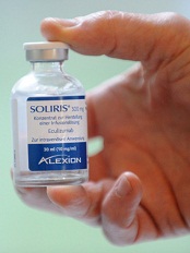
Credit: Globovision
The manufacturer of the recently approved eculizumab (Soliris) issued a voluntary US recall on June 2 of a single affected lot of the drug, although it included 8 additional lots in the recall.
The manufacturer, Alexion Pharmaceuticals, found visible particulates in the 300 mg/30 mL concentrated solution for intravenous infusion, which could cause an immune reaction and blood clots in patients receiving the drug.
The single affected Soliris lot, distributed in the US only, is #1007A. Also included in the U.S. recall are lots 10002-1, 00006-1, 10003A, 10004A, 10005A, 10005AR, 10006A, and 10008A.
All lots in the recall were manufactured using the same process component.
Alexion believes it has identified the part of the process that has resulted in the visible particles in the solution and has changed the process component.
An earlier recall of eculizumab, also for particulate matter, occurred in December 2013.
Alexion does not anticipate any interruption to the patient supply of eculizumab.
Eculizumab recently received full US Food and Drug Administration (FDA) approval to treat adult and pediatric patients with atypical hemolytic uremic syndrome (aHUS). It had received accelerated approval for this indication in 2011.
Eculizumab is also FDA-approved to treat patients with paroxysmal nocturnal hemoglobinuria.
For more information on the recall, visit the company website.
Healthcare professionals and patients should report adverse events or side effects related to Soliris to the FDA's MedWatch Safety Information and Adverse Event Reporting Program. ![]()
Study explains why pneumococcal vaccines fall short in SCD

Credit: St Jude Children’s
Research Hospital
A new study reveals differences in the pneumococcal genome that explain why current vaccines do not sufficiently protect children with sickle cell disease (SCD) from pneumococcal infections.
Researchers performed whole-genome sequencing of hundreds of pneumococcal bacteria collected from healthy subjects and patients with SCD.
The team found that disease-causing strains of the bacteria differed between the 2 groups.
And the pneumococcal strains from the SCD patients differed from the 13 pneumococcal strains included in the current vaccine recommended for children age 5 and younger.
“The results help explain why current vaccines haven’t been as successful at protecting children with sickle cell disease from pneumococcal infections as they have in protecting other children,” said Joshua Wolf, MD, of St Jude Children’s Research Hospital.
Dr Wolf and his colleagues detailed these results in Cell Host & Microbe.
The researchers had compared the genomes of 322 pneumococcal bacteria collected from SCD patients between 1994 and 2011 to DNA from 327 strains obtained from individuals without SCD.
The analysis revealed that, over time, the genomes of bacteria isolated from SCD patients shrank, as genes and the corresponding DNA were discarded or combined. The changes reflected bacterial adaptation to the SCD host and contributed to the bacteria’s ability to persist despite advances in preventive care.
The researchers then used transposon sequencing to compare the bacterial fitness of pneumococcal genes in mice with and without SCD. This revealed 60 genes whose transposon disruption resulted in fitness differences between the 2 types of mice.
So the bacteria faced different conditions in animals with and without SCD. The bloodstream of normal mice was a more hostile environment for pneumococcal bacteria than the bloodstream of mice with SCD.
When the researchers evaluated the aforementioned 60 genes in bacteria isolated from SCD patients, they found 6 that were missing or altered in a significant percentage of samples (P<0.05). This included SP0511, SP0946, SP1032, SP1449, SP1483, and SP1835.
These genes are involved in transporting iron into bacteria, bacterial metabolism, and other processes that are likely altered in patients with SCD.
“We demonstrated that genes necessary to cause disease in the general public are expendable in patients with sickle cell disease,” said study author Jason Rosch, PhD, also of St Jude.
The researchers believe these findings will aid efforts to improve vaccine effectiveness and inform research into new ways to protect young SCD patients from life-threatening pneumococcal infections that can lead to pneumonia, meningitis, bloodstream infections, and other problems. ![]()
Credit: St Jude Children’s
Research Hospital
A new study reveals differences in the pneumococcal genome that explain why current vaccines do not sufficiently protect children with sickle cell disease (SCD) from pneumococcal infections.
Researchers performed whole-genome sequencing of hundreds of pneumococcal bacteria collected from healthy subjects and patients with SCD.
The team found that disease-causing strains of the bacteria differed between the 2 groups.
And the pneumococcal strains from the SCD patients differed from the 13 pneumococcal strains included in the current vaccine recommended for children age 5 and younger.
“The results help explain why current vaccines haven’t been as successful at protecting children with sickle cell disease from pneumococcal infections as they have in protecting other children,” said Joshua Wolf, MD, of St Jude Children’s Research Hospital.
Dr Wolf and his colleagues detailed these results in Cell Host & Microbe.
The researchers had compared the genomes of 322 pneumococcal bacteria collected from SCD patients between 1994 and 2011 to DNA from 327 strains obtained from individuals without SCD.
The analysis revealed that, over time, the genomes of bacteria isolated from SCD patients shrank, as genes and the corresponding DNA were discarded or combined. The changes reflected bacterial adaptation to the SCD host and contributed to the bacteria’s ability to persist despite advances in preventive care.
The researchers then used transposon sequencing to compare the bacterial fitness of pneumococcal genes in mice with and without SCD. This revealed 60 genes whose transposon disruption resulted in fitness differences between the 2 types of mice.
So the bacteria faced different conditions in animals with and without SCD. The bloodstream of normal mice was a more hostile environment for pneumococcal bacteria than the bloodstream of mice with SCD.
When the researchers evaluated the aforementioned 60 genes in bacteria isolated from SCD patients, they found 6 that were missing or altered in a significant percentage of samples (P<0.05). This included SP0511, SP0946, SP1032, SP1449, SP1483, and SP1835.
These genes are involved in transporting iron into bacteria, bacterial metabolism, and other processes that are likely altered in patients with SCD.
“We demonstrated that genes necessary to cause disease in the general public are expendable in patients with sickle cell disease,” said study author Jason Rosch, PhD, also of St Jude.
The researchers believe these findings will aid efforts to improve vaccine effectiveness and inform research into new ways to protect young SCD patients from life-threatening pneumococcal infections that can lead to pneumonia, meningitis, bloodstream infections, and other problems.
Team reports way to reduce sickling, SCD progression

Credit: Graham Colm
Through a series of preclinical experiments, scientists discovered they could reduce the sickling of red blood cells (RBCs) and slow the progression of sickle cell disease (SCD).
In a mouse model of SCD and blood samples from SCD patients, the researchers reduced sickling by manipulating sphingosine-1-phosphate (S1P) and sphingosine kinase 1 (SPHK1).
Yang Xia, MD, PhD, of The University of Texas Health Science Center at Houston, and colleagues described this work in The Journal of Clinical Investigation.
The scientists first discovered that S1P, a lipid enriched and stored in RBCs, is elevated in mice with SCD. Further investigation revealed that elevated SPHK1 activity underlies the increased levels of S1P and contributes to RBC sickling.
To confirm SPHK1’s role in SCD, the researchers tested 2 SPHK1 inhibitors, SK1-I and PF-543, in cells from mice with SCD. Both agents successfully inhibited SPHK1 activity and reduced S1P levels in a dose-dependent manner, but PF-543 demonstrated greater potency.
In subsequent experiments, PF-543 decreased intravascular hemolysis and reduced inflammation in mice with SCD. The treatment also decreased tissue injury and splenomegaly and increased survival in the mice.
When the scientists knocked down SPHK1 in hematopoietic stem cells (HSCs), they observed decreased sickling and reticulocytes in SCD chimeras, resulting from a reduction in S1P levels.
Knocking down SPHK1 in HSCs also decreased intravascular hemolysis, prolonged RBC life span, reduced inflammation, decreased splenomegaly and tissue injury, and increased survival in the mice.
Experiments in cells from patients with SCD showed that SPHK1 activity and S1P levels were elevated and directly contributed to sickling. So the researchers decided to evaluate how PF-543 would affect these cells.
Treating cells with PF-543 significantly inhibited hypoxia-induced SPHK1 activity and prevented the elevation of S1P in a dose-dependent manner. The treatment also significantly reduced the percentage of sickled cells in a dose-dependent manner.
Finally, the scientists found that S1P-induced sickling was independent of S1P receptor activation. S1P receptor antagonists did not inhibit hypoxia-induced sickling in cells from SCD patients. And treatment with S1P did not enhance sickling under hypoxic conditions.
“This work could lead to novel treatments for sickle cell disease,” said study author Harinder Juneja, MD, of The University of Texas Health Science Center. “The study provides a better understanding of the pathogenesis of the disease and reveals a new therapeutic target.”
Coauthor Rod Kellems, PhD, also of The University of Texas Health Science Center, added, “This research provides insight into how red blood cells work, revealing that SPHK1-mediated elevation of S1P contributes to sickling and promotes disease progression and highlights potential therapeutic opportunities for sickle cell disease.”
Group finds cancer stem cells in MDS patients

Credit: NIH
A new study provides “conclusive evidence” to support the existence of cancer stem cells in patients with myelodysplastic syndromes (MDS), according to researchers.
The group analyzed malignant cells in the bone marrow of MDS patients and identified a distinct subset of cells that showed all the hallmarks of cancer stem cells.
Only these MDS stem cells—none of the other malignant cells—were able to propagate the disease.
The researchers reported these discoveries in Cancer Cell.
Petter Woll, PhD, of the University of Oxford in the UK, and his colleagues conducted this research, analyzing bone marrow cells from 15 patients with low- or intermediate-risk MDS.
The team set out to establish in which cells cancer-driving mutations originated. This led them to a rare subset of MDS cells—Lin-CD34+CD38-CD90+CD45RA- cells—that appeared to have all the properties of cancer stem cells.
These cells sat at the top of a hierarchy of MDS cells, could sustain themselves and replenish the other MDS cells, and were the origin of all stable DNA changes and mutations that drove the progression of MDS.
“This is conclusive evidence for the existence of cancer stem cells in myelodysplastic syndromes,” Dr Woll said. “We have identified a subset of cancer cells [and] shown that these rare cells are invariably the cells in which the cancer originates and also are the only cancer-propagating cells in the patients.”
Study author Sten Eirik W. Jacobsen, MD, PhD, also of the University of Oxford, noted that cancer stem cells have already been reported in a number of malignancies.
But previous findings have remained controversial, since the lab tests used to establish the identity of cancer stem cells have been shown to be unreliable.
“In our studies, we avoided the problem of unreliable lab tests by tracking the origin and development of cancer-driving mutations in MDS patients,” Dr Jacobsen said.
Dr Woll added that this research provides a target for the development of more efficient therapies for MDS.
“We need to understand more about what makes these cancer stem cells unique, what makes them different to all the other cancer cells,” he said. “If we can find biological pathways that are specifically dysregulated in cancer stem cells, we might be able to target them with new drugs.”