User login
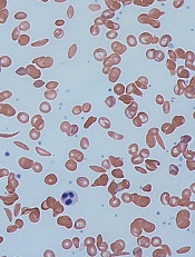
Tests use cell density to diagnose sickle cell disease

and a sickled cell
Credit: Betty Pace
Two new tests can diagnose sickle cell disease (SCD) quickly, simply, and cheaply, researchers have reported in PNAS.
The tests separate red blood cells based on density and can provide an SCD diagnosis in less than 12 minutes for as little as 50 cents.
When run against clinical samples, both tests showed high sensitivity and specificity for SCD. And one of the tests could distinguish between the two main subclasses of SCD.
Ashok A. Kumar, PhD, of Harvard University in Cambridge, Massachusetts, and his colleagues developed the tests by connecting two ideas scientists have understood for decades.
The first is the notion that blood cells affected by SCD are denser than normal cells, and the second is that many polymers, when mixed in water, automatically separate into layers ordered by density.
Dr Kumar and his colleagues discovered the layers could be used to separate red blood cells by density.
So the researchers designed 2 tests to separate red blood cells into multiple bins of density. The presence or absence of cells in the bins distinguished individuals with the most prevalent forms of SCD (Hb SS or Hb SC) from individuals with either normal hemoglobin (Hb AA) or sickle-cell trait (Hb AS).
“We wanted to make the test[s] as simple as possible,” Dr Kumar noted. “The idea was to make [them] something you could run from just a finger prick. Because these gradients assemble on their own, that meant we could make them in whatever volume we wanted, even a small capillary tube.”
The design the team settled on is barely larger than a toothpick. Running the tests is as simple as uncapping a tube, pricking a patient’s finger, and allowing the blood to wick into the tube.
The researchers evaluated the tests in 59 subjects, 33 who were negative for SCD and 26 who were positive for SCD. Both tests identified SCD-positive samples with a sensitivity greater than 90% and a specificity greater than 88%.
The simpler test, called SCD-AMPS-2, involves 2 phases. It identified Hb SS and Hb SC with a sensitivity of 90% (73%–98%) and a specificity of 97% (86%–100%).
The higher-resolution test, called SCD-AMPS-3, involves 3 phases. It identified the 2 types of SCD with a sensitivity of 91% (78%–98%) and a specificity of 88% (74%–98%). This test could also distinguish between Hb SS and Hb SC.
Despite these promising results, Dr Kumar said additional testing will be needed to determine whether the tests are truly accurate enough to use in the field. ![]()
FDA approves generic decitabine for MDS

Credit: Bill Branson
The US Food and Drug Administration (FDA) has approved decitabine for injection, a generic version of Dacogen, to treat patients with myelodysplastic syndromes (MDS).
Decitabine is indicated for previously treated and untreated patients with de novo and secondary MDS of all French-American-British subtypes—refractory anemia, refractory anemia with ringed sideroblasts, refractory anemia with excess blasts, refractory anemia with excess blasts in transformation, and chronic myelomonocytic leukemia—as well as intermediate-1, intermediate-2, and high-risk International Prognostic Scoring System groups.
Decitabine will be marketed in 20 mL single-dose glass vials containing 50 mg decitabine—the same size and strength as the brand name drug. The dosing regimen is identical as well.
InnoPharma developed the generic formulation of decitabine and entered into an agreement with Sandoz Inc. Sandoz will sell, market, and distribute decitabine in the US. InnoPharma is set to be acquired by Pfizer Inc., but the transaction is subject to US regulatory approval.
The FDA approved another generic form of decitabine for the treatment of MDS in July 2013. That drug is a product of Dr Reddy’s Laboratories Limited.
Dacogen has been FDA-approved to treat MDS since May 2006. Dacogen is a registered trademark used by Eisai Inc. under license from Astex Pharmaceuticals Inc. ![]()
Drug granted orphan status for PNH in EU

A novel compound has received orphan status in the Europe Union to treat paroxysmal nocturnal hemoglobinuria (PNH), a life-threatening disease that causes severe anemia and confers a high risk of thrombosis.
The compound, AMY-101, works by inhibiting C3, a central component of the complement immune system.
AMY-101 was developed by John Lambris, PhD, of the University of Pennsylvania, and subsequently licensed to Amyndas Pharmaceuticals.
AMY-101’s orphan status provides Amyndas with benefits such as tax incentives, market exclusivity for 10 years, possibilities for additional research funding, and additional guidance from the European Medicines Agency during clinical development.
How AMY-101 works
PNH is caused by the defective expression of regulatory proteins on the surface of blood cells, which leaves them vulnerable to complement attack. This can lead to hemolysis, which results in severe anemia and contributes to a high risk of thrombosis.
The monoclonal antibody eculizumab is often successful in treating PNH, but roughly a third of patients do not respond well to the drug and still require blood transfusions to manage their anemia.
Research has suggested this lack of response is due to fragments of complement C3 proteins on the surface of the patients’ red blood cells, which are eventually attacked by immune cells.
In an attempt to overcome this problem, Dr Lambris and his colleagues developed AMY-101. The drug is designed to inhibit the complement cascade, thereby preventing hemolysis and immune cell recognition.
The researchers have investigated the effects of AMY-101 on self-attack and the resulting hemolysis in human PNH cells and found the drug to be active.
These results have not been published, but the group has published results with a C3 inhibitor known as Cp40, and AMY-101 is based on Cp40.
The researchers reported in Blood that Cp40 and its long-acting form, PEG-Cp40, effectively inhibited hemolysis and efficiently prevented the deposition of C3 fragments on red blood cells from patients with PNH. ![]()
Drug can treat inflammation-induced anemia

An experimental drug designed to help regulate the blood’s iron supply may be a viable treatment option for inflammation-induced anemia, according to a study published in Blood.
The only current treatment strategy for this type of anemia involves targeting the underlying disease or infection.
However, recent research has sought to explore additional options for patients whose inflammation is difficult to control or when the cause of inflammation is unknown.
A hepcidin inhibitor called lexaptepid pegol (lexaptepid) has demonstrated efficacy in treating inflammation-induced anemia in animal studies. Lexaptepid inactivates hepcidin, thereby maintaining the transport of iron to the bloodstream.
To evaluate lexaptepid’s potential in humans, investigators caused inflammation-induced anemia in 24 healthy male adults and randomized them to receive lexaptepid or placebo.
Subjects received a low dose of Escherichia coli endotoxin to induce controlled inflammation and received either lexaptepid or placebo 30 minutes later.
After 9 hours, serum iron had decreased by 8.3±9.0 μmol/L in controls but increased by 15.9±9.8 μmol/L in lexaptepid-treated subjects (P<0.0001).
In addition to evaluating whether lexaptepid interfered with hepcidin production, the researchers also sought to determine whether the drug influenced the immune response.
Results suggested it did not. Treated subjects and controls alike experienced flu-like symptoms, increased body temperature and white blood cell counts, and higher concentrations of inflammatory and signaling proteins.
“It is quite encouraging that lexaptepid helped maintain appropriate levels of iron in the bloodstream of healthy volunteers without compromising the immune response,” said lead study author Lucas van Eijk, MD, of Radboud University Medical Center in Nijmegen, The Netherlands.
“We are hopeful that, with further study, this first-of-its-kind therapy could significantly improve quality of life for patients suffering from chronic illnesses.”
Results of a phase 2 study testing lexaptepid in anemic cancer patients were presented at the AACR Annual Meeting 2014. ![]()
FDA approves eltrombopag for SAA
The US Food and Drug Administration (FDA) has approved eltrombopag (Promacta) for use in patients with severe aplastic anemia (SAA) who have had an insufficient response to immunosuppressive therapy (IST).
Eltrombopag is an oral thrombopoietin receptor agonist that helps to induce the proliferation and differentiation of hematopoietic stem cells to increase blood cell production.
Eltrombopag is already FDA approved to treat patients with chronic immune thrombocytopenia who have had an insufficient response to corticosteroids, immunoglobulins, or splenectomy.
The drug is also approved to treat thrombocytopenia in patients with chronic hepatitis C to allow for the initiation and maintenance of interferon-based therapy.
The latest FDA approval is based on results from an investigator-sponsored phase 2 study (09-H-0154) conducted by the National Heart, Lung and Blood Institute. Results of this trial were previously published in The New England Journal of Medicine.
Eltrombopag in SAA: Latest trial results
In this study, researchers evaluated eltrombopag in 43 SAA patients who had an insufficient response to at least 1 prior IST and a platelet count of 30 x 109/L or less.
At baseline, the median platelet count was 20 x 109/L, hemoglobin was 8.4 g/dL, the absolute neutrophil count was 0.58 x 10⁹/L, and absolute reticulocyte count was 24.3 x 10⁹/L.
Patients had a median age of 45 years (range, 17 to 77 years), and 56% were male. The majority of patients (84%) received at least 2 prior ISTs.
Patients received eltrombopag at an initial dose of 50 mg once daily for 2 weeks. The dose increased over 2-week periods to a maximum of 150 mg once daily.
The study’s primary endpoint was hematologic response, which was initially assessed after 12 weeks of treatment. Treatment was discontinued after 16 weeks in patients who did not exhibit a hematologic response.
Forty percent of patients (N=17) experienced a hematologic response in at least one lineage—platelets, red blood cells (RBCs), or white blood cells—after week 12.
In the extension phase of the study, 8 patients achieved a multilineage response. Four of these patients subsequently tapered off treatment and maintained the response. The median follow up was 8.1 months (range, 7.2 to 10.6 months).
Ninety-one percent of patients were platelet-transfusion-dependent at baseline. Patients who responded to eltrombopag did not require platelet transfusions for a median of 200 days (range, 8 to 1096 days).
Eighty-six percent of patients were RBC-transfusion-dependent at baseline. Patients who responded to eltrombopag did not require RBC transfusions for a median of 208 days (range, 15 to 1082 days).
The most common adverse events (≥20%) associated with eltrombopag were nausea (33%), fatigue (28%), cough (23%), diarrhea (21%), and headache (21%).
Patients also had bone marrow aspirates evaluated for cytogenetic abnormalities. Eight patients had a new cytogenetic abnormality after treatment, including 5 patients who had complex changes in chromosome 7.
Patients who develop new cytogenetic abnormalities while on eltrombopag may need to be taken off treatment.
Eltrombopag is marketed by GlaxoSmithKline under the brand name Promacta in the US and Revolade in most other countries. For more information on eltrombopag, see the prescribing information. ![]()
Study links gene dysfunction to Fanconi anemia, AML

Credit: Tom Ellenberger
Researchers say they’ve discovered “an intimate link” between RUNX genes and Fanconi anemia, a finding that also has implications for treating acute myeloid leukemia (AML).
The group found that RUNX1 and RUNX3 interact with Fanconi anemia group D2 (FANCD2), a protein involved in the repair of DNA damage.
The RUNX proteins facilitate the recruitment of FANCD2 to sites of DNA damage in both Fanconi anemia and AML.
Motomi Osato, MD, PhD, of the Cancer Science Institute of Singapore, and his colleagues recounted these findings in Cell Reports.
The researchers began by studying RUNX deficiency in mice. They found that knockdown of both RUNX1 and RUNX3 led to bone marrow failure or myeloproliferative disorders in the mice.
These clinical manifestations are seen in inherited bone marrow failure syndromes such as Fanconi anemia, which is caused by the disruption of gene products that participate in the repair of DNA interstrand crosslinks (ICLs).
With this in mind, the researchers decided to investigate RUNX function in the ICL repair pathway. And they found RUNX proteins play a critical role in the pathway by facilitating the recruitment of FANCD2 to sites of DNA damage.
To explore the clinical relevance of RUNX participation in DNA damage repair, the team conducted experiments in Kasumi-1 and SKNO-1 cells. These AML cell lines express RUNX1-ETO, which is thought to suppress the expression and/or function of RUNX1 and RUNX3 simultaneously.
The researchers showed that Kasumi-1 and SKNO-1 cells were sensitive to the ICL-inducing agent mytomycin C, and knocking down RUNX1-ETO reduced this sensitivity. Depleting RUNX1-ETO also led to increased FANCD2 recruitment to chromatin.
The team said these results suggest that RUNX1-ETO might repress FANCD2 foci formation and ICL repair in AML cells. And they predicted that RUNX dysfunction would sensitize the cells to PARP inhibitors.
So they tested 2 PARP inhibitors—olaparib and rucaparib—in Kasumi-1 cells and observed sensitivity to both drugs. Knocking down RUNX1-ETO partially reduced this sensitivity, while adding mytomycin C increased sensitivity.
“PARP inhibitors have been with us for quite some time, but nobody has realized their application for leukemia,” Dr Osato said. “Our study has shed light on the possibility of a more effective treatment using a combined therapy with PARP inhibitors, which can potentially be extended to other types of common cancers.”
The researchers are now conducting additional drug testing in xenograft models. ![]()
Monthly transfusions may prevent stroke recurrence in SCD

Credit: St Jude Children’s
Research Hospital
Monthly blood transfusions can reduce the risk of silent or overt stroke among children with sickle cell disease (SCD) who previously had a silent stroke, according to a study published in The New England Journal of Medicine.
Children with evidence of silent cerebral infarcts who received monthly blood transfusions for 3 years had a 58% lower risk of suffering repeat silent or overt
strokes than children who did not receive transfusions.
In fact, researchers said the actual benefit of transfusion therapy may be even higher, as 15% of the children who were assigned to receive transfusions either did not receive them or only received them for a brief period.
“The results of our study show that blood transfusions can play a critical role in preventing this insidious and potentially devastating condition,” said study author James F. Casella, MD, of the Johns Hopkins Children’s Center in Baltimore, Maryland.
“They also highlight the importance of intervening early to preclude ongoing or further brain injury among these youngsters. Most importantly, our findings suggest a much-needed treatment option for clinicians and families of children with sickle cell disease who have had silent strokes.”
Previous studies have suggested that blood transfusions may help prevent stroke in patients with SCD by increasing the number of normal red blood cells and decreasing the likelihood of blocked blood vessels.
But Dr Casella and his colleagues wanted to determine if monthly blood transfusions would help prevent stroke in children with SCD who had evidence of a previous silent cerebral infarct, as well as whether the benefits of transfusion outweigh the risks.
The researchers analyzed 196 children, ages 5 to 15 years, who were diagnosed with SCD and had infarct-like lesions on their MRI scans. The children were randomized to an observation arm or to receive blood transfusions every month for 3 years.
Six percent (6/99) of children who received regular transfusions suffered another silent or overt stroke. One of the patients had a stroke, and 5 had new or enlarged silent cerebral infarcts.
In comparison, 14% (14/97) of children in the observation arm experienced a silent or overt stroke. Seven had a stroke, and 7 had new or enlarged silent cerebral infarcts.
So children who did not receive transfusions were more than twice as likely as their peers to have repeat strokes.
Children who did not receive transfusions were also more likely to suffer a range of other SCD-related problems, such as episodes of extreme pain. There were 295 pain episodes among children who did not receive transfusions and 126 episodes among transfused patients.
An unexpected result, according to the researchers, was that intelligence measures were not different between the 2 treatment arms. Previous studies suggested that silent strokes are associated with a 5-point reduction in IQ. The researchers said they plan to explore this finding further.
Nevertheless, this study provides “clear evidence” that transfusions can decrease the progression of silent strokes in children with SCD, said study author Michael R. DeBaun, MD, of Vanderbilt University in Nashville, Tennessee.
“These results suggest that children who have this disease should be screened early for silent strokes, at least by the time they begin elementary school, to help them manage the disease and to ensure minimal impact on school performance,” he added.
Dr DeBaun and his colleagues said children with SCD should have a surveillance MRI, preferably without sedation, at a young age. Most children with SCD who are at risk for a silent stroke will have one by age 6 years.
The researchers also noted that healthcare providers should discuss treatment options with families to determine if transfusion therapy is appropriate, as there is a risk of transfusion reactions and iron overload.
The decision to transfuse should be made by factoring in each child’s overall health, medical history, and the ability to take time from school for monthly procedures.
The researchers said further study is needed to identify which children with a history of silent strokes are at greatest risk for recurrence so transfusion therapy can be targeted to them.
An editorial related to this study also calls for additional research to determine if the findings can be translated to clinical practice. ![]()
Credit: St Jude Children’s
Research Hospital
Monthly blood transfusions can reduce the risk of silent or overt stroke among children with sickle cell disease (SCD) who previously had a silent stroke, according to a study published in The New England Journal of Medicine.
Children with evidence of silent cerebral infarcts who received monthly blood transfusions for 3 years had a 58% lower risk of suffering repeat silent or overt
strokes than children who did not receive transfusions.
In fact, researchers said the actual benefit of transfusion therapy may be even higher, as 15% of the children who were assigned to receive transfusions either did not receive them or only received them for a brief period.
“The results of our study show that blood transfusions can play a critical role in preventing this insidious and potentially devastating condition,” said study author James F. Casella, MD, of the Johns Hopkins Children’s Center in Baltimore, Maryland.
“They also highlight the importance of intervening early to preclude ongoing or further brain injury among these youngsters. Most importantly, our findings suggest a much-needed treatment option for clinicians and families of children with sickle cell disease who have had silent strokes.”
Previous studies have suggested that blood transfusions may help prevent stroke in patients with SCD by increasing the number of normal red blood cells and decreasing the likelihood of blocked blood vessels.
But Dr Casella and his colleagues wanted to determine if monthly blood transfusions would help prevent stroke in children with SCD who had evidence of a previous silent cerebral infarct, as well as whether the benefits of transfusion outweigh the risks.
The researchers analyzed 196 children, ages 5 to 15 years, who were diagnosed with SCD and had infarct-like lesions on their MRI scans. The children were randomized to an observation arm or to receive blood transfusions every month for 3 years.
Six percent (6/99) of children who received regular transfusions suffered another silent or overt stroke. One of the patients had a stroke, and 5 had new or enlarged silent cerebral infarcts.
In comparison, 14% (14/97) of children in the observation arm experienced a silent or overt stroke. Seven had a stroke, and 7 had new or enlarged silent cerebral infarcts.
So children who did not receive transfusions were more than twice as likely as their peers to have repeat strokes.
Children who did not receive transfusions were also more likely to suffer a range of other SCD-related problems, such as episodes of extreme pain. There were 295 pain episodes among children who did not receive transfusions and 126 episodes among transfused patients.
An unexpected result, according to the researchers, was that intelligence measures were not different between the 2 treatment arms. Previous studies suggested that silent strokes are associated with a 5-point reduction in IQ. The researchers said they plan to explore this finding further.
Nevertheless, this study provides “clear evidence” that transfusions can decrease the progression of silent strokes in children with SCD, said study author Michael R. DeBaun, MD, of Vanderbilt University in Nashville, Tennessee.
“These results suggest that children who have this disease should be screened early for silent strokes, at least by the time they begin elementary school, to help them manage the disease and to ensure minimal impact on school performance,” he added.
Dr DeBaun and his colleagues said children with SCD should have a surveillance MRI, preferably without sedation, at a young age. Most children with SCD who are at risk for a silent stroke will have one by age 6 years.
The researchers also noted that healthcare providers should discuss treatment options with families to determine if transfusion therapy is appropriate, as there is a risk of transfusion reactions and iron overload.
The decision to transfuse should be made by factoring in each child’s overall health, medical history, and the ability to take time from school for monthly procedures.
The researchers said further study is needed to identify which children with a history of silent strokes are at greatest risk for recurrence so transfusion therapy can be targeted to them.
An editorial related to this study also calls for additional research to determine if the findings can be translated to clinical practice.
NICE supports lenalidomide for MDS

The UK’s National Institute for Health and Care Excellence (NICE) has issued a final draft guidance recommending lenalidomide (Revlimid) as an option for treating myelodysplastic syndromes (MDS) characterized by 5q deletion.
Lenalidomide is approved in the European Union to treat transfusion-dependent anemia caused by low- or intermediate-1 risk MDS characterized by 5q deletion when other therapeutic options are insufficient or inadequate.
However, the main treatment option for this patient population in the UK is best supportive care, which involves regular red blood cell transfusions.
In earlier draft guidances, NICE did not support lenalidomide use in MDS patients with 5q deletion. Although data suggested the drug is effective for these patients, a NICE advisory committee was not convinced the drug provided a survival benefit.
But now, the committee has concluded that lenalidomide is a clinically effective treatment for these patients because it is associated with a statistically significant improvement in transfusion independence and health-related quality of life compared with placebo.
Furthermore, the committee said it is plausible that lenalidomide can indirectly improve overall survival by improving transfusion independence.
“The committee heard from clinical experts that lenalidomide is an effective therapy,” said Sir Andrew Dillon, NICE chief executive.
“Celgene–who market lenalidomide–worked with us to provide enough evidence to make it possible for us to recommend it for this group of people. Celgene provided a revised analysis and further information on their proposal for a reduction in the cost of the drug to the NHS [National Health Service].”
This patient access scheme involves the NHS paying for lenalidomide treatment for up to 26 monthly cycles. And Celgene will provide the drug free of charge for those people who receive more than 26 monthly cycles.
Lenalidomide is available in 21-day packs of 10 mg and 5 mg capsules at net prices of £3780 and £3570, respectively. The cost of a 28-day cycle of treatment with 10 mg of lenalidomide (excluding value-added tax) is £3780.
The committee noted that the incremental cost-effectiveness ratio for lenalidomide compared with best supportive care is uncertain because the proportion of people who might need treatment beyond 26 cycles is uncertain.
However, the committee accepted that a commitment from Celgene to publish data on the proportion of people receiving treatment beyond 26 cycles would provide reassurance that lenalidomide is a cost-effective use of NHS resources in MDS patients with 5q deletion.
NICE’s final draft guidance is now with consultees, who have the opportunity to appeal against it. Until NICE issues a final guidance, NHS bodies should make decisions locally on the funding of specific treatments.
Method may help treat SCD, other disorders
Credit: Graham Beards
Scientists have discovered that manipulating gene regulation can cause red blood cells to produce fetal hemoglobin, a finding that may have implications for sickle cell disease (SCD) and other hemoglobinopathies.
The researchers used protein-engineering techniques to force chromatin fiber into looped structures that contact DNA at specific sites to preferentially activate genes that regulate hemoglobin.
The team described the work in Cell. The research was previously presented at the 2013 ASH Annual Meeting.
Key to the researcher’s method is a developmental transition that normally occurs in the blood of newborns. A biological switch regulates a changeover from fetal hemoglobin to adult hemoglobin as it begins to silence the genes that produce fetal hemoglobin.
Hematologists have long known that SCD patients with elevated levels of fetal hemoglobin have a milder form of the disease.
“This observation has been a major driver in the field to understand the molecular basis of the mechanisms that control the biological switch, with the ultimate goal to reverse it,” said study author Gerd A. Blobel, MD, PhD, of The Children’s Hospital of Philadelphia in Pennsylvania.
In previous research, his team used bioengineering techniques to adapt zinc-finger proteins to latch onto specific DNA sites far apart on a chromosome. The chromatin loop that results transmits regulatory signals for specific genes.
In their current work, the researchers custom-designed zinc fingers to flip the biological switch in hematopoietic stem cells, reactivating the genes expressing fetal hemoglobin at the expense of the genes expressing adult hemoglobin.
The team achieved these results in cultured blood cells from adult mice and adult humans.
The researchers are now planning to test the approach in animal models of SCD. If this strategy corrects the disease in animals, it may set the stage to move to human trials.
Dr Blobel also noted that, in principle, the forced chromatin looping approach could be applied to other hemoglobin-related disorders, such as certain forms of thalassemia.
ESA recalled due to particulates

Amgen is recalling lots of the erythropoiesis-stimulating agent Aranesp (darbepoetin alfa) that were distributed in several countries outside the US.
The recall applies to 9 lots of Aranesp 500 mcg prefilled syringes from non-US distributors, wholesalers, and hospital pharmacies.
A routine quality examination revealed cellulose and/or polyester particles in a small number of syringes, so Amgen is recalling the 9 lots as a precautionary measure.
To date, there have been no complaints or adverse events that can be attributed to the presence of these particles.
The presence of particulate matter could elicit inflammatory and allergic responses, both chronic and acute, and may be life-threatening.
However, health implications may vary depending on the route of drug administration, the amount of particulate matter injected into the patient, the size of the particles, the patient’s underlying medical condition, and the presence of a right-to-left cardiac shunt.
The products impacted by the recall are Aranesp 500 mcg prefilled syringes. A single lot of Aranesp was packaged for different countries into 9 lots: 1042847, 1044141A, 1044141C, 1044141D, 1046891A, 1046891B, 1047394A, 1047622A, and 1047996A.
The impacted syringes were distributed in Belgium, Denmark, Finland, France, Ireland, Italy, Kuwait, Luxemburg, Russia, Saudi Arabia, Slovenia, Sweden, Switzerland, and the UK.
Consumers in the US who have questions regarding this recall can contact Amgen at 1-800-77-AMGEN (open 24 hours per day, 7 days per week).
Adverse events or quality problems associated with Aranesp can be reported to the US Food and Drug Administration’s MedWatch Adverse Event Reporting Program.
In the US, Aranesp is indicated for the treatment of anemia associated with chronic renal failure, including patients on dialysis and patients not on dialysis, or in patients with non-myeloid malignancies where anemia results from concomitantly administered chemotherapy.