User login
First target doesn’t affect survival in NSCLC with brain metastases
“The findings of our study highlight the importance of adopting a personalized, case-based approach when treating each patient” instead of always treating the brain or lung first, lead author Arvind Kumar, a medical student at Icahn School of Medicine at Mount Sinai, New York, said in an interview.
The study was released at European Lung Cancer Congress 2023.
According to the author, current guidelines recommend treating the brain first in patients with non–small cell lung cancer and a tumor that has spread to the brain.
“Determining whether the brain or body gets treated first depends on where the symptoms are coming from, how severe the symptoms are, how bulky the disease is, and how long the treatment to each is expected to take,” radiation oncologist Henry S. Park, MD, MPH, chief of the thoracic radiotherapy program at Yale University, New Haven, Conn., said in an interview. “Often the brain is treated first since surgery is used for both diagnosis of metastatic disease as well as removal of the brain metastasis, especially if it is causing symptoms. The radiosurgery that follows tends to occur within a day or a few days.”
However, he said, “if the brain disease is small and not causing symptoms, and the lung disease is more problematic, then we will often treat the body first and fit in the brain treatment later.”
For the new study, researchers identified 1,044 patients in the National Cancer Database with non–small cell lung cancer and brain metastases who received systemic therapy plus surgery, brain stereotactic radiosurgery, or lung radiation. All were treated from 2010 to 2019; 79.0% received brain treatment first, and the other 21.0% received lung treatment first.
There was no statistically significant difference in overall survival between those whose brains were treated first and those whose lungs were treated first (hazard ratio, 1.24, 95% confidence interval [CI], 0.91-1.70, P = .17). A propensity score–matched analysis turned up no difference in 5-year survival (38.2% of those whose brains were treated first, 95% CI, 27.5-34.4, vs. 38.0% of those whose lungs were treated first, 95% CI, 29.9-44.7, P = .32.)
“These results were consistent regardless of which combination of treatment modalities the patient received – neurosurgery versus brain stereotactic radiosurgery, thoracic surgery versus thoracic radiation,” the author said.
He cautioned that “our study only included patients who were considered candidates for either surgery or radiation to both the brain and lung. The results of our study should therefore be cautiously interpreted for patients who may have contraindications to such treatment.”
Dr. Park, who didn’t take part in the study, said “the results are consistent with what I would generally expect.”
He added: “The take-home message for clinicians should be that there is no one correct answer in how to manage non–small cell lung cancer with synchronous limited metastatic disease in only the brain. If the brain disease is bulky and/or causes symptoms while the body disease isn’t – or if a biopsy or surgery is required to prove that the patient in fact has metastatic disease – then the brain disease should be treated first. On the other hand, if the body disease is bulky and/or causing symptoms while the brain disease isn’t – and there is no need for surgery but rather only a biopsy of the brain – then the body disease can be treated first.”
No funding was reported. The study authors and Dr. Park reported no financial conflicts or other disclosures.
“The findings of our study highlight the importance of adopting a personalized, case-based approach when treating each patient” instead of always treating the brain or lung first, lead author Arvind Kumar, a medical student at Icahn School of Medicine at Mount Sinai, New York, said in an interview.
The study was released at European Lung Cancer Congress 2023.
According to the author, current guidelines recommend treating the brain first in patients with non–small cell lung cancer and a tumor that has spread to the brain.
“Determining whether the brain or body gets treated first depends on where the symptoms are coming from, how severe the symptoms are, how bulky the disease is, and how long the treatment to each is expected to take,” radiation oncologist Henry S. Park, MD, MPH, chief of the thoracic radiotherapy program at Yale University, New Haven, Conn., said in an interview. “Often the brain is treated first since surgery is used for both diagnosis of metastatic disease as well as removal of the brain metastasis, especially if it is causing symptoms. The radiosurgery that follows tends to occur within a day or a few days.”
However, he said, “if the brain disease is small and not causing symptoms, and the lung disease is more problematic, then we will often treat the body first and fit in the brain treatment later.”
For the new study, researchers identified 1,044 patients in the National Cancer Database with non–small cell lung cancer and brain metastases who received systemic therapy plus surgery, brain stereotactic radiosurgery, or lung radiation. All were treated from 2010 to 2019; 79.0% received brain treatment first, and the other 21.0% received lung treatment first.
There was no statistically significant difference in overall survival between those whose brains were treated first and those whose lungs were treated first (hazard ratio, 1.24, 95% confidence interval [CI], 0.91-1.70, P = .17). A propensity score–matched analysis turned up no difference in 5-year survival (38.2% of those whose brains were treated first, 95% CI, 27.5-34.4, vs. 38.0% of those whose lungs were treated first, 95% CI, 29.9-44.7, P = .32.)
“These results were consistent regardless of which combination of treatment modalities the patient received – neurosurgery versus brain stereotactic radiosurgery, thoracic surgery versus thoracic radiation,” the author said.
He cautioned that “our study only included patients who were considered candidates for either surgery or radiation to both the brain and lung. The results of our study should therefore be cautiously interpreted for patients who may have contraindications to such treatment.”
Dr. Park, who didn’t take part in the study, said “the results are consistent with what I would generally expect.”
He added: “The take-home message for clinicians should be that there is no one correct answer in how to manage non–small cell lung cancer with synchronous limited metastatic disease in only the brain. If the brain disease is bulky and/or causes symptoms while the body disease isn’t – or if a biopsy or surgery is required to prove that the patient in fact has metastatic disease – then the brain disease should be treated first. On the other hand, if the body disease is bulky and/or causing symptoms while the brain disease isn’t – and there is no need for surgery but rather only a biopsy of the brain – then the body disease can be treated first.”
No funding was reported. The study authors and Dr. Park reported no financial conflicts or other disclosures.
“The findings of our study highlight the importance of adopting a personalized, case-based approach when treating each patient” instead of always treating the brain or lung first, lead author Arvind Kumar, a medical student at Icahn School of Medicine at Mount Sinai, New York, said in an interview.
The study was released at European Lung Cancer Congress 2023.
According to the author, current guidelines recommend treating the brain first in patients with non–small cell lung cancer and a tumor that has spread to the brain.
“Determining whether the brain or body gets treated first depends on where the symptoms are coming from, how severe the symptoms are, how bulky the disease is, and how long the treatment to each is expected to take,” radiation oncologist Henry S. Park, MD, MPH, chief of the thoracic radiotherapy program at Yale University, New Haven, Conn., said in an interview. “Often the brain is treated first since surgery is used for both diagnosis of metastatic disease as well as removal of the brain metastasis, especially if it is causing symptoms. The radiosurgery that follows tends to occur within a day or a few days.”
However, he said, “if the brain disease is small and not causing symptoms, and the lung disease is more problematic, then we will often treat the body first and fit in the brain treatment later.”
For the new study, researchers identified 1,044 patients in the National Cancer Database with non–small cell lung cancer and brain metastases who received systemic therapy plus surgery, brain stereotactic radiosurgery, or lung radiation. All were treated from 2010 to 2019; 79.0% received brain treatment first, and the other 21.0% received lung treatment first.
There was no statistically significant difference in overall survival between those whose brains were treated first and those whose lungs were treated first (hazard ratio, 1.24, 95% confidence interval [CI], 0.91-1.70, P = .17). A propensity score–matched analysis turned up no difference in 5-year survival (38.2% of those whose brains were treated first, 95% CI, 27.5-34.4, vs. 38.0% of those whose lungs were treated first, 95% CI, 29.9-44.7, P = .32.)
“These results were consistent regardless of which combination of treatment modalities the patient received – neurosurgery versus brain stereotactic radiosurgery, thoracic surgery versus thoracic radiation,” the author said.
He cautioned that “our study only included patients who were considered candidates for either surgery or radiation to both the brain and lung. The results of our study should therefore be cautiously interpreted for patients who may have contraindications to such treatment.”
Dr. Park, who didn’t take part in the study, said “the results are consistent with what I would generally expect.”
He added: “The take-home message for clinicians should be that there is no one correct answer in how to manage non–small cell lung cancer with synchronous limited metastatic disease in only the brain. If the brain disease is bulky and/or causes symptoms while the body disease isn’t – or if a biopsy or surgery is required to prove that the patient in fact has metastatic disease – then the brain disease should be treated first. On the other hand, if the body disease is bulky and/or causing symptoms while the brain disease isn’t – and there is no need for surgery but rather only a biopsy of the brain – then the body disease can be treated first.”
No funding was reported. The study authors and Dr. Park reported no financial conflicts or other disclosures.
FROM ELCC 2023
Type of insurance linked to length of survival after lung surgery
The study used public insurance status as a marker for low socioeconomic status (SES) and suggests that patients with combined insurance may constitute a separate population that deserves more attention.
Lower SES has been linked to later stage diagnoses and worse outcomes in NSCLC. Private insurance is a generally-accepted indicator of higher SES, while public insurance like Medicare or Medicaid, alone or in combination with private supplementary insurance, is an indicator of lower SES.
Although previous studies have found associations between patients having public health insurance and experiencing later-stage diagnoses and worse overall survival, there have been few studies of surgical outcomes, and almost no research has examined combination health insurance, according to Allison O. Dumitriu Carcoana, who presented the research during a poster session at the European Lung Cancer Congress 2023.
“This is an important insurance subgroup for us because the majority of our patients fall into this subgroup by being over 65 years old and thus qualifying for Medicare while also paying for a private supplement,” said Ms. Dumitriu Carcoana, who is a medical student at University of South Florida Health Morsani College of Medicine, Tampa.
A previous analysis by the group found an association between private insurance status and better discharge status, as well as higher 5-year overall survival. After accumulating an additional 278 patients, the researchers examined 10-year survival outcomes.
In the new analysis, 52% of 711 participants had combination insurance, while 28% had private insurance, and 20% had public insurance. The subgroups all had similar demographic and histological characteristics. The study was unique in that it found no between-group differences in higher stage at diagnosis, whereas previous studies have found a greater risk of higher stage diagnosis among individuals with public insurance. As expected, patients in the combined insurance group had a higher mean age (P less than .0001) and higher Charlson comorbidity index scores (P = .0014), which in turn was associated with lower 10-year survival. The group also had the highest percentage of former smokers, while the public insurance group had the highest percentage of current smokers (P = .0003).
At both 5 and 10 years, the private insurance group had better OS than the group with public (P less than .001) and the combination insurance group (P = .08). Public health insurance was associated with worse OS at 5 years (hazard ratio, 1.83; P less than .005) but not at 10 years (HR, 1.18; P = .51), while combination insurance was associated with worse OS at 10 years (HR, 1.72; P = .02).
“We think that patients with public health insurance having the worst 5-year overall survival, despite their lower ages and fewer comorbid conditions, compared with patients with combination insurance, highlights the impact of lower socioeconomic status on health outcomes. These patients had the same tumor characteristics, BMI, sex, and race as our patients in the other two insurance groups. The only other significant risk factor [the group had besides having a higher proportion of patients with lower socioeconomic status was that it had a higher proportion of current smokers]. But the multivariate analyses showed that insurance status was an independent predictor of survival, regardless of smoking status or other comorbidities,” said Ms. Dumitriu Carcoana.
“At 10 years post-operatively, the survival curves have shifted and the combination patients had the worst 10-year overall survival. We attribute this to their higher number of comorbid conditions and increased age. In practice, [this means that] the group of patients with public insurance type, but no supplement, should be identified clinically, and the clinical team can initiate a discussion,” Ms. Dumitriu Carcoana said.
“Do these patients feel that they can make follow-up appointments, keep up with medication costs, and make the right lifestyle decisions postoperatively on their current insurance plan? If not, can they afford a private supplement? In our cohort specifically, it may also be important to do more preoperative counseling on the importance of smoking cessation,” she added.
The study is interesting, but it has some important limitations, according to Raja Flores, MD, who was not involved with the study. The authors stated that there was no difference between the insurance groups with respect to mortality or cancer stage, which is the most important predictor of survival. However, the poster didn't include details of the authors' analysis, making it difficult to interpret, Dr. Flores said.
The fact that the study includes a single surgeon has some disadvantages in terms of broader applicability, but it also controls for surgical technique. “Different surgeons have different ways of doing things, so if you had the same surgeon doing it the same way every time, you can look at other variables like insurance (status) and stage,” said Dr. Flores.
The results may also provide an argument against using robotic surgery in patients who do not have insurance, especially since they have not been proven to be better than standard minimally invasive surgery with no robotic assistance. With uninsured patients, “you’re using taxpayer money for a more expensive procedure that isn’t proving to be any better,” Dr. Flores explained.
The study was performed at a single center and cannot prove causation due to its retrospective nature.
Ms. Dumitriu Carcoana and Dr. Flores have no relevant financial disclosures.
*This article was updated on 4/13/2023.
The study used public insurance status as a marker for low socioeconomic status (SES) and suggests that patients with combined insurance may constitute a separate population that deserves more attention.
Lower SES has been linked to later stage diagnoses and worse outcomes in NSCLC. Private insurance is a generally-accepted indicator of higher SES, while public insurance like Medicare or Medicaid, alone or in combination with private supplementary insurance, is an indicator of lower SES.
Although previous studies have found associations between patients having public health insurance and experiencing later-stage diagnoses and worse overall survival, there have been few studies of surgical outcomes, and almost no research has examined combination health insurance, according to Allison O. Dumitriu Carcoana, who presented the research during a poster session at the European Lung Cancer Congress 2023.
“This is an important insurance subgroup for us because the majority of our patients fall into this subgroup by being over 65 years old and thus qualifying for Medicare while also paying for a private supplement,” said Ms. Dumitriu Carcoana, who is a medical student at University of South Florida Health Morsani College of Medicine, Tampa.
A previous analysis by the group found an association between private insurance status and better discharge status, as well as higher 5-year overall survival. After accumulating an additional 278 patients, the researchers examined 10-year survival outcomes.
In the new analysis, 52% of 711 participants had combination insurance, while 28% had private insurance, and 20% had public insurance. The subgroups all had similar demographic and histological characteristics. The study was unique in that it found no between-group differences in higher stage at diagnosis, whereas previous studies have found a greater risk of higher stage diagnosis among individuals with public insurance. As expected, patients in the combined insurance group had a higher mean age (P less than .0001) and higher Charlson comorbidity index scores (P = .0014), which in turn was associated with lower 10-year survival. The group also had the highest percentage of former smokers, while the public insurance group had the highest percentage of current smokers (P = .0003).
At both 5 and 10 years, the private insurance group had better OS than the group with public (P less than .001) and the combination insurance group (P = .08). Public health insurance was associated with worse OS at 5 years (hazard ratio, 1.83; P less than .005) but not at 10 years (HR, 1.18; P = .51), while combination insurance was associated with worse OS at 10 years (HR, 1.72; P = .02).
“We think that patients with public health insurance having the worst 5-year overall survival, despite their lower ages and fewer comorbid conditions, compared with patients with combination insurance, highlights the impact of lower socioeconomic status on health outcomes. These patients had the same tumor characteristics, BMI, sex, and race as our patients in the other two insurance groups. The only other significant risk factor [the group had besides having a higher proportion of patients with lower socioeconomic status was that it had a higher proportion of current smokers]. But the multivariate analyses showed that insurance status was an independent predictor of survival, regardless of smoking status or other comorbidities,” said Ms. Dumitriu Carcoana.
“At 10 years post-operatively, the survival curves have shifted and the combination patients had the worst 10-year overall survival. We attribute this to their higher number of comorbid conditions and increased age. In practice, [this means that] the group of patients with public insurance type, but no supplement, should be identified clinically, and the clinical team can initiate a discussion,” Ms. Dumitriu Carcoana said.
“Do these patients feel that they can make follow-up appointments, keep up with medication costs, and make the right lifestyle decisions postoperatively on their current insurance plan? If not, can they afford a private supplement? In our cohort specifically, it may also be important to do more preoperative counseling on the importance of smoking cessation,” she added.
The study is interesting, but it has some important limitations, according to Raja Flores, MD, who was not involved with the study. The authors stated that there was no difference between the insurance groups with respect to mortality or cancer stage, which is the most important predictor of survival. However, the poster didn't include details of the authors' analysis, making it difficult to interpret, Dr. Flores said.
The fact that the study includes a single surgeon has some disadvantages in terms of broader applicability, but it also controls for surgical technique. “Different surgeons have different ways of doing things, so if you had the same surgeon doing it the same way every time, you can look at other variables like insurance (status) and stage,” said Dr. Flores.
The results may also provide an argument against using robotic surgery in patients who do not have insurance, especially since they have not been proven to be better than standard minimally invasive surgery with no robotic assistance. With uninsured patients, “you’re using taxpayer money for a more expensive procedure that isn’t proving to be any better,” Dr. Flores explained.
The study was performed at a single center and cannot prove causation due to its retrospective nature.
Ms. Dumitriu Carcoana and Dr. Flores have no relevant financial disclosures.
*This article was updated on 4/13/2023.
The study used public insurance status as a marker for low socioeconomic status (SES) and suggests that patients with combined insurance may constitute a separate population that deserves more attention.
Lower SES has been linked to later stage diagnoses and worse outcomes in NSCLC. Private insurance is a generally-accepted indicator of higher SES, while public insurance like Medicare or Medicaid, alone or in combination with private supplementary insurance, is an indicator of lower SES.
Although previous studies have found associations between patients having public health insurance and experiencing later-stage diagnoses and worse overall survival, there have been few studies of surgical outcomes, and almost no research has examined combination health insurance, according to Allison O. Dumitriu Carcoana, who presented the research during a poster session at the European Lung Cancer Congress 2023.
“This is an important insurance subgroup for us because the majority of our patients fall into this subgroup by being over 65 years old and thus qualifying for Medicare while also paying for a private supplement,” said Ms. Dumitriu Carcoana, who is a medical student at University of South Florida Health Morsani College of Medicine, Tampa.
A previous analysis by the group found an association between private insurance status and better discharge status, as well as higher 5-year overall survival. After accumulating an additional 278 patients, the researchers examined 10-year survival outcomes.
In the new analysis, 52% of 711 participants had combination insurance, while 28% had private insurance, and 20% had public insurance. The subgroups all had similar demographic and histological characteristics. The study was unique in that it found no between-group differences in higher stage at diagnosis, whereas previous studies have found a greater risk of higher stage diagnosis among individuals with public insurance. As expected, patients in the combined insurance group had a higher mean age (P less than .0001) and higher Charlson comorbidity index scores (P = .0014), which in turn was associated with lower 10-year survival. The group also had the highest percentage of former smokers, while the public insurance group had the highest percentage of current smokers (P = .0003).
At both 5 and 10 years, the private insurance group had better OS than the group with public (P less than .001) and the combination insurance group (P = .08). Public health insurance was associated with worse OS at 5 years (hazard ratio, 1.83; P less than .005) but not at 10 years (HR, 1.18; P = .51), while combination insurance was associated with worse OS at 10 years (HR, 1.72; P = .02).
“We think that patients with public health insurance having the worst 5-year overall survival, despite their lower ages and fewer comorbid conditions, compared with patients with combination insurance, highlights the impact of lower socioeconomic status on health outcomes. These patients had the same tumor characteristics, BMI, sex, and race as our patients in the other two insurance groups. The only other significant risk factor [the group had besides having a higher proportion of patients with lower socioeconomic status was that it had a higher proportion of current smokers]. But the multivariate analyses showed that insurance status was an independent predictor of survival, regardless of smoking status or other comorbidities,” said Ms. Dumitriu Carcoana.
“At 10 years post-operatively, the survival curves have shifted and the combination patients had the worst 10-year overall survival. We attribute this to their higher number of comorbid conditions and increased age. In practice, [this means that] the group of patients with public insurance type, but no supplement, should be identified clinically, and the clinical team can initiate a discussion,” Ms. Dumitriu Carcoana said.
“Do these patients feel that they can make follow-up appointments, keep up with medication costs, and make the right lifestyle decisions postoperatively on their current insurance plan? If not, can they afford a private supplement? In our cohort specifically, it may also be important to do more preoperative counseling on the importance of smoking cessation,” she added.
The study is interesting, but it has some important limitations, according to Raja Flores, MD, who was not involved with the study. The authors stated that there was no difference between the insurance groups with respect to mortality or cancer stage, which is the most important predictor of survival. However, the poster didn't include details of the authors' analysis, making it difficult to interpret, Dr. Flores said.
The fact that the study includes a single surgeon has some disadvantages in terms of broader applicability, but it also controls for surgical technique. “Different surgeons have different ways of doing things, so if you had the same surgeon doing it the same way every time, you can look at other variables like insurance (status) and stage,” said Dr. Flores.
The results may also provide an argument against using robotic surgery in patients who do not have insurance, especially since they have not been proven to be better than standard minimally invasive surgery with no robotic assistance. With uninsured patients, “you’re using taxpayer money for a more expensive procedure that isn’t proving to be any better,” Dr. Flores explained.
The study was performed at a single center and cannot prove causation due to its retrospective nature.
Ms. Dumitriu Carcoana and Dr. Flores have no relevant financial disclosures.
*This article was updated on 4/13/2023.
FROM ELCC 2023
Outpatient costs top drug costs in some insured, working women with breast cancer
Among a sample of younger women with invasive breast cancer and employer-sponsored insurance, outpatient-related out-of-pocket (OOP) costs were greater than drug costs.
For these same patients, prescriptions were largely for nonproprietary anticancer drugs and entailed limited OOP costs. For women with high-deductible health plans (HDHPs) and commercially driven health plans (CDHPs), OOP costs were higher, compared with coverage by more generous plans, according to the Research Letter published in JAMA Network Open.
“You would expect that people undergoing cancer treatment should not have to face very high out-of-pocket costs associated with care regardless of treatment modality because their treatment is largely guideline-indicated, and they have no choices,” stated corresponding author Rena Conti, PhD, associate professor with the school of business, Boston University, in an interview. “If you are diagnosed with cancer and undergoing treatment, you’re following the recommendation of your doctor, and your doctor is following standard protocols for treatment. In that scenario, Economics 101 suggests that people should not have to pay anything or [should pay] very little, especially for things that are cheap and are known to be effective, because there’s no overuse. Where normally we think that out-of-pocket costs are meant to control overuse, people with breast cancer are not opting to get more than indicated chemotherapy or radiation.”
The analysis of 25,224 women with invasive breast cancer diagnosis and claims for 1 or more of 14 oral anticancer drugs revealed that OOP costs for nondrug outpatient claims represented 79.0% of total costs. OOP drug costs were modest, with a 30-day supply ranging from $0.57-$0.60 for tamoxifen to $134.08-$141.07 for palbociclib.
“We were interested in understanding to what extent women who are insured with private insurance are exposed to out-of-pocket costs for standard breast cancer treatment, both in looking at drugs, but also the other aspects of the treatments they undergo.”
High OOP costs for the oral anticancer prescription drugs that are central to breast cancer treatment are associated with treatment nonadherence and discontinuation. Little has been known, however, about OOP costs of treatment associated with invasive breast cancer among employer-insured women younger than 65 years, the paper says.
“This population may face significant financial burdens related to long-term hormonal-based prevention and enrollment in high-deductible health plans and consumer-driven health plans,” the authors state in their paper.
In the cross-sectional study, which used the national 2018 Marative MarketScan database, 23.1% were HDHP- or CDHP-insured. Fifty-one percent had no OOP costs for drugs. The total mean estimated OOP cost, however, was $1,502.23 per patient, with inpatient costs representing only $112.41 (95% confidence interval, $112.40-$112.42); outpatient costs were $1,186.27 (95% CI, $1,185.67-$1,188.16). Pharmaceutical costs were $203.55 (95% CI, $203.34-$203.78).“We were surprised to find that the vast majority were getting breast cancer treatment with older, very effective, very safe, relatively inexpensive drugs and had limited out-of-pocket costs with some variation – higher costs for the few receiving newer, expensive drugs. The backbone of treatment is the older, generic drugs, which are cheap for both the insurers and the patients. But we found also that women are facing high out-of-pocket costs for nondrug-based therapy – specifically for doctor visits, getting check-ups, diagnostic scans, and maybe other types of treatment, as well. ... It’s a very different story than the one typically being told about the preponderance of out-of-pocket costs being drug-related,” Dr. Conti said.
The explanation may be that progress in breast cancer treatment over the last decades has led to effective treatments that are largely now inexpensive. The situation is different with ovarian cancer and many blood cancers such as chronic lymphocytic leukemia and multiple myeloma. For them, the new, innovative, safe, and effective drugs are very expensive, she noted.
“I think that insurers can modulate the out-of-pocket costs associated with drug treatment through formulary design and other tools they have. It’s less easy for them to modulate out-of-pocket costs associated with other modalties of care. Still, for medical care that is obviously necessary, there needs to be a cap on what women should have to pay,” Dr. Conti said.
A further concern raised by Dr. Conti is shrinking Medicaid coverage with the expiration of COVID-specific expanded Medicaid eligibility.
“Policy folks are closely watching the size of uninsured populations and also the growing importance of the high deductible and consumer-driven plans in which patients face high out-of-pocket first dollar coverage for care. With Medicaid rolls shrinking, we’ll see more people in low-premium, not well-insured plans. Americans’ exposure to higher costs for guideline-recommended care might grow, especially as more of them are independent contractors in the gig economy and not working for big corporations.”
“We worry that if and when they get a diagnosis of breast cancer, which is common among younger women, they are going to be faced with costs associated with their care that are going to have to be paid out-of-pocket – and it’s not going to be for the drug, it’s the other types of care. Doctors should know that the younger patient population that they are serving might be facing burdens associated with their care.”
Dr. Conti added, “Among women who are underinsured, there is a clear burden associated with cancer treatment. Reform efforts have largely focused on reducing out-of-pocket costs for seniors and have not focused much on guideline-consistent care for those under 65 who are working. Their burden can be quite onerous and cause financial harm for them and their families, resulting in worse health,” she continued, “Policy attention should go to unburdening people who have a serious diagnosis and who really have to be treated. There’s very good evidence that imposing additional out-of-pocket costs for guideline-consistent care causes people to make really hard decisions about paying rent versus paying for meds, about splitting pills and not doing all the things their physician is recommending, and about staying in jobs they don’t love but are locked into [because of health coverage].”
Dr. Conti concluded, “The good news is that, in breast cancer, the drugs work and are cheap. But the bad news is that there are many people who are underinsured and therefore, their care still has a high out-of-pocket burden. ACA radically changed working age people’s ability to qualify for insurance and be insured, but that didn’t mean that they are really well-covered when they become sick. They are still in peril over high out-of-pocket costs because of the proliferation of plans that are very skimpy. Women think they are insured until they get a diagnosis.”
Noting study limitations, Dr. Conti said that OOP costs cited are an underestimate, because many patients will also be treated for other comorbidities and complications related to treatment.
The authors disclosed no conflicts of interest. The study was funded by the American Cancer Society.
Among a sample of younger women with invasive breast cancer and employer-sponsored insurance, outpatient-related out-of-pocket (OOP) costs were greater than drug costs.
For these same patients, prescriptions were largely for nonproprietary anticancer drugs and entailed limited OOP costs. For women with high-deductible health plans (HDHPs) and commercially driven health plans (CDHPs), OOP costs were higher, compared with coverage by more generous plans, according to the Research Letter published in JAMA Network Open.
“You would expect that people undergoing cancer treatment should not have to face very high out-of-pocket costs associated with care regardless of treatment modality because their treatment is largely guideline-indicated, and they have no choices,” stated corresponding author Rena Conti, PhD, associate professor with the school of business, Boston University, in an interview. “If you are diagnosed with cancer and undergoing treatment, you’re following the recommendation of your doctor, and your doctor is following standard protocols for treatment. In that scenario, Economics 101 suggests that people should not have to pay anything or [should pay] very little, especially for things that are cheap and are known to be effective, because there’s no overuse. Where normally we think that out-of-pocket costs are meant to control overuse, people with breast cancer are not opting to get more than indicated chemotherapy or radiation.”
The analysis of 25,224 women with invasive breast cancer diagnosis and claims for 1 or more of 14 oral anticancer drugs revealed that OOP costs for nondrug outpatient claims represented 79.0% of total costs. OOP drug costs were modest, with a 30-day supply ranging from $0.57-$0.60 for tamoxifen to $134.08-$141.07 for palbociclib.
“We were interested in understanding to what extent women who are insured with private insurance are exposed to out-of-pocket costs for standard breast cancer treatment, both in looking at drugs, but also the other aspects of the treatments they undergo.”
High OOP costs for the oral anticancer prescription drugs that are central to breast cancer treatment are associated with treatment nonadherence and discontinuation. Little has been known, however, about OOP costs of treatment associated with invasive breast cancer among employer-insured women younger than 65 years, the paper says.
“This population may face significant financial burdens related to long-term hormonal-based prevention and enrollment in high-deductible health plans and consumer-driven health plans,” the authors state in their paper.
In the cross-sectional study, which used the national 2018 Marative MarketScan database, 23.1% were HDHP- or CDHP-insured. Fifty-one percent had no OOP costs for drugs. The total mean estimated OOP cost, however, was $1,502.23 per patient, with inpatient costs representing only $112.41 (95% confidence interval, $112.40-$112.42); outpatient costs were $1,186.27 (95% CI, $1,185.67-$1,188.16). Pharmaceutical costs were $203.55 (95% CI, $203.34-$203.78).“We were surprised to find that the vast majority were getting breast cancer treatment with older, very effective, very safe, relatively inexpensive drugs and had limited out-of-pocket costs with some variation – higher costs for the few receiving newer, expensive drugs. The backbone of treatment is the older, generic drugs, which are cheap for both the insurers and the patients. But we found also that women are facing high out-of-pocket costs for nondrug-based therapy – specifically for doctor visits, getting check-ups, diagnostic scans, and maybe other types of treatment, as well. ... It’s a very different story than the one typically being told about the preponderance of out-of-pocket costs being drug-related,” Dr. Conti said.
The explanation may be that progress in breast cancer treatment over the last decades has led to effective treatments that are largely now inexpensive. The situation is different with ovarian cancer and many blood cancers such as chronic lymphocytic leukemia and multiple myeloma. For them, the new, innovative, safe, and effective drugs are very expensive, she noted.
“I think that insurers can modulate the out-of-pocket costs associated with drug treatment through formulary design and other tools they have. It’s less easy for them to modulate out-of-pocket costs associated with other modalties of care. Still, for medical care that is obviously necessary, there needs to be a cap on what women should have to pay,” Dr. Conti said.
A further concern raised by Dr. Conti is shrinking Medicaid coverage with the expiration of COVID-specific expanded Medicaid eligibility.
“Policy folks are closely watching the size of uninsured populations and also the growing importance of the high deductible and consumer-driven plans in which patients face high out-of-pocket first dollar coverage for care. With Medicaid rolls shrinking, we’ll see more people in low-premium, not well-insured plans. Americans’ exposure to higher costs for guideline-recommended care might grow, especially as more of them are independent contractors in the gig economy and not working for big corporations.”
“We worry that if and when they get a diagnosis of breast cancer, which is common among younger women, they are going to be faced with costs associated with their care that are going to have to be paid out-of-pocket – and it’s not going to be for the drug, it’s the other types of care. Doctors should know that the younger patient population that they are serving might be facing burdens associated with their care.”
Dr. Conti added, “Among women who are underinsured, there is a clear burden associated with cancer treatment. Reform efforts have largely focused on reducing out-of-pocket costs for seniors and have not focused much on guideline-consistent care for those under 65 who are working. Their burden can be quite onerous and cause financial harm for them and their families, resulting in worse health,” she continued, “Policy attention should go to unburdening people who have a serious diagnosis and who really have to be treated. There’s very good evidence that imposing additional out-of-pocket costs for guideline-consistent care causes people to make really hard decisions about paying rent versus paying for meds, about splitting pills and not doing all the things their physician is recommending, and about staying in jobs they don’t love but are locked into [because of health coverage].”
Dr. Conti concluded, “The good news is that, in breast cancer, the drugs work and are cheap. But the bad news is that there are many people who are underinsured and therefore, their care still has a high out-of-pocket burden. ACA radically changed working age people’s ability to qualify for insurance and be insured, but that didn’t mean that they are really well-covered when they become sick. They are still in peril over high out-of-pocket costs because of the proliferation of plans that are very skimpy. Women think they are insured until they get a diagnosis.”
Noting study limitations, Dr. Conti said that OOP costs cited are an underestimate, because many patients will also be treated for other comorbidities and complications related to treatment.
The authors disclosed no conflicts of interest. The study was funded by the American Cancer Society.
Among a sample of younger women with invasive breast cancer and employer-sponsored insurance, outpatient-related out-of-pocket (OOP) costs were greater than drug costs.
For these same patients, prescriptions were largely for nonproprietary anticancer drugs and entailed limited OOP costs. For women with high-deductible health plans (HDHPs) and commercially driven health plans (CDHPs), OOP costs were higher, compared with coverage by more generous plans, according to the Research Letter published in JAMA Network Open.
“You would expect that people undergoing cancer treatment should not have to face very high out-of-pocket costs associated with care regardless of treatment modality because their treatment is largely guideline-indicated, and they have no choices,” stated corresponding author Rena Conti, PhD, associate professor with the school of business, Boston University, in an interview. “If you are diagnosed with cancer and undergoing treatment, you’re following the recommendation of your doctor, and your doctor is following standard protocols for treatment. In that scenario, Economics 101 suggests that people should not have to pay anything or [should pay] very little, especially for things that are cheap and are known to be effective, because there’s no overuse. Where normally we think that out-of-pocket costs are meant to control overuse, people with breast cancer are not opting to get more than indicated chemotherapy or radiation.”
The analysis of 25,224 women with invasive breast cancer diagnosis and claims for 1 or more of 14 oral anticancer drugs revealed that OOP costs for nondrug outpatient claims represented 79.0% of total costs. OOP drug costs were modest, with a 30-day supply ranging from $0.57-$0.60 for tamoxifen to $134.08-$141.07 for palbociclib.
“We were interested in understanding to what extent women who are insured with private insurance are exposed to out-of-pocket costs for standard breast cancer treatment, both in looking at drugs, but also the other aspects of the treatments they undergo.”
High OOP costs for the oral anticancer prescription drugs that are central to breast cancer treatment are associated with treatment nonadherence and discontinuation. Little has been known, however, about OOP costs of treatment associated with invasive breast cancer among employer-insured women younger than 65 years, the paper says.
“This population may face significant financial burdens related to long-term hormonal-based prevention and enrollment in high-deductible health plans and consumer-driven health plans,” the authors state in their paper.
In the cross-sectional study, which used the national 2018 Marative MarketScan database, 23.1% were HDHP- or CDHP-insured. Fifty-one percent had no OOP costs for drugs. The total mean estimated OOP cost, however, was $1,502.23 per patient, with inpatient costs representing only $112.41 (95% confidence interval, $112.40-$112.42); outpatient costs were $1,186.27 (95% CI, $1,185.67-$1,188.16). Pharmaceutical costs were $203.55 (95% CI, $203.34-$203.78).“We were surprised to find that the vast majority were getting breast cancer treatment with older, very effective, very safe, relatively inexpensive drugs and had limited out-of-pocket costs with some variation – higher costs for the few receiving newer, expensive drugs. The backbone of treatment is the older, generic drugs, which are cheap for both the insurers and the patients. But we found also that women are facing high out-of-pocket costs for nondrug-based therapy – specifically for doctor visits, getting check-ups, diagnostic scans, and maybe other types of treatment, as well. ... It’s a very different story than the one typically being told about the preponderance of out-of-pocket costs being drug-related,” Dr. Conti said.
The explanation may be that progress in breast cancer treatment over the last decades has led to effective treatments that are largely now inexpensive. The situation is different with ovarian cancer and many blood cancers such as chronic lymphocytic leukemia and multiple myeloma. For them, the new, innovative, safe, and effective drugs are very expensive, she noted.
“I think that insurers can modulate the out-of-pocket costs associated with drug treatment through formulary design and other tools they have. It’s less easy for them to modulate out-of-pocket costs associated with other modalties of care. Still, for medical care that is obviously necessary, there needs to be a cap on what women should have to pay,” Dr. Conti said.
A further concern raised by Dr. Conti is shrinking Medicaid coverage with the expiration of COVID-specific expanded Medicaid eligibility.
“Policy folks are closely watching the size of uninsured populations and also the growing importance of the high deductible and consumer-driven plans in which patients face high out-of-pocket first dollar coverage for care. With Medicaid rolls shrinking, we’ll see more people in low-premium, not well-insured plans. Americans’ exposure to higher costs for guideline-recommended care might grow, especially as more of them are independent contractors in the gig economy and not working for big corporations.”
“We worry that if and when they get a diagnosis of breast cancer, which is common among younger women, they are going to be faced with costs associated with their care that are going to have to be paid out-of-pocket – and it’s not going to be for the drug, it’s the other types of care. Doctors should know that the younger patient population that they are serving might be facing burdens associated with their care.”
Dr. Conti added, “Among women who are underinsured, there is a clear burden associated with cancer treatment. Reform efforts have largely focused on reducing out-of-pocket costs for seniors and have not focused much on guideline-consistent care for those under 65 who are working. Their burden can be quite onerous and cause financial harm for them and their families, resulting in worse health,” she continued, “Policy attention should go to unburdening people who have a serious diagnosis and who really have to be treated. There’s very good evidence that imposing additional out-of-pocket costs for guideline-consistent care causes people to make really hard decisions about paying rent versus paying for meds, about splitting pills and not doing all the things their physician is recommending, and about staying in jobs they don’t love but are locked into [because of health coverage].”
Dr. Conti concluded, “The good news is that, in breast cancer, the drugs work and are cheap. But the bad news is that there are many people who are underinsured and therefore, their care still has a high out-of-pocket burden. ACA radically changed working age people’s ability to qualify for insurance and be insured, but that didn’t mean that they are really well-covered when they become sick. They are still in peril over high out-of-pocket costs because of the proliferation of plans that are very skimpy. Women think they are insured until they get a diagnosis.”
Noting study limitations, Dr. Conti said that OOP costs cited are an underestimate, because many patients will also be treated for other comorbidities and complications related to treatment.
The authors disclosed no conflicts of interest. The study was funded by the American Cancer Society.
FROM JAMA NETWORK OPEN
Routine third-trimester ultrasounds can detect likely breech births
Implementing universal ultrasound during the third trimester of pregnancy significantly reduced the number of undiagnosed breech presentations, according to a study published in PLOS Medicine. The effects held if sonographers used a traditional ultrasound machine or if midwives used a handheld ultrasound tool to perform what is known as a point-of-care ultrasound (POCUS) procedure.
“Giving pregnant women a third-trimester scan reduces the rate of undetected breech in labor by over two-thirds, which reduces the chances of harm to the baby,” said Asma Khalil, MBBCh, MD, professor of obstetrics and maternal-fetal medicine at the University of London’s St. George’s Hospital, and a coauthor of the new study.
Routine ultrasounds typically are performed from the 10th to the 13th week of pregnancy, not during the third trimester, when the risk for a breech birth would be most apparent. Breech births occur in 3%-4% of pregnancies, raising the risk that babies will experience broken bones or hemorrhage. Knowing that breech is possible before birth enables physicians to discuss options with the pregnant woman in advance, Dr. Khalil said. These steps include rotating the baby in the uterus or conducting a cesarean delivery. Such counseling is not possible if breech is undetected until spontaneous or induced labor.
“Breech presentation at term is not very common, but diagnosing it prior to the onset of labor or induction of labor offers patients much more flexibility in terms of options and planning,” said Cecilia B. Leggett, MD, a resident in obstetrics and gynecology at Cedars-Sinai in Los Angeles. Dr. Leggett, who was not involved in the study, has shown that handheld devices are as accurate at assessing fetal weight as are standard ultrasound machines.
Two tools, same result
Dr. Khalil and her colleagues compared the rates of undiagnosed breech presentations before and after implementing universal third-semester ultrasound at two hospitals in the United Kingdom. The requirement began in 2020; the study compared the rate of undiagnosed breeches from the period of 2016-2020 with that of 2020-2021.
St. George’s Hospital in London used a traditional ultrasound machine that is read by a sonographer, whereas the Norfolk and Norwich University Hospitals, in Norwich, England, employed midwives to use a handheld ultrasound device.
The rate of undiagnosed breech cases declined from 14.2% at St. George’s before the universal ultrasound requirement (82 missed cases of 578 breech births) to 2.8% after the requirement began (7 missed cases of 251 breech births). The story was similar at Norfolk and Norwich, where 16.2% missed breech cases occurred before the requirement (27 of 167) and 3.5% missed cases were reported after it (5 of 142).
The increased accuracy of breech diagnosis before labor probably led to fewer cases of impaired blood flow to a baby’s brain at birth, Dr. Khalil’s group reported, as well as a probable reduction in the number of stillborn babies or those who die extremely young.
Traditional ultrasound scans read by sonographers are expensive, Dr. Khalil noted, whereas the portable handheld devices are much cheaper and could be used widely to improve detection of breech births. That step would require robust training about how to properly use these devices, Dr. Leggett said.
“As we see more and more studies come out about technology for POCUS, I think it’s important to keep in mind that we need the education about the tools to be as accessible as the tools themselves,” she said.
Dr. Leggett had no relevant financial relationships. Dr. Khalil is a vice president of the Royal College of Obstetricians and Gynaecologists, is a trustee and the treasurer of the International Society of Ultrasound in Obstetrics and Gynecology, and has lectured at and consulted in several ultrasound-based projects, webinars, and educational events.
A version of this article first appeared on Medscape.com.
Implementing universal ultrasound during the third trimester of pregnancy significantly reduced the number of undiagnosed breech presentations, according to a study published in PLOS Medicine. The effects held if sonographers used a traditional ultrasound machine or if midwives used a handheld ultrasound tool to perform what is known as a point-of-care ultrasound (POCUS) procedure.
“Giving pregnant women a third-trimester scan reduces the rate of undetected breech in labor by over two-thirds, which reduces the chances of harm to the baby,” said Asma Khalil, MBBCh, MD, professor of obstetrics and maternal-fetal medicine at the University of London’s St. George’s Hospital, and a coauthor of the new study.
Routine ultrasounds typically are performed from the 10th to the 13th week of pregnancy, not during the third trimester, when the risk for a breech birth would be most apparent. Breech births occur in 3%-4% of pregnancies, raising the risk that babies will experience broken bones or hemorrhage. Knowing that breech is possible before birth enables physicians to discuss options with the pregnant woman in advance, Dr. Khalil said. These steps include rotating the baby in the uterus or conducting a cesarean delivery. Such counseling is not possible if breech is undetected until spontaneous or induced labor.
“Breech presentation at term is not very common, but diagnosing it prior to the onset of labor or induction of labor offers patients much more flexibility in terms of options and planning,” said Cecilia B. Leggett, MD, a resident in obstetrics and gynecology at Cedars-Sinai in Los Angeles. Dr. Leggett, who was not involved in the study, has shown that handheld devices are as accurate at assessing fetal weight as are standard ultrasound machines.
Two tools, same result
Dr. Khalil and her colleagues compared the rates of undiagnosed breech presentations before and after implementing universal third-semester ultrasound at two hospitals in the United Kingdom. The requirement began in 2020; the study compared the rate of undiagnosed breeches from the period of 2016-2020 with that of 2020-2021.
St. George’s Hospital in London used a traditional ultrasound machine that is read by a sonographer, whereas the Norfolk and Norwich University Hospitals, in Norwich, England, employed midwives to use a handheld ultrasound device.
The rate of undiagnosed breech cases declined from 14.2% at St. George’s before the universal ultrasound requirement (82 missed cases of 578 breech births) to 2.8% after the requirement began (7 missed cases of 251 breech births). The story was similar at Norfolk and Norwich, where 16.2% missed breech cases occurred before the requirement (27 of 167) and 3.5% missed cases were reported after it (5 of 142).
The increased accuracy of breech diagnosis before labor probably led to fewer cases of impaired blood flow to a baby’s brain at birth, Dr. Khalil’s group reported, as well as a probable reduction in the number of stillborn babies or those who die extremely young.
Traditional ultrasound scans read by sonographers are expensive, Dr. Khalil noted, whereas the portable handheld devices are much cheaper and could be used widely to improve detection of breech births. That step would require robust training about how to properly use these devices, Dr. Leggett said.
“As we see more and more studies come out about technology for POCUS, I think it’s important to keep in mind that we need the education about the tools to be as accessible as the tools themselves,” she said.
Dr. Leggett had no relevant financial relationships. Dr. Khalil is a vice president of the Royal College of Obstetricians and Gynaecologists, is a trustee and the treasurer of the International Society of Ultrasound in Obstetrics and Gynecology, and has lectured at and consulted in several ultrasound-based projects, webinars, and educational events.
A version of this article first appeared on Medscape.com.
Implementing universal ultrasound during the third trimester of pregnancy significantly reduced the number of undiagnosed breech presentations, according to a study published in PLOS Medicine. The effects held if sonographers used a traditional ultrasound machine or if midwives used a handheld ultrasound tool to perform what is known as a point-of-care ultrasound (POCUS) procedure.
“Giving pregnant women a third-trimester scan reduces the rate of undetected breech in labor by over two-thirds, which reduces the chances of harm to the baby,” said Asma Khalil, MBBCh, MD, professor of obstetrics and maternal-fetal medicine at the University of London’s St. George’s Hospital, and a coauthor of the new study.
Routine ultrasounds typically are performed from the 10th to the 13th week of pregnancy, not during the third trimester, when the risk for a breech birth would be most apparent. Breech births occur in 3%-4% of pregnancies, raising the risk that babies will experience broken bones or hemorrhage. Knowing that breech is possible before birth enables physicians to discuss options with the pregnant woman in advance, Dr. Khalil said. These steps include rotating the baby in the uterus or conducting a cesarean delivery. Such counseling is not possible if breech is undetected until spontaneous or induced labor.
“Breech presentation at term is not very common, but diagnosing it prior to the onset of labor or induction of labor offers patients much more flexibility in terms of options and planning,” said Cecilia B. Leggett, MD, a resident in obstetrics and gynecology at Cedars-Sinai in Los Angeles. Dr. Leggett, who was not involved in the study, has shown that handheld devices are as accurate at assessing fetal weight as are standard ultrasound machines.
Two tools, same result
Dr. Khalil and her colleagues compared the rates of undiagnosed breech presentations before and after implementing universal third-semester ultrasound at two hospitals in the United Kingdom. The requirement began in 2020; the study compared the rate of undiagnosed breeches from the period of 2016-2020 with that of 2020-2021.
St. George’s Hospital in London used a traditional ultrasound machine that is read by a sonographer, whereas the Norfolk and Norwich University Hospitals, in Norwich, England, employed midwives to use a handheld ultrasound device.
The rate of undiagnosed breech cases declined from 14.2% at St. George’s before the universal ultrasound requirement (82 missed cases of 578 breech births) to 2.8% after the requirement began (7 missed cases of 251 breech births). The story was similar at Norfolk and Norwich, where 16.2% missed breech cases occurred before the requirement (27 of 167) and 3.5% missed cases were reported after it (5 of 142).
The increased accuracy of breech diagnosis before labor probably led to fewer cases of impaired blood flow to a baby’s brain at birth, Dr. Khalil’s group reported, as well as a probable reduction in the number of stillborn babies or those who die extremely young.
Traditional ultrasound scans read by sonographers are expensive, Dr. Khalil noted, whereas the portable handheld devices are much cheaper and could be used widely to improve detection of breech births. That step would require robust training about how to properly use these devices, Dr. Leggett said.
“As we see more and more studies come out about technology for POCUS, I think it’s important to keep in mind that we need the education about the tools to be as accessible as the tools themselves,” she said.
Dr. Leggett had no relevant financial relationships. Dr. Khalil is a vice president of the Royal College of Obstetricians and Gynaecologists, is a trustee and the treasurer of the International Society of Ultrasound in Obstetrics and Gynecology, and has lectured at and consulted in several ultrasound-based projects, webinars, and educational events.
A version of this article first appeared on Medscape.com.
FROM PLOS MEDICINE
Thoracic cancer approvals differ at FDA, EMA
The findings of this new study suggest that patients in Europe may face delayed access to new therapies, the authors wrote in a poster presentation at the European Lung Cancer Congress 2023.
They also noted that some FDA approvals occurred before pivotal trial data became available, which can leave doubt about efficacy.
“Effective cancer management relies on availability of therapies which improve patient outcomes, such as immunotherapy. The two largest regulators involved in approving immunotherapies are the FDA and the EMA and therefore we aimed to compare the approval timings between both to see if a difference in approval timings was present,” coauthor Aakash Desai, MD, said in an interview.
Previously, the researchers conducted a study of cancer approval patterns at the FDA and EMA between 2010 and 2019, and found U.S. patients gain access to new cancer therapeutics more quickly than do European patients. Of 89 new therapies approved in that time span, the FDA approval occurred first in 85 cases (95%), though just 72% were submitted to FDA first. The median increased time it took for EMA approval compared with the FDA was 241 days. Thirty-nine percent of U.S. approvals came before the publication of the pivotal clinical trial, versus 9% of EMA approvals.
The new study focuses on thoracic oncology, where lung cancer is the leading cause of death. “As such, prompt approval timings for immunotherapies are crucial for effective treatment. Furthermore, lung cancer immunotherapies target certain biomarkers, of which, PD1 and PD-L1 are key,” said Dr. Desai, a fellow at Mayo Clinic, Rochester, Minn.
Still, Dr. Desai sounded a note of caution. “Just because a therapy is approved more quickly does not necessarily mean it is efficacious, as the clinical trials involving these drugs may not have been completed or fully reported at the time of authorization. [Drug developers] need to have a more global and coordinated approach to evaluating evidence and approval of drugs so the care received by a particular patient is not a factor of where they live,” he said.
The researchers surveyed approvals of seven immune checkpoint inhibitors (ICIs) approved by both the FDA and the EMA for thoracic malignancies, including non–small cell lung cancer (NSCLC), small cell lung cancer (SCLC), and mesothelioma. The FDA approved 22 indications for the novel ICIs in thoracic malignancies, compared with 16 indications at the EMA. The difference in median approval times was larger for SCLC (179 versus 308 days) and mesothelioma (39 versus 280 days) than for NSCLC (242 versus 272 days).
“There are two discrepancies in biomarker requirements between the FDA and EMA, whereby the FDA has a broader requirement, despite these being ranked fairly consistently in terms of evidence of benefit by [European Society for Medical Oncology Magnitude of Clinical Benefit Scale and National Comprehensive Cancer Network] frameworks,” said Dr. Desai. In the case of atezolizumab for adjuvant NSCLC, the FDA required PDL1 levels of 1% or higher, while the EMA required 50% or higher. For durvalumab in unresectable NSCLC, the FDA had no PDL1 requirement, while the EMA required 1% or higher.
Dr. Desai suggested a need for further investigation into the differences between the two agencies. Asked why the two agencies might have different views on the biomarkers, Dr. Desai responded: “That is the million-dollar question. My guess is [the] EMA weighs subgroup data more than [the] FDA.”
Dr. Desai has no relevant financial disclosures.
The findings of this new study suggest that patients in Europe may face delayed access to new therapies, the authors wrote in a poster presentation at the European Lung Cancer Congress 2023.
They also noted that some FDA approvals occurred before pivotal trial data became available, which can leave doubt about efficacy.
“Effective cancer management relies on availability of therapies which improve patient outcomes, such as immunotherapy. The two largest regulators involved in approving immunotherapies are the FDA and the EMA and therefore we aimed to compare the approval timings between both to see if a difference in approval timings was present,” coauthor Aakash Desai, MD, said in an interview.
Previously, the researchers conducted a study of cancer approval patterns at the FDA and EMA between 2010 and 2019, and found U.S. patients gain access to new cancer therapeutics more quickly than do European patients. Of 89 new therapies approved in that time span, the FDA approval occurred first in 85 cases (95%), though just 72% were submitted to FDA first. The median increased time it took for EMA approval compared with the FDA was 241 days. Thirty-nine percent of U.S. approvals came before the publication of the pivotal clinical trial, versus 9% of EMA approvals.
The new study focuses on thoracic oncology, where lung cancer is the leading cause of death. “As such, prompt approval timings for immunotherapies are crucial for effective treatment. Furthermore, lung cancer immunotherapies target certain biomarkers, of which, PD1 and PD-L1 are key,” said Dr. Desai, a fellow at Mayo Clinic, Rochester, Minn.
Still, Dr. Desai sounded a note of caution. “Just because a therapy is approved more quickly does not necessarily mean it is efficacious, as the clinical trials involving these drugs may not have been completed or fully reported at the time of authorization. [Drug developers] need to have a more global and coordinated approach to evaluating evidence and approval of drugs so the care received by a particular patient is not a factor of where they live,” he said.
The researchers surveyed approvals of seven immune checkpoint inhibitors (ICIs) approved by both the FDA and the EMA for thoracic malignancies, including non–small cell lung cancer (NSCLC), small cell lung cancer (SCLC), and mesothelioma. The FDA approved 22 indications for the novel ICIs in thoracic malignancies, compared with 16 indications at the EMA. The difference in median approval times was larger for SCLC (179 versus 308 days) and mesothelioma (39 versus 280 days) than for NSCLC (242 versus 272 days).
“There are two discrepancies in biomarker requirements between the FDA and EMA, whereby the FDA has a broader requirement, despite these being ranked fairly consistently in terms of evidence of benefit by [European Society for Medical Oncology Magnitude of Clinical Benefit Scale and National Comprehensive Cancer Network] frameworks,” said Dr. Desai. In the case of atezolizumab for adjuvant NSCLC, the FDA required PDL1 levels of 1% or higher, while the EMA required 50% or higher. For durvalumab in unresectable NSCLC, the FDA had no PDL1 requirement, while the EMA required 1% or higher.
Dr. Desai suggested a need for further investigation into the differences between the two agencies. Asked why the two agencies might have different views on the biomarkers, Dr. Desai responded: “That is the million-dollar question. My guess is [the] EMA weighs subgroup data more than [the] FDA.”
Dr. Desai has no relevant financial disclosures.
The findings of this new study suggest that patients in Europe may face delayed access to new therapies, the authors wrote in a poster presentation at the European Lung Cancer Congress 2023.
They also noted that some FDA approvals occurred before pivotal trial data became available, which can leave doubt about efficacy.
“Effective cancer management relies on availability of therapies which improve patient outcomes, such as immunotherapy. The two largest regulators involved in approving immunotherapies are the FDA and the EMA and therefore we aimed to compare the approval timings between both to see if a difference in approval timings was present,” coauthor Aakash Desai, MD, said in an interview.
Previously, the researchers conducted a study of cancer approval patterns at the FDA and EMA between 2010 and 2019, and found U.S. patients gain access to new cancer therapeutics more quickly than do European patients. Of 89 new therapies approved in that time span, the FDA approval occurred first in 85 cases (95%), though just 72% were submitted to FDA first. The median increased time it took for EMA approval compared with the FDA was 241 days. Thirty-nine percent of U.S. approvals came before the publication of the pivotal clinical trial, versus 9% of EMA approvals.
The new study focuses on thoracic oncology, where lung cancer is the leading cause of death. “As such, prompt approval timings for immunotherapies are crucial for effective treatment. Furthermore, lung cancer immunotherapies target certain biomarkers, of which, PD1 and PD-L1 are key,” said Dr. Desai, a fellow at Mayo Clinic, Rochester, Minn.
Still, Dr. Desai sounded a note of caution. “Just because a therapy is approved more quickly does not necessarily mean it is efficacious, as the clinical trials involving these drugs may not have been completed or fully reported at the time of authorization. [Drug developers] need to have a more global and coordinated approach to evaluating evidence and approval of drugs so the care received by a particular patient is not a factor of where they live,” he said.
The researchers surveyed approvals of seven immune checkpoint inhibitors (ICIs) approved by both the FDA and the EMA for thoracic malignancies, including non–small cell lung cancer (NSCLC), small cell lung cancer (SCLC), and mesothelioma. The FDA approved 22 indications for the novel ICIs in thoracic malignancies, compared with 16 indications at the EMA. The difference in median approval times was larger for SCLC (179 versus 308 days) and mesothelioma (39 versus 280 days) than for NSCLC (242 versus 272 days).
“There are two discrepancies in biomarker requirements between the FDA and EMA, whereby the FDA has a broader requirement, despite these being ranked fairly consistently in terms of evidence of benefit by [European Society for Medical Oncology Magnitude of Clinical Benefit Scale and National Comprehensive Cancer Network] frameworks,” said Dr. Desai. In the case of atezolizumab for adjuvant NSCLC, the FDA required PDL1 levels of 1% or higher, while the EMA required 50% or higher. For durvalumab in unresectable NSCLC, the FDA had no PDL1 requirement, while the EMA required 1% or higher.
Dr. Desai suggested a need for further investigation into the differences between the two agencies. Asked why the two agencies might have different views on the biomarkers, Dr. Desai responded: “That is the million-dollar question. My guess is [the] EMA weighs subgroup data more than [the] FDA.”
Dr. Desai has no relevant financial disclosures.
FROM ELCC 2023
New insight into the growing problem of gaming disorder
A team of international researchers led by Orsolya Király, PhD, of the Institute of Psychology, Eötvös Loránd University, Budapest, reviewed the characteristics and etiology of GD. They concluded that its genesis arises from the interaction of environmental factors, game-specific factors and individual factors, including personality traits, comorbid psychopathology, and genetic predisposition.
“The development of GD is a complex process and we identified three major factors involved,” study coauthor Mark Griffiths, PhD, distinguished professor of behavioral addiction and director of the international gaming research unit, psychology department, Nottingham (England) Trent University, said in an interview. Because of this complexity, “prevention and intervention in GD require multiprofessional action.”
The review was published in Comprehensive Psychiatry.
In a second paper, published online in Frontiers in Psychiatry, Chinese investigators reviewing randomized controlled trials (RCTs) presented “compelling evidence” to support four effective interventions for GD: group counseling, acceptance and cognitive restructuring intervention program (ACRIP), short-term cognitive-behavioral therapy (CBT), and craving behavioral intervention (CBI).
A third paper, published online in the Journal of Behavioral Addictions, in which researchers analyzed close to 50 studies of GD, found that the concept of “recovery” is rarely mentioned in GD research. Lead author Belle Gavriel-Fried, PhD, senior professor, Bob Shapell School of Social Work, Tel Aviv University, said in an interview that recovery is a “holistic concept that taps into many aspects of life.”
Understanding the “differences in the impact and availability” of negative and positive human resources and their effect on recovery “can help clinicians to customize treatment,” she said.
Complex interplay
GD is garnering increasing attention in the clinical community, especially since 2019, when the World Health Organization included it in the ICD-11.
“Although for most individuals, gaming is a recreational activity or even a passion, a small group of gamers experiences negative symptoms which impact their mental and physical health and cause functional impairment,” wrote Dr. Király and colleagues.
Dr. Griffiths explained that his team wanted to provide an “up-to-date primer – a ‘one-stop shop’ – on all things etiologic concerning gaming disorder for academics and practitioners” as well as others, such as health policy makers, teachers, and individuals in the gaming industry.
The researchers identified three factors that increase the risk of developing GD, the first being gaming-related factors, which make video games “addictive in a way that vulnerable individuals may develop GD.”
For example, GD is more prevalent among online versus offline game players, possibly because online multiplayer games “provide safe environments in which players can fulfill their social needs while remaining invisible and anonymous.”
Game genre also matters, with massively multiplayer online role-playing games, first-person/third-person shooter games, real-time strategy games, and multiplayer online battle arena games most implicated in problematic gaming. Moreover, the “monetization techniques” of certain games also increase their addictive potential.
The researchers point to individual factors that increase the risk of developing GD, including male sex and younger age, personality traits like impulsivity and sensation-seeking, and comorbidities including ADHD, anxiety, and depression.
Poor self-esteem and lack of social competencies make gaming “an easy and efficient way to compensate for these deficiencies, which in turn, heightens the risk for developing GD,” they add. Neurobiological processes and genetic predisposition also play a role.
Lastly, the authors mentioned environmental factors, including family and peer-group issues, problems at work or school, and cultural factors.
“The take-home messages are that problematic gaming has had a long history of empirical research; that the psychiatric community now views GD as a legitimate mental health issue; and that the reasons for GD are complex, with many different factors involved in the acquisition, development, and maintenance of GD,” said Dr. Griffiths.
Beneficial behavioral therapies
Yuzhou Chen and colleagues, Southwest University, Chongqing, China, conducted a systematic review of RCTs investigating interventions for treating GD. Despite the “large number of intervention approaches developed over the past decade, as yet, there are no authoritative guidelines for what makes an effective GD intervention,” they wrote.
Few studies have focused specifically on GD but instead have focused on a combination of internet addiction and GD. But the interventions used to treat internet addiction may not apply to GD. And few studies have utilized an RCT design. The researchers therefore set out to review studies that specifically used an RCT design to investigate interventions for GD.
They searched six databases to identify RCTs that tested GD interventions from the inception of each database until the end of 2021. To be included, participants had to be diagnosed with GD and receive either a “complete and systematic intervention” or be in a comparator control group receiving no intervention or placebo.
Seven studies met the inclusion criteria (n = 332 participants). The studies tested five interventions:
- Group counseling with three different themes (interpersonal interaction, acceptance and commitment, cognition and behavior)
- CBI, which addresses cravings
- Transcranial direct current stimulation (tDCS)
- ACRIP with the main objectives of reducing GD symptoms and improving psychological well-being
- Short-term CBT, which addresses maladaptive cognitions
The mean duration of the interventions ranged from 3 to 15 weeks.
The primary outcome was GD severity, with secondary outcomes including depression, anxiety, cognition, game time, self-esteem, self-compassion, shyness, impulsivity, and psychological well-being.
Group counseling, CBI, ACRIP, and short-term CBT interventions had “a significant effect on decreasing the severity of GD,” while tDCS had “no significant effect.”
Behavioral therapy “exerts its effect on the behavioral mechanism of GD; for example, by reducing the association between game-related stimuli and the game player’s response to them,” the authors suggested.
Behavioral therapy “exerts its effect on the behavioral mechanism of GD; for example, by reducing the association between game-related stimuli and the game-player’s response to them,” the authors suggested.
Recovery vs. pathology
Recovery “traditionally represents the transition from trauma and illness to health,” Dr. Gavriel-Fried and colleagues noted.
Two paradigms of recovery are “deficit based” and “strength based.” The first assesses recovery in terms of abstinence, sobriety, and symptom reduction; and the second focuses on “growth, rather than a reduction in pathology.”
But although recovery is “embedded within mental health addiction policies and practice,” the concept has received “scant attention” in GD research.
The researchers therefore aimed to “map and summarize the state of the art on recovery from GD,” defining “recovery” as the “ability to handle conflicting feelings and emotions without external mediation.”
They conducted a scoping review of all literature regarding GD or internet GD published before February 2022 (47 studies, 2,924 participants with GD; mean age range, 13-26 years).
Most studies (n = 32) consisted of exclusively male subjects. Only 10 included both sexes, and female participants were in the minority.
Most studies (n = 42) did not address the concept of recovery, although all studies did report significant improvements in gaming-related pathology. Typical terminology used to describe changes in participants’ GD were “reduction” and/or “decrease” in symptom severity.
Although 18 studies mentioned the word “recovery,” only 5 actually discussed issues related to the notion of recovery, and only 5 used the term “abstinence.”
In addition, only 13 studies examined positive components of life in patients with GD, such as increased psychological well-being, life satisfaction, quality of life, improved emotional state, relational skills, and executive control, as well as improved self-care, hygiene, sleep, and interest in school studies.
“As a person and researcher who believes that words shape the way we perceive things, I think we should use the word ‘recovery’ rather than ‘pathology’ much more in research, therapy, and policy,” said Dr. Gavriel-Fried.
She noted that, because GD is a “relatively new behavioral addictive disorder, theories are still being developed and definitions of the symptoms are still being fine-tuned.”
“The field as a whole will benefit from future theoretical work that will lead to practical solutions for treating GD and ways to identify the risk factors,” Dr. Gavriel-Fried said.
Filling a research gap
In a comment, David Greenfield, MD, founder and medical director of the Connecticut-based Center for Internet and Technology Addiction, noted that 3 decades ago, there was almost no research into this area.
“The fact that we have these reviews and studies is good because all of the research adds to the science providing more data about an area we still don’t know that much about, where research is still in its infancy,” said Dr. Greenfield, who was not involved with the present study.
“Although we have definitions, there’s no complete agreement about the definitions of GD, and we do not yet have a unified approach,” continued Dr. Greenfield, who wrote the books Overcoming Internet Addiction for Dummies and Virtual Addiction.
He suggested that “recovery” is rarely used as a concept in GD research perhaps because there’s a “bifurcation in the field of addiction medicine in which behavioral addictions are not seen as equivalent to substance addictions,” and, particularly with GD, the principles of “recovery” have not yet matured.
“Recovery means meaningful life away from the screen, not just abstinence from the screen,” said Dr. Greenfield.
The study by Mr. Chen and colleagues was supported by grants from the National Social Science Foundation of China, the Chongqing Research Program of Basic Research and Frontier Technology, and the Fundamental Research Funds for the Central Universities. Dr. Griffiths has reported receiving research funding from Norsk Tipping (the gambling operator owned by the Norwegian government). The study by Dr. Király and colleagues received support from the Hungarian National Research Development and Innovation Office and the Janos Bolyai Research Scholarship Academy of Sciences to individual investigators. The study by Dr. Gavriel-Fried and colleagues received support from the Hungarian National Research Development and Innovation Office and the Janos Bolyai Research Scholarship Academy of Sciences to individual investigators. Dr. Gavriel-Fried has reported receiving grants from the Israel National Insurance Institute and the Committee for Independent Studies of the Israel Lottery. Dr. Greenfield reported no relevant financial relationships.
A version of this article first appeared on Medscape.com.
A team of international researchers led by Orsolya Király, PhD, of the Institute of Psychology, Eötvös Loránd University, Budapest, reviewed the characteristics and etiology of GD. They concluded that its genesis arises from the interaction of environmental factors, game-specific factors and individual factors, including personality traits, comorbid psychopathology, and genetic predisposition.
“The development of GD is a complex process and we identified three major factors involved,” study coauthor Mark Griffiths, PhD, distinguished professor of behavioral addiction and director of the international gaming research unit, psychology department, Nottingham (England) Trent University, said in an interview. Because of this complexity, “prevention and intervention in GD require multiprofessional action.”
The review was published in Comprehensive Psychiatry.
In a second paper, published online in Frontiers in Psychiatry, Chinese investigators reviewing randomized controlled trials (RCTs) presented “compelling evidence” to support four effective interventions for GD: group counseling, acceptance and cognitive restructuring intervention program (ACRIP), short-term cognitive-behavioral therapy (CBT), and craving behavioral intervention (CBI).
A third paper, published online in the Journal of Behavioral Addictions, in which researchers analyzed close to 50 studies of GD, found that the concept of “recovery” is rarely mentioned in GD research. Lead author Belle Gavriel-Fried, PhD, senior professor, Bob Shapell School of Social Work, Tel Aviv University, said in an interview that recovery is a “holistic concept that taps into many aspects of life.”
Understanding the “differences in the impact and availability” of negative and positive human resources and their effect on recovery “can help clinicians to customize treatment,” she said.
Complex interplay
GD is garnering increasing attention in the clinical community, especially since 2019, when the World Health Organization included it in the ICD-11.
“Although for most individuals, gaming is a recreational activity or even a passion, a small group of gamers experiences negative symptoms which impact their mental and physical health and cause functional impairment,” wrote Dr. Király and colleagues.
Dr. Griffiths explained that his team wanted to provide an “up-to-date primer – a ‘one-stop shop’ – on all things etiologic concerning gaming disorder for academics and practitioners” as well as others, such as health policy makers, teachers, and individuals in the gaming industry.
The researchers identified three factors that increase the risk of developing GD, the first being gaming-related factors, which make video games “addictive in a way that vulnerable individuals may develop GD.”
For example, GD is more prevalent among online versus offline game players, possibly because online multiplayer games “provide safe environments in which players can fulfill their social needs while remaining invisible and anonymous.”
Game genre also matters, with massively multiplayer online role-playing games, first-person/third-person shooter games, real-time strategy games, and multiplayer online battle arena games most implicated in problematic gaming. Moreover, the “monetization techniques” of certain games also increase their addictive potential.
The researchers point to individual factors that increase the risk of developing GD, including male sex and younger age, personality traits like impulsivity and sensation-seeking, and comorbidities including ADHD, anxiety, and depression.
Poor self-esteem and lack of social competencies make gaming “an easy and efficient way to compensate for these deficiencies, which in turn, heightens the risk for developing GD,” they add. Neurobiological processes and genetic predisposition also play a role.
Lastly, the authors mentioned environmental factors, including family and peer-group issues, problems at work or school, and cultural factors.
“The take-home messages are that problematic gaming has had a long history of empirical research; that the psychiatric community now views GD as a legitimate mental health issue; and that the reasons for GD are complex, with many different factors involved in the acquisition, development, and maintenance of GD,” said Dr. Griffiths.
Beneficial behavioral therapies
Yuzhou Chen and colleagues, Southwest University, Chongqing, China, conducted a systematic review of RCTs investigating interventions for treating GD. Despite the “large number of intervention approaches developed over the past decade, as yet, there are no authoritative guidelines for what makes an effective GD intervention,” they wrote.
Few studies have focused specifically on GD but instead have focused on a combination of internet addiction and GD. But the interventions used to treat internet addiction may not apply to GD. And few studies have utilized an RCT design. The researchers therefore set out to review studies that specifically used an RCT design to investigate interventions for GD.
They searched six databases to identify RCTs that tested GD interventions from the inception of each database until the end of 2021. To be included, participants had to be diagnosed with GD and receive either a “complete and systematic intervention” or be in a comparator control group receiving no intervention or placebo.
Seven studies met the inclusion criteria (n = 332 participants). The studies tested five interventions:
- Group counseling with three different themes (interpersonal interaction, acceptance and commitment, cognition and behavior)
- CBI, which addresses cravings
- Transcranial direct current stimulation (tDCS)
- ACRIP with the main objectives of reducing GD symptoms and improving psychological well-being
- Short-term CBT, which addresses maladaptive cognitions
The mean duration of the interventions ranged from 3 to 15 weeks.
The primary outcome was GD severity, with secondary outcomes including depression, anxiety, cognition, game time, self-esteem, self-compassion, shyness, impulsivity, and psychological well-being.
Group counseling, CBI, ACRIP, and short-term CBT interventions had “a significant effect on decreasing the severity of GD,” while tDCS had “no significant effect.”
Behavioral therapy “exerts its effect on the behavioral mechanism of GD; for example, by reducing the association between game-related stimuli and the game player’s response to them,” the authors suggested.
Behavioral therapy “exerts its effect on the behavioral mechanism of GD; for example, by reducing the association between game-related stimuli and the game-player’s response to them,” the authors suggested.
Recovery vs. pathology
Recovery “traditionally represents the transition from trauma and illness to health,” Dr. Gavriel-Fried and colleagues noted.
Two paradigms of recovery are “deficit based” and “strength based.” The first assesses recovery in terms of abstinence, sobriety, and symptom reduction; and the second focuses on “growth, rather than a reduction in pathology.”
But although recovery is “embedded within mental health addiction policies and practice,” the concept has received “scant attention” in GD research.
The researchers therefore aimed to “map and summarize the state of the art on recovery from GD,” defining “recovery” as the “ability to handle conflicting feelings and emotions without external mediation.”
They conducted a scoping review of all literature regarding GD or internet GD published before February 2022 (47 studies, 2,924 participants with GD; mean age range, 13-26 years).
Most studies (n = 32) consisted of exclusively male subjects. Only 10 included both sexes, and female participants were in the minority.
Most studies (n = 42) did not address the concept of recovery, although all studies did report significant improvements in gaming-related pathology. Typical terminology used to describe changes in participants’ GD were “reduction” and/or “decrease” in symptom severity.
Although 18 studies mentioned the word “recovery,” only 5 actually discussed issues related to the notion of recovery, and only 5 used the term “abstinence.”
In addition, only 13 studies examined positive components of life in patients with GD, such as increased psychological well-being, life satisfaction, quality of life, improved emotional state, relational skills, and executive control, as well as improved self-care, hygiene, sleep, and interest in school studies.
“As a person and researcher who believes that words shape the way we perceive things, I think we should use the word ‘recovery’ rather than ‘pathology’ much more in research, therapy, and policy,” said Dr. Gavriel-Fried.
She noted that, because GD is a “relatively new behavioral addictive disorder, theories are still being developed and definitions of the symptoms are still being fine-tuned.”
“The field as a whole will benefit from future theoretical work that will lead to practical solutions for treating GD and ways to identify the risk factors,” Dr. Gavriel-Fried said.
Filling a research gap
In a comment, David Greenfield, MD, founder and medical director of the Connecticut-based Center for Internet and Technology Addiction, noted that 3 decades ago, there was almost no research into this area.
“The fact that we have these reviews and studies is good because all of the research adds to the science providing more data about an area we still don’t know that much about, where research is still in its infancy,” said Dr. Greenfield, who was not involved with the present study.
“Although we have definitions, there’s no complete agreement about the definitions of GD, and we do not yet have a unified approach,” continued Dr. Greenfield, who wrote the books Overcoming Internet Addiction for Dummies and Virtual Addiction.
He suggested that “recovery” is rarely used as a concept in GD research perhaps because there’s a “bifurcation in the field of addiction medicine in which behavioral addictions are not seen as equivalent to substance addictions,” and, particularly with GD, the principles of “recovery” have not yet matured.
“Recovery means meaningful life away from the screen, not just abstinence from the screen,” said Dr. Greenfield.
The study by Mr. Chen and colleagues was supported by grants from the National Social Science Foundation of China, the Chongqing Research Program of Basic Research and Frontier Technology, and the Fundamental Research Funds for the Central Universities. Dr. Griffiths has reported receiving research funding from Norsk Tipping (the gambling operator owned by the Norwegian government). The study by Dr. Király and colleagues received support from the Hungarian National Research Development and Innovation Office and the Janos Bolyai Research Scholarship Academy of Sciences to individual investigators. The study by Dr. Gavriel-Fried and colleagues received support from the Hungarian National Research Development and Innovation Office and the Janos Bolyai Research Scholarship Academy of Sciences to individual investigators. Dr. Gavriel-Fried has reported receiving grants from the Israel National Insurance Institute and the Committee for Independent Studies of the Israel Lottery. Dr. Greenfield reported no relevant financial relationships.
A version of this article first appeared on Medscape.com.
A team of international researchers led by Orsolya Király, PhD, of the Institute of Psychology, Eötvös Loránd University, Budapest, reviewed the characteristics and etiology of GD. They concluded that its genesis arises from the interaction of environmental factors, game-specific factors and individual factors, including personality traits, comorbid psychopathology, and genetic predisposition.
“The development of GD is a complex process and we identified three major factors involved,” study coauthor Mark Griffiths, PhD, distinguished professor of behavioral addiction and director of the international gaming research unit, psychology department, Nottingham (England) Trent University, said in an interview. Because of this complexity, “prevention and intervention in GD require multiprofessional action.”
The review was published in Comprehensive Psychiatry.
In a second paper, published online in Frontiers in Psychiatry, Chinese investigators reviewing randomized controlled trials (RCTs) presented “compelling evidence” to support four effective interventions for GD: group counseling, acceptance and cognitive restructuring intervention program (ACRIP), short-term cognitive-behavioral therapy (CBT), and craving behavioral intervention (CBI).
A third paper, published online in the Journal of Behavioral Addictions, in which researchers analyzed close to 50 studies of GD, found that the concept of “recovery” is rarely mentioned in GD research. Lead author Belle Gavriel-Fried, PhD, senior professor, Bob Shapell School of Social Work, Tel Aviv University, said in an interview that recovery is a “holistic concept that taps into many aspects of life.”
Understanding the “differences in the impact and availability” of negative and positive human resources and their effect on recovery “can help clinicians to customize treatment,” she said.
Complex interplay
GD is garnering increasing attention in the clinical community, especially since 2019, when the World Health Organization included it in the ICD-11.
“Although for most individuals, gaming is a recreational activity or even a passion, a small group of gamers experiences negative symptoms which impact their mental and physical health and cause functional impairment,” wrote Dr. Király and colleagues.
Dr. Griffiths explained that his team wanted to provide an “up-to-date primer – a ‘one-stop shop’ – on all things etiologic concerning gaming disorder for academics and practitioners” as well as others, such as health policy makers, teachers, and individuals in the gaming industry.
The researchers identified three factors that increase the risk of developing GD, the first being gaming-related factors, which make video games “addictive in a way that vulnerable individuals may develop GD.”
For example, GD is more prevalent among online versus offline game players, possibly because online multiplayer games “provide safe environments in which players can fulfill their social needs while remaining invisible and anonymous.”
Game genre also matters, with massively multiplayer online role-playing games, first-person/third-person shooter games, real-time strategy games, and multiplayer online battle arena games most implicated in problematic gaming. Moreover, the “monetization techniques” of certain games also increase their addictive potential.
The researchers point to individual factors that increase the risk of developing GD, including male sex and younger age, personality traits like impulsivity and sensation-seeking, and comorbidities including ADHD, anxiety, and depression.
Poor self-esteem and lack of social competencies make gaming “an easy and efficient way to compensate for these deficiencies, which in turn, heightens the risk for developing GD,” they add. Neurobiological processes and genetic predisposition also play a role.
Lastly, the authors mentioned environmental factors, including family and peer-group issues, problems at work or school, and cultural factors.
“The take-home messages are that problematic gaming has had a long history of empirical research; that the psychiatric community now views GD as a legitimate mental health issue; and that the reasons for GD are complex, with many different factors involved in the acquisition, development, and maintenance of GD,” said Dr. Griffiths.
Beneficial behavioral therapies
Yuzhou Chen and colleagues, Southwest University, Chongqing, China, conducted a systematic review of RCTs investigating interventions for treating GD. Despite the “large number of intervention approaches developed over the past decade, as yet, there are no authoritative guidelines for what makes an effective GD intervention,” they wrote.
Few studies have focused specifically on GD but instead have focused on a combination of internet addiction and GD. But the interventions used to treat internet addiction may not apply to GD. And few studies have utilized an RCT design. The researchers therefore set out to review studies that specifically used an RCT design to investigate interventions for GD.
They searched six databases to identify RCTs that tested GD interventions from the inception of each database until the end of 2021. To be included, participants had to be diagnosed with GD and receive either a “complete and systematic intervention” or be in a comparator control group receiving no intervention or placebo.
Seven studies met the inclusion criteria (n = 332 participants). The studies tested five interventions:
- Group counseling with three different themes (interpersonal interaction, acceptance and commitment, cognition and behavior)
- CBI, which addresses cravings
- Transcranial direct current stimulation (tDCS)
- ACRIP with the main objectives of reducing GD symptoms and improving psychological well-being
- Short-term CBT, which addresses maladaptive cognitions
The mean duration of the interventions ranged from 3 to 15 weeks.
The primary outcome was GD severity, with secondary outcomes including depression, anxiety, cognition, game time, self-esteem, self-compassion, shyness, impulsivity, and psychological well-being.
Group counseling, CBI, ACRIP, and short-term CBT interventions had “a significant effect on decreasing the severity of GD,” while tDCS had “no significant effect.”
Behavioral therapy “exerts its effect on the behavioral mechanism of GD; for example, by reducing the association between game-related stimuli and the game player’s response to them,” the authors suggested.
Behavioral therapy “exerts its effect on the behavioral mechanism of GD; for example, by reducing the association between game-related stimuli and the game-player’s response to them,” the authors suggested.
Recovery vs. pathology
Recovery “traditionally represents the transition from trauma and illness to health,” Dr. Gavriel-Fried and colleagues noted.
Two paradigms of recovery are “deficit based” and “strength based.” The first assesses recovery in terms of abstinence, sobriety, and symptom reduction; and the second focuses on “growth, rather than a reduction in pathology.”
But although recovery is “embedded within mental health addiction policies and practice,” the concept has received “scant attention” in GD research.
The researchers therefore aimed to “map and summarize the state of the art on recovery from GD,” defining “recovery” as the “ability to handle conflicting feelings and emotions without external mediation.”
They conducted a scoping review of all literature regarding GD or internet GD published before February 2022 (47 studies, 2,924 participants with GD; mean age range, 13-26 years).
Most studies (n = 32) consisted of exclusively male subjects. Only 10 included both sexes, and female participants were in the minority.
Most studies (n = 42) did not address the concept of recovery, although all studies did report significant improvements in gaming-related pathology. Typical terminology used to describe changes in participants’ GD were “reduction” and/or “decrease” in symptom severity.
Although 18 studies mentioned the word “recovery,” only 5 actually discussed issues related to the notion of recovery, and only 5 used the term “abstinence.”
In addition, only 13 studies examined positive components of life in patients with GD, such as increased psychological well-being, life satisfaction, quality of life, improved emotional state, relational skills, and executive control, as well as improved self-care, hygiene, sleep, and interest in school studies.
“As a person and researcher who believes that words shape the way we perceive things, I think we should use the word ‘recovery’ rather than ‘pathology’ much more in research, therapy, and policy,” said Dr. Gavriel-Fried.
She noted that, because GD is a “relatively new behavioral addictive disorder, theories are still being developed and definitions of the symptoms are still being fine-tuned.”
“The field as a whole will benefit from future theoretical work that will lead to practical solutions for treating GD and ways to identify the risk factors,” Dr. Gavriel-Fried said.
Filling a research gap
In a comment, David Greenfield, MD, founder and medical director of the Connecticut-based Center for Internet and Technology Addiction, noted that 3 decades ago, there was almost no research into this area.
“The fact that we have these reviews and studies is good because all of the research adds to the science providing more data about an area we still don’t know that much about, where research is still in its infancy,” said Dr. Greenfield, who was not involved with the present study.
“Although we have definitions, there’s no complete agreement about the definitions of GD, and we do not yet have a unified approach,” continued Dr. Greenfield, who wrote the books Overcoming Internet Addiction for Dummies and Virtual Addiction.
He suggested that “recovery” is rarely used as a concept in GD research perhaps because there’s a “bifurcation in the field of addiction medicine in which behavioral addictions are not seen as equivalent to substance addictions,” and, particularly with GD, the principles of “recovery” have not yet matured.
“Recovery means meaningful life away from the screen, not just abstinence from the screen,” said Dr. Greenfield.
The study by Mr. Chen and colleagues was supported by grants from the National Social Science Foundation of China, the Chongqing Research Program of Basic Research and Frontier Technology, and the Fundamental Research Funds for the Central Universities. Dr. Griffiths has reported receiving research funding from Norsk Tipping (the gambling operator owned by the Norwegian government). The study by Dr. Király and colleagues received support from the Hungarian National Research Development and Innovation Office and the Janos Bolyai Research Scholarship Academy of Sciences to individual investigators. The study by Dr. Gavriel-Fried and colleagues received support from the Hungarian National Research Development and Innovation Office and the Janos Bolyai Research Scholarship Academy of Sciences to individual investigators. Dr. Gavriel-Fried has reported receiving grants from the Israel National Insurance Institute and the Committee for Independent Studies of the Israel Lottery. Dr. Greenfield reported no relevant financial relationships.
A version of this article first appeared on Medscape.com.
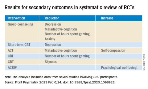
ECMO for refractory asthma exacerbations
The overnight shift in the MCU began as it does for many intensivists, by hearing about ED admissions, transfers from outside hospitals, sick floor patients, and high-risk patients in the MICU. Earlier in the day, the MICU team had admitted a 39-year-old woman with a severe asthma attack that required endotracheal intubation and mechanical ventilation in the ED for hypercarbic respiratory failure. After intubation, she had no audible air movement on chest exam, severe hypercarbic respiratory acidosis determined by an arterial blood gas, a clear chest radiograph, and negative findings on a respiratory viral panel. Her family said that she had run out of her steroid inhaler a month earlier and could not afford a refill. She had been using increasing amounts of albuterol over the past week before developing severe shortness of breath on the day of admission. The ED and MICU teams aggressively treated her with high-dose inhaled albuterol, ipratropium, and IV magnesium sulfate for bronchodilation; methylprednisolone for airway inflammation; and continuous ketamine for sedation, analgesia, and bronchodilation (Rehder KJ, et al. Respir Care. 2017;62[6]:849). Her airway pressures continued to be high despite using lung protective ventilation, so she was shifted to a permissive hypercapnia ventilation strategy using neuromuscular blockade, deep sedation, and low minute-ventilation (Laher AE, et al. J Intensive Care Med. 2018;33[9]:491).

Two hours into the shift, the bedside nurse noted that the patient had become hypotensive. Her ventilator pressures remained stable with peak inspiratory pressures of 38-42 cm H2O, plateau pressures of 28-30 cm H2O, auto-positive end-expiratory pressure (auto-PEEP) of 10-12 cm H2O, and fractional inspiratory oxygen (FiO2) of 40%. A repeat chest radiograph showed no signs of barotrauma, but arterial blood gas values showed severe respiratory acidosis with a pH of 7.05 and a PCO2 > 100 mm Hg. Her condition stabilized when she received a continuous infusion of bicarbonate to control her acidosis and low-dose IV norepinephrine for blood pressure control. It was at that moment that the bedside nurse astutely asked whether we should consider starting ECMO for the patient, as coauthor Dr. Arun Kannappan had done for a similar patient with asthma a month earlier. Dr. Vandivier notes, “My first response was that ECMO was not needed, because our patient had stabilized, and I had taken care of many patients like this in the past. But as I considered the situation more carefully, it was clear that . In short, my ‘traditional’ approach left little room for error in a patient with high ventilator pressures and hemodynamic instability.”

ECMO is a technique used to add oxygen or remove CO2 from the blood of people with different forms of respiratory failure (Fan E, et al. Intensive Care Med. 2016;2:712) that was first used by Hill and colleagues in 1966 for trauma-induced ARDS (Hill JD, et al. N Engl J Med. 1972;286:629). The ECMO circuit pumps blood from the venous system into an oxygenator that adds oxygen and removes CO2 before blood is returned to either the venous or arterial circulation (Intensive Care Med. 2016;42:712). Venovenous ECMO (vvECMO) is used in clinical scenarios where only oxygenation and/or CO2 removal is needed, whereas venoarterial ECMO (vaECMO) is reserved for situations where additional hemodynamic support is necessary. ECMO is traditionally thought of as a means to increase blood oxygenation, but it is less widely appreciated that ECMO is particularly effective at removing blood CO2. In addition to ECMO helping to normalize oxygenation or eliminate CO2, it can also be used to lower tidal volumes, decrease airway pressures, and allow “lungs to rest” with the goal of avoiding ventilator-induced lung injury (VILI).

Standing at the bedside, it seemed to the authors that it was the right time to think about instituting a salvage therapy. But was there evidence that ECMO could improve survival? Were there clear guidelines for when to initiate ECMO, and was ECMO more effective than other salvage therapies such as inhaled volatile anesthetics?
Since McDonnell and colleagues first described the use of ECMO for a severe asthma exacerbation in 1981 (Ann Thoracic Surg. 1981;31[2]:171), about 95 articles have been published. Other than two registry studies and a recent epidemiologic study, all of these publications were case reports, case series, and reviews. Mikkelsen and colleagues (ASAIO J. 2009;55[1]:47) performed a retrospective, cohort study using the International Extracorporeal Life Support (ECLS) Organization Registry to determine whether ECMO use for status asthmaticus was associated with greater survival than the use of ECMO for other causes of respiratory failure. From 1986 through 2006, a total of 2,127 cases of respiratory failure were identified that required ECMO, including 27 for status asthmaticus and 1,233 for other causes. Their analysis showed that 83.3% of asthmatics treated with ECMO survived to hospital discharge, compared with 50.8% of people treated with ECMO for respiratory failure not due to asthma, with an odds ratio (OR) of 4.86 favoring survival of asthmatics (OR = 4.86; 95% CI, 1.65-14.31, P = .004).
Yeo and colleagues (Yeo HJ, et al. Critical Care. 2017;21:297) also used the ECLS Organization Registry to measure survival to hospital discharge, complications, and clinical factors associated with in-hospital mortality for asthmatics treated with ECMO. They included 272 people treated with ECMO for asthma between 1992 and 2016, after excluding people treated with ECMO for cardiopulmonary resuscitation or cardiac dysfunction. ECMO was associated with improvements in ventilator mechanics, including a reduction in respiratory rate, FiO2, peak inspiratory pressure, mean airway pressure, and driving pressure. Use of ECMO for status asthmaticus was also associated with an 83.5% survival to hospital discharge, similar to the study by Mikkelsen and colleagues. Hemorrhage, the most common complication, occurred in roughly a quarter of people treated with ECMO. In the multivariate analysis, age, bleeding, pre-ECMO PEEP, post-ECMO FiO2, and driving pressure were all associated with higher in-hospital mortality.
Although there are no formal criteria to guide use of ECMO for asthma exacerbations with respiratory failure, a number of physicians and a physician organization have recommended that ECMO be considered for persistently high ventilator pressures, uncontrolled respiratory acidosis, or hemodynamic instability. Because our patient qualified for ECMO based on all three suggested criteria, we consulted cardiac surgery who quickly started her on vvECMO. She remained on ECMO for 4 days until she was decannulated, extubated, and discharged home.
Despite this positive outcome, the lack of a high-quality, controlled study to help guide our decision was surprising given the ability of ECMO to efficiently remove CO2 and to decrease ventilator pressures. The lack of guidance prompted us to perform a retrospective, epidemiologic cohort study to determine whether treatment with ECMO for asthma exacerbations with respiratory failure was associated with reduced mortality, compared with people treated without ECMO (Zakrajsek JK, Chest. 2023;163[1]:38). The study included 13,714 people admitted to an ECMO-capable hospital with respiratory failure that required invasive ventilation because of an asthma exacerbation between 2010 and 2020, of which 127 were treated with ECMO and 13,587 were not. During this period, use of ECMO as a salvage therapy for severe asthma exacerbations was a rare event, but it became more common over time. With the limitation that 40% of asthma patients were transferred from an outside hospital, 74% were started on ECMO in the first 2 hospital days, and 94% were started within the first week of hospitalization. Once started, ECMO was continued for a median of 1.0 day and range of 1-49 days. Hospital mortality was 14.6% in the ECMO group versus 26.2% in the no ECMO group, which equated to an 11.6% absolute risk reduction (P = 0.03) and 52% relative risk reduction (P = 0.04) in mortality. ECMO was associated with hospital costs that were $114,000 higher per patient, compared with the no ECMO group, but did not affect intensive care unit length of stay, hospital length of stay, or time on invasive mechanical ventilation.
We were pleased that our patient had a good outcome, and were reassured by our study results. But we were left to wonder whether ECMO really was the best salvage therapy for asthma exacerbations with respiratory failure, and if it was initiated for the right indications at the best time. These are important treatment considerations that take on new urgency given that physicians are increasingly looking to ECMO as a salvage therapy for refractory asthma, and the recent FDA approval of low-flow, extracorporeal CO2 removal systems that could make CO2 removal a more available, and perhaps less expensive, strategy. Despite promising epidemiological data, it will be important that these questions are answered with well-designed clinical trials so that physicians can be armed with the knowledge needed to navigate complex clinical scenarios, and ultimately to prevent unfortunate deaths from a reversible disease.
The overnight shift in the MCU began as it does for many intensivists, by hearing about ED admissions, transfers from outside hospitals, sick floor patients, and high-risk patients in the MICU. Earlier in the day, the MICU team had admitted a 39-year-old woman with a severe asthma attack that required endotracheal intubation and mechanical ventilation in the ED for hypercarbic respiratory failure. After intubation, she had no audible air movement on chest exam, severe hypercarbic respiratory acidosis determined by an arterial blood gas, a clear chest radiograph, and negative findings on a respiratory viral panel. Her family said that she had run out of her steroid inhaler a month earlier and could not afford a refill. She had been using increasing amounts of albuterol over the past week before developing severe shortness of breath on the day of admission. The ED and MICU teams aggressively treated her with high-dose inhaled albuterol, ipratropium, and IV magnesium sulfate for bronchodilation; methylprednisolone for airway inflammation; and continuous ketamine for sedation, analgesia, and bronchodilation (Rehder KJ, et al. Respir Care. 2017;62[6]:849). Her airway pressures continued to be high despite using lung protective ventilation, so she was shifted to a permissive hypercapnia ventilation strategy using neuromuscular blockade, deep sedation, and low minute-ventilation (Laher AE, et al. J Intensive Care Med. 2018;33[9]:491).
Two hours into the shift, the bedside nurse noted that the patient had become hypotensive. Her ventilator pressures remained stable with peak inspiratory pressures of 38-42 cm H2O, plateau pressures of 28-30 cm H2O, auto-positive end-expiratory pressure (auto-PEEP) of 10-12 cm H2O, and fractional inspiratory oxygen (FiO2) of 40%. A repeat chest radiograph showed no signs of barotrauma, but arterial blood gas values showed severe respiratory acidosis with a pH of 7.05 and a PCO2 > 100 mm Hg. Her condition stabilized when she received a continuous infusion of bicarbonate to control her acidosis and low-dose IV norepinephrine for blood pressure control. It was at that moment that the bedside nurse astutely asked whether we should consider starting ECMO for the patient, as coauthor Dr. Arun Kannappan had done for a similar patient with asthma a month earlier. Dr. Vandivier notes, “My first response was that ECMO was not needed, because our patient had stabilized, and I had taken care of many patients like this in the past. But as I considered the situation more carefully, it was clear that . In short, my ‘traditional’ approach left little room for error in a patient with high ventilator pressures and hemodynamic instability.”
ECMO is a technique used to add oxygen or remove CO2 from the blood of people with different forms of respiratory failure (Fan E, et al. Intensive Care Med. 2016;2:712) that was first used by Hill and colleagues in 1966 for trauma-induced ARDS (Hill JD, et al. N Engl J Med. 1972;286:629). The ECMO circuit pumps blood from the venous system into an oxygenator that adds oxygen and removes CO2 before blood is returned to either the venous or arterial circulation (Intensive Care Med. 2016;42:712). Venovenous ECMO (vvECMO) is used in clinical scenarios where only oxygenation and/or CO2 removal is needed, whereas venoarterial ECMO (vaECMO) is reserved for situations where additional hemodynamic support is necessary. ECMO is traditionally thought of as a means to increase blood oxygenation, but it is less widely appreciated that ECMO is particularly effective at removing blood CO2. In addition to ECMO helping to normalize oxygenation or eliminate CO2, it can also be used to lower tidal volumes, decrease airway pressures, and allow “lungs to rest” with the goal of avoiding ventilator-induced lung injury (VILI).
Standing at the bedside, it seemed to the authors that it was the right time to think about instituting a salvage therapy. But was there evidence that ECMO could improve survival? Were there clear guidelines for when to initiate ECMO, and was ECMO more effective than other salvage therapies such as inhaled volatile anesthetics?
Since McDonnell and colleagues first described the use of ECMO for a severe asthma exacerbation in 1981 (Ann Thoracic Surg. 1981;31[2]:171), about 95 articles have been published. Other than two registry studies and a recent epidemiologic study, all of these publications were case reports, case series, and reviews. Mikkelsen and colleagues (ASAIO J. 2009;55[1]:47) performed a retrospective, cohort study using the International Extracorporeal Life Support (ECLS) Organization Registry to determine whether ECMO use for status asthmaticus was associated with greater survival than the use of ECMO for other causes of respiratory failure. From 1986 through 2006, a total of 2,127 cases of respiratory failure were identified that required ECMO, including 27 for status asthmaticus and 1,233 for other causes. Their analysis showed that 83.3% of asthmatics treated with ECMO survived to hospital discharge, compared with 50.8% of people treated with ECMO for respiratory failure not due to asthma, with an odds ratio (OR) of 4.86 favoring survival of asthmatics (OR = 4.86; 95% CI, 1.65-14.31, P = .004).
Yeo and colleagues (Yeo HJ, et al. Critical Care. 2017;21:297) also used the ECLS Organization Registry to measure survival to hospital discharge, complications, and clinical factors associated with in-hospital mortality for asthmatics treated with ECMO. They included 272 people treated with ECMO for asthma between 1992 and 2016, after excluding people treated with ECMO for cardiopulmonary resuscitation or cardiac dysfunction. ECMO was associated with improvements in ventilator mechanics, including a reduction in respiratory rate, FiO2, peak inspiratory pressure, mean airway pressure, and driving pressure. Use of ECMO for status asthmaticus was also associated with an 83.5% survival to hospital discharge, similar to the study by Mikkelsen and colleagues. Hemorrhage, the most common complication, occurred in roughly a quarter of people treated with ECMO. In the multivariate analysis, age, bleeding, pre-ECMO PEEP, post-ECMO FiO2, and driving pressure were all associated with higher in-hospital mortality.
Although there are no formal criteria to guide use of ECMO for asthma exacerbations with respiratory failure, a number of physicians and a physician organization have recommended that ECMO be considered for persistently high ventilator pressures, uncontrolled respiratory acidosis, or hemodynamic instability. Because our patient qualified for ECMO based on all three suggested criteria, we consulted cardiac surgery who quickly started her on vvECMO. She remained on ECMO for 4 days until she was decannulated, extubated, and discharged home.
Despite this positive outcome, the lack of a high-quality, controlled study to help guide our decision was surprising given the ability of ECMO to efficiently remove CO2 and to decrease ventilator pressures. The lack of guidance prompted us to perform a retrospective, epidemiologic cohort study to determine whether treatment with ECMO for asthma exacerbations with respiratory failure was associated with reduced mortality, compared with people treated without ECMO (Zakrajsek JK, Chest. 2023;163[1]:38). The study included 13,714 people admitted to an ECMO-capable hospital with respiratory failure that required invasive ventilation because of an asthma exacerbation between 2010 and 2020, of which 127 were treated with ECMO and 13,587 were not. During this period, use of ECMO as a salvage therapy for severe asthma exacerbations was a rare event, but it became more common over time. With the limitation that 40% of asthma patients were transferred from an outside hospital, 74% were started on ECMO in the first 2 hospital days, and 94% were started within the first week of hospitalization. Once started, ECMO was continued for a median of 1.0 day and range of 1-49 days. Hospital mortality was 14.6% in the ECMO group versus 26.2% in the no ECMO group, which equated to an 11.6% absolute risk reduction (P = 0.03) and 52% relative risk reduction (P = 0.04) in mortality. ECMO was associated with hospital costs that were $114,000 higher per patient, compared with the no ECMO group, but did not affect intensive care unit length of stay, hospital length of stay, or time on invasive mechanical ventilation.
We were pleased that our patient had a good outcome, and were reassured by our study results. But we were left to wonder whether ECMO really was the best salvage therapy for asthma exacerbations with respiratory failure, and if it was initiated for the right indications at the best time. These are important treatment considerations that take on new urgency given that physicians are increasingly looking to ECMO as a salvage therapy for refractory asthma, and the recent FDA approval of low-flow, extracorporeal CO2 removal systems that could make CO2 removal a more available, and perhaps less expensive, strategy. Despite promising epidemiological data, it will be important that these questions are answered with well-designed clinical trials so that physicians can be armed with the knowledge needed to navigate complex clinical scenarios, and ultimately to prevent unfortunate deaths from a reversible disease.
The overnight shift in the MCU began as it does for many intensivists, by hearing about ED admissions, transfers from outside hospitals, sick floor patients, and high-risk patients in the MICU. Earlier in the day, the MICU team had admitted a 39-year-old woman with a severe asthma attack that required endotracheal intubation and mechanical ventilation in the ED for hypercarbic respiratory failure. After intubation, she had no audible air movement on chest exam, severe hypercarbic respiratory acidosis determined by an arterial blood gas, a clear chest radiograph, and negative findings on a respiratory viral panel. Her family said that she had run out of her steroid inhaler a month earlier and could not afford a refill. She had been using increasing amounts of albuterol over the past week before developing severe shortness of breath on the day of admission. The ED and MICU teams aggressively treated her with high-dose inhaled albuterol, ipratropium, and IV magnesium sulfate for bronchodilation; methylprednisolone for airway inflammation; and continuous ketamine for sedation, analgesia, and bronchodilation (Rehder KJ, et al. Respir Care. 2017;62[6]:849). Her airway pressures continued to be high despite using lung protective ventilation, so she was shifted to a permissive hypercapnia ventilation strategy using neuromuscular blockade, deep sedation, and low minute-ventilation (Laher AE, et al. J Intensive Care Med. 2018;33[9]:491).
Two hours into the shift, the bedside nurse noted that the patient had become hypotensive. Her ventilator pressures remained stable with peak inspiratory pressures of 38-42 cm H2O, plateau pressures of 28-30 cm H2O, auto-positive end-expiratory pressure (auto-PEEP) of 10-12 cm H2O, and fractional inspiratory oxygen (FiO2) of 40%. A repeat chest radiograph showed no signs of barotrauma, but arterial blood gas values showed severe respiratory acidosis with a pH of 7.05 and a PCO2 > 100 mm Hg. Her condition stabilized when she received a continuous infusion of bicarbonate to control her acidosis and low-dose IV norepinephrine for blood pressure control. It was at that moment that the bedside nurse astutely asked whether we should consider starting ECMO for the patient, as coauthor Dr. Arun Kannappan had done for a similar patient with asthma a month earlier. Dr. Vandivier notes, “My first response was that ECMO was not needed, because our patient had stabilized, and I had taken care of many patients like this in the past. But as I considered the situation more carefully, it was clear that . In short, my ‘traditional’ approach left little room for error in a patient with high ventilator pressures and hemodynamic instability.”
ECMO is a technique used to add oxygen or remove CO2 from the blood of people with different forms of respiratory failure (Fan E, et al. Intensive Care Med. 2016;2:712) that was first used by Hill and colleagues in 1966 for trauma-induced ARDS (Hill JD, et al. N Engl J Med. 1972;286:629). The ECMO circuit pumps blood from the venous system into an oxygenator that adds oxygen and removes CO2 before blood is returned to either the venous or arterial circulation (Intensive Care Med. 2016;42:712). Venovenous ECMO (vvECMO) is used in clinical scenarios where only oxygenation and/or CO2 removal is needed, whereas venoarterial ECMO (vaECMO) is reserved for situations where additional hemodynamic support is necessary. ECMO is traditionally thought of as a means to increase blood oxygenation, but it is less widely appreciated that ECMO is particularly effective at removing blood CO2. In addition to ECMO helping to normalize oxygenation or eliminate CO2, it can also be used to lower tidal volumes, decrease airway pressures, and allow “lungs to rest” with the goal of avoiding ventilator-induced lung injury (VILI).
Standing at the bedside, it seemed to the authors that it was the right time to think about instituting a salvage therapy. But was there evidence that ECMO could improve survival? Were there clear guidelines for when to initiate ECMO, and was ECMO more effective than other salvage therapies such as inhaled volatile anesthetics?
Since McDonnell and colleagues first described the use of ECMO for a severe asthma exacerbation in 1981 (Ann Thoracic Surg. 1981;31[2]:171), about 95 articles have been published. Other than two registry studies and a recent epidemiologic study, all of these publications were case reports, case series, and reviews. Mikkelsen and colleagues (ASAIO J. 2009;55[1]:47) performed a retrospective, cohort study using the International Extracorporeal Life Support (ECLS) Organization Registry to determine whether ECMO use for status asthmaticus was associated with greater survival than the use of ECMO for other causes of respiratory failure. From 1986 through 2006, a total of 2,127 cases of respiratory failure were identified that required ECMO, including 27 for status asthmaticus and 1,233 for other causes. Their analysis showed that 83.3% of asthmatics treated with ECMO survived to hospital discharge, compared with 50.8% of people treated with ECMO for respiratory failure not due to asthma, with an odds ratio (OR) of 4.86 favoring survival of asthmatics (OR = 4.86; 95% CI, 1.65-14.31, P = .004).
Yeo and colleagues (Yeo HJ, et al. Critical Care. 2017;21:297) also used the ECLS Organization Registry to measure survival to hospital discharge, complications, and clinical factors associated with in-hospital mortality for asthmatics treated with ECMO. They included 272 people treated with ECMO for asthma between 1992 and 2016, after excluding people treated with ECMO for cardiopulmonary resuscitation or cardiac dysfunction. ECMO was associated with improvements in ventilator mechanics, including a reduction in respiratory rate, FiO2, peak inspiratory pressure, mean airway pressure, and driving pressure. Use of ECMO for status asthmaticus was also associated with an 83.5% survival to hospital discharge, similar to the study by Mikkelsen and colleagues. Hemorrhage, the most common complication, occurred in roughly a quarter of people treated with ECMO. In the multivariate analysis, age, bleeding, pre-ECMO PEEP, post-ECMO FiO2, and driving pressure were all associated with higher in-hospital mortality.
Although there are no formal criteria to guide use of ECMO for asthma exacerbations with respiratory failure, a number of physicians and a physician organization have recommended that ECMO be considered for persistently high ventilator pressures, uncontrolled respiratory acidosis, or hemodynamic instability. Because our patient qualified for ECMO based on all three suggested criteria, we consulted cardiac surgery who quickly started her on vvECMO. She remained on ECMO for 4 days until she was decannulated, extubated, and discharged home.
Despite this positive outcome, the lack of a high-quality, controlled study to help guide our decision was surprising given the ability of ECMO to efficiently remove CO2 and to decrease ventilator pressures. The lack of guidance prompted us to perform a retrospective, epidemiologic cohort study to determine whether treatment with ECMO for asthma exacerbations with respiratory failure was associated with reduced mortality, compared with people treated without ECMO (Zakrajsek JK, Chest. 2023;163[1]:38). The study included 13,714 people admitted to an ECMO-capable hospital with respiratory failure that required invasive ventilation because of an asthma exacerbation between 2010 and 2020, of which 127 were treated with ECMO and 13,587 were not. During this period, use of ECMO as a salvage therapy for severe asthma exacerbations was a rare event, but it became more common over time. With the limitation that 40% of asthma patients were transferred from an outside hospital, 74% were started on ECMO in the first 2 hospital days, and 94% were started within the first week of hospitalization. Once started, ECMO was continued for a median of 1.0 day and range of 1-49 days. Hospital mortality was 14.6% in the ECMO group versus 26.2% in the no ECMO group, which equated to an 11.6% absolute risk reduction (P = 0.03) and 52% relative risk reduction (P = 0.04) in mortality. ECMO was associated with hospital costs that were $114,000 higher per patient, compared with the no ECMO group, but did not affect intensive care unit length of stay, hospital length of stay, or time on invasive mechanical ventilation.
We were pleased that our patient had a good outcome, and were reassured by our study results. But we were left to wonder whether ECMO really was the best salvage therapy for asthma exacerbations with respiratory failure, and if it was initiated for the right indications at the best time. These are important treatment considerations that take on new urgency given that physicians are increasingly looking to ECMO as a salvage therapy for refractory asthma, and the recent FDA approval of low-flow, extracorporeal CO2 removal systems that could make CO2 removal a more available, and perhaps less expensive, strategy. Despite promising epidemiological data, it will be important that these questions are answered with well-designed clinical trials so that physicians can be armed with the knowledge needed to navigate complex clinical scenarios, and ultimately to prevent unfortunate deaths from a reversible disease.

2023 GOLD update: Changes in COPD nomenclature and initial therapy
Airways Disorders Network
Asthma & COPD Section
The 2023 GOLD committee proposed changes in nomenclature and therapy for various subgroups of patients with COPD.
The mainstay of initial treatment for symptomatic COPD should include combination LABA/LAMA bronchodilators in a single inhaler. For patients with features of concomitant asthma or eosinophils greater than or equal to 300 cells/microliter, an ICS/LABA/LAMA combination inhaler is recommended.
People with “young COPD” develop respiratory symptoms and meet spirometric criteria for COPD between the ages of 25 and 50 years old. Other terminology changes center around those with functional and/or structural changes suggesting COPD, but who do not meet the postbronchodilator spirometric criteria to confirm the COPD diagnosis.
Those with “pre-COPD” have normal spirometry, including the FEV1 and FEV1/FVC ratio, but have functional and/or structural changes concerning for COPD. Functional changes include air trapping and/or hyperinflation on PFTs, low diffusion capacity, and/or decline in FEV1 of > 40 mL per year.
Structural changes include emphysematous changes and/or bronchial wall changes on CT scans. “PRISm” stands for preserved ratio with impaired spirometry, where the postbronchodilator FEV1/FVC is greater than or equal to 0.70, but FEV1 is < 80% predicted with similar functional and/or structural changes to those with “pre-COPD.” People with PRISm have increased all-cause mortality. Not all people with pre-COPD or PRISm progress clinically and spirometrically to COPD; however, they should be treated because they have symptoms as well as functional and/or structural abnormalities. Despite increasing data regarding pre-COPD and PRISm, many gaps remain regarding optimal management.
Maria Ashar, MD, MBBS
Airways Disorders Network
Asthma & COPD Section
The 2023 GOLD committee proposed changes in nomenclature and therapy for various subgroups of patients with COPD.
The mainstay of initial treatment for symptomatic COPD should include combination LABA/LAMA bronchodilators in a single inhaler. For patients with features of concomitant asthma or eosinophils greater than or equal to 300 cells/microliter, an ICS/LABA/LAMA combination inhaler is recommended.
People with “young COPD” develop respiratory symptoms and meet spirometric criteria for COPD between the ages of 25 and 50 years old. Other terminology changes center around those with functional and/or structural changes suggesting COPD, but who do not meet the postbronchodilator spirometric criteria to confirm the COPD diagnosis.
Those with “pre-COPD” have normal spirometry, including the FEV1 and FEV1/FVC ratio, but have functional and/or structural changes concerning for COPD. Functional changes include air trapping and/or hyperinflation on PFTs, low diffusion capacity, and/or decline in FEV1 of > 40 mL per year.
Structural changes include emphysematous changes and/or bronchial wall changes on CT scans. “PRISm” stands for preserved ratio with impaired spirometry, where the postbronchodilator FEV1/FVC is greater than or equal to 0.70, but FEV1 is < 80% predicted with similar functional and/or structural changes to those with “pre-COPD.” People with PRISm have increased all-cause mortality. Not all people with pre-COPD or PRISm progress clinically and spirometrically to COPD; however, they should be treated because they have symptoms as well as functional and/or structural abnormalities. Despite increasing data regarding pre-COPD and PRISm, many gaps remain regarding optimal management.
Maria Ashar, MD, MBBS
Airways Disorders Network
Asthma & COPD Section
The 2023 GOLD committee proposed changes in nomenclature and therapy for various subgroups of patients with COPD.
The mainstay of initial treatment for symptomatic COPD should include combination LABA/LAMA bronchodilators in a single inhaler. For patients with features of concomitant asthma or eosinophils greater than or equal to 300 cells/microliter, an ICS/LABA/LAMA combination inhaler is recommended.
People with “young COPD” develop respiratory symptoms and meet spirometric criteria for COPD between the ages of 25 and 50 years old. Other terminology changes center around those with functional and/or structural changes suggesting COPD, but who do not meet the postbronchodilator spirometric criteria to confirm the COPD diagnosis.
Those with “pre-COPD” have normal spirometry, including the FEV1 and FEV1/FVC ratio, but have functional and/or structural changes concerning for COPD. Functional changes include air trapping and/or hyperinflation on PFTs, low diffusion capacity, and/or decline in FEV1 of > 40 mL per year.
Structural changes include emphysematous changes and/or bronchial wall changes on CT scans. “PRISm” stands for preserved ratio with impaired spirometry, where the postbronchodilator FEV1/FVC is greater than or equal to 0.70, but FEV1 is < 80% predicted with similar functional and/or structural changes to those with “pre-COPD.” People with PRISm have increased all-cause mortality. Not all people with pre-COPD or PRISm progress clinically and spirometrically to COPD; however, they should be treated because they have symptoms as well as functional and/or structural abnormalities. Despite increasing data regarding pre-COPD and PRISm, many gaps remain regarding optimal management.
Maria Ashar, MD, MBBS
In metastatic NSCLC, better QoL outcomes tied to better outcomes
The authors, including Fabio Salomone of the University of Naples Federico II, department of clinical medicine and surgery, also observed trends toward an association between QoL improvement and PFS among patients treated with chemotherapy and immunotherapy.
The new research was presented during a poster session at European Lung Cancer Congress 2023.
“The findings of the study support the thesis that QoL and survival in patients with NSCLC are linked. Although this is documented in the literature, this study sums up the evidence of a large number of RCTs, and provides detail in the QoL/survival relationship by treatment type. The subgroup analysis by treatment type is a key strength of the study showing that the QoL/survival link is stronger and more reliable in target(ed) therapies,” George Kypriotakis, PhD, who was not involved with the study, said in an interview.
Combining efficacy and quality of life improvement is an important consideration in clinical practice. “It is important that clinicians provide therapies that are also palliative and improve QoL,” said Dr. Kypriotakis, assistant professor of behavioral sciences at University of Texas MD Anderson Cancer Center, Houston. He noted that the finding of a PFS benefit is a good indicator of overall benefit, which is important since OS outcomes require a larger number of patients and longer follow-up to determine.
“PFS can still be a valid surrogate for OS, especially when it is positively associated with QoL,” noted Dr. Kypriotakis.
The study included 81 trials. Sixteen of the studies investigated immunotherapy, 50 investigated targeted therapy, and 17 investigated chemotherapy regimens. Thirty-seven percent of the trials found an improvement in QoL in the treatment arm compared with the control arm, 59.3% found no difference between arms, and 3.7% found a worse QoL in the treatment arm. There was no statistically significant association between an improvement in OS and QoL among the trials (P = .368).
Improved QoL tied to improved PFS
The researchers found an association between improved QoL and improved PFS. Among 60 trials that showed improved PFS, 43.3% found a superior QoL in the treatment arm, 53.3% showed no difference, and 3.3% showed reduced QoL. Among 20 trials that found no improvement in PFS, 20% demonstrated an improved QoL, 75% found no change, and 5% showed worse QoL (P = .0473).
A subanalysis of 48 targeted therapy trials found a correlation between PFS and QoL improvement (P = .0196). Among 25 trials involving patients receiving epidermal growth factor receptor (EGFR) and anaplastic lymphoma kinase (ALK) inhibitors showing an improved PFS, 60% showed improved QoL, 36% showed no difference, and 4% showed worsening (P = .0077). Seven of these trials showed no PFS benefit and no change in QoL.
Industry sponsorship may affect QOL results
The researchers found potential evidence that industry sponsorship may lead to a spin on QoL outcomes. Among 51 trials that showed no QoL benefit associated with treatment, the description of the QoL outcome in 37 industry-sponsored was judged to be neutral and coherent with the study findings in 26 cases, but unjustifiably favorable in 11 cases. Among 14 with nonprofit support, descriptions of QoL results were found to be neutral in all cases (P = .0232).
“Obviously, industry may be motivated to overemphasize treatment benefits, especially in measures that also have a qualitative/subjective dimension such as QoL. Assuming that the authors used a reliable criterion to evaluate “inappropriateness,” industry may be more likely to emphasize QoL improvements as a surrogate for OS, especially when seeking drug approval,” Dr. Kypriotakis said.
The study is retrospective and cannot prove causation.
Dr. Salomone and Dr. Kypriotakis have no relevant financial disclosures.
The authors, including Fabio Salomone of the University of Naples Federico II, department of clinical medicine and surgery, also observed trends toward an association between QoL improvement and PFS among patients treated with chemotherapy and immunotherapy.
The new research was presented during a poster session at European Lung Cancer Congress 2023.
“The findings of the study support the thesis that QoL and survival in patients with NSCLC are linked. Although this is documented in the literature, this study sums up the evidence of a large number of RCTs, and provides detail in the QoL/survival relationship by treatment type. The subgroup analysis by treatment type is a key strength of the study showing that the QoL/survival link is stronger and more reliable in target(ed) therapies,” George Kypriotakis, PhD, who was not involved with the study, said in an interview.
Combining efficacy and quality of life improvement is an important consideration in clinical practice. “It is important that clinicians provide therapies that are also palliative and improve QoL,” said Dr. Kypriotakis, assistant professor of behavioral sciences at University of Texas MD Anderson Cancer Center, Houston. He noted that the finding of a PFS benefit is a good indicator of overall benefit, which is important since OS outcomes require a larger number of patients and longer follow-up to determine.
“PFS can still be a valid surrogate for OS, especially when it is positively associated with QoL,” noted Dr. Kypriotakis.
The study included 81 trials. Sixteen of the studies investigated immunotherapy, 50 investigated targeted therapy, and 17 investigated chemotherapy regimens. Thirty-seven percent of the trials found an improvement in QoL in the treatment arm compared with the control arm, 59.3% found no difference between arms, and 3.7% found a worse QoL in the treatment arm. There was no statistically significant association between an improvement in OS and QoL among the trials (P = .368).
Improved QoL tied to improved PFS
The researchers found an association between improved QoL and improved PFS. Among 60 trials that showed improved PFS, 43.3% found a superior QoL in the treatment arm, 53.3% showed no difference, and 3.3% showed reduced QoL. Among 20 trials that found no improvement in PFS, 20% demonstrated an improved QoL, 75% found no change, and 5% showed worse QoL (P = .0473).
A subanalysis of 48 targeted therapy trials found a correlation between PFS and QoL improvement (P = .0196). Among 25 trials involving patients receiving epidermal growth factor receptor (EGFR) and anaplastic lymphoma kinase (ALK) inhibitors showing an improved PFS, 60% showed improved QoL, 36% showed no difference, and 4% showed worsening (P = .0077). Seven of these trials showed no PFS benefit and no change in QoL.
Industry sponsorship may affect QOL results
The researchers found potential evidence that industry sponsorship may lead to a spin on QoL outcomes. Among 51 trials that showed no QoL benefit associated with treatment, the description of the QoL outcome in 37 industry-sponsored was judged to be neutral and coherent with the study findings in 26 cases, but unjustifiably favorable in 11 cases. Among 14 with nonprofit support, descriptions of QoL results were found to be neutral in all cases (P = .0232).
“Obviously, industry may be motivated to overemphasize treatment benefits, especially in measures that also have a qualitative/subjective dimension such as QoL. Assuming that the authors used a reliable criterion to evaluate “inappropriateness,” industry may be more likely to emphasize QoL improvements as a surrogate for OS, especially when seeking drug approval,” Dr. Kypriotakis said.
The study is retrospective and cannot prove causation.
Dr. Salomone and Dr. Kypriotakis have no relevant financial disclosures.
The authors, including Fabio Salomone of the University of Naples Federico II, department of clinical medicine and surgery, also observed trends toward an association between QoL improvement and PFS among patients treated with chemotherapy and immunotherapy.
The new research was presented during a poster session at European Lung Cancer Congress 2023.
“The findings of the study support the thesis that QoL and survival in patients with NSCLC are linked. Although this is documented in the literature, this study sums up the evidence of a large number of RCTs, and provides detail in the QoL/survival relationship by treatment type. The subgroup analysis by treatment type is a key strength of the study showing that the QoL/survival link is stronger and more reliable in target(ed) therapies,” George Kypriotakis, PhD, who was not involved with the study, said in an interview.
Combining efficacy and quality of life improvement is an important consideration in clinical practice. “It is important that clinicians provide therapies that are also palliative and improve QoL,” said Dr. Kypriotakis, assistant professor of behavioral sciences at University of Texas MD Anderson Cancer Center, Houston. He noted that the finding of a PFS benefit is a good indicator of overall benefit, which is important since OS outcomes require a larger number of patients and longer follow-up to determine.
“PFS can still be a valid surrogate for OS, especially when it is positively associated with QoL,” noted Dr. Kypriotakis.
The study included 81 trials. Sixteen of the studies investigated immunotherapy, 50 investigated targeted therapy, and 17 investigated chemotherapy regimens. Thirty-seven percent of the trials found an improvement in QoL in the treatment arm compared with the control arm, 59.3% found no difference between arms, and 3.7% found a worse QoL in the treatment arm. There was no statistically significant association between an improvement in OS and QoL among the trials (P = .368).
Improved QoL tied to improved PFS
The researchers found an association between improved QoL and improved PFS. Among 60 trials that showed improved PFS, 43.3% found a superior QoL in the treatment arm, 53.3% showed no difference, and 3.3% showed reduced QoL. Among 20 trials that found no improvement in PFS, 20% demonstrated an improved QoL, 75% found no change, and 5% showed worse QoL (P = .0473).
A subanalysis of 48 targeted therapy trials found a correlation between PFS and QoL improvement (P = .0196). Among 25 trials involving patients receiving epidermal growth factor receptor (EGFR) and anaplastic lymphoma kinase (ALK) inhibitors showing an improved PFS, 60% showed improved QoL, 36% showed no difference, and 4% showed worsening (P = .0077). Seven of these trials showed no PFS benefit and no change in QoL.
Industry sponsorship may affect QOL results
The researchers found potential evidence that industry sponsorship may lead to a spin on QoL outcomes. Among 51 trials that showed no QoL benefit associated with treatment, the description of the QoL outcome in 37 industry-sponsored was judged to be neutral and coherent with the study findings in 26 cases, but unjustifiably favorable in 11 cases. Among 14 with nonprofit support, descriptions of QoL results were found to be neutral in all cases (P = .0232).
“Obviously, industry may be motivated to overemphasize treatment benefits, especially in measures that also have a qualitative/subjective dimension such as QoL. Assuming that the authors used a reliable criterion to evaluate “inappropriateness,” industry may be more likely to emphasize QoL improvements as a surrogate for OS, especially when seeking drug approval,” Dr. Kypriotakis said.
The study is retrospective and cannot prove causation.
Dr. Salomone and Dr. Kypriotakis have no relevant financial disclosures.
FROM ELCC 2023
Refined incidence rate of HCC with alcohol-associated cirrhosis encourages surveillance
Incidence rates were higher for cohorts that underwent HCC surveillance versus those that did not undergo surveillance, suggesting that such programs offer significant benefit, lead author Daniel Q. Huang, MBBS, of the University of California, San Diego, and colleagues reported.

“A systematic review of the incidence of HCC among patients with alcohol-associated cirrhosis has not been reported,” the investigators wrote in Clinical Gastroenterology and Hepatology, prompting the present research.
Previous studies have described a broad range of annual incidence findings for HCC in this population, from 0.6% to 5.6%, suggesting that a systematic approach was needed.
To this end, Dr. Huang and colleagues analyzed data from 18 studies that involved 148,333 patients with alcohol-associated cirrhosis. The primary analysis aimed to determine cumulative incidence rates over time, while the secondary analysis characterized the impact of participation in HCC surveillance programs.
“This meta-analysis used reconstructed individual participant data, which is considered to be the gold standard for reporting survival data because it accounts for censoring of events,” the investigators noted. “The current study provides important data that are useful for clinical practice and clinical trial design.”
The cumulative incidence rates of HCC were 1%, 3%, and 9% at 1 year, 5 years, and 10 years, respectively. Among 12 of the risk factors studied, smoking, diabetes, and decompensation were all significantly associated with rate of HCC.
“Therefore, patients with alcohol-associated cirrhosis should be screened for diabetes to identify the patients at high risk for HCC development,” the investigators wrote. “In addition, patients with alcohol-associated cirrhosis should be advised to stop smoking, while patients with hepatic decompensation should be monitored carefully for the development of HCC if clinically appropriate.”
The secondary analysis showed that HCC incidence rates were higher among patients participating in HCC surveillance programs than those who did not participate (18.6 vs. 4.8 per 1,000 person-years; P = .001).
“Patients with alcohol-associated cirrhosis are known to have lower HCC surveillance rates, which may be related to poor disease awareness, clinic time constraints caused by other active medical issues, and provider beliefs regarding the likelihood of adherence,” the investigators noted.
Increased efforts are needed to promote surveillance in this population, they added, suggesting a range of communication pathways, including social media, traditional news outlets, and direct mailing.
Dr. Huang and colleagues also suggested that the findings should be validated in large prospective studies.
The study was funded by the National Institute on Alcohol Abuse and Alcoholism, the National Institute of Environmental Health Sciences, the National Center for Advancing Translational Sciences, and others. Dr. Huang disclosed funding from the Singapore Ministry of Health’s National Medical Research Council.
The association between cirrhosis and hepatocellular carcinoma (HCC) risk is well known and therefore routine surveillance is recommended by the American Association for the Study of Liver Diseases. More recent data has shown alcohol use to be an independent risk factor for HCC along with various other cancers.

Quite frequently, the focus of management in patients with alcohol-associated liver disease is alcohol cessation to prevent further decompensation, with screening often being overlooked. Previous studies have shown, however, that earlier detection is associated with improved survival. Another interesting finding of this study was that those patients who had concomitant smoking use, diabetes, and hepatic decompensation were more likely to develop HCC. When managing patients with alcohol related liver disease, confounding risk factors should be mitigated (that is, encouragement of smoking cessation, enhanced screening for diabetes, and more rigorous screening in decompensated patients).
This study brings to light the need for improved screening and concomitant risk factor mitigation for hepatocellular carcinoma given higher rates of detection in those undergoing surveillance. Larger, prospective studies are needed, however, to validate the findings in this study given the recent overall increase in rates of alcohol-associated liver disease.
Priya Maddur MD, is a visiting clinical associate professor of medicine, University of Arizona, Tucson. Dr. Maddur has no relevant disclosures.
The association between cirrhosis and hepatocellular carcinoma (HCC) risk is well known and therefore routine surveillance is recommended by the American Association for the Study of Liver Diseases. More recent data has shown alcohol use to be an independent risk factor for HCC along with various other cancers.
Quite frequently, the focus of management in patients with alcohol-associated liver disease is alcohol cessation to prevent further decompensation, with screening often being overlooked. Previous studies have shown, however, that earlier detection is associated with improved survival. Another interesting finding of this study was that those patients who had concomitant smoking use, diabetes, and hepatic decompensation were more likely to develop HCC. When managing patients with alcohol related liver disease, confounding risk factors should be mitigated (that is, encouragement of smoking cessation, enhanced screening for diabetes, and more rigorous screening in decompensated patients).
This study brings to light the need for improved screening and concomitant risk factor mitigation for hepatocellular carcinoma given higher rates of detection in those undergoing surveillance. Larger, prospective studies are needed, however, to validate the findings in this study given the recent overall increase in rates of alcohol-associated liver disease.
Priya Maddur MD, is a visiting clinical associate professor of medicine, University of Arizona, Tucson. Dr. Maddur has no relevant disclosures.
The association between cirrhosis and hepatocellular carcinoma (HCC) risk is well known and therefore routine surveillance is recommended by the American Association for the Study of Liver Diseases. More recent data has shown alcohol use to be an independent risk factor for HCC along with various other cancers.
Quite frequently, the focus of management in patients with alcohol-associated liver disease is alcohol cessation to prevent further decompensation, with screening often being overlooked. Previous studies have shown, however, that earlier detection is associated with improved survival. Another interesting finding of this study was that those patients who had concomitant smoking use, diabetes, and hepatic decompensation were more likely to develop HCC. When managing patients with alcohol related liver disease, confounding risk factors should be mitigated (that is, encouragement of smoking cessation, enhanced screening for diabetes, and more rigorous screening in decompensated patients).
This study brings to light the need for improved screening and concomitant risk factor mitigation for hepatocellular carcinoma given higher rates of detection in those undergoing surveillance. Larger, prospective studies are needed, however, to validate the findings in this study given the recent overall increase in rates of alcohol-associated liver disease.
Priya Maddur MD, is a visiting clinical associate professor of medicine, University of Arizona, Tucson. Dr. Maddur has no relevant disclosures.
Incidence rates were higher for cohorts that underwent HCC surveillance versus those that did not undergo surveillance, suggesting that such programs offer significant benefit, lead author Daniel Q. Huang, MBBS, of the University of California, San Diego, and colleagues reported.
“A systematic review of the incidence of HCC among patients with alcohol-associated cirrhosis has not been reported,” the investigators wrote in Clinical Gastroenterology and Hepatology, prompting the present research.
Previous studies have described a broad range of annual incidence findings for HCC in this population, from 0.6% to 5.6%, suggesting that a systematic approach was needed.
To this end, Dr. Huang and colleagues analyzed data from 18 studies that involved 148,333 patients with alcohol-associated cirrhosis. The primary analysis aimed to determine cumulative incidence rates over time, while the secondary analysis characterized the impact of participation in HCC surveillance programs.
“This meta-analysis used reconstructed individual participant data, which is considered to be the gold standard for reporting survival data because it accounts for censoring of events,” the investigators noted. “The current study provides important data that are useful for clinical practice and clinical trial design.”
The cumulative incidence rates of HCC were 1%, 3%, and 9% at 1 year, 5 years, and 10 years, respectively. Among 12 of the risk factors studied, smoking, diabetes, and decompensation were all significantly associated with rate of HCC.
“Therefore, patients with alcohol-associated cirrhosis should be screened for diabetes to identify the patients at high risk for HCC development,” the investigators wrote. “In addition, patients with alcohol-associated cirrhosis should be advised to stop smoking, while patients with hepatic decompensation should be monitored carefully for the development of HCC if clinically appropriate.”
The secondary analysis showed that HCC incidence rates were higher among patients participating in HCC surveillance programs than those who did not participate (18.6 vs. 4.8 per 1,000 person-years; P = .001).
“Patients with alcohol-associated cirrhosis are known to have lower HCC surveillance rates, which may be related to poor disease awareness, clinic time constraints caused by other active medical issues, and provider beliefs regarding the likelihood of adherence,” the investigators noted.
Increased efforts are needed to promote surveillance in this population, they added, suggesting a range of communication pathways, including social media, traditional news outlets, and direct mailing.
Dr. Huang and colleagues also suggested that the findings should be validated in large prospective studies.
The study was funded by the National Institute on Alcohol Abuse and Alcoholism, the National Institute of Environmental Health Sciences, the National Center for Advancing Translational Sciences, and others. Dr. Huang disclosed funding from the Singapore Ministry of Health’s National Medical Research Council.
Incidence rates were higher for cohorts that underwent HCC surveillance versus those that did not undergo surveillance, suggesting that such programs offer significant benefit, lead author Daniel Q. Huang, MBBS, of the University of California, San Diego, and colleagues reported.
“A systematic review of the incidence of HCC among patients with alcohol-associated cirrhosis has not been reported,” the investigators wrote in Clinical Gastroenterology and Hepatology, prompting the present research.
Previous studies have described a broad range of annual incidence findings for HCC in this population, from 0.6% to 5.6%, suggesting that a systematic approach was needed.
To this end, Dr. Huang and colleagues analyzed data from 18 studies that involved 148,333 patients with alcohol-associated cirrhosis. The primary analysis aimed to determine cumulative incidence rates over time, while the secondary analysis characterized the impact of participation in HCC surveillance programs.
“This meta-analysis used reconstructed individual participant data, which is considered to be the gold standard for reporting survival data because it accounts for censoring of events,” the investigators noted. “The current study provides important data that are useful for clinical practice and clinical trial design.”
The cumulative incidence rates of HCC were 1%, 3%, and 9% at 1 year, 5 years, and 10 years, respectively. Among 12 of the risk factors studied, smoking, diabetes, and decompensation were all significantly associated with rate of HCC.
“Therefore, patients with alcohol-associated cirrhosis should be screened for diabetes to identify the patients at high risk for HCC development,” the investigators wrote. “In addition, patients with alcohol-associated cirrhosis should be advised to stop smoking, while patients with hepatic decompensation should be monitored carefully for the development of HCC if clinically appropriate.”
The secondary analysis showed that HCC incidence rates were higher among patients participating in HCC surveillance programs than those who did not participate (18.6 vs. 4.8 per 1,000 person-years; P = .001).
“Patients with alcohol-associated cirrhosis are known to have lower HCC surveillance rates, which may be related to poor disease awareness, clinic time constraints caused by other active medical issues, and provider beliefs regarding the likelihood of adherence,” the investigators noted.
Increased efforts are needed to promote surveillance in this population, they added, suggesting a range of communication pathways, including social media, traditional news outlets, and direct mailing.
Dr. Huang and colleagues also suggested that the findings should be validated in large prospective studies.
The study was funded by the National Institute on Alcohol Abuse and Alcoholism, the National Institute of Environmental Health Sciences, the National Center for Advancing Translational Sciences, and others. Dr. Huang disclosed funding from the Singapore Ministry of Health’s National Medical Research Council.
FROM CLINICAL GASTROENTEROLOGY AND HEPATOLOGY