User login
Lusutrombopag is effective for thrombocytopenia in liver disease
Lusutrombopag, an oral thrombopoietin receptor agonist, was found to reduce the need for platelet transfusion in patients with chronic liver disease with a planned invasive procedure, according to results in an abstract of a phase III trial that will be presented as a latebreaker at the annual meeting of the American Association for the Study of Liver Disease in San Francisco.
The novel therapy, which upregulates platelet production, was “efficacious and well tolerated,” producing a reduced risk of overall adverse events, including bleeding events, according to Dr. Namiki Izumi, Musashino Red Cross Hospital, Tokyo.

In this ongoing global phase III trial, called L-PLUS 2, 96 patients with chronic liver disease, a platelet count less than 50,000/microL, and a planned invasive procedure were randomized to receive a once-daily 3-mg tablet of lusutrombopag or a matching placebo for 7 days. The primary endpoint was the need for a preoperative platelet transfusion.
“The proportion of patients who required no preoperative platelet transfusion was significantly greater with lusutrombopag [29.2% vs. 12.5%; P less than .0001],” Dr. Izumi reported. The proportion of responders, defined by a platelet count greater than or equal to 50,000/microL and a greater than or equal to 20,000/microL-increase from baseline, was also significantly greater in the lusutrombopag arm (77.1% vs. 6.3%; P less than .0001).
In addition, the median time with a platelet count greater than or equal to 50,000/microL was 22.1 days in those who received lusutrombopag but no platelet transfusion versus 3.3 days in the placebo patients who did receive transfusion (P less than 0.0001).
Many adverse events occurred less frequently in the arm randomized to lusutrombopag. This included bleeding events (14.6% vs. 27.1%) and postoperative fever (39.6% vs. 56.3%). The rates of procedural hypertension (41.7% vs. 37.5%) and procedural pain (45.8% vs. 41.7%) were slightly greater in the group randomized to lusutrombopag, but elevations in liver enzymes, such as aspartate aminotransferase (22.9% vs. 31.3%) were somewhat lower.
“Protocol-required imaging revealed one thromboembolic event of the portal venous system in each study arm, neither of which was related to platelets,” according to Dr. Izumi, who reported that no patient discontinued therapy as a result of an adverse event.
Because of the frequency with which thrombocytopenia is observed in patients with chronic liver disease, platelet transfusion is considered a standard procedure when an invasive intervention is planned, according to Dr. Izumi. The data from this trial suggest that preoperative treatment with lusutrombopag may be an alternative.
Dr. Izumi reported financial relationships with Bayer, Daiichi Sankyo, Gilead, Merck, and Shionogi.
Lusutrombopag, an oral thrombopoietin receptor agonist, was found to reduce the need for platelet transfusion in patients with chronic liver disease with a planned invasive procedure, according to results in an abstract of a phase III trial that will be presented as a latebreaker at the annual meeting of the American Association for the Study of Liver Disease in San Francisco.
The novel therapy, which upregulates platelet production, was “efficacious and well tolerated,” producing a reduced risk of overall adverse events, including bleeding events, according to Dr. Namiki Izumi, Musashino Red Cross Hospital, Tokyo.
In this ongoing global phase III trial, called L-PLUS 2, 96 patients with chronic liver disease, a platelet count less than 50,000/microL, and a planned invasive procedure were randomized to receive a once-daily 3-mg tablet of lusutrombopag or a matching placebo for 7 days. The primary endpoint was the need for a preoperative platelet transfusion.
“The proportion of patients who required no preoperative platelet transfusion was significantly greater with lusutrombopag [29.2% vs. 12.5%; P less than .0001],” Dr. Izumi reported. The proportion of responders, defined by a platelet count greater than or equal to 50,000/microL and a greater than or equal to 20,000/microL-increase from baseline, was also significantly greater in the lusutrombopag arm (77.1% vs. 6.3%; P less than .0001).
In addition, the median time with a platelet count greater than or equal to 50,000/microL was 22.1 days in those who received lusutrombopag but no platelet transfusion versus 3.3 days in the placebo patients who did receive transfusion (P less than 0.0001).
Many adverse events occurred less frequently in the arm randomized to lusutrombopag. This included bleeding events (14.6% vs. 27.1%) and postoperative fever (39.6% vs. 56.3%). The rates of procedural hypertension (41.7% vs. 37.5%) and procedural pain (45.8% vs. 41.7%) were slightly greater in the group randomized to lusutrombopag, but elevations in liver enzymes, such as aspartate aminotransferase (22.9% vs. 31.3%) were somewhat lower.
“Protocol-required imaging revealed one thromboembolic event of the portal venous system in each study arm, neither of which was related to platelets,” according to Dr. Izumi, who reported that no patient discontinued therapy as a result of an adverse event.
Because of the frequency with which thrombocytopenia is observed in patients with chronic liver disease, platelet transfusion is considered a standard procedure when an invasive intervention is planned, according to Dr. Izumi. The data from this trial suggest that preoperative treatment with lusutrombopag may be an alternative.
Dr. Izumi reported financial relationships with Bayer, Daiichi Sankyo, Gilead, Merck, and Shionogi.
FROM THE LIVER MEETING 2015
Key clinical point: In a phase III trial, lusutrombopag was found to reduce the need for platelet transfusion in chronic liver disease patients requiring surgery.
Major finding: Prior to a planned invasive procedure, only 20.8% of lusutrombopag versus 87.5% of placebo patients (P less than .0001) required platelet transfusion.
Data source: A multicenter, double-blind phase III trial.
Disclosures: Dr. Izumi reported financial relationships with Bayer, Daiichi Sankyo, Gilead, Merck, and Shionogi.

Eight-week, three-drug regimen produces SVR over 90% in HCV
A triple antiviral combination with direct-acting antiviral (DAA) agents was associated with high rates of sustained virologic response (SVR) across all three hepatitis C virus (HCV) genotypes evaluated, according to combined data from two phase II studies described in an abstract to be presented in a late-breaker session at the annual meeting of the American Association for the Study of Liver Disease.
The robust SVR rates “support further evaluation of this three-drug combination among a diverse population of HCV-infected patients, including those with additional HCV genotypes, cirrhosis, prior treatment, and HIV/HCV coinfection,” reported Dr. Edward J. Gane, Auckland (New Zealand) District Health Board.

In the ongoing, open-label, dose-ranging studies from which the results were drawn, patients with genotypes (GT) 1, 2, or 3 HCV were treated with the NS3/4A protease inhibitor grazoprevir and the NS5B polymerase inhibitor MK-3682 plus one of two NS5A inhibitors, elbasvir or MK-84084. The three-drug combinations were administered orally once daily for 8 weeks.
In the 93 GT1 patients, the overall SVR rate 12 weeks after completion of the 8-week treatment course was 98% whether patients had HCV GT1a (46 patients) or GT1b (47 patients). There did not appear to be any significant differences in efficacy across the three-drug combinations or doses evaluated. In the two relapsers among GT1 patients, sequencing did not reveal large shifts in resistance-associated variants for NS3, NS5B, or NS5A following relapse.
In the 61 GT2 patients, responses were less homogeneous. Specifically, SVR rates 12 weeks after completion of therapy fell from 94% to rates ranging from 61% to 70% when the dose of MK-3682 or elbasvir was reduced from 450 mg to 300 mg. Although no treatment-emergent resistance-associated variants emerged on treatment, an L31M/I NS5A variant at baseline was associated with more relapses.
In the 86 GT3 patients, the overall SVR rate 12 weeks after completion of therapy was 91% with a range of 86%-95% for the various regimens evaluated, which Dr. Gane characterized as comparable. Baseline mutations A30K, L31M, or Y93H on NS5A were associated with lower rates of SVR (45% vs. 97% in the absence of these resistance-associated variants). Two of the eight GT3 relapsers acquired Y93H.
The most frequent adverse events that were considered drug related were headache, fatigue, nausea, diarrhea, flatulence, and insomnia. There were no serious adverse events observed, and no patient discontinued therapy due to an adverse event.
The data suggest that 8 weeks of a three-drug regimen that contains the NS3/4A protease inhibitor grazoprevir, the NS5B polymerase inhibitor MK-3682, and one additional NS5A inhibitor is “highly effective and well tolerated” in GT1, GT2, or GT3 HCV infection, according to Dr. Gane. Although this study was conducted in patients who were both treatment naive and noncirrhotic, Dr. Gane indicated that the results encourage further studies in more challenging HCV-infected populations.
Dr. Gane reports financial relationships with Achillion, Abbvie, Gilead, Janssen, Merck, and Novira, and Tekmira.
A triple antiviral combination with direct-acting antiviral (DAA) agents was associated with high rates of sustained virologic response (SVR) across all three hepatitis C virus (HCV) genotypes evaluated, according to combined data from two phase II studies described in an abstract to be presented in a late-breaker session at the annual meeting of the American Association for the Study of Liver Disease.
The robust SVR rates “support further evaluation of this three-drug combination among a diverse population of HCV-infected patients, including those with additional HCV genotypes, cirrhosis, prior treatment, and HIV/HCV coinfection,” reported Dr. Edward J. Gane, Auckland (New Zealand) District Health Board.
In the ongoing, open-label, dose-ranging studies from which the results were drawn, patients with genotypes (GT) 1, 2, or 3 HCV were treated with the NS3/4A protease inhibitor grazoprevir and the NS5B polymerase inhibitor MK-3682 plus one of two NS5A inhibitors, elbasvir or MK-84084. The three-drug combinations were administered orally once daily for 8 weeks.
In the 93 GT1 patients, the overall SVR rate 12 weeks after completion of the 8-week treatment course was 98% whether patients had HCV GT1a (46 patients) or GT1b (47 patients). There did not appear to be any significant differences in efficacy across the three-drug combinations or doses evaluated. In the two relapsers among GT1 patients, sequencing did not reveal large shifts in resistance-associated variants for NS3, NS5B, or NS5A following relapse.
In the 61 GT2 patients, responses were less homogeneous. Specifically, SVR rates 12 weeks after completion of therapy fell from 94% to rates ranging from 61% to 70% when the dose of MK-3682 or elbasvir was reduced from 450 mg to 300 mg. Although no treatment-emergent resistance-associated variants emerged on treatment, an L31M/I NS5A variant at baseline was associated with more relapses.
In the 86 GT3 patients, the overall SVR rate 12 weeks after completion of therapy was 91% with a range of 86%-95% for the various regimens evaluated, which Dr. Gane characterized as comparable. Baseline mutations A30K, L31M, or Y93H on NS5A were associated with lower rates of SVR (45% vs. 97% in the absence of these resistance-associated variants). Two of the eight GT3 relapsers acquired Y93H.
The most frequent adverse events that were considered drug related were headache, fatigue, nausea, diarrhea, flatulence, and insomnia. There were no serious adverse events observed, and no patient discontinued therapy due to an adverse event.
The data suggest that 8 weeks of a three-drug regimen that contains the NS3/4A protease inhibitor grazoprevir, the NS5B polymerase inhibitor MK-3682, and one additional NS5A inhibitor is “highly effective and well tolerated” in GT1, GT2, or GT3 HCV infection, according to Dr. Gane. Although this study was conducted in patients who were both treatment naive and noncirrhotic, Dr. Gane indicated that the results encourage further studies in more challenging HCV-infected populations.
Dr. Gane reports financial relationships with Achillion, Abbvie, Gilead, Janssen, Merck, and Novira, and Tekmira.
FROM THE LIVER MEETING 2015
Key clinical point: In genotypes 1, 2, and 3 hepatitis C virus, a three-drug combination produced encouraging sustained viral response rates after just 8 weeks of treatment.
Major finding: At 8 weeks, SVR rates on a combination of three direct-acting antivirals were 98%, 94%, and 91% in genotypes 1, 2, and 3, respectively.
Data source: Multicenter, randomized, open-label, phase II study.
Disclosures: Dr. Gane reports financial relationships with Achillion, Abbvie, Gilead, Janssen, Merck, and Novira, and Tekmira.

AHA: Mixed results for mitral valve replacement vs. repair
Patients undergoing mitral valve replacement had a lower risk of regurgitation and heart failure–related adverse events at 2 years than those undergoing valve repair, according to the results of a trial presented at the American Heart Association scientific sessions and published simultaneously in the New England Journal of Medicine.
The results of the trial appear to associate mitral valve replacement with clinical advantages over mitral valve repair after 2 years of follow-up. However, replacement held no significant advantages over repair in the primary endpoint of left ventricular end-systolic volume index (LVESVI) or in overall survival, said Dr. Daniel Goldstein of the department of cardiothoracic surgery at Montefiore Medical Center, New York.
In the trial conducted by the Cardiothoracic Surgical Trials Network (CTSN), 251 patients with chronic severe ischemic mitral regurgitation were randomly assigned to undergo surgical repair of the mitral valve or to receive a mitral valve replacement with a prosthetic and procedure selected at the discretion of the surgeon.
In addition to the primary endpoint of LVESVI, the two approaches were also compared for survival, regurgitation recurrence, and heart failure events.
At 2 years, the mean change from baseline in LVESVI, a measure of remodeling, did not differ significantly between the repair and replacement arms (–9.0 vs. –6.5 mL/m2, respectively). In addition, although the 2-year mortality rate was numerically lower in the repair arm relative to the replacement arm (19% vs. 23.2%, respectively), it was also not statistically different (P = .39).
However, the rate of recurrence of moderate or severe mitral regurgitation favored replacement over repair and was significant (3.8% vs. 58.8%, respectively; P less than .001). In addition, the rate of cardiovascular readmissions was significantly lower in the replacement group (P = .01).
For those in the repair group, there were significant trends for more serious adverse events related to heart failure (P = .05) and for a lower quality of life improvement (P = .07) on the Minnesota Living With Heart Failure questionnaire. There were no significant differences in rates of all serious adverse events or overall readmissions.
All of the differences between groups observed at 2 years amplify differences previously reported after 12 months (N Engl J Med. 2014 Jan 2;370[1]:23-32). For example, the difference in the rate of moderate to severe regurgitation favoring replacement over repair was already significant at that time (2.3% vs. 32.6%, respectively; P less than .001), even though the mortality rates were then, as now, numerically lower in the repair group versus the replacement group (14.3% vs. 17.6%, respectively; P = .45).
Dr. Goldstein reported no relevant financial relationships.
FROM THE AHA SCIENTIFIC SESSIONS
Key clinical point: Mitral valve replacement reduced regurgitation better than valve repair, but it didn’t significantly improve left ventricular function or survival.
Major finding: In patients with severe ischemic mitral regurgitation, regurgitation occurred more frequently after mitral valve repair than after valve replacement (58.8% vs. 3.8%; P less than .001), but left ventricular end-systolic volume indexes and survival rates were not significantly different.
Data source: A randomized, multicenter trial with 251 patients.
Disclosures: Dr. Goldstein reported no relevant financial relationships.
New targeted Crohn’s therapy performs well in phase III trial
HONOLULU – Ustekinumab, a monoclonal antibody targeted against interleukins 12 and 23 (IL-12 and IL-23), met its primary endpoint for control of Crohn’s disease in a multinational phase III trial presented at the annual meeting of the American College of Gastroenterology.
The trial, called UNITI-2, enrolled patients with moderate to severe Crohn’s disease who had failed traditional therapies but were naive to or at least had not failed a tumor necrosis factor (TNF) inhibitor, reported Dr. Brian Feagan, professor of medicine, University of Western Ontario, London. Results of a second and parallel phase III trial with ustekinumab, called UNITI-1, which enrolled TNF inhibitor failures, have not yet been reported.
In UNITI-2, 628 patients were randomized to placebo, 130 mg of ustekinumab in a fixed subcutaneous dose of 130 mg, or a weight-based dose of 6 mg/kg of subcutaneous ustekinumab. Major enrollment criteria, other than failure of a non-TNF inhibitor therapy, included a Crohn’s disease activity index (CDAI) score between 220 and 450. The primary endpoint was a CDAI reduction of at least 100 points at 6 weeks. Clinical remission at 8 weeks, defined as CDAI less than 150, was a secondary endpoint.
The primary endpoint was reached by 28.7% randomized to placebo, 51.7% of those randomized to the fixed dose of ustekinumab, and 55.5% of those randomized to weight-based dosing. The advantage for the active treatment arms was statistically significant (both P less than .001). For the secondary endpoint of clinical remission at 8 weeks, the rates were 19.6% for placebo, 30.6% (P = .009 vs. placebo) for fixed-dose ustekinumab, and 40.2% (P less than .001 vs. placebo) for the weight-based dose.
The anti-inflammatory effect of ustekinumab, which inhibits signal transduction of IL-12 and IL-23 by binding to the p40 subunit that both cytokines employ for receptor binding, was reflected in the rapid reduction in C-reactive protein (CRP) concentrations observed with both doses but not with placebo, according to Dr. Feagan. He noted that the greater CRP reduction on the weight-based dosing, which nearly doubles ustekinumab exposure for some individuals, supports the dose-response anti-inflammatory mechanism of the drug.
Ustekinumab was well tolerated with similar rates and types of adverse events reported in the active treatment and placebo groups. This included infections, serious infections, and infusion reactions. There was a theoretical concern about an increased risk for cardiovascular events that was not substantiated in this study. No malignancies were observed in this short-term study.
Nearly 70% of the patients were naive to a biologic treatment. Of the 31.4% with prior exposure to a TNF inhibitor, none had met criteria for failure. About 80% had failed corticosteroids after what was considered to be an adequate course, while nearly 70% had failed one or more immunomodulators. About half had failed both.
Details about quality of life measures were not presented at the ACG, but Dr. Feagan said, summarizing the data, they “also confirmed the clinical efficacy of ustekinumab,” which was approved for the treatment of psoriatic arthritis more than 2 years ago.
The data, which suggest a level of efficacy comparable to other targeted therapies in Crohn’s disease, appear to encourage a filing for Food and Drug Administration approval in Crohn’s disease. However, data about the relative efficacy of this drug in those who have failed a TNF inhibitor is expected to provide greater insight about how this agent will add to current options.
“It is encouraging to see disease control with agents targeting novel cytokines in the inflammatory pathway, but we need to begin thinking about how these biologics fit with each other,” said Dr. David Rubin, chief, section of gastroenterology, hepatology, and nutrition, University of Chicago. Dr. Rubin was a moderator of the ACG session in which the UNITI-2 data were presented.
Dr. Feagan has financial relationships with Millennium, Merck, Centocor, Elan/Biogen, Janssen-Ortho, Teva Pharmaceuticals, Bristol-Myers Squibb, Celgene, and many other pharmaceutical companies.
AT ACG 2015
Key clinical point: Ustekinumab, a novel monoclonal antibody for the treatment of Crohn’s disease, met its primary endpoint in a phase III trial.
Major finding: A reduction of 100 points or greater in the Crohn’s disease activity index (CDAI) was achieved by more than 50% patients on either dosing strategy of ustekinumab versus 28.7% of those in the placebo group (P less than .001).
Data source: Double-blind, placebo-controlled multicenter phase III trial.
Disclosures: The trial was sponsored by Janssen. Dr. Feagan has financial relationships with Millennium, Merck, Centocor, Elan/Biogen, Janssen-Ortho, Teva Pharmaceuticals, Bristol-Myers Squibb, and many others.
Early TIPS tied to mortality reduction in esophageal bleeds
HONOLULU – Early use of a transjugular intrahepatic portosystemic shunt (TIPS) is associated with substantial reductions in mortality, according to an analysis of a national inpatient database.
Based on this study, “early use of TIPS, together with patient and physician education on current guidelines and protocols, should continue to be a priority to improve patient outcomes” in patients with hepatic cirrhosis and risk of recurrent esophageal variceal bleeds, reported Dr. Basile Njei, a gastroenterology fellow at Yale University, New Haven, Conn.
In this study, the Nationwide Inpatient Sample database was queried by ICD-9 codes to identify patients with esophageal variceal bleeding treated between the years 2000 and 2010. The goal was to compare early use of TIPS, defined as TIPS administered within 72 hours of the bleeding, relative to rescue TIPS, defined as TIPS after two or more episodes of bleeding or one bleeding episode followed by another endoscopic intervention, such as balloon tamponade or surgery.
Over the period of study, a Poisson regression analysis used to control for multiple variables associated any TIPS utilization with an inverse association with overall mortality, producing a relative risk of 0.88 (95% confidence interval, 0.83-0.92). In the context of timing of TIPS, in-hospital mortality fell from 5.6% for those who received rescue TIPS to 1.5% in those who underwent early TIPS.
On multivariate analysis, an advantage was observed for early TIPS relative to rescue TIPS for in-hospital mortality (RR, 0.85; P less than .01), in-hospital rebleeding (RR, 0.57; P less than .01), and length of hospital stay (RR, 0.87; P less than .01). Rates of sepsis (RR, 0.83; P = .32) and hepatic encephalopathy (RR, 0.87; P = .22) were not significantly lower in the early TIPS group, but they were also not increased. For early TIPS versus no TIPS, the advantages on multivariate analysis were similar for both in-hospital deaths (RR, 0.87; P less than .01) and in-hospital rebleeding (RR, 0.57; P less than .01), but no advantage was seen for length of stay for TIPS versus no TIPS (RR, 0.99; P = .18).
Overall, there was a steady decline in mortality associated with esophageal variceal bleeding over the period of evaluation, falling incrementally over time from 656 deaths per 100,000 hospitalizations in 2000 to 412 deaths per 100,000 in 2010. This 37.2% reduction was statistically significant (P less than .01). The reduction in mortality was inversely associated with an increasing use of TIPS over the study period.
The data from this analysis are consistent with a multicenter randomized trial conducted several years ago in Europe (N Engl J Med. 2010;362:2370-9). In that study 63 patients with hepatic cirrhosis and acute variceal bleeding who had been treated with vasoactive drugs plus endoscopic therapy were randomized to early TIPS or rescue TIPS. At 1 year, 86% of those in the early TIPS group were alive versus 61% (P = .01) of those randomized to receive TIPS as a rescue strategy.
Relative to the previous study, the key finding of this study is that early TIPS “is associated with significant short-term reductions in rebleeding and mortality without a significant increase in encephalopathy in real world U.S. clinical practice,” according to Dr. Njei. It substantiates the European study and encourages a protocol that emphasizes early TIPS, particularly in those with a high risk of repeat esophageal variceal bleeding.
In the discussion that followed the presentation of these results at the annual meeting of the American College of Gastroenterology, the moderator, Dr. Paul Y. Kwo, medical director of liver transplantation, Indiana University, Indianapolis, pointed out, that some of those in the rescue TIPS group might simply have been poor candidates for this intervention. Although he praised the methodology of this study, which won the 2015 ACG Fellows-In-Training Award, he questioned whether rescue TIPS was a last resort salvage therapy in those initially considered poor risks for TIPS. Dr. Njei responded that the multivariate analysis was specifically designed to control for variables such as risk status to diminish this potential bias. Indeed, he said he believes TIPS is underemployed.
“The relatively small percentage of eligible cases receiving early TIPS suggests that there is room for further improvement in the treatment of patients with decompensated cirrhosis and esophageal variceal bleeding,” Dr. Njei concluded.
Dr. Njei reported that he had no relevant financial relationships to disclose.
AT ACG 2015
Key clinical point:Early use of a transjugular intrahepatic portosystemic shunt to reduce the risk of esophageal variceal rebleeding is associated with reduced mortality.
Major finding: In those receiving early TIPS (TIPS administered within 72 hours of the bleeding) mortality was 1.5% vs. 5.6% for those receiving TIPS as rescue therapy.
Data source: A retrospective evaluation of a national inpatient database.
Disclosures: Dr. Njei reported that he had no relevant financial relationships to disclose.
Disparity found in PPI risk perception among physicians
HONOLULU – A survey of almost 500 physicians found that primary care physicians (PCPs) are far more concerned about the reported adverse effects of proton pump inhibitors (PPIs) than are gastroenterologists and use them more sparingly. The results of the survey were presented at the 2015 American College of Gastroenterology (ACG) Annual Scientific Meeting and Postgraduate Course.
“We asked physicians about a broad array of adverse effects from long-term use of PPIs and PCPs expressed greater concern for all of them,” reported Dr. Samir Kapadia, division of gastroenterology and hepatology, State University of New York at Stony Brook. “Alternatively, significantly more gastroenterologists responded that they really had no concerns for any of these adverse effects.”

The evidence may be on the side of the gastroenterologists, according to Dr. Kapadia. Although PPIs have been associated with hypomagnesemia, iron deficiency, vitamin B12 deficiency, diarrhea caused by Clostridium difficile infection, and interactions with the platelet inhibitor clopidogrel, Dr. Kapadia noted that few associations have been made on the basis of prospective trials.
“Much of the available literature is observational or based on studies that are heterogeneous and small,” Dr. Kapadia. “Confounding factors in these studies also limit interpretation.”
In this study for which surveys are still being collected, a 19-item questionnaire was distributed to 384 gastroenterologists and 88 PCPs. In addition to demographic information, the surveys were designed to capture opinions about the safety of PPIs as well as elicit information about how these agents are being used in clinical practice.
Of side effects associated with PPIs, significantly more PCPs than gastroenterologists expressed concern about hypomagnesemia (41.7% vs. 6.3%; P less than .001), iron deficiency (33.3% vs. 11.4%; P = .014) and vitamin B12 deficiency (47.6% vs. 17.3%; P = .005). From the other perspective, when asked about their concern for these and other safety issues, the answer was “none of the above” for 26.2% of PCPs and 67.1% of gastroenterologists (P less than .001).
When given specific risk scenarios, PCPs were consistently more prepared to discontinue PPI therapy than were gastroenterologists. For example, in a hypothetical 65-year-old with GERD symptoms expressing concern about risk of hip fracture, 64.5% of PCPs vs. 30.7% of gastroenterologists (P less than .001) responded that they would discontinue the PPI. In a patient of the same age about to start broad-spectrum antibiotics for cellulitis, 16.1% of PCPs, but only 4.3% of gastroenterologists (P = .001) reported that they would discontinue PPIs. Conversely, 68.5% of gastroenterologists vs. 54.2% of PCPs (P = .028) would continue therapy.
For a hypothetical 65-year-old with symptomatic gastroesophageal reflux disease (GERD) initiating clopidogrel, 50% of PCPs vs. 27.6% of gastroenterologists (P = .001) would switch to an H2-receptor antagonist. Only 27.3% of PCPs vs. 46.4% of gastroenterologists (P = .001) would continue the PPI. When the age of the hypothetical patient is raised to 75 years, PCPs, but not gastroenterologists, were even more likely to discontinue PPI therapy.
Using PPIs appropriately is an important goal, Dr. Kapadia emphasized. However, he suggested that many warnings about the risks of PPIs, including those issued by the Food and Drug Administration, are incompletely substantiated and are not being evaluated with an appropriate attention to benefit-to-risk ratio of a drug that not only controls symptoms but may also reduce risk of GI bleeding. Others share this point of view.
“The pendulum has moved too far in regard to the fear of potential side effects,” agreed Dr. Philip Katz, chairman, division of gastroenterology, Albert Einstein Medical Center, Philadelphia. First author of the 2013 ACG guidelines on GERD, which addresses the safety of PPIs (Am J Gastroenterol. 2013;108:308-28), Dr. Katz said in an interview that the data generated by this survey suggest that PCPs are misinterpreting the relative risks and need to be given more information about indications in which benefits are well established.
Making the same point, Dr. Nicholas J. Shaheen, chief, division of gastroenterology and hepatology, University of North Carolina, Chapel Hill, suggested “This may be a failure on our part [as gastroenterologists] to educate our colleagues about the role of these drugs.”
Dr. Kapadia reported no potential conflicts.
HONOLULU – A survey of almost 500 physicians found that primary care physicians (PCPs) are far more concerned about the reported adverse effects of proton pump inhibitors (PPIs) than are gastroenterologists and use them more sparingly. The results of the survey were presented at the 2015 American College of Gastroenterology (ACG) Annual Scientific Meeting and Postgraduate Course.
“We asked physicians about a broad array of adverse effects from long-term use of PPIs and PCPs expressed greater concern for all of them,” reported Dr. Samir Kapadia, division of gastroenterology and hepatology, State University of New York at Stony Brook. “Alternatively, significantly more gastroenterologists responded that they really had no concerns for any of these adverse effects.”
The evidence may be on the side of the gastroenterologists, according to Dr. Kapadia. Although PPIs have been associated with hypomagnesemia, iron deficiency, vitamin B12 deficiency, diarrhea caused by Clostridium difficile infection, and interactions with the platelet inhibitor clopidogrel, Dr. Kapadia noted that few associations have been made on the basis of prospective trials.
“Much of the available literature is observational or based on studies that are heterogeneous and small,” Dr. Kapadia. “Confounding factors in these studies also limit interpretation.”
In this study for which surveys are still being collected, a 19-item questionnaire was distributed to 384 gastroenterologists and 88 PCPs. In addition to demographic information, the surveys were designed to capture opinions about the safety of PPIs as well as elicit information about how these agents are being used in clinical practice.
Of side effects associated with PPIs, significantly more PCPs than gastroenterologists expressed concern about hypomagnesemia (41.7% vs. 6.3%; P less than .001), iron deficiency (33.3% vs. 11.4%; P = .014) and vitamin B12 deficiency (47.6% vs. 17.3%; P = .005). From the other perspective, when asked about their concern for these and other safety issues, the answer was “none of the above” for 26.2% of PCPs and 67.1% of gastroenterologists (P less than .001).
When given specific risk scenarios, PCPs were consistently more prepared to discontinue PPI therapy than were gastroenterologists. For example, in a hypothetical 65-year-old with GERD symptoms expressing concern about risk of hip fracture, 64.5% of PCPs vs. 30.7% of gastroenterologists (P less than .001) responded that they would discontinue the PPI. In a patient of the same age about to start broad-spectrum antibiotics for cellulitis, 16.1% of PCPs, but only 4.3% of gastroenterologists (P = .001) reported that they would discontinue PPIs. Conversely, 68.5% of gastroenterologists vs. 54.2% of PCPs (P = .028) would continue therapy.
For a hypothetical 65-year-old with symptomatic gastroesophageal reflux disease (GERD) initiating clopidogrel, 50% of PCPs vs. 27.6% of gastroenterologists (P = .001) would switch to an H2-receptor antagonist. Only 27.3% of PCPs vs. 46.4% of gastroenterologists (P = .001) would continue the PPI. When the age of the hypothetical patient is raised to 75 years, PCPs, but not gastroenterologists, were even more likely to discontinue PPI therapy.
Using PPIs appropriately is an important goal, Dr. Kapadia emphasized. However, he suggested that many warnings about the risks of PPIs, including those issued by the Food and Drug Administration, are incompletely substantiated and are not being evaluated with an appropriate attention to benefit-to-risk ratio of a drug that not only controls symptoms but may also reduce risk of GI bleeding. Others share this point of view.
“The pendulum has moved too far in regard to the fear of potential side effects,” agreed Dr. Philip Katz, chairman, division of gastroenterology, Albert Einstein Medical Center, Philadelphia. First author of the 2013 ACG guidelines on GERD, which addresses the safety of PPIs (Am J Gastroenterol. 2013;108:308-28), Dr. Katz said in an interview that the data generated by this survey suggest that PCPs are misinterpreting the relative risks and need to be given more information about indications in which benefits are well established.
Making the same point, Dr. Nicholas J. Shaheen, chief, division of gastroenterology and hepatology, University of North Carolina, Chapel Hill, suggested “This may be a failure on our part [as gastroenterologists] to educate our colleagues about the role of these drugs.”
Dr. Kapadia reported no potential conflicts.
FROM THE AMERICAN COLLEGE OF GASTROENTEROLOGY 2015 SCIENTIFIC MEETING AND POSTGRADUATE COURSE
Key clinical point: Primary care physicians used proton pump inhibitors more sparingly, were more concerned about reported adverse effects than were gastroenterologists, but are perhaps too cautious in the cost-benefit analysis.
Major finding: Primary care physicians (PCPs) are far more concerned about the reported adverse effects of proton pump inhibitors than are gastroenterologists.
Data source: A survey of nearly 500 physicians, weighted toward gastroenterologists.
Disclosures: Dr. Kapadia reported no potential conflicts of interest.

DNA-based pancreatic cyst test guides treatment
HONOLULU – A DNA-based molecular diagnostic test that stratifies pancreatic cysts for their malignant potential better than current alternatives can help guide management, according to two sets of corroborating data presented at the annual meeting of the American College of Gastroenterology.
The test was associated with a high degree of accuracy for identifying those pancreatic cysts with a high as well as those with a low potential for malignant transformation, according to Dr. David E. Loren, codirector, pancreaticobiliary section, Thomas Jefferson University, Philadelphia.

“This test can be recommended across the board to patients with a pancreatic cyst. The exception would be those with a positive cytology for malignancy in which no further diagnostic study is needed,” said Dr. Loren.
The proprietary test, marketed under the name PancraGEN (Interpace Diagnostics), uses genetic and molecular information, such as oncogene mutations and loss of heterozygosity (LOH) markers, to stratify pancreatic cysts as high risk on the basis of malignant transformation potential or low risk on the basis of characteristics predicting a benign course. It is commercially available and has been used in more than 14,000 patients worldwide, according to Dr. Loren.
In the first of two studies presented at ACG 2015, the performance of the diagnostic test was compared to AGA criteria for managing pancreatic cysts, which were published earlier this year (Gastroenterology 2015;148:819-22). Using registry data with known outcomes, researchers compared the performance of the AGA criteria and the PancraGEN test in 417 patients.
The AGA criteria, which require assessment of cytological atypia and endoscopic ultrasound imaging features, were falsely negative in 57% of cases and falsely positive in 4%, producing a positive predictive value of 59% and a negative predictive value of 93%. Of the 27 malignancies missed by the AGA criteria, the PancraGEN test identified 21. As a result, it generated far greater sensitivity (87% vs. 43%; P less than .0001) even though the specificity of the PancraGEN test and the AGA criteria were similar (92% vs. 96%, respectively).
“The DNA-based approach helped identify false-negative cases otherwise missed by AGA-recommended imaging and cytology,” Dr. Loren confirmed.
In a second study using the same registry data, the goal was to determine whether the DNA-based diagnostic test would provide more accurate management decisions. In this study, the DNA-based diagnostic test was compared to the International Consensus Guidelines (ICG), which were most recently updated in 2012 (Pancreatology 2012;12:183-197). The study evaluated outcomes in the context of recommendations by the ICG as well as the risk status established with the PancraGEN test.
In cysts recommended for surveillance by ICG but identified as high-risk by PancraGEN testing, 57% had malignant outcomes. In cysts recommended for Surgery by ICG guidelines but identified as low-risk by PancraGEN test, 99% had a benign outcome.
Whether compared to AGA or ICG criteria, the DNA-based test is more accurate for detecting malignant potential “without drastically overdiagnosing malignancy in patients with benign lesions,” said Dr. Loren, who said the second study confirmed its utility “in real-life management decisions.”
Cost efficacy data were not presented at the ACG meeting, but Dr. Loren said that models predict cost savings from PancraGEN testing because of more appropriate care, particularly a reduction in unnecessary surgeries. Although it is also theoretically possible that a reduction in false-negative diagnoses could lead to a mortality benefit through earlier treatment of lesions with a high likelihood of malignant transformation, Dr. Loren cautioned that this advantage would be difficult to show without extended follow-up because of the slow growth of many tumors.
It is estimated that about 120,000 pancreatic cysts are identified annually. On cytology, results are often ambiguous, complicating the decision to incur the risks and costs of surgery relative to a watch-and-wait approach. A more precise test has the potential both to avoid unnecessary surgeries and to ensure that those at high risk are treated appropriately. Overall, Dr. Loren said he believes that the PancraGEN test will improve care in this population.
“We think this is a better approach relative to current alternatives,” he asserted.
Dr. Loren received research support from Interpace Diagnostics
HONOLULU – A DNA-based molecular diagnostic test that stratifies pancreatic cysts for their malignant potential better than current alternatives can help guide management, according to two sets of corroborating data presented at the annual meeting of the American College of Gastroenterology.
The test was associated with a high degree of accuracy for identifying those pancreatic cysts with a high as well as those with a low potential for malignant transformation, according to Dr. David E. Loren, codirector, pancreaticobiliary section, Thomas Jefferson University, Philadelphia.
“This test can be recommended across the board to patients with a pancreatic cyst. The exception would be those with a positive cytology for malignancy in which no further diagnostic study is needed,” said Dr. Loren.
The proprietary test, marketed under the name PancraGEN (Interpace Diagnostics), uses genetic and molecular information, such as oncogene mutations and loss of heterozygosity (LOH) markers, to stratify pancreatic cysts as high risk on the basis of malignant transformation potential or low risk on the basis of characteristics predicting a benign course. It is commercially available and has been used in more than 14,000 patients worldwide, according to Dr. Loren.
In the first of two studies presented at ACG 2015, the performance of the diagnostic test was compared to AGA criteria for managing pancreatic cysts, which were published earlier this year (Gastroenterology 2015;148:819-22). Using registry data with known outcomes, researchers compared the performance of the AGA criteria and the PancraGEN test in 417 patients.
The AGA criteria, which require assessment of cytological atypia and endoscopic ultrasound imaging features, were falsely negative in 57% of cases and falsely positive in 4%, producing a positive predictive value of 59% and a negative predictive value of 93%. Of the 27 malignancies missed by the AGA criteria, the PancraGEN test identified 21. As a result, it generated far greater sensitivity (87% vs. 43%; P less than .0001) even though the specificity of the PancraGEN test and the AGA criteria were similar (92% vs. 96%, respectively).
“The DNA-based approach helped identify false-negative cases otherwise missed by AGA-recommended imaging and cytology,” Dr. Loren confirmed.
In a second study using the same registry data, the goal was to determine whether the DNA-based diagnostic test would provide more accurate management decisions. In this study, the DNA-based diagnostic test was compared to the International Consensus Guidelines (ICG), which were most recently updated in 2012 (Pancreatology 2012;12:183-197). The study evaluated outcomes in the context of recommendations by the ICG as well as the risk status established with the PancraGEN test.
In cysts recommended for surveillance by ICG but identified as high-risk by PancraGEN testing, 57% had malignant outcomes. In cysts recommended for Surgery by ICG guidelines but identified as low-risk by PancraGEN test, 99% had a benign outcome.
Whether compared to AGA or ICG criteria, the DNA-based test is more accurate for detecting malignant potential “without drastically overdiagnosing malignancy in patients with benign lesions,” said Dr. Loren, who said the second study confirmed its utility “in real-life management decisions.”
Cost efficacy data were not presented at the ACG meeting, but Dr. Loren said that models predict cost savings from PancraGEN testing because of more appropriate care, particularly a reduction in unnecessary surgeries. Although it is also theoretically possible that a reduction in false-negative diagnoses could lead to a mortality benefit through earlier treatment of lesions with a high likelihood of malignant transformation, Dr. Loren cautioned that this advantage would be difficult to show without extended follow-up because of the slow growth of many tumors.
It is estimated that about 120,000 pancreatic cysts are identified annually. On cytology, results are often ambiguous, complicating the decision to incur the risks and costs of surgery relative to a watch-and-wait approach. A more precise test has the potential both to avoid unnecessary surgeries and to ensure that those at high risk are treated appropriately. Overall, Dr. Loren said he believes that the PancraGEN test will improve care in this population.
“We think this is a better approach relative to current alternatives,” he asserted.
Dr. Loren received research support from Interpace Diagnostics
EXPERT ANALYSIS FROM ACG 2015

Risk score assesses likelihood of fecal transplant failure
HONOLULU – Fecal transplant was shown to be effective in the treatment of recurrent or refractory episodes of Clostridium difficile infection in a double-blind trial for the first time, and a second study isolated predictors of treatment failure.
The results of the double-blind study were expected. Several studies, although none double blind, have previously shown this therapy to be efficacious. The study of predictors of failure may be of more immediate practical application, because it is relevant to patient selection, according to the investigators, some of whom were involved in both studies.
In the double-blind study, 46 patients were randomized at two centers to receive a fecal microbiota transplant from donor stool or to a placebo, which was a transplant of the patient’s own stool.
The primary outcome was clinical cure at the end of 8 weeks, achieved in 91% of those randomized to the active therapy and 63% of those randomized to placebo (P = .024). Most of those who had treatment failure, which occurred on average 10 days after the procedure (range, 1-62 days), received the active therapy subsequently and also achieved clinical cure after donor transplantation.

“There were no significant differences in any adverse event rates between the donor transplant and placebo groups,” Dr. Colleen Kelly reported at the annual meeting of the American College of Gastroenterology. Dr. Kelly, a gastroenterologist with the Miriam Hospital, Brown University, Providence, R.I., was also a coauthor of the study of risk factors for the failure of fecal transplant.
Underlining the importance of a double-blind trial to provide confirmation of the efficacy of fecal transplant, Dr. Christina M. Surawicz, who moderated the ACG session at which both studies were presented, said, “We have been waiting for these results.”
However, a disparity in results at the two participating centers is notable. In Providence, cure was achieved in 90% of those receiving fecal transplant versus 43% receiving placebo (P = .019). At the other participating institution, Montefiore Medical Center, New York, the cure rate was 92% with fecal transplant, but 90% of the placebo patients were also cured (P = .89). The data were not intended for a separate analysis, so Dr. Kelly said this did not affect her final conclusion that this trial confirmed the efficacy of this treatment.
In the separate study evaluating predictors of failure to achieve cure after fecal transplant, the retrospective analysis was performed with 345 patients treated for C. difficile infection with fecal transplant. In this population, the overall failure rate was 23.7%, but there was a wide variability observed upon univariate and multivariate analysis of risk factors, Dr. Monika Fischer, a gastroenterologist at Indiana University Hospital, Indianapolis, said at the meeting.
For example, in patients with nonsevere recurrent C. difficile infection, the failure rate was 18%. In contrast, the failure rate was approximately 50% among those with severe refractory C. difficile infection.
Upon multivariate analysis, three risk factors were independent predictors of fecal transplant failure: inpatient status, which was associated with an odds ratio of 6.92 (P = .001); being immunosuppressed (OR, 3.28; P = .0004); and previous hospitalization for C. difficile infection (OR, 1.45 for each prior hospitalization; P less than .001).
With the use of these three factors, a risk score was found to be highly predictive of failure. In this risk score, 5 points were attributed to inpatient status, 3 points to immunosuppression, and 1 point to each prior hospitalization for C. difficile infection. On a scale of 0-12 points, the risk of failure was 13% in those with 0 points, 17% in those with 1-3 points, and 44% in those with 4 or more points.
“Our data suggest that the risk of fecal transplant failure is predictable based on pretransplant characteristics. We hope that this risk stratification model will prove helpful,” said Dr. Fischer, whose coauthors included Dr. Kelly.
Although she did not directly endorse this risk stratification tool for patient management, Dr. Surawicz called this presentation “very clinically relevant.” Practical tools are needed to guide the use of this therapy as it becomes more widely used in the treatment of C. difficile, said Dr. Surawicz, professor of gastroenterology, University of Washington, Seattle.
Dr. Kelly reported no relevant financial conflicts. Dr. Fischer reported a financial relationship with Rebiotix.
HONOLULU – Fecal transplant was shown to be effective in the treatment of recurrent or refractory episodes of Clostridium difficile infection in a double-blind trial for the first time, and a second study isolated predictors of treatment failure.
The results of the double-blind study were expected. Several studies, although none double blind, have previously shown this therapy to be efficacious. The study of predictors of failure may be of more immediate practical application, because it is relevant to patient selection, according to the investigators, some of whom were involved in both studies.
In the double-blind study, 46 patients were randomized at two centers to receive a fecal microbiota transplant from donor stool or to a placebo, which was a transplant of the patient’s own stool.
The primary outcome was clinical cure at the end of 8 weeks, achieved in 91% of those randomized to the active therapy and 63% of those randomized to placebo (P = .024). Most of those who had treatment failure, which occurred on average 10 days after the procedure (range, 1-62 days), received the active therapy subsequently and also achieved clinical cure after donor transplantation.
“There were no significant differences in any adverse event rates between the donor transplant and placebo groups,” Dr. Colleen Kelly reported at the annual meeting of the American College of Gastroenterology. Dr. Kelly, a gastroenterologist with the Miriam Hospital, Brown University, Providence, R.I., was also a coauthor of the study of risk factors for the failure of fecal transplant.
Underlining the importance of a double-blind trial to provide confirmation of the efficacy of fecal transplant, Dr. Christina M. Surawicz, who moderated the ACG session at which both studies were presented, said, “We have been waiting for these results.”
However, a disparity in results at the two participating centers is notable. In Providence, cure was achieved in 90% of those receiving fecal transplant versus 43% receiving placebo (P = .019). At the other participating institution, Montefiore Medical Center, New York, the cure rate was 92% with fecal transplant, but 90% of the placebo patients were also cured (P = .89). The data were not intended for a separate analysis, so Dr. Kelly said this did not affect her final conclusion that this trial confirmed the efficacy of this treatment.
In the separate study evaluating predictors of failure to achieve cure after fecal transplant, the retrospective analysis was performed with 345 patients treated for C. difficile infection with fecal transplant. In this population, the overall failure rate was 23.7%, but there was a wide variability observed upon univariate and multivariate analysis of risk factors, Dr. Monika Fischer, a gastroenterologist at Indiana University Hospital, Indianapolis, said at the meeting.
For example, in patients with nonsevere recurrent C. difficile infection, the failure rate was 18%. In contrast, the failure rate was approximately 50% among those with severe refractory C. difficile infection.
Upon multivariate analysis, three risk factors were independent predictors of fecal transplant failure: inpatient status, which was associated with an odds ratio of 6.92 (P = .001); being immunosuppressed (OR, 3.28; P = .0004); and previous hospitalization for C. difficile infection (OR, 1.45 for each prior hospitalization; P less than .001).
With the use of these three factors, a risk score was found to be highly predictive of failure. In this risk score, 5 points were attributed to inpatient status, 3 points to immunosuppression, and 1 point to each prior hospitalization for C. difficile infection. On a scale of 0-12 points, the risk of failure was 13% in those with 0 points, 17% in those with 1-3 points, and 44% in those with 4 or more points.
“Our data suggest that the risk of fecal transplant failure is predictable based on pretransplant characteristics. We hope that this risk stratification model will prove helpful,” said Dr. Fischer, whose coauthors included Dr. Kelly.
Although she did not directly endorse this risk stratification tool for patient management, Dr. Surawicz called this presentation “very clinically relevant.” Practical tools are needed to guide the use of this therapy as it becomes more widely used in the treatment of C. difficile, said Dr. Surawicz, professor of gastroenterology, University of Washington, Seattle.
Dr. Kelly reported no relevant financial conflicts. Dr. Fischer reported a financial relationship with Rebiotix.
AT ACG 2015

GI bleeds in obese patients more complicated but no more fatal
HONOLULU – Obese patients who develop an upper GI bleed receive more treatment, are more likely to develop hemorrhagic shock, and are more likely to require admission to an intensive care unit than those who are not obese, but they do not have higher in-hospital mortality, according to analysis of a large national database that was presented at the annual meeting of the American College of Gastroenterology.
The significantly greater odds ratio of most major complications from upper GI bleeds in patients with obesity relative to those who are not obese was expected but so was an increased rate of in-hospital mortality, according to the first author of the study, Dr. Marwan S. Abou Gergi of Catalyst Medical Consulting, Baltimore.
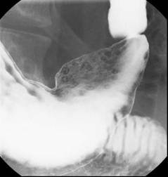
“In this study, patients with obesity received more frequent endoscopic interventions, which could explain why mortality rates were not significantly higher,” Dr. Abou Gergi reported.
In the analysis, characterized as the first study to evaluate the impact of obesity on outcomes in upper GI hemorrhage, data were drawn from the 2012 Nationwide Inpatient Sample database, which is considered to provide a representative sample of U.S. hospital experience. Drawn from more than 7 million hospitalizations, the study focused on patients 18 or over with a primary ICD-9 code for upper GI hemorrhage. The primary outcome was mortality. Secondary outcomes included interventions, ICU admissions, and length of stay.
Of the 132,545 discharges with upper GI hemorrhage, 11,220 (8.5%) were identified as obese. The in-hospital mortality overall was 1.97%, but the proportion of those who died was slightly lower among patients identified as obese, producing a nonsignificant adjusted odds ratio (OR) of 0.87 (P greater than .1). Yet the rates of hemorrhage shock (OR 1.31; P = .02) and admission to the ICU (OR 1.35; P less than .02) were greater in the obese. The median length of stay of 0.35 days for obese patients was also significantly longer.
One reason for the lower rate of mortality may be more aggressive treatment. In particular, repeat endoscopy therapy was more common in those who were obese (P less than .01), suggesting, “Our treatments are working,” Dr. Abou Gergi said.
The proportion of patients with a score of 3 or greater on the Charlson comorbidity index was higher in the obese than in those not identified as obese (49% vs. 39%; P less than .01). This along with previously published evidence that obese patients take more anticoagulants, take more antiplatelets, and may face delays in endoscopy due to greater difficulty in administering sedation, were among considerations predicting a higher mortality, according to Dr. Abou Gergi.
However, there are several potential explanations for these unexpected findings. One is that mortality rates overall were low, making it difficult to show differences on this outcome. In addition, ICD-9 codes may be effective for isolating a group with obesity but not in identifying a control group without obesity.
“It is likely that not all patients who are obese received this code, so we may be seeing a population of obese patients be compared to another population that includes at least some patients who also have obesity,” explained Dr. John R. Saltzman, director of endoscopy, Brigham and Women’s Hospital, Boston. A coauthor of this study, Dr. Saltzman noted that despite a comparable rate of in-hospital mortality, most of the findings in the study argued that patients who develop upper GI bleeding have a more difficult course. He noted that this is reflected in the cost of care, which was significantly higher in those who were obese.
Although he acknowledged that he was surprised that this was “essentially a negative study,” he believes that mortality may have been “too tough” as a primary endpoint for demonstrating a difference. However, he also believes that it may be appropriate to give credit for effective treatments.
“I think that may be the key. We are just getting better at taking care of these patients,” Dr. Saltzman said.
Dr. Abou Gergi reported he has no relevant financial relationships.
HONOLULU – Obese patients who develop an upper GI bleed receive more treatment, are more likely to develop hemorrhagic shock, and are more likely to require admission to an intensive care unit than those who are not obese, but they do not have higher in-hospital mortality, according to analysis of a large national database that was presented at the annual meeting of the American College of Gastroenterology.
The significantly greater odds ratio of most major complications from upper GI bleeds in patients with obesity relative to those who are not obese was expected but so was an increased rate of in-hospital mortality, according to the first author of the study, Dr. Marwan S. Abou Gergi of Catalyst Medical Consulting, Baltimore.
“In this study, patients with obesity received more frequent endoscopic interventions, which could explain why mortality rates were not significantly higher,” Dr. Abou Gergi reported.
In the analysis, characterized as the first study to evaluate the impact of obesity on outcomes in upper GI hemorrhage, data were drawn from the 2012 Nationwide Inpatient Sample database, which is considered to provide a representative sample of U.S. hospital experience. Drawn from more than 7 million hospitalizations, the study focused on patients 18 or over with a primary ICD-9 code for upper GI hemorrhage. The primary outcome was mortality. Secondary outcomes included interventions, ICU admissions, and length of stay.
Of the 132,545 discharges with upper GI hemorrhage, 11,220 (8.5%) were identified as obese. The in-hospital mortality overall was 1.97%, but the proportion of those who died was slightly lower among patients identified as obese, producing a nonsignificant adjusted odds ratio (OR) of 0.87 (P greater than .1). Yet the rates of hemorrhage shock (OR 1.31; P = .02) and admission to the ICU (OR 1.35; P less than .02) were greater in the obese. The median length of stay of 0.35 days for obese patients was also significantly longer.
One reason for the lower rate of mortality may be more aggressive treatment. In particular, repeat endoscopy therapy was more common in those who were obese (P less than .01), suggesting, “Our treatments are working,” Dr. Abou Gergi said.
The proportion of patients with a score of 3 or greater on the Charlson comorbidity index was higher in the obese than in those not identified as obese (49% vs. 39%; P less than .01). This along with previously published evidence that obese patients take more anticoagulants, take more antiplatelets, and may face delays in endoscopy due to greater difficulty in administering sedation, were among considerations predicting a higher mortality, according to Dr. Abou Gergi.
However, there are several potential explanations for these unexpected findings. One is that mortality rates overall were low, making it difficult to show differences on this outcome. In addition, ICD-9 codes may be effective for isolating a group with obesity but not in identifying a control group without obesity.
“It is likely that not all patients who are obese received this code, so we may be seeing a population of obese patients be compared to another population that includes at least some patients who also have obesity,” explained Dr. John R. Saltzman, director of endoscopy, Brigham and Women’s Hospital, Boston. A coauthor of this study, Dr. Saltzman noted that despite a comparable rate of in-hospital mortality, most of the findings in the study argued that patients who develop upper GI bleeding have a more difficult course. He noted that this is reflected in the cost of care, which was significantly higher in those who were obese.
Although he acknowledged that he was surprised that this was “essentially a negative study,” he believes that mortality may have been “too tough” as a primary endpoint for demonstrating a difference. However, he also believes that it may be appropriate to give credit for effective treatments.
“I think that may be the key. We are just getting better at taking care of these patients,” Dr. Saltzman said.
Dr. Abou Gergi reported he has no relevant financial relationships.
AT ACG 2015

Online neurocognitive test is versatile clinical tool
NEW YORK – An online neurocognitive test designed to provide reliable objective information about memory, concentration, and emotional well-being is a highly effective tool not only for guiding patients to appropriate therapy but for establishing the credibility of the clinician, according to the description of one such tool at the American Psychiatric Association’s Institute on Psychiatric Services.
The neurocognitive evaluation, which most patients complete on their home computers, “employs explicitly measured scales that reassure patients that I have considered multiple factors and am not just giving my off-the-hip guess and impression,” reported Dr. Joseph J. Parks, director of MOHealthNet, the Medicaid authority for Missouri in Jefferson City.
Several proprietary evaluation systems are available, according to Dr. Parks, but his experience has been with a tool called WebNeuro. Physicians develop an account on this online system and then assign a unique code to patients for access. The assessment, which employs validated and standardized tests of cognitive function and related psychometric fields, such as mood and coping skills, takes about 35 minutes to complete.
Results, which are based on comparisons with normative data in a large database, are generated almost immediately. For typical patients, Dr. Parks estimated that it takes 3-5 minutes to interpret the results. Validation studies include one published in 2007 (Behav Res Methods. 2007 Nov;39[4]:940-9).
This assessment tool has a broad number of options over other alternatives, according to Dr. Parks, who called traditional bedside neurocognitive tests “lame.” He suggested that the information gleaned from asking patients to spell words backward or perform uncommon math problems is “unimpressive.’ Ultimately, “you don’t perform these tests most of the time because you have no respect for their value.”
Referring patients for assessment by a neurocognitive psychologist or psychiatrist is another option, but Dr. Parks noted that these specialists are in short supply and not always willing to perform evaluations not linked to treatment. Moreover, it might take weeks for a referral to lead to an appointment and then another delay before the specialist provides a report that is “longer than I need.”
In contrast, the online neurocognitive tests are available at any time and generate data in hard numbers, showing individual patient performance on a variety of scales relative to normal performance. Dr. Parks reports that he uses this evaluation to gain insight on a broad array of potential diagnoses, such as attention-deficit/hyperactivity disorder (ADHD) and dementia. The test allows the clinician to document impaired memory or concentration, and provide information about relative severity. The data are particularly helpful in cases when the patient is not convinced.
“When patients are not happy that I am not prescribing the stimulant that they requested, I can point to the results to say you don’t have ADHD,” Dr. Parks explained. He described patients complaining of problems of concentration who can be reassured when the data suggest that, in fact, their performance is on the upper limits of normal.
“When we discuss the results, having taken the test provides the patient with a much clearer understanding of what I mean by concentration or memory,” Dr. Parks said. “Results of the test make both sides of the conversation more explicit.”
The test also can be repeated to demonstrate the effect of treatment, according to Dr. Parks. Again, an improvement in key scores provides reassurance to the patient when subjective assessments of improvement are doubted.
The test is available in several languages, including Spanish, and it is geared for a third- or fourth-grade reading level. Although Dr. Parks estimated that about 80% of his patients take the test at home, 20% complete the evaluation on a computer at his facility or with assistance from a proctor, such as a behavioral therapist. Dr. Parks said the test is reimbursed by Medicare and about two-thirds of health insurers in Missouri.
Online neurocognitive testing addresses a large number of the deficiencies associated with previous options, according to Dr. Parks. “This is an area that has been so ripe for change.”
Dr. Parks reported no relevant financial relationships.
EXPERT ANALYSIS AT THE INSTITUTE ON PSYCHIATRIC SERVICES