User login
FDA inspection findings rarely reported in literature

Photo by Esther Dyson
A new study suggests that, when the US Food and Drug Administration (FDA) identifies problems at clinical trial sites, those findings are seldom reported in peer-reviewed articles later written about the research.
Only 4% of the articles analyzed in this study disclosed that an FDA inspection revealed significant problems at a trial site.
Charles Seife, of the Carter Institute of Journalism at New York University in New York, New York, reported this finding in JAMA Internal Medicine.
Seife noted that the FDA classifies its inspections based on the severity of the violations, and the most severe is “official action indicated (OAI),” which means objectionable conditions or practices that warrant regulatory action.
During the 2013 fiscal year, about 2% of the 644 inspections the FDA conducted at trial sites were classified as OAI.
With this in mind, Seife and his students set out to identify published trials in which an FDA inspection revealed significant problems and determine whether there was mention of it in the medical literature.
The group identified 57 trials in which an FDA inspection uncovered the following problems:
- Falsification or submission of false information (22 trials, 39%)
- Problems with adverse events reporting (14 trials, 25%)
- Protocol violations (42 trials, 74%)
- Inadequate or inaccurate recordkeeping (35 trials, 61%)
- Failure to protect the safety of patients and/or issues with oversight or informed consent (30 trials, 53%)
- Violations that were not otherwise characterized (20 trials, 35%).
Only 3 of the 78 publications (4%) that resulted from these trials mentioned the objectionable conditions or practices.
“The FDA does not typically notify journals when a site participating in a published clinical trial receives an OAI inspection, nor does it generally make any announcement intended to alert the public about the research misconduct that it finds,” Seife wrote.
“The documents the agency discloses tend to be heavily redacted. As a result, it is usually very difficult, or even impossible, to determine which published clinical trials are implicated by the FDA’s allegations of research misconduct.”
In a related editorial, Robert Steinbrook, MD, of the Yale School of Medicine in New Haven, Connecticut, and Rita F. Redberg, MD, of the University of California, San Francisco, wrote that Seife’s study highlights an important area for improved reporting of clinical trials and enhanced transparency at the FDA.
“A central responsibility of medical journals is maintaining and improving trust in the medical literature,” they wrote. “Journals should expect that investigators and sponsors of clinical trials would promptly notify them of substantial findings from FDA and other regulatory agency inspections and modify their reports of clinical trials as needed, either before or after publication.” ![]()
Study supports iron supplementation after blood donation

Photo by Charles Haymond
Low-dose oral iron supplementation can reduce the time to hemoglobin recovery after blood donation, according to a study published in JAMA.
Researchers found that a daily dose of ferrous gluconate (37.5 mg of elemental iron) reduced blood donors’ time to 80% hemoglobin recovery, whether the donors were iron-depleted (with ferritin levels of 26 ng/mL or lower) or iron-replete (with ferritin levels higher than 26 ng/mL).
Joseph E. Kiss, MD, of the Institute for Transfusion Medicine in Pittsburgh, Pennsylvania, and his colleagues conducted this study at 4 regional blood centers in the US.
The researchers randomized 215 subjects (who had not donated whole blood or red blood cells within 4 months) to receive one tablet of ferrous gluconate daily or no iron for 24 weeks after donating a unit of whole blood (500 mL).
The study’s primary outcomes were time to recovery of 80% of the post-donation decrease in hemoglobin and recovery of baseline ferritin levels.
The researchers found that subjects who received iron supplementation achieved 80% hemoglobin recovery more quickly than subjects who did not receive iron, regardless of ferritin levels.
In the low-ferritin group, the mean time to 80% hemoglobin recovery was 32 days in subjects who received iron and 158 days in those who did not (P<0.001). In the higher-ferritin group, the mean time to 80% hemoglobin recovery was 31 days in subjects who received iron and 78 days in those who did not (P=0.02).
In the low-ferritin group, the median time to recovery of baseline ferritin levels was 21 days in subjects who received iron and more than 168 days in subjects who did not. In the higher-ferritin group, the median time to recovery of baseline ferritin levels was 107 days in subjects who received iron and more than 168 days in those who did not.
The median time to recovery of iron stores was 76 days in all subjects who received iron supplements and more than 168 days in those who did not (P<0.001). Sixty-seven percent of subjects who did not receive iron had failed to recover their iron stores by 168 days.
The researchers said these findings raise important considerations regarding the 8-week minimum waiting period between blood donations that is required in the US and Canada. This is a shorter period than those allowed in many other countries.
The study suggests that hemoglobin recovery differs according to a donor’s pre-donation ferritin level, but, even in iron-replete donors, the mean recovery was only 70% at 8 weeks.
Prolonging the minimum wait time between blood donations would allow for more thorough recovery, the researchers noted, but it could also compromise the blood supply. Furthermore, increasing the waiting period might not be adequate for donors who do not take iron supplements. ![]()
Computer model simulates blood development

Photo courtesy of
University of Cambridge
A new computer model that simulates blood cell development could aid the discovery of novel treatments for hematologic malignancies, according to researchers.
“With this new computer model, we can carry out simulated experiments in seconds that would take many weeks to perform in the laboratory, dramatically speeding up research into blood development and the genetic mutations that cause leukemia,” said Bertie Gottgens, PhD, of the University of Cambridge in the UK.
Dr Gottgens and his colleagues explained this research in Nature Biotechnology.
To start, the researchers measured the activity of 48 genes in 3934 hematopoietic progenitor cells. They used the resulting dataset to construct the computer model of blood cell development, using computational approaches originally developed at Microsoft Research for the synthesis of computer code.
Subsequent lab experiments validated the accuracy of the model.
The researchers noted that the model can be used to simulate the activity of key genes implicated in hematologic malignancies. For example, around 1 in 5 children who develop leukemia have a faulty version of the RUNX1 gene, as do a similar proportion of adults with acute myeloid leukemia.
The computer model shows how RUNX1 interacts with other genes to control blood cell development. The gene produces the RUNX1 protein, which, in healthy patients, activates a network of key genes. In leukemia patients, an altered form of the protein is thought to suppress the network.
If the researchers change the “rules” in the network model, they can simulate the formation of abnormal leukemia cells. By tweaking the leukemia model until the behavior of the network reverts back to normal, the team can identify pathways that, potentially, could be targeted with drugs.
“Because the computer simulations are very fast, we can quickly screen through lots of possibilities to pick the most promising ones as pathways for drug development,” Dr Gottgens said.
“The cost of developing a new drug is enormous, and much of this cost comes from new candidate drugs failing late in the drug development process. Our model could significantly reduce the risk of failure, with the potential to make drug discovery faster and cheaper.” ![]()
Anticoagulant now available in US pharmacies

Photo by Rhoda Baer
The oral factor Xa inhibitor edoxaban (Savaysa) is now available in US pharmacies, according to Daiichi Sankyo Company, Limited, the company developing the drug.
Savaysa is approved by the US Food and Drug Administration to reduce the risk of stroke and systemic embolism (SE) in patients with non-valvular atrial fibrillation (NVAF), as well as for the treatment of deep vein thrombosis and pulmonary embolism following 5 to 10 days of initial therapy with a parenteral anticoagulant.
According to Savaysa’s label, it should not be used in NVAF patients with creatinine clearance levels greater than 95 mL/min because, in that population, there is an increased risk of ischemic stroke with the drug compared to warfarin.
Daiichi Sankyo has developed resources for physicians and patients to help ensure patients can begin and/or remain on Savaysa per physician instructions.
The Savaysa Savings Plus program will include a reimbursement hotline to assist patients and prescribers who request help understanding a patient’s available coverage. Eligible patients who are prescribed Savaysa can enroll in a copay savings program and pay $4 per month through the Savaysa Savings Card.
Vouchers will also be available to provide patients and doctors with a way to try Savaysa at no cost to see if it is right for the patient.
In addition, the Savaysa Patient Assistance Program will offer assistance to qualified individuals, providing free product to eligible patients who are prescribed Savaysa, are uninsured, and are unable to identify alternative payment sources.
The approval of Savaysa in the US is based on data from the ENGAGE AF-TIMI 48 trial and the Hokusai-VTE trial. ![]()
FDA clears new blood-draw device
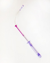
Photo courtesy of
Velano Vascular
The US Food and Drug Administration (FDA) has cleared for marketing a device that reduces the need for venipunctures for in-hospital blood draws.
Velano Vascular’s blood-draw device resembles a common syringe.
It allows peripheral intravenous catheters to be repurposed to draw blood from patients, thereby reducing the need for additional needle sticks among patients receiving medications and hydration via intravenous delivery.
The single-use device will soon be used for clinical evaluation in select hospitals, including the University of Pennsylvania in Philadelphia and University Hospitals Case Medical Center of Cleveland in Ohio.
“A fundamental benefit of this technology is reducing the ‘pin cushion effect,’ in which hospitalized patients are ‘stuck’ several times daily to obtain blood tests,” said Eric M. Stone, co-founder and chief executive officer of Velano Vascular, the company developing the blood-draw device.
“Oftentimes, the draw procedure is plagued by multiple failed attempts. The FDA’s clearance of this novel technology validates the existing clinical need and will allow us to expedite our efforts to bring this innovation to patients, healthcare providers, and hospitals around the world.”
According to research conducted by Velano Vascular, 1 of every 3 hospital patients is stuck 2 or more times daily for blood draws, with a significant subset of these patients receiving 3 or more blood draws, along with numerous needle sticks.
Twenty-eight percent of adult venipunctures and 44% of pediatric venipunctures require more than one stick to successfully draw blood.
“Traditional blood draws are one of the most common and most problematic healthcare procedures,” said Karen Daley, PhD, RN, past president of the American Nurses Association and a healthcare worker safety advocate.
“It is an antiquated technology that creates pain and anxiety for many patients, a significant safety risk for healthcare professionals, and a real inefficiency in our healthcare system. Velano Vascular has developed a common-sense solution to this pervasive, long-standing problem.” ![]()
Though costly, blood cancer drugs appear cost-effective

Photo by Bill Branson
A new analysis indicates that certain high-cost therapies for hematologic malignancies provide reasonable value for money spent.
Most cost-effectiveness ratios were lower than thresholds commonly used to establish cost-effectiveness in the US—$50,000 or $100,000 per quality-adjusted life year (QALY) gained.
The median cost-effectiveness ratio was highest for chronic myeloid leukemia (CML), at $55,000/QALY, and lowest for non-Hodgkin lymphoma (NHL), at $21,500/QALY.
Researchers presented these data in Blood.
“Given the increased discussion about the high cost of these treatments, we were somewhat surprised to discover that their cost-effectiveness ratios were lower than expected,” said study author Peter J. Neumann, ScD, of Tufts Medical Center in Boston.
“Our analysis had a small sample size and included both industry- and non-industry-funded studies. In addition, cost-effectiveness ratios may have changed over time as associated costs or benefits have changed. However, the study underscores that debates in healthcare should consider the value of breakthrough drugs and not just costs.”
With that issue in mind, Dr Neumann and his colleagues had conducted a systematic review of studies published between 1996 and 2012 that examined the cost utility of agents for hematologic malignancies. The cost utility of a drug was depicted as a ratio of a drug’s total cost per patient QALY gained.
The researchers identified 29 studies, 22 of which were industry-funded. Nine studies were conducted from a US perspective, 6 from the UK, 3 from Norway, 3 from Sweden, 2 from France, 1 from Canada, 1 from Finland, and 4 from “other” countries.
The team grouped studies according to malignancy—CML, chronic lymphocytic leukemia (CLL), NHL, and multiple myeloma (MM)—as well as by treatment—α interferon, alemtuzumab, bendamustine, bortezomib, dasatinib, imatinib, lenalidomide, rituximab alone or in combination, and thalidomide.
The studies reported 44 cost-effectiveness ratios, most concerning interventions for NHL (41%) or CML (30%). Most ratios pertained to rituximab (43%), α interferon (18%), or imatinib (16%), and the most common intervention-disease combination was rituximab (alone or in combination) for NHL (36%).
The median cost-effectiveness ratios fluctuated over time, rising from $35,000/QALY (1996-2002) to $52,000/QALY (2003-2006), then falling to $22,000/QALY (2007-2012).
The median cost-effectiveness ratio reported by industry-funded studies was lower ($26,000/QALY) than for non-industry-funded studies ($33,000/QALY).
Four cost-effectiveness ratios, 1 from an industry-funded study, exceeded $100,000/QALY. This included 2 studies of bortezomib in MM, 1 of α interferon in CML, and 1 of imatinib in CML.
The researchers said these results suggest that many new treatments for hematologic malignancies may confer reasonable value for money spent. The distribution of cost-effectiveness ratios is comparable to those for cancers overall and for other healthcare fields, they said.
This study was funded by internal resources at the Center for the Evaluation of Value and Risk in Health. The center receives funding from federal, private foundation, and pharmaceutical industry sources. ![]()
EC approves bortezomib for MCL
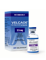
Photo courtesy of Millennium
The European Commission (EC) has approved bortezomib (Velcade) in combination with rituximab, cyclophosphamide, doxorubicin, and prednisone (VR-CAP) to treat adults with previously untreated mantle cell lymphoma (MCL) in whom hematopoietic stem cell transplant (HSCT) is considered unsuitable.
Now, bortezomib can be marketed for this indication in all 28 countries of the European Union (EU).
Bortezomib is already approved in the EU to treat multiple myeloma (MM), either as monotherapy or in combination with other agents.
The EC’s approval of bortezomib in MCL is based on data from a phase 3 study known as LYM-3002.
This randomized trial included 487 patients with newly diagnosed MCL who were ineligible, or not considered, for HSCT. Patients were randomized to receive VR-CAP or R-CHOP (rituximab, cyclophosphamide, doxorubicin, vincristine, and prednisone).
The VR-CAP regimen significantly improved progression-free survival (PFS), the primary endpoint, when compared to R-CHOP.
According to an independent review committee, there was a 59% improvement in PFS for the VR-CAP arm compared to the R-CHOP arm, with median times of 24.7 months and 14.4 months, respectively (hazard ratio=0.63; P<0.001).
Study investigators reported a 96% increase in PFS with VR-CAP compared to R-CHOP, with median times of 30.7 months and 16.1 months, respectively (hazard ratio=0.51, P<0.001).
VR-CAP was associated with additional, but manageable, toxicity when compared to R-CHOP. Serious adverse events (AEs) were reported in 38% and 30% of patients, respectively. And grade 3 or higher AEs were reported in 93% and 85% of patients, respectively.
Treatment discontinuation due to AEs occurred in 9% of patients in the VR-CAP arm and 7% in the R-CHOP arm. On-treatment, drug-related deaths occurred in 2% and 3% of patients, respectively.
About bortezomib
Bortezomib works by reversibly interrupting the normal working of cell proteasomes, inducing cancerous cells to stop growing and die.
In addition to the new MCL indication, the drug is approved in the EU to treat various stages of MM. It’s approved in combination with melphalan and prednisone to treat previously untreated adults with MM who are unsuitable for high-dose chemotherapy with HSCT.
Bortezomib is also approved in combination with dexamethasone, or with dexamethasone plus thalidomide, to treat previously untreated MM patients set to receive high-dose chemotherapy followed by HSCT.
And the drug is approved as monotherapy or in combination with pegylated liposomal doxorubicin or dexamethasone to treat adults with MM whose disease has progressed after at least one other treatment and who have already had, or cannot undergo, HSCT.
Bortezomib is approved in more than 90 countries and has been used to treat more than 550,000 patients worldwide.
The product is co-developed by Millennium, the Takeda Oncology Company, a wholly owned subsidiary of Takeda Pharmaceutical Company Limited, and Janssen Pharmaceutical Companies.
Millennium is responsible for commercialization in the US. Janssen Pharmaceutical Companies are responsible for commercialization in Europe and the rest of the world. Takeda Pharmaceutical Company Limited and Janssen Pharmaceutical K.K. co-promote the drug in Japan. ![]()
Photo courtesy of Millennium
The European Commission (EC) has approved bortezomib (Velcade) in combination with rituximab, cyclophosphamide, doxorubicin, and prednisone (VR-CAP) to treat adults with previously untreated mantle cell lymphoma (MCL) in whom hematopoietic stem cell transplant (HSCT) is considered unsuitable.
Now, bortezomib can be marketed for this indication in all 28 countries of the European Union (EU).
Bortezomib is already approved in the EU to treat multiple myeloma (MM), either as monotherapy or in combination with other agents.
The EC’s approval of bortezomib in MCL is based on data from a phase 3 study known as LYM-3002.
This randomized trial included 487 patients with newly diagnosed MCL who were ineligible, or not considered, for HSCT. Patients were randomized to receive VR-CAP or R-CHOP (rituximab, cyclophosphamide, doxorubicin, vincristine, and prednisone).
The VR-CAP regimen significantly improved progression-free survival (PFS), the primary endpoint, when compared to R-CHOP.
According to an independent review committee, there was a 59% improvement in PFS for the VR-CAP arm compared to the R-CHOP arm, with median times of 24.7 months and 14.4 months, respectively (hazard ratio=0.63; P<0.001).
Study investigators reported a 96% increase in PFS with VR-CAP compared to R-CHOP, with median times of 30.7 months and 16.1 months, respectively (hazard ratio=0.51, P<0.001).
VR-CAP was associated with additional, but manageable, toxicity when compared to R-CHOP. Serious adverse events (AEs) were reported in 38% and 30% of patients, respectively. And grade 3 or higher AEs were reported in 93% and 85% of patients, respectively.
Treatment discontinuation due to AEs occurred in 9% of patients in the VR-CAP arm and 7% in the R-CHOP arm. On-treatment, drug-related deaths occurred in 2% and 3% of patients, respectively.
About bortezomib
Bortezomib works by reversibly interrupting the normal working of cell proteasomes, inducing cancerous cells to stop growing and die.
In addition to the new MCL indication, the drug is approved in the EU to treat various stages of MM. It’s approved in combination with melphalan and prednisone to treat previously untreated adults with MM who are unsuitable for high-dose chemotherapy with HSCT.
Bortezomib is also approved in combination with dexamethasone, or with dexamethasone plus thalidomide, to treat previously untreated MM patients set to receive high-dose chemotherapy followed by HSCT.
And the drug is approved as monotherapy or in combination with pegylated liposomal doxorubicin or dexamethasone to treat adults with MM whose disease has progressed after at least one other treatment and who have already had, or cannot undergo, HSCT.
Bortezomib is approved in more than 90 countries and has been used to treat more than 550,000 patients worldwide.
The product is co-developed by Millennium, the Takeda Oncology Company, a wholly owned subsidiary of Takeda Pharmaceutical Company Limited, and Janssen Pharmaceutical Companies.
Millennium is responsible for commercialization in the US. Janssen Pharmaceutical Companies are responsible for commercialization in Europe and the rest of the world. Takeda Pharmaceutical Company Limited and Janssen Pharmaceutical K.K. co-promote the drug in Japan.
Aggressiveness of CLL linked to genetic variability

The genetic variability of chronic lymphocytic leukemia (CLL) appears to predict its aggressiveness, according to a study published in Genome Medicine.
Investigators found evidence suggesting that greater variability in gene expression is associated with more aggressive disease.
The team analyzed gene expression in two cohorts of CLL patients—those with IgVH mutations and a good prognosis and those with unmutated CLL who have more aggressive disease.
The researchers examined 70 mutated and 52 unmutated CLL samples, as well as 20 control samples taken from healthy individuals.
Unmutated, aggressive CLL showed increased gene expression variability across individuals, whereas gene expression variability was lower in less aggressive, mutated CLL.
The investigators validated these observations by comparing them against a second sample group consisting of 24 mutated and 36 unmutated CLL samples.
The results suggested that CLL aggressiveness is specifically determined by a set of 500 genes showing increased expression variability across individuals. The genes are involved in processes such as adaptation to the environment, cell death, tumor growth, and drug resistance.
“Our conclusion is that the coefficient of variation for gene expression in CLL efficiently predicts its aggressiveness,” said study author Alfonso Valencia, PhD, of Centro Nacional de Investigaciones Oncologicas (CNIO) in Madrid, Spain.
“More importantly, if further research confirms these findings, a classifier based on the measurement of gene expression variability could be created to predict the disease subtype of CLL patients.”
The researchers said their next step is to discover the mechanisms responsible for the high levels of expression variability for a given gene across individuals.
Understanding the mechanisms underlying this phenomenon is of crucial relevance for oncology research, the investigators said, as it is linked to tumor heterogeneity, a key feature of cancer progression and drug resistance.
The greater the genetic variability in a tumor, the better it will adapt to its environment, and the more probabilities for this tumor to spread, develop resistance to cancer therapies, and metastasize to distant organs.
Risk of HBV reactivation ‘underappreciated’

Credit: CDC
Reactivation of the hepatitis B virus (HBV) may be more of a risk than we anticipated, investigators have reported in Hepatology.
Their research indicates that HBV reactivation is associated with the use of chemotherapy, high-dose corticosteroids, biologics targeting tumor necrosis factor-alpha (TNF-α), and agents that aren’t really considered immunosuppressive.
HBV reactivation is also fairly common after organ transplant and hematopoietic stem cell transplant (HSCT).
As reactivation of HBV can be fatal, the study authors suggest routine screening of HBV in all patients prior to the start of these treatments.
The researchers noted that, in September 2013, the US Food and Drug Administration (FDA) issued a Drug Safety Communication in an attempt to decrease the risk of HBV reactivation. The communication advised healthcare professionals to screen patients for HBV prior to the administration of ofatumumab or rituximab.
“[T]his may be just the tip of the iceberg,” said Adrian Di Bisceglie, MD, of Saint Louis University School of Medicine in Missouri.
“Our research suggests that the issue of HBV reactivation may be an underappreciated clinical challenge that extends well beyond the use of just two anti-CD20 medications.”
After a systematic literature review, Dr Di Bisceglie and his colleagues identified 504 studies pertaining to reactivation of HBV.
The investigators reviewed 14 studies in which the antiviral agent lamivudine was used to prevent HBV reactivation in HBsAg-positive patients receiving chemotherapy. Among patients who did not receive lamivudine, HBV reactivation occurred in 32%. Thirteen percent of patients experienced liver failure, and 7% died.
The researchers also looked at patients undergoing HSCT. In one study, 61 patients had resolved HBV infection before HSCT. But 12 of these patients (20%) developed reverse seroconversion (reappearance of HBsAg in a person who was HBsAg-negative, anti-HBc-positive prior to HSCT).
The cumulative probability of reverse seroconversion was 9% a year after HSCT, 21.7% at 2 years, and 42.9% at 4 years.
The investigators also noted that high-dose corticosteroids carry a significant risk of HBV reactivation, both as part of combination treatment for malignancies and when used alone to treat benign conditions.
In addition, the researchers found data showing that HBV reactivation has occurred with antitumor agents that are not thought to be particularly immunosuppressive, such as imatinib and thalidomide. The team said this raises questions about the mechanisms by which drugs are causing HBV reactivation.
The investigators also looked at data from 257 patients with active or recovered HBV infection who received treatment with biological therapies targeting TNF-α.
Forty-two percent of the patients had elevations in serum aminotransferase levels, 39% had reappearance of HBV DNA, 16% had signs and symptoms of liver disease, and 5% died of liver failure.
HBV reactivation was more frequent among patients receiving infliximab than etanercept. It was 7-fold higher among patients who were HBsAg-positive (38%) than those who were HBsAg-negative but anti-HBc-positive (5%).
While it remains unclear how HBV reactivation occurs, experts believe a loss of immune control over viral replication may trigger the process.
“Further study and cooperation between various medical disciplines will help broaden understanding of HBV reactivation,” Dr Di Bisceglie concluded.
Vaccine gets orphan designation for ATLL
The European Medicines Agency (EMA) has granted orphan drug designation for a therapeutic vaccine candidate known as THV02 to treat adult T-cell leukemia/lymphoma (ATLL).
THV02 is an experimental treatment composed of 2 lentiviral vectors to be used in a prime/boost regimen in ATLL patients infected by the HTLV-1 virus.
Both investigational drugs encode the same antigens, derived from 4 proteins of the HTLV-1 virus.
THV02 is intended to induce an immune response against HTLV antigens born by ATLL with the aim of enabling the patients’ immune system to fight leukemic cells.
Preclinical evaluation has suggested that THV02 is safe, and the vaccine has presented an “unprecedented” immunogenicity profile in several models, according to THERAVECTYS, the company developing THV02.
“Preclinical immunogenicity results obtained to date are very promising, and we are really excited by the prospect of bringing a safe and better-tolerated alternative to patients who are desperately in need of a treatment,” said Déborah Revaud, the senior scientist in charge of developing THV02.
The EMA grants orphan designation to drugs in development intended for the treatment, prevention, or diagnosis of life-threatening or chronically debilitating diseases occurring in fewer than 5 in 10,000 people.
The designation allows sponsors to benefit from an accelerated development process, financial incentives, and a 10-year period of market exclusivity once the drug is on the market.
“We are extremely pleased that the European Medicines Agency has granted an orphan drug status to our vaccine candidate against ATLL,” said Emmanuelle Sabbah-Petrover, PhD, head of regulatory affairs at THERAVECTYS.
“We expect to recruit our first patients towards the end of Q3 2015 in Europe and advance further developments in the US and in Japan in 2016.”
Should THV02 demonstrate a convincing safety and efficacy profile during its development against ATLL, THERAVECTYS said it will consider developing the vaccine for HTLV-related infections as treatment and possibly as prophylaxis.