User login
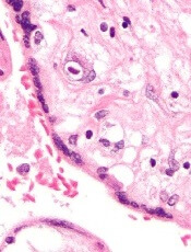
Team creates new cells for modeling malaria

infection in iPSC-derived liver
cells 8 days after infection
Credit: Shengyong Ng et al.
Researchers say they’ve found a way to grow liver-like cells from induced pluripotent stem cells (iPSCs).
The liver-like cells can be infected with several strains of the malaria parasite and respond to existing drugs the same way mature human liver cells do.
The new cells, described in Stem Cell Reports, could allow scientists to test drugs on cells from people with different genetic backgrounds, who may respond differently to malaria infection and treatment.
Modeling infection
Until now, malaria researchers have not had many reliable ways to test new drugs in liver tissue.
“What’s historically been done is people have tried to make do with the systems that were available,” said study author Sangeeta Bhatia, MD, PhD, of the Massachusetts Institute of Technology in Cambridge.
In 2013, Dr Bhatia and her colleagues showed they could model malaria infection in hepatocytes from human donors. However, this generates only a limited supply from each donor, and not all of the cells work well for drug studies.
The researchers then turned to iPSCs, which can be generated from human skin cells by adding reprogramming factors. To create liver cells, the researchers added a series of growth factors, including hepatocyte growth factor, to the iPSCs.
The team generated these cells in 2012 and used them to model infection of hepatitis C. However, these cells, known as hepatocyte-like cells, did not seem to be as mature as real adult liver cells.
In the current study, the researchers found these cells could be infected with several strains of malaria. But, initially, the cells did not respond to drugs in the same way as adult liver cells.
In particular, they were not sensitive to primaquine, which works only if cells have a certain set of drug-metabolism enzymes found in mature liver cells.
To induce the cells to become more mature and turn on these metabolic enzymes, the researchers added a molecule they had identified in a previous study. This compound, which the researchers call a “maturin,” stimulated the cells to turn on those enzymes, which made them sensitive to primaquine.
Toward better drugs
The team is now working with the nonprofit foundation Medical Malaria Ventures to test about 10 potential malaria drugs that are in the pipeline, first using adult donor liver cells and then the hepatocyte-like cells generated in this study.
These cells could also prove useful to help identify new drug targets, the researchers said. In this study, they found the liver-like cells can be infected with malaria when they are still in the equivalent of fetal stages of development, when they become hepatoblasts, which are precursors to hepatocytes.
In future studies, the researchers plan to investigate which genes get turned on when the cells become susceptible to infection, which may suggest new targets for malaria drugs.
They also hope to compare the genes needed for malaria infection with those needed for hepatitis infection, in hopes of identifying common pathways to target for both diseases. ![]()
Memory T cells can fight CMV infection
CMV infection
Transplanting a small number of antiviral memory T cells along with donor hematopoietic stem cells can fight and may even prevent cytomegalovirus (CMV) disease in transplant recipients, according to preclinical research published in the Journal of Immunology.
To date, researchers have focused on developing anti-CMV immunotherapy with effector-phenotype CD8+ T cells (TEFF cells), which attack and kill virally infected host cells.
But Christopher Snyder, PhD, of Thomas Jefferson University in Philadelphia, Pennsylvania, and his colleagues found that CMV-specific TEFF cells divide poorly in response to CMV infection or reactivation in mouse models.
So they wondered if CMV-specific memory-phenotype CD8+ T cells (TM cells)—which act more like stem cells—could help control the infection long-term.
The group showed that a small number of TM cells were enough to produce and repeatedly replenish all of the TEFF cells needed to fight CMV.
The infused TM cells became major contributors to the recipient antiviral immune response, persisting for at least 3 months’ time and producing TEFF cells at a steady stream.
To determine whether these cells have counterparts in humans, Dr Snyder and his colleagues compared the genomic fingerprints of mouse and human TM cells that were specific for CMV. Results showed the two had similar profiles.
“This suggested that human and mouse CMV-specific memory T cells are very similar populations,” said study author Michael Quinn, an MD/PhD student at Thomas Jefferson University.
“Therefore, infusing similar cells into humans could improve on immunotherapeutic methods for controlling CMV infection. This may be a valuable approach to keep the disease from emerging in people.”
Dr Snyder added, “Our data argue for developing new clinical trials focused specifically on using these T memory cells, in order to determine if it would indeed be better than current therapeutic options.” ![]()
mAb shows ‘modest activity’ in rel/ref MM

Photo by Linda Bartlett
The monoclonal IgM antibody PAT-SM6 was “well-tolerated” and showed “modest clinical activity” in patients with relapsed or refractory multiple myeloma (MM), researchers reported in haematologica.
Adverse events occurred in all 12 patients enrolled in the phase 1/2a study, but most were considered unrelated to treatment.
A third of patients, all of whom had progressive disease upon study entry, achieved stable disease after receiving PAT-SM6. The remaining patients progressed.
Leo Rasche, MD, of University Hospital Wurzburg in Germany, and his colleagues conducted this study. It was funded, in part, by Patrys Limited, the company developing PAT-SM6.
The study included 12 heavily pretreated MM patients. They had a median age of 69.5 years and a long-standing history of MM (range, 3.25 to 15.75 years). They had received a median of 3.9 prior lines of therapy (range, 2-7).
Patients received 4 escalating doses of PAT-SM6, over a period of 2 weeks, via intravenous infusions at 0.3 mg/kg, 1 mg/kg, 3 mg/kg, and 6 mg/kg.
Safety data
There were 54 treatment-emergent adverse events in all 12 patients. However, there were no dose-limiting toxicities and no deaths. The maximum tolerated dose has not been reached.
More than 80% of the adverse events were of mild to moderate intensity. Two patients (16.6%) each experienced a single serious event. One patient had acute back pain, and one had a bile duct stone. Neither of these events was considered treatment-related.
Twenty-one adverse events were considered treatment-related. This included leukopenia (66.6%), neutropenia (50%), hypertension (16.6%), catheter-related thrombophlebitis (8.3%), injection site erythema (8.3%), slight headache (8.3%), C-reactive protein increase (8.3%), and hypertriglyceridemia (8.3%).
Efficacy data
Most patients progressed following treatment, but 4 (33.3%) had stable disease. The investigators noted that stable disease is not necessarily connected with a clinical benefit, so they analyzed the 4 patients in detail.
Patient 4, who received PAT-SM6 at 1 mg/kg, entered the study with high-risk disease. The patient had 13q deletion and 1q21 gain, had received 5 prior lines of therapy, and was refractory to novel agents, including pomalidomide and bortezomib.
The patient was treatment-free for 1 month prior to receiving PAT-SM6. During treatment, there were no symptoms of active myeloma, and the patient asked to continue salvage therapy 1 week after the end of the study.
Patient 7, who received PAT-SM6 at 3 mg/kg, had been diagnosed with MM for 15 years and had received 4 prior lines of therapy.
The patient’s treatment-free interval before receiving PAT-SM6 was 30 months. After PAT-SM6, the patient was therapy-free for 4.6 months.
Patient 10, who received PAT-SM6 at 6mg/kg, had 6 prior lines of therapy, including tandem autologous stem cell transplant and several polychemotherapeutic regimens.
The patient was therapy-free for 4 months prior to receiving PAT-SM6. The patient requested salvage therapy 1 month after receiving PAT-SM6.
Patient 11, who received PAT-SM6 at 6mg/kg, had 4 prior lines of therapy and was refractory to both lenalidomide and thalidomide.
The patient’s treatment-free interval prior to PAT-SM6 was 12 months. After PAT-SM6, the patient was therapy-free for 5.2 months.
The researchers said additional trials testing PAT-SM6 in combination with other MM therapies are planned. PAT-SM6 has received orphan drug designation for MM in the US and the European Union. ![]()
Vitamin A as malaria prophylaxis

Photo by Sarah Mattison
New research suggests vitamin A may reduce the risk of malaria in young children.
The study showed that children under 5 living in sub-Saharan Africa were 54% less likely to develop malaria if they had been given a single, large dose of vitamin A.
The finding, published in eLife, indicates that vitamin A may be able to protect children from the malaria parasite, especially if administered during the wet season, when malaria-infected mosquitos are most prevalent.
“Now, we need to test vitamin A in a randomized, controlled, clinical trial to better understand whether this could really be an effective way to prevent this disease,” said study author Maria-Graciela Hollm-Delgado, PhD, of the Johns Hopkins Bloomberg School of Public Health in Baltimore, Maryland.
Dr Hollm-Delgado and her colleagues analyzed national survey data from 4 sub-Saharan countries—Burkina Faso, Mozambique, Rwanda, and Senegal—on children between the ages of 6 months and 59 months.
The goal was to determine the risk of Plasmodium parasitemia (n=8390) and Plasmodium falciparum-specific antigenemia (n=6121) following vitamin A supplementation and vaccinations.
The researchers found the measles and polio vaccines were not associated with malaria. And Bacille Calmette Guerin vaccination was associated with an increased risk of antigenemia (relative risk [RR]=4.06) but not parasitemia.
Only vitamin A was protective against malaria. Children who received vitamin A were less likely to present with parasitemia (RR=0.46) and antigenemia (RR=0.23).
Vitamin A appeared to be more protective under certain circumstances, including when administered during the rainy season, as well as when given to older children and when more time had passed since supplementation.
The researchers aren’t certain why vitamin A would reduce the rate of malaria infection, but they suspect it is because vitamin A, which is known to boost immunity and improve the ability to fight off infection, may help the body clear out the malaria parasite more quickly.
Only 62% of children in the study had received vitamin A supplementation, even though World Health Organization guidelines recommend that all children in sub-Saharan Africa receive a single, large dose of vitamin A.
Rates were higher for many vaccinations, Dr Hollm-Delgado said, noting that the guidelines for vitamin A aren’t as specific as they are for most vaccinations. ![]()
Balanced transfusion strategy may be better
![]()
Credit: UAB Hospital
A new study indicates that transfusing a balanced ratio of plasma, platelets, and red blood cells (RBCs) can decrease bleeding better than blood products with a higher ratio of RBCs, but this does not seem to affect mortality rates.
Patients who received blood products with a plasma-platelet-RBC ratio of 1:1:1 were more likely to achieve hemostasis and less likely to experience exsanguination than patients who received blood products with a ratio of 1:1:2.
However, there was no significant difference between the two transfusion strategies in overall death rates at 24 hours or at 30 days.
John B. Holcomb, MD, of the University of Texas Health Science Center at Houston, and his colleagues reported these results in JAMA.
The team conducted a study of 680 severely injured patients who arrived at 1 of 12 Level 1 trauma centers. The patients were predicted to require massive transfusion and were randomly assigned to receive blood products with ratios of 1:1:1 or 1:1:2 during active resuscitation, in addition to all local standard-of-care interventions.
The researchers found no significant differences for the primary outcomes of the study: mortality at 24 hours—12.7% in the 1:1:1 group and 17.0% in the 1:1:2 group (P=0.12)—or at 30 days—22.4% and 26.1%, respectively (P=0.26).
However, the incidence of exsanguination, which was the predominant cause of death within the first 24 hours, was significantly lower in the 1:1:1 group than in the 1:1:2 group—9.2% and 14.6%, respectively (P=0.03).
And more patients in the 1:1:1 group achieved hemostasis than in the 1:1:2 group—86% and 78%, respectively (P=0.006).
On the other hand, there were no significant differences between the two groups for rates of multiple inflammatory-mediated complications, such as acute respiratory distress syndrome, multiple organ failure, infection, sepsis, venous thromboembolism, and transfusion-related complications.
Based on these results, Dr Holcomb and his colleagues recommended that clinicians consider using a 1:1:1 transfusion protocol, starting with the initial units transfused while patients are actively bleeding, and then transitioning to laboratory-guided treatment once they’ve achieved hemorrhage control.
The team added that future studies should concentrate on the physiologically relevant period of active bleeding after injury and use acute complications and later deaths as safety endpoints. ![]()
mAb prompts responses in pretreated MM

Credit: Linda Bartlett
Single-agent daratumumab can elicit responses in patients with multiple myeloma (MM) who have failed treatment with proteasome inhibitors and immunomodulatory agents (IMiDs), preliminary results of a phase 2 study suggest.
The study enrolled MM patients who have received at least 3 different lines of therapy, including both a proteasome inhibitor and an IMiD, and patients who are double-refractory to a proteasome inhibitor and an IMiD.
Daratumumab, an anti-CD38 monoclonal antibody, has breakthrough therapy designation from the US Food and Drug Administration to treat this patient population.
Genmab A/S, the company that discovered daratumumab and licensed it to Janssen Biotech, Inc. for development, announced results from the phase 2 trial (Sirius MMY2002) yesterday.
This 2-part study enrolled 124 MM patients. The goal of part 1 was to define an optimal daratumumab regimen going forward, and part 2 was an expansion based on the optimal regimen.
Patients were randomized to receive daratumumab at 8 mg/kg every 4 weeks continuously by intravenous infusion or at 16 mg/kg administered at weekly intervals for 8 weeks, then every 2 weeks for an additional 16 weeks, and every 4 weeks thereafter by intravenous infusion.
Genmab reported that the overall response rate was 29.2% in the 16 mg/kg dosing group. And the median duration of response was 7.4 months, as determined by an independent review committee.
Daratumumab also showed a manageable safety profile, according to Genmab. The company said the data will be discussed with health authorities at upcoming meetings, pending their agreement. ![]()
Product deemed breakthrough for beta-thalassemia major

The US Food and Drug Administration (FDA) has granted breakthrough therapy designation to LentiGlobin® BB305 for the treatment of transfusion-dependent patients with beta-thalassemia major.
The product is created by inserting a functional human beta-globin gene into a patient’s hematopoietic stem cells ex vivo. The cells are returned to the patient via transplant.
The product is intended to treat sickle cell disease as well as beta-thalassemia major.
“The FDA’s breakthrough designation of LentiGlobin highlights that new therapies are needed for the treatment of patients with beta-thalassemia major, especially treatments with the potential to meaningfully reduce or liberate patients from transfusion dependence,” said David Davidson, MD, chief medical officer of bluebird bio, the company developing LentiGlobin.
“Our early clinical data investigating the use of LentiGlobin in patients with multiple genotypes of beta-thalassemia major . . . are very encouraging, and we remain on track to complete enrollment in the Northstar and HGB-205 studies in 2015.”
Early study results
The breakthrough designation is supported by data from the ongoing phase 1/2 Northstar (HGB-204) and HGB-205 studies. Findings in 8 subjects from both studies were presented at the 2014 ASH Annual Meeting.
As of December 1, five subjects with beta-thalassemia major had received LentiGlobin in the Northstar Study. The first 2 subjects were producing steadily increasing amounts of beta-T87Q-globin and were free of the need for transfusion for 5 months and 3 months, respectively.
Three additional subjects have received LentiGlobin as well, but researchers said it is too early to draw any meaningful conclusions on clinical efficacy.
As of December 1, two subjects with beta-thalassemia major received LentiGlobin as part of the HGB-205 study. Both achieved rapid transfusion independence with near-normal hemoglobin levels. And they were free from the need for transfusions for 12 months and 9 months, respectively.
The third treated subject, the first individual with sickle cell disease ever to be treated with gene therapy, has achieved neutrophil engraftment. But researchers said it is too early to draw any meaningful conclusions on clinical efficacy.
About breakthrough designation
The FDA’s breakthrough therapy designation is intended to expedite the development and review of a drug candidate intended to treat a serious or life-threatening condition.
For a drug to gain the designation, preliminary clinical evidence must suggest the drug could offer substantial improvement over existing therapies on one or more clinically significant endpoints.
The benefits of breakthrough designation include the same benefits as fast track designation (priority review, rolling review, etc.), plus an organizational commitment involving the FDA’s senior managers with more intensive guidance from the FDA.
Breakthrough therapy designation does not change the standards for approval. ![]()
The US Food and Drug Administration (FDA) has granted breakthrough therapy designation to LentiGlobin® BB305 for the treatment of transfusion-dependent patients with beta-thalassemia major.
The product is created by inserting a functional human beta-globin gene into a patient’s hematopoietic stem cells ex vivo. The cells are returned to the patient via transplant.
The product is intended to treat sickle cell disease as well as beta-thalassemia major.
“The FDA’s breakthrough designation of LentiGlobin highlights that new therapies are needed for the treatment of patients with beta-thalassemia major, especially treatments with the potential to meaningfully reduce or liberate patients from transfusion dependence,” said David Davidson, MD, chief medical officer of bluebird bio, the company developing LentiGlobin.
“Our early clinical data investigating the use of LentiGlobin in patients with multiple genotypes of beta-thalassemia major . . . are very encouraging, and we remain on track to complete enrollment in the Northstar and HGB-205 studies in 2015.”
Early study results
The breakthrough designation is supported by data from the ongoing phase 1/2 Northstar (HGB-204) and HGB-205 studies. Findings in 8 subjects from both studies were presented at the 2014 ASH Annual Meeting.
As of December 1, five subjects with beta-thalassemia major had received LentiGlobin in the Northstar Study. The first 2 subjects were producing steadily increasing amounts of beta-T87Q-globin and were free of the need for transfusion for 5 months and 3 months, respectively.
Three additional subjects have received LentiGlobin as well, but researchers said it is too early to draw any meaningful conclusions on clinical efficacy.
As of December 1, two subjects with beta-thalassemia major received LentiGlobin as part of the HGB-205 study. Both achieved rapid transfusion independence with near-normal hemoglobin levels. And they were free from the need for transfusions for 12 months and 9 months, respectively.
The third treated subject, the first individual with sickle cell disease ever to be treated with gene therapy, has achieved neutrophil engraftment. But researchers said it is too early to draw any meaningful conclusions on clinical efficacy.
About breakthrough designation
The FDA’s breakthrough therapy designation is intended to expedite the development and review of a drug candidate intended to treat a serious or life-threatening condition.
For a drug to gain the designation, preliminary clinical evidence must suggest the drug could offer substantial improvement over existing therapies on one or more clinically significant endpoints.
The benefits of breakthrough designation include the same benefits as fast track designation (priority review, rolling review, etc.), plus an organizational commitment involving the FDA’s senior managers with more intensive guidance from the FDA.
Breakthrough therapy designation does not change the standards for approval.
Corticosteroids increase risk of VTE in IBD

Credit: Kevin MacKenzie
Corticosteroid use may increase the risk of venous thromboembolism (VTE) in patients with inflammatory bowel disease (IBD), according to a study published in Clinical Gastroenterology and Hepatology.
The study showed that IBD patients who received corticosteroids alone or in combination with biologic therapy had a significantly higher risk of developing VTE than patients who received only biologic therapy.
“Venous thromboembolism is common in IBD and can lead to significant morbidity, increased death, and high rates of recurrent blood clots,” said lead study author Peter D.R. Higgins, MD, PhD, of the University of Michigan in Ann Arbor.
“The importance of understanding what causes this complication in this patient group cannot be understated.”
With that in mind, Dr Higgins and his colleagues conducted a retrospective analysis of adults with IBD identified from the Truven Health MarketScan® Databases.
The researchers assessed the rates of VTE over a 12-month period in 15,100 patients who were treated with biologics, corticosteroids, or a combination of the two.
In all, there were 325 VTEs. They occurred in 2.25% of patients who received only corticosteroids, 0.44% of patients who received biologics only, and 2.49% of patients who received combination therapy.
So the unadjusted risk of VTE within 12 months of an index prescription was 5-fold higher in patients who received corticosteroids alone and in combination with biologics than in patients who received biologics alone (P=0.028).
In a multivariate analysis in which corticosteroid-treated patients served as the reference, the odds ratio for VTE was 0.21 in patients who received biologics only (P<0.05) and 1.01 in patients treated with combination therapy (no significant difference).
“Combination therapy with corticosteroids and biologics was associated with nearly the same risk as corticosteroids alone, validating our conclusion that corticosteroids may truly increase venous thromboembolism risk and eliminate the potential benefit (for venous thromboembolic events) of inducing remission with biologics alone,” Dr Higgins said.
He and his colleagues noted that, although the association between active IBD flares and VTE has been well established, this is the first study to show a strong, independent association between corticosteroid use and VTE.
The study was funded by AbbVie Inc.
Platelet transfusions ineffective for TBI
![]()
Platelet transfusions may not be appropriate treatment for patients with traumatic brain injury (TBI), new research suggests.
More than 1.7 million people in the US suffer a TBI every year, and as many as half of them experience progression of bleeding inside or around the brain, which is associated with an increased risk of death.
Platelet transfusions and desmopressin (DDAVP) are commonly used to prevent bleeding—and therefore death—in these patients.
But a new study published in the Journal of Neurotrauma suggests neither treatment is effective.
“Previous studies of platelet transfusion have looked only at mortality, and few studies have addressed the effect of DDAVP on bleeding in patients with TBI,” said study author Dennis Yong Kim, MD, of LA BioMed in Los Angeles, California.
“Our study found that the administration of platelets and DDAVP is no more effective in preventing progression of hemorrhage or death than was the use of none of these medications, irrespective of whether or not patients were on antiplatelet medications, such as aspirin, prior to their TBI.”
“Given the limited availability and potential for complications associated with transfusion of blood products like platelets, we believe that physicians should take a step back and re-think the necessity and efficacy of such treatments in patients with TBI.”
Dr Kim and his colleagues conducted a 3-year retrospective study of patients admitted to a Level 1 trauma center with TBI between January 1, 2010, and December 31, 2012. Of 408 patients, 126 received platelet transfusions and DDAVP, and 282 did not.
Overall, 37% of the patients demonstrated progression of traumatic intracranial hemorrhage within 4 hours of admission.
The researchers compared outcomes for the patients who received platelet transfusions and DDAVP to patients who did not receive the therapies.
A univariate analysis showed no difference in the incidence of hemorrhage progression, which occurred in 43.7% of patients who received transfusions and DDAVP and 34.2% of patients who received neither treatment (P=0.07).
And multivariate analyses showed that platelet transfusions and DDAVP were not associated with a decreased risk of hemorrhage progression (odds ratio=1.40, P=0.2) or mortality (odds ratio=1.50, P=0.4).
NICE gives conditional support for eculizumab
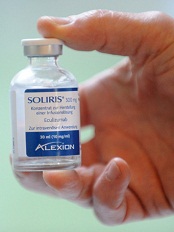
Credit: Globovision
The UK’s National Institute for Health and Care Excellence (NICE) has issued a final guidance recommending eculizumab (Soliris) as a treatment for atypical hemolytic uremic syndrome (aHUS), but only if certain conditions are met.
The guidance is the first to be produced as part of NICE’s highly specialized technologies program to evaluate treatments for very rare conditions.
aHUS affects around 200 people in England, with 20 to 30 new patients diagnosed with the condition each year.
The condition causes inflammation of blood vessels and thrombus formation throughout the body. So aHUS patients are at constant risk of sudden and progressive damage to, and failure of, vital organs.
“aHUS is a very distressing condition that imposes a significant burden both on those with the condition and their carers and families,” said NICE Chief Executive Sir Andrew Dillon.
“[A committee advising NICE] accepted that eculizumab is a step change in the management of aHUS and can be considered a significant innovation for a disease with a high unmet clinical need. It offers people with aHUS the possibility of avoiding end-stage renal failure, dialysis, and kidney transplantation, as well as other organ damage.”
“The drug is, however, very expensive. The committee felt that the budget impact of eculizumab would be lower if the potential for adjusting the dose of the drug and stopping treatment was explored. This is reflected in the guidance, which recommends eculizumab should be funded only if important conditions are met, including the development of rules for starting and stopping treatment for clinical reasons. In the meantime, NHS England and the company should consider what opportunities might exist to reduce the cost of eculizumab to the NHS.”
The conditions for funding eculizumab are as follows:
- Use of the drug must be coordinated through an expert center.
- Monitoring systems must record the number of people with aHUS, the number who receive eculizumab, and the dose and duration of treatment.
- A national protocol must be developed for starting and stopping eculizumab for clinical reasons.
- A research program is needed with robust methods to evaluate when stopping treatment or dose adjustment might occur.
Eculizumab usage and costs
Eculizumab is given intravenously in adults as initial treatment at a dose of 900 mg for 4 weeks, then as maintenance treatment at a dose of 1200 mg on week 5 and then every 12 to 16 days.
The summary of product characteristics for eculizumab states that “treatment is recommended to continue for a patient’s lifetime, unless discontinuation of treatment is clinically indicated.” Eculizumab costs £3150 per 30 mL vial (excluding tax).
The net budget impact of eculizumab is confidential. However, NICE has prepared an illustration of the possible budget impact of eculizumab for aHUS, using information that is available in the public domain.
This is based on a treatment cost of £340,200 per adult patient in the first year (based on the acquisition cost of the drug and the recommended dosing for an adult) and assumes a patient cohort of 170, as estimated by NHS England.
If it is assumed that all of these adults with aHUS are treated with eculizumab, the budget impact for the first year would be £57.8 million.
If an additional 20 new patients are treated the following year (based on a worldwide incidence of 0.4 per million), the budget impact will rise to £62.5 million in year 2, assuming all new patients are treated and all existing patients continue to be treated at the maintenance cost of £327,600 per year.
Using the same assumptions, the budget impact will rise to £69 million in year 3 (190 existing and 20 new patients), £75 million in year 4 (210 existing and 20 new patients), and £82 million in year 5 (230 existing and 20 new patients).
NHS England has indicated that the amount of the budget allocated for highly specialized services in 2013/14 was £544 million, and the money spent on high-cost drugs was £156 million.
The committee acknowledged that the estimate of the incremental cost of eculizumab made by Alexion Pharmaceuticals (the company developing eculizumab) compared with standard care was considerable. And incremental costs estimated by the evidence review group were higher still (although results are confidential).
Alexion estimated that eculizumab produced 25.22 additional quality-adjusted life-years (QALYs) per patient compared with standard care. Although the QALYs estimated in the evidence review group’s analysis were markedly lower than those calculated by the company, the advisory committee said both analyses produced substantial QALY gains of a magnitude that is rarely seen for any new drug.
For more information, see the guidance on the NICE website.