User login
Combo proves ‘beneficial’ for ‘unfit’ CLL patients

STOCKHOLM—Obinutuzumab plus chlorambucil (G-Clb) is a “valid and beneficial” frontline treatment option for “unfit” patients with chronic lymphocytic leukemia (CLL), according to a speaker at the 23rd Congress of the European Hematology Association (EHA).
Final results from the CLL11 study have revealed additional benefits of G-Clb over rituximab plus chlorambucil (R-Clb) in patients with previously untreated CLL and comorbidities.
Prior results from this study showed that G-Clb produced higher response rates and prolonged progression-fee survival (PFS) compared to R-Clb.
Now, with a median follow-up of 5 years, researchers have found that G-Clb prolongs overall survival (OS) and time to next treatment (TTNT) as well.
Valentin Goede, MD, of the University Hospital Cologne in Germany, presented these results during the Presidential Symposium of the EHA Congress (abstract S151).
The study was sponsored by Hoffmann-La Roche.
CLL11 enrolled patients with previously untreated CLL and coexisting medical conditions. They were randomized to receive six 28-day cycles of Clb alone, G-Clb, or R-Clb.
In stage 1, researchers compared G-Clb (n=238) to Clb alone (n=118) and R-Clb (n=233) to Clb alone (n=118). In stage 2, they compared G-Clb (n=333) and R-Clb (n=330).
“The treatment arms were well-balanced, not just with regard to patient [characteristics] but also with disease characteristics,” Dr Goede said.
Overall, the median age was 73 (range, 39-90), the median Cumulative Illness Rating Scale score was 8, and the median creatinine clearance was 62 mL/min.
Efficacy: G-Clb vs Clb
The median observation time for G-Clb vs Clb was 62.5 months.
The median PFS was 31.1 months in the G-Clb arm and 11.1 months in the Clb arm. The 5-year PFS rates were 25% and 2%, respectively. The hazard ratio (HR) was 0.21 (P<0.0001).
The median OS was not reached in the G-Clb arm and was 66.7 months in the Clb arm. The 5-year OS rates were 66% and 53%, respectively. The HR was 0.68 (P=0.0196).
Thirty-nine percent of the G-Clb arm died, as did 49% of the Clb arm. The main causes of death were adverse events (AEs) and disease progression.
Efficacy: G-Clb vs R-Clb
The median observation time for G-Clb vs R-Clb was 59.4 months.
The median PFS was 28.9 months in the G-Clb arm and 15.7 months in the R-Clb arm. The 5-year PFS rates were 23% and 9%, respectively. The HR was 0.49 (P<0.0001).
“The median PFS was nearly doubled, from approximately 15 months in the rituximab arm to almost 30 months in the obinutuzumab arm,” Dr Goede said. “And this translated into a clinically meaningful prolongation of time to next treatment.”
The median TTNT was 56.4 months in the G-Clb arm and 34.9 months in the R-Clb arm. At 5 years, TTNT rates were 49% and 32%, respectively. The HR was 0.58 (P<0.0001).
“In the rituximab arm, the median time to next treatment was a little greater than 2.5 years, and, in the obinutuzumab arm, it was almost 5 years,” Dr Goede said. “From a clinical perspective, I would consider treatment-free intervals of that duration as highly relevant and beneficial in an elderly population.”
The median OS was not reached in the G-Clb arm and was 73.1 months in the R-Clb arm. The 5-year OS rates were 66% and 57%, respectively. The HR was 0.76 (P=0.0245).
“This difference is clinically meaningful, and it is also remarkable in the context of the long follow-up, given the fact that approximately half of the patients have received at least 1 salvage treatment in the meantime,” Dr Goede said.
In all, 37% of the G-Clb arm died, as did 45% of the R-Clb arm. Again, the main causes of death were AEs and disease progression.
Safety
Dr Goede said no new safety signals or late-onset toxicities were detected.
“Adverse events of any grade, but particularly grade 3-5 and serious adverse events, were more frequent in the obinutuzumab arm compared to the other 2 arms,” he noted. “[This] was mainly driven by more infusion reactions and some greater hematological toxicity.”
“Importantly, the rate of fatal adverse events, during treatment but also during follow-up, was not higher in the obinutuzumab arm. And the most common fatal adverse events were second malignancies.”
G-Clb vs Clb
Ninety-five percent of patients in the G-Clb arm and 83% of those in the Clb arm had at least 1 AE. The rates of grade 3-5 AEs were 74% and 51%, respectively. The rates of serious AEs were 47% and 39%, respectively. The rates of fatal AEs were 8% and 11%, respectively.
Seventeen percent of patients in the G-Clb arm and 11% of those in the Clb arm had late-onset neutropenia. The rates of prolonged neutropenia were 3% and 9%, respectively.
Fourteen percent of patients in the G-Clb arm and 7% of those in the Clb arm had second malignancies (starting 6 months after treatment initiation). The most common of these were squamous cell carcinoma (2% vs 0%) and basal cell carcinoma (2% vs <1%).
G-Clb vs R-Clb
Ninety-four percent of patients in the G-Clb arm and 90% of those in the R-Clb arm had at least 1 AE. The rates of grade 3-5 AEs were 72% and 60%, respectively. The rates of serious AEs were 45% and 39%, respectively. The rates of fatal AEs were 7% and 10%, respectively.
Fifteen percent of patients in the G-Clb arm and 12% of those in the R-Clb arm had late-onset neutropenia. The rates of prolonged neutropenia were 2% and 4%, respectively.
Eleven percent of patients in the G-Clb arm and 10% of those in the Clb arm had second malignancies. Squamous cell carcinoma occurred in 2% of patients in both arms. Basal cell carcinoma occurred in 2% of G-Clb recipients and 1% of R-Clb recipients.
Umbralisib can revitalize ruxolitinib in MF
STOCKHOLM—The PI3K delta inhibitor umbralisib can “augment or resurrect” responses to ruxolitinib in patients with myelofibrosis (MF), according to a speaker at the 23rd Congress of the European Hematology Association (EHA).
Results of a phase 1 study showed that adding umbralisib to treatment with ruxolitinib could induce responses in MF patients who had a suboptimal or lost response to ruxolitinib.
Of the 23 patients who received the combination, 2 achieved a complete remission (CR), 11 had clinical improvement, and 8 had stable disease.
In addition, umbralisib plus ruxolitinib was considered well-tolerated. The most common adverse event (AE) was anemia.
Tamara K. Moyo, MD, PhD, of Vanderbilt University Medical Center in Nashville, Tennessee, presented these results at the EHA Congress as abstract S133. The research was sponsored by TG Therapeutics.
Patients
Dr Moyo reported results in 23 MF patients who had a suboptimal response, lost a response, or had no response while on a stable dose of ruxolitinib for at least 8 weeks. Their median age was 67 (range, 49-83), and 61% were male.
Patients had primary MF (30%), post-essential thrombocythemia (ET) MF (43%), or post-polycythemia vera (PV) MF (26%). Forty-three percent of patients had JAK2 V617F, 30% had CALR mutations, 17% had MPL mutations, and 13% were triple-negative. One patient had co-occurring CALR and MPL mutations.
Most patients had an ECOG performance score of 0 (39%) or 1 (52%). All had intermediate-1 (35%), intermediate-2 (35%), or high-risk disease (30%) according to DIPSS Plus.
Sixty-one percent of patients had splenomegaly.
Treatment
In stage 1, the patients received stable ruxolitinib and escalating umbralisib. In stage 2, patients received escalating ruxolitinib and umbralisib at the maximum tolerated dose (MTD) established from stage 1.
Patients could then proceed to expansion cohorts in which they would receive any dose of ruxolitinib and umbralisib at the MTD. The expansion cohorts include patients with treatment-naïve MF, PV, chronic myelomonocytic leukemia, and myelodysplastic syndromes/myeloproliferative neoplasms.
However, Dr Moyo reported only on the 23 ruxolitinib-experienced MF patients.
Safety
There were 2 dose-limiting toxicities of asymptomatic, grade 3 amylase/lipase elevations. One occurred in a patient receiving 800 mg of umbralisib daily and 10 mg of ruxolitinib twice daily. The other occurred in a patient receiving 800 mg of umbralisib daily and 15 mg of ruxolitinib twice daily.
Therefore, 600 mg daily was deemed the MTD of umbralisib.
Seventeen patients had at least 1 AE. There were 17 grade 3 or higher AEs in 13 patients.
AEs of any grade included anemia (n=10), neutrophil decrease (n=2), platelet decrease (n=5), AST increase (n=6), ALT increase (n=3), amylase increase (n=3), lipase increase (n=3), diarrhea (n=2), colitis (n=1), dyspnea (n=1), upper respiratory infection (n=2), pneumonia (n=4), other infections (n=6), and sepsis (n=1).
Grade 3 AEs included anemia (n=3), neutrophil decrease (n=2), amylase increase (n=2), lipase increase (n=2), diarrhea (n=2), colitis (n=1), dyspnea (n=1), pneumonia (n=1), and other infections (n=2). The case of sepsis was the only grade 4 AE.
Dr Moyo noted that anemia—the most common AE—was commonly attributed to disease rather than study treatment.
The case of colitis, which was grade 3, was deemed possibly related to treatment, so the patient was removed from the study.
Thirteen patients had discontinued study treatment at the time of analysis. Aside from the patient who discontinued due to colitis, 2 patients went off study due to dose-limiting toxicities, 3 due to progressive disease, 6 due to physician or patient decision, and 1 due to transplant.
Efficacy
Two patients could not be assessed for efficacy, and 8 had stable disease on umbralisib and ruxolitinib.
The combination produced clinical improvement—reduction in spleen volume, increase in hemoglobin, and improvement in MF-related symptoms—in 11 patients (48%).
And 2 patients (9%) achieved a CR. Dr Moyo said there were “few commonalities” between these 2 patients.
Both had intermediate-1-risk disease as well as persistent or progressive MF-related symptoms and thrombocytosis at baseline. However, 1 patient had post-ET MF, and 1 had post-PV MF.
The post-ET MF patient had an MPL driver mutation. She received ruxolitinib at 20 mg twice daily and umbralisib at 400 mg daily. The patient achieved a CR at cycle 15 and remained on study 2 years before proceeding to transplant. The patient is now about 1 year from her transplant with no evidence of disease.
The post-PV patient had a JAK2 V617F driver mutation. She received ruxolitinib at 15 mg twice daily and umbralisib at 600 mg daily. The patient achieved a CR at cycle 5 and remains on study, currently receiving cycle 12 of treatment.
Dr Moyo said these results suggest “the addition of umbralisib to ruxolitinib can augment or resurrect a response in MF patients who have had suboptimal or lost response to ruxolitinib alone, and this treatment combination warrants further investigation.”
Gene signature might identify patients at risk of CAR T-associated neurotoxicity
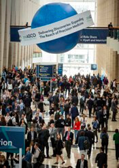
CHICAGO—A specific gene signature might be able to identify patients at risk of CD19 CAR T-cell associated neurotoxicity, according to results of an exploratory analysis presented at the 2018 ASCO Annual Meeting.
The analysis, based on bone marrow samples from patients with relapsed/refractory B-cell acute lymphoblastic leukemia (ALL) treated with JCAR015 in the ROCKET trial, helped identify a set of neurotoxicity-associated genes that separated patients based on molecular subtype.
“These findings suggest that patient risk stratification by molecular subtype of disease or gene expression signature may play a role in identifying patients at elevated risk of neurotoxicity,” said Jae Park, MD, of Memorial Sloan Kettering Cancer Center, New York, New York, in a presentation of the findings (abstract 7007).
The phase 2 ROCKET study included adult patients with relapsed or refractory morphological (>5% blasts in bone marrow) CD-19 positive disease in first salvage or greater, including post allogeneic hematopoietic stem cell transplantation (HSCT). Prior blinatumomab was allowed.
The tumor gene expression study presented at ASCO was based on sequenced RNA from pre-apheresis bone marrow samples available for 31 patients in the ROCKET study.
Investigators identified a set of 10 genes expressed more frequently in bone marrow samples from patients in ROCKET with low (grade 0-1) neurotoxicity, and 7 that were more frequent in those who had severe (grade 4-5) neurotoxicity.
Looking at B-cell ALL samples in public datasets by molecular subtype, they found genes highly expressed in the low neurotoxicity ROCKET patients were also highly expressed in Philadelphia chromosome-positive (Ph+) and Ph-like subtypes.
Conversely, the genes highly expressed in the severe neurotoxicity patients were also highly expressed in non-Ph-like samples.
A total of 16 ROCKET patients were classified as having Ph-like gene expression and 15 as having non-Ph-like expression.
There were no grade 4-5 neurotoxicity events in the Ph-like patients, while both grade 3+ and grade 4+ neurotoxicity were significantly more prevalent in the non-Ph-like patients, investigators reported.
One of the most differentially expressed genes in the set was CCL17, which was higher in the low-neurotoxicity tumor samples, and likewise highly expressed in Ph-like B-cell ALL, according to the report.
“[CCL17] may serve as an early biomarker for differentiating severe neurotoxicity,” Dr Park said.
These findings are now being validated in the previously mentioned data set, as well as other studies to see if the findings can be replicated, according to Dr Park.
Juno Therapeutics, a Celgene company, shut down the phase 2 ROCKET trial of JCAR015 in 2017 after 2 clinical holds in 2016 and 5 patient deaths.
FDA places SB-generated CAR T-cell therapy on clinical hold

The US Food and Drug Administrated (FDA) placed a clinical hold on the phase 1 trial of the Sleeping Beauty (SB)-generated CAR T-cell therapy in relapsed or refractory leukemia and lymphoma patients.
The Sleeping Beauty platform was designed to very rapidly manufacture CD19-specific CAR T cells at the point of care.
All SB-CAR T-cell processing is planned to take place within 2 days at the healthcare facility, thus eliminating shipping cells from hospitals to production sites and back again.
The FDA is requesting more chemistry, manufacturing, and control (CMC) information before allowing the trial to proceed.
The Sleeping Beauty technology, a non-viral transposon/transposase system, has the potential to reduce the costs and complexity associated with recombinant viral vector-based immunotherapy, according to developers.
Ziopharm Oncology, Precigen, Inc, a wholly owned subsidiary of Intrexon Corporation, and the University of Texas MD Anderson Cancer Center, are developing the Sleeping Beauty CAR T cell therapy.
“We know what is needed to address the hold issues and are looking forward to responding to the agency in a timely manner,” said Laurence Cooper, MD, PhD, chief executive officer of Ziopharm, in a corporate release.
“We are undertaking cutting-edge science and are on the verge of a paradigm shift based on our approach to very rapidly manufacture CD19-specific T cells within 2 days using our non-viral approach to CAR-T therapy based on the Sleeping Beauty platform.”
The phase 1 trial in question is a third-generation trial in which the CAR T cells are designed to co-express CD19-specific CAR, membrane-bound interleukin 15, and a safety switch.
The findings from earlier generation phase 1 trials have been previously reported in The Journal of Clinical Investigation.
Quizartinib can prolong OS in rel/ref, FLT3-ITD AML
STOCKHOLM—Phase 3 results suggest the FLT3 inhibitor quizartinib can prolong overall survival (OS) in patients with relapsed/refractory, FLT3-ITD acute myeloid leukemia (AML).
In the QuANTUM-R study, patients who received single-agent quizartinib had a significantly longer median OS than patients who received salvage chemotherapy.
There was a trend toward improved event-free survival (EFS) with quizartinib as well.
“QuANTUM-R represents the first study that shows a significant improvement in overall survival for a single agent—a FLT3 inhibitor or any other targeted agent—in this population of FLT3-mutated AML patients with refractory or relapsed disease . . .,” said study investigator Jorge Cortes, MD, of MD Anderson Cancer Center in Houston, Texas.
Dr Cortes presented results from QuANTUM-R at the 23rd Congress of the European Hematology Association (EHA). The research was selected as the best late-breaking abstract (LB2600).
The study was funded by Daiichi Sankyo, Inc., and Dr Cortes is a consultant for the company.
Patients and treatment
QuANTUM-R enrolled adults with FLT3-ITD AML (at least 3% FLT3-ITD allelic ratio) who had refractory disease or had relapsed within 6 months of their first complete remission. They had received at least 1 cycle of an induction regimen containing standard-dose anthracycline or mitoxantrone.
Patients were randomized to receive once-daily treatment with quizartinib (n=245) or a salvage chemotherapy regimen (n=122)—low-dose cytarabine (LoDAC, n=29); combination mitoxantrone, etoposide, and cytarabine (MEC, n=40); or combination fludarabine, cytarabine, and idarubicin (FLAG-IDA, n=53).
Responders could proceed to hematopoietic stem cell transplant (HSCT), and those in the quizartinib arm could resume quizartinib after HSCT.
Baseline characteristics were similar between the treatment arms. The median age was 55 (range, 19-81) for patients receiving quizartinib and 58 (range, 18-78) for those receiving chemotherapy.
Thirty-three percent of the quizartinib arm had refractory disease, and 67% had relapsed disease. Thirty-four percent of the chemotherapy arm had refractory disease, and 66% had relapsed disease.
The percentage of patients with a prior allogeneic HSCT was 25% in the quizartinib arm and 23% in the chemotherapy arm. Most patients in both arms had intermediate-risk cytogenetics—78% of the quizartinib arm and 66% of the chemotherapy arm.
In all, 241 patients received quizartinib, and 94 received salvage chemotherapy—LoDAC (n=22), MEC (n=25), and FLAG-IDA (n=47). Of the 28 patients in the chemotherapy group who were not treated, most withdrew consent.
The median treatment duration was 4 cycles (range, 1-3) in the quizartinib arm and 1 cycle (range, 1-2) for patients who received LoDAC, MEC, and FLAG-IDA.
The most common reason for discontinuation of chemotherapy was lack of response/progression (n=49), followed by death (n=6). Twenty-four patients completed salvage chemotherapy.
In the quizarinib arm, the most common reasons for treatment discontinuation were HSCT (n=79), relapse (n=60), or lack of response/progression (n=47).
Thirty-two percent of quizartinib-treated patients and 12% of the chemotherapy group went on to HSCT.
Results
The median follow-up was 23.5 months. The efficacy results include all randomized patients, and the safety results include only those who received their assigned treatment.
The study’s primary endpoint was OS. The median OS was 6.2 months in the quizartinib arm and 4.7 months in the chemotherapy arm (hazard ratio=0.76, P=0.0177). The 1-year OS rate was 27% and 20%, respectively.
The median EFS was 6.0 weeks in the quizartinib arm and 3.7 weeks in the chemotherapy arm (hazard ratio=0.90, P=0.1071). Dr Cortes noted that patients who did not receive treatment were censored on day 1, and partial responses were counted as failures in the EFS analysis.
The overall response rate was 69% in the quizartinib arm and 30% in the chemotherapy arm.
The composite complete response (CR) rate was 48% in the quizartinib arm and 27% in the chemotherapy arm. This includes the CR rate (4% and 1%, respectively), the rate of CR with incomplete platelet recovery (4% and 0%, respectively), and the rate of CR with incomplete hematologic recovery (40% and 26%, respectively). The rate of partial response was 21% and 3%, respectively.
Dr Cortes said rates of treatment-emergent adverse events (TEAEs) were similar between the treatment arms.
Grade 3 or higher hematologic TEAEs occurring in at least 5% of patients (in the quizartinib and chemotherapy groups, respectively) included thrombocytopenia (35% and 34%), anemia (30% and 29%), neutropenia (32% and 25%), febrile neutropenia (31% and 21%), and leukopenia (17% and 16%).
Grade 3 or higher nonhematologic TEAEs occurring in at least 5% of patients (in the quizartinib and chemotherapy groups, respectively) included fatigue (8% and 1%), hypokalemia (12% and 9%), sepsis/septic shock (16% and 18%), dyspnea (5% for both), hypophosphatemia (5% for both), and pneumonia (12% and 9%).
Three percent of patients in the quizartinib arm had grade 3 QTcF prolongation, but there were no grade 4 cases. Two patients discontinued quizartinib due to QTcF prolongation.
“The safety of this drug has remained constant across over 1600 patients that have been treated with quizartinib across a variety of studies,” Dr Cortes said.
He added that QuANTUM-R results open up the possibility that quizartinib could be used in other settings. Researchers are already testing standard chemotherapy with and without quizartinib in a phase 3 trial of patients with newly diagnosed, FLT-ITD AML (QuANTUM-First).
Peripheral blood MRD correlates with treatment benefit in CLL
CHICAGO—Minimal residual disease (MRD) kinetics confirms the high, durable MRD-negativity with venetoclax plus rituximab in relapsed/refractory chronic lymphocytic leukemia (CLL), according to a further examination of the phase 3 MURANO study.
Undetectable MRD-negativity is associated with extended progression-free survival (PFS) and overall survival in patients receiving chemoimmunotherapy for CLL.
“Attainment of MRD-negativity in relapsed/refractory CLL is also a desired trial endpoint due to the subjectivity of complete response definition regarding pathologic lymph node size,” said Peter Hillmen, MD, of St James’s University Hospital, Leeds, United Kingdom, at the 2018 ASCO Annual Meeting.
Dr Hillmen reported new data on MRD response in cytogenetic and molecular risk groups, MRD sustainability and kinetics, and MRD conversion in the MURANO trial (abstract 7508).
MURANO trial (NCT02005471)
In the trial, venetoclax-rituximab showed superior PFS and peripheral blood and bone marrow MRD-negativity as compared to bendamustine plus rituximab (BR) in relapsed/refractory CLL patients.
Patients were randomized to venetoclax-rituximab for 6 months, followed by single-agent venetoclax for up to 1.5 years, or BR for 6 months. Peripheral blood samples were serially collected and bone marrow was collected at the end of combination treatment or at best response.
MRD findings
The new results show higher concordance in MRD-negativity between bone marrow and peripheral blood in venetoclax-rituximab (45 of 50 patients, 90%) vs BR (3 of 10 patients, 30%) in paired samples.
Focusing on peripheral blood MRD, Dr Hillmen said the best MRD-negativity rates were higher with venetoclax-rituximab (84%) than BR (23%). These results were independent of high-risk factors—such as del 17p, IGVH unmutated, and mutated TP53—only for venetoclax-rituximab treated patients.
“The superior peripheral blood MRD response with venetoclax-rituximab was consistent across subgroups at the end of completion of treatment,” Dr Hillmen said. “Most patients who achieved peripheral blood MRD-negativity on venetoclax-rituximab remained MRD-negative and were progression-free.”
Among 121 of 194 (62%) patients on venetoclax-rituximab who achieved MRD-negativity at the end of combination therapy, 100 (83%) patients maintained MRD-negativity and were progression-free at a median follow-up of 13.8 months. Two patients developed progressive disease and 2 patients died (unrelated to CLL).
Two patients developed Richter’s disease (with one MRD-positive directly before therapy) and 15 (12%) patients converted to confirmed MRD-positive at a median MRD-positive follow-up of 5.6 months.
“High peripheral blood MRD-negativity at the end of combination treatment and concordance with bone marrow MRD with venetoclax-rituximab,” Dr Hillmen said, “confirms the value of peripheral blood MRD for evaluation of treatment benefit in relapsed/refractory CLL patients. The high rate of peripheral blood MRD-negativity at end of combination treatment with venetoclax-rituximab was attained regardless of risk features.”
Some conversion to MRD-positivity occurred only in a small proportion of patients. Most cases were of intermediate level and remained progression-free, he said.
“MRD kinetics indicate that peripheral blood MRD-negativity with venetoclax-rituximab occurs early and is maintained over time with current follow-up,” Dr Hillmen added. The MRD data now provide a framework for designing response adaptive therapy.
The US Food and Drug Administration recently approved venetoclax-rituximab for CLL or small lymphocytic lymphoma for patients with or without del 17p.
Venetoclax is being developed by Genentech and Abbvie.
Efficacy of KTE-C19 CAR T cells not compromised by prior blinatumomab
CHICAGO—Prior exposure to blinatumomab does not appear to affect the successful manufacture of KTE-C19 or its efficacy in patients with relapsed/refractory acute lymphoblastic leukemia (ALL), an investigator reported at the 2018 ASCO Annual Meeting.
The clinical benefit of KTE-C19, an anti-CD19 chimeric antigen receptor (CAR) T-cell therapy, was preserved regardless of whether patients were exposed to blinatumomab, said Bijal D. Shah, MD, of Moffitt Cancer Center in Tampa, Florida.
High rates of complete response and undetectable minimal residual disease (MRD) were independent of exposure to blinatumomab, a CD19/CD3 bispecific T-cell engager.
“We feel the results of these data support KTE-C19 as an effective option for adults with refractory leukemia, regardless of prior exposure to CD19-directed therapy,” Dr Shah reported at the meeting (abstract 7006*).
The current standard of care for adults with relapsed/refractory ALL includes blinatumomab, raising the question of whether prior exposure to this CD19-directed treatment could influence the manufacture or efficacy of KTE-C19.
Sara Cooley, MD, of Masonic Medical Center, University of Minnesota in Minneapolis, said results of this analysis suggest prior blinatumomab should not be a contraindication or concern in the context of KTE-C19.
“This remains to be shown with other CAR T-cell therapies,” she said in a presentation at ASCO on cellular therapy in leukemia.
The analysis by Dr Shah and co-investigators was based on ZUMA-3 (NCT02614066), a phase 1 study including adults with relapsed/refractory ALL who received KTE-C19 at doses of 0.5, 1, or 2 x 10⁶ cells/kg.
They excluded patients in the highest dose cohort, who were required to be blinatumomab naïve, per protocol. That left 23 patients who received 0.5 or 1 x 10⁶ cells/kg, of whom 11 had prior blinatumomab exposure and 12 did not.
Best overall response appeared to be independent of prior blinatumomab treatment, with a CR rate of 72% overall, and 63% and 80% for blinatumomab-exposed and blinatumomab-naïve patients, respectively.
The rate of undetectable MRD was likewise high at 88% in the prior blinatumomab group and 100% in the no-blinatumomab group.
Product characteristics did not vary according to blinatumomab exposure, though there was a trend toward a more differentiated phenotype in those patients who had received prior CD19-directed treatment, he said.
There were no significant differences between groups in the rate of grade 3 or greater cytokine release syndrome. Neurologic events were higher in the blinatumomab-naïve patients, though a higher percentage of those patients received the 1 x 10⁶ cells/kg dose, Dr Shah reported.
Investigators also looked at CAR T levels by treatment.
“We cannot appreciate any significant differences between the blinatumomab-naïve and the blinatumomab-exposed groups,” Dr Shah told ASCO attendees.
The ZUMA-3 trial was sponsored by Kite, a Gilead Company.
*Data in the abstract differ from the presentation.
‘Excellent’ survival with HCT despite early treatment failure in FL

Autologous and allogeneic hematopoietic stem cell transplantation (HCT) both offer excellent long-term survival in follicular lymphoma (FL) patients who experience early treatment failure, an analysis of a large transplant registry suggests.
Five-year survival rates exceeded 70% for patients who received autologous or matched sibling donor (MSD) transplants, according to the analysis of the Center for International Blood and Marrow Transplant Research (CIBMTR) database. The database included 440 patients who underwent a procedure between 2002 and 2014.
“Until better risk-stratification tools are available for FL, auto-HCT and MSD allo-HCT should be considered as effective treatment options with excellent long-term survival for high-risk patients as defined by early treatment failure,” Sonali M. Smith, MD, of the University of Chicago, and co-investigators wrote in the journal Cancer.
Early treatment failure in FL is associated with worse overall survival. In the National LymphoCare Study (NLCS), patients who received upfront R-CHOP therapy and progressed within 24 months had a 5-year overall survival of 50%, versus 90% for patients without early progression.
By contrast, survival figures in the present study are “provocatively higher” than those in the NLCS, in which only 8 out of 110 patients underwent HCT, Dr Smith and co-authors said.
Dr Smith’s study showed that with a median follow-up of 69 to 73 months, adjusted probability of 5-year overall survival was 70% for autologous and 73% for MSD HCT, versus 49% for matched unrelated donor HCT (P=0.0008).
Ryan C. Lynch, MD, and Ajay K. Gopal, MD, of the Fred Hutchinson Cancer Research Center in Seattle, Washington, said that the finding “convincingly demonstrates” the benefit of transplant in the setting of early treatment failure.
“Select patients (particularly younger patients) with chemoresponsive disease who understand the risk-benefit ratio in comparison with currently approved and experimental therapies still remain good candidates for autologous HCT,” Drs Lynch and Gopal said in an editorial.
“For older patients or patients with comorbidities, we would continue to recommend clinical trials or treatment with an approved PI3K inhibitor,” they added.
The study by Dr Smith and colleagues is not the first to show a benefit of HCT in this clinical scenario. In a recent NLCS/CIBMTR analysis of FL patients, 5-year overall survival was 73% for those undergoing autologous HCT done within a year of early treatment failure, versus 60% for those who did not (P=0.05).
The two studies “collectively suggest that transplantation should be considered in this high-risk group of patients with early relapse,” Dr Smith and co-authors wrote.
Interim PET scans identify HL patients with better outcomes
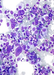
CHICAGO—Interim PET scans can identify a subset of Hodgkin lymphoma (HL) patients with a better outcome suitable for de-escalation treatment after upfront BEACOPP without impairing disease control, according to final results of the AHL2011-LYSA study.
BEACOPP, compared to ABVD, improves progression-free survival (PFS) but not overall survival (OS) and is associated with a higher risk of myelodysplasia, acute leukemia, and infertility.
Investigators evaluated whether some patients might be able to reduce treatment intensity without compromising the effectiveness of their therapy.
Olivier Casasnovas, MD, of CHU Le Bocage Service d'Hématologie Clinique, Dijon, France, presented the final analysis at the 2018 ASCO Annual Meeting (abstract 7503).
AHL2011-LYSA study (NCT01358747)
The randomized phase 3 study compared an early PET-driven treatment de-escalation to a non-PET-monitored strategy in patients with advanced-stage HL.
The study included 823 previously untreated patients, median age 30 years (range 16 – 60), with stage III, IV, or high-risk IIB HL.
The PET-driven strategy consisted of 2 BEACOPP* cycles (PET2), followed by 4 cycles of ABVD** for PET2-negative patients, and 4 cycles of BEACOPP for PET2-positive patients.
The experimental PET-driven strategy (410 patients) was randomly compared to a standard treatment delivering 6 cycles of BEACOPP (413 patients). PFS was the primary endpoint with a hypothesis of non-inferiority of the PET-driven arm compared to the standard arm.
Patients characteristics were well balanced between the arms, Dr Casasnovas said. PET2-positivity rate was similar in both arms (experimental 13%, standard 12%).
Based on PET2 results, 346 (84%) patients received 4 cycles of ABVD and 51 (12%) patients received 4 additional cycles of BEACOPP in the experimental arm.
Results
With a median follow-up of 50 months, the 5-year PFS was similar in the standard (86.2%) and the PET-driven arms (85.7%). The 5-year PFS for PET 2-negative/PET 4-negative patients was 90.9%, for PET 2-positive/PET4-negative patients was 75.4%, and for PET 4-positive patients was 46.5%.
The 5-year OS was similar in both arms (96.4% experimental, 95.2% standard).
The treatment toxicity was significantly higher in patients receiving 6 cycles of BEACOPP as compared to those who received 2 cycles of BEACOPP plus 4 cycles of ABVD.
Those who received more cycles of BEACOPP had more frequent grade 3 or higher adverse events than those with fewer cycles, including anemia (11% vs 2%), leukopenia (85% vs 74%), thrombocytopenia (44% vs 15%), and sepsis (7% vs 3%), as well as in serious adverse events (45% vs 28%).
“After 4 cycles of chemotherapy, it [PET positivity] identifies a subset of patients with a particularly poor outcome,” Dr Casasnovas said, “encouraging researchers to develop new treatment options in these patients.”
“PET performed after 2 cycles of BEACOPP escalation can be safely used to guide subsequent treatment,” he concluded.
“This approach allows clinicians to reduce the treatment-related immediate toxicity in most patients,” he added, “and provides similar patient outcomes compared to standard BEACOPP escalation treatment.”
* Bleomycin, etoposide, doxorubicin, cyclophosphamide, vincristine, procarbazine, prednisone
**Adriamycin (doxorubicin), bleomycin, vinblastine, dacarbazine
PU-H71 receives orphan drug designation for myelofibrosis
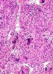
The US Food and Drug Administration (FDA) has granted orphan drug designation to PU-H71 to treat patients with myelofibrosis.
The drug specifically targets the epichaperome, a network of high-molecular-weight complexes found in multiple diseases, including cancer and neurologic disorders. These complexes enhance cellular survival, irrespective of tissue of origin or genetic background.
According to research published in Nature Reviews Cancer, Pu-H71 interferes with the epichaperome function in diseases and does not affect normal cells.
PU-H71 is being evaluated in a phase 1b trial in myelofibrosis and advanced metastatic breast cancer.
“In myelofibrosis, the epichaperome plays a central role in optimizing the JAK-STAT pathway,” said Srdan Verstovsek, MD, PhD, “allowing JAK2 to form dimers that evade inhibition with a JAK2 inhibitor such as ruxolitinib.”
“By inhibiting epichaperome function and breaking this mechanism, we believe PU-H71 can increase anti-cancer activity of JAK2 inhibitors,” he said. Dr Verstovsek, of the MD Anderson Cancer Center in Houston, Texas, is lead clinical research advisor for the phase 1b myelofibrosis study.
Phase 1b Study (NCT01393509)
This is a multicenter study designed to assess the safety, tolerability, pharmacokinetic and preliminary efficacy of PU-H71 in patients taking concomitant ruxolitinib.
The safety expansion phase of the trial is open for accrual only to patients with myeloproliferative neoplasms (MPNs).
These patients must have been on ruxolitinib for at least 3 months, be on a stable dose for at least 1 month prior to enrollment and be taking at least 5 mg twice daily.
Patients must have persistent disease manifestations, despite ruxolitinib therapy. These include persistent splenomegaly, abnormal blood counts, persistent constitutional symptoms, residual fibrosis in bone marrow (2+ or greater), or measurable allele burden as evidenced by clonal JAK2 or MPL mutation.
Samus Therapeutics, the developer of PU-H71, announced, simultaneously with the orphan drug designation, the dosing of the first patient in the phase 1b myelofibrosis study.
“Targeting the epichaperome offers an exciting new avenue for treating myelofibrosis and related diseases,” Dr Verstovsek said.
“I look forward to seeing how the combination of these therapies can affect outcomes in patients for whom this resistance is associated with poor prognoses.”